
Pathology of the Male BreastBệnh lý vú nam
Ngày đăng / cập nhật: 2009-03-15
Bản gốc trên radiologyassistant.nl ↗ · tải về lúc 2026-11-19 20:19 · bản dịch máy có chuẩn hóa thuật ngữ chẩn đoán hình ảnh
This review is based on a presentation given by Leonard Glassman and adapted for the Radiology Assistant by Robin Smithuis.
We will discuss:Bài tổng quan này dựa trên một bài thuyết trình của Leonard Glassman và được Robin Smithuis chuyển thể cho Trợ lý X quang.
Chúng ta sẽ thảo luận:
- Normal male mammogram Chụp X-quang tuyến vú nam giới bình thường
- Classic features of gynecomastia Đặc điểm cổ điển của vú to ở nam giới
- Lesions that require a biopsyCác tổn thương cần sinh thiết
by Leonard M. Glassman Tác giả Leonard M. Glassman
Normal Male MammogramChụp X-quang tuyến vú nam bình thường
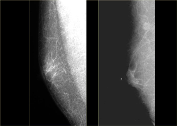 Normal male mammogram Click on image to enlargeX-quang tuyến vú nam bình thường Nhấp vào hình ảnh để phóng to
When you do breast imaging in a male, always stick to the following rule:
'If it is not normal, gynecomastia or classically benign,
it needs a biopsy'.
Khi bạn chụp hình ảnh vú ở nam giới, luôn tuân theo quy tắc sau:
Nếu nó không bình thường, vú to ở nam giới hoặc lành tính cổ điển,
cần phải làm sinh thiết.
So, first we're going to give you a couple of examples of the normal male breast, just to get used to what normal looks like.
Then we will discuss gynecomastia and finally discuss some specific tumors.
Obviously, the lesions in this last category will need a biopsy, unless you are sure that it is classically benign, like for instance a lymph node or lipoma.
Đầu tiên chúng ta sẽ cho bạn một vài ví dụ về vú của nam giới bình thường, chỉ để làm quen với những gì bình thường trông giống như thế nào.
Sau đó chúng ta sẽ thảo luận về vú to nam giới và cuối cùng thảo luận về một số khối u cụ thể.
Rõ ràng, các tổn thương trong loại cuối cùng này sẽ cần sinh thiết, trừ khi bạn chắc chắn rằng nó là lành tính cổ điển, ví dụ như hạch bạch huyết hoặc u mỡ.
On the left two examples of a normal male mammogram.
The left image shows normal skin, a nipple and a small amount of connective tissue behind the nipple.
The image on the right shows a bit more connective tissue, but this is still normal.
Ở bên trái hai ví dụ về chụp X-quang tuyến vú ở nam giới bình thường.
Hình ảnh bên trái cho thấy da bình thường, núm vú và một lượng nhỏ mô liên kết phía sau núm vòm.
Hình ảnh bên phải cho thấy mô liên kết nhiều hơn một chút, nhưng điều này vẫn bình thường.
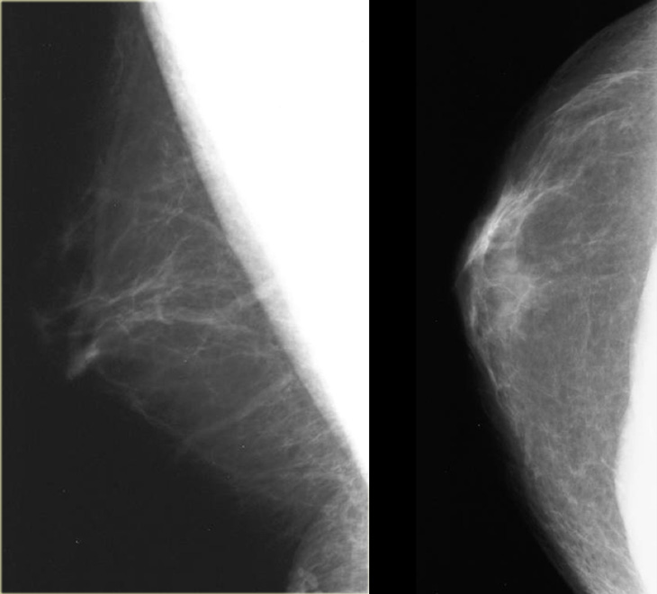 Normal male mammogramChụp X-quang tuyến vú nam giới bình thường
On the left another normal male breast.
There is more fatty tissue and there are a number of blood vessels.
There is a small amount of fibrous connective tissue, but basically most of this breast is just fat.Bên trái một vú nam bình thường khác.
Có nhiều mô mỡ hơn và có một số mạch máu.
Có một lượng nhỏ mô liên kết xơ, nhưng về cơ bản hầu hết vú này chỉ là mỡ.
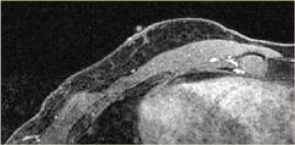 MR of the male breast T1WI with fat suppressionCộng hưởng từ vú nam T1WI với xóa mỡ
On the left a MRI of the normal male breast.
There is a small amount of connective tissue behind the nipple.Bên trái là hình ảnh chụp MRI vú nam giới bình thường.
Có một lượng nhỏ mô liên kết phía sau núm vú.
GynecomastiaNữ hóa tuyến vú
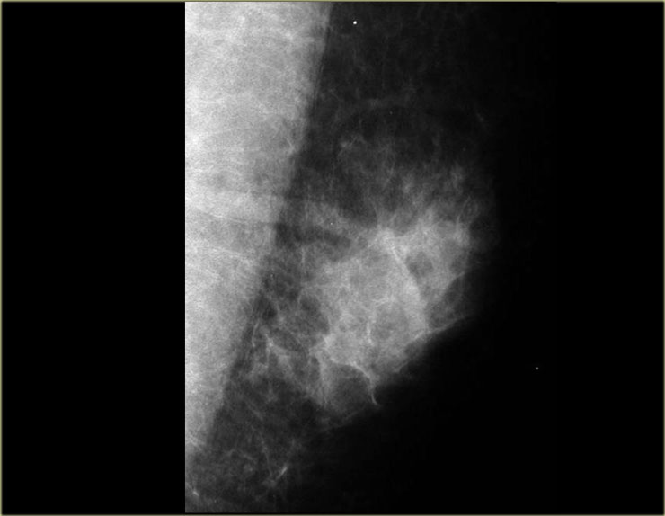 GynecomastiaNữ hóa tuyến vú
Gynecomastia is the most common abnormality in the male breast.
Clinically, it presents as a soft mobile tender subareolar mass.
Every word in this sentence is critical:
soft - mobile - tender - subareolar .
So it has to be soft and mobile.
It is tender in the acute phase, but not in the chronic phase.
Gynecomastia must be subareolar!
Any mass that is not subareolar is not gynecomastia.Vú to nam giới là bất thường phổ biến nhất ở vú nam giới.
Trên lâm sàng, nó biểu hiện dưới dạng một khối dưới da mềm, di động.
Mỗi từ trong câu này đều quan trọng:
mềm - di động - mềm - dưới ống sống.
Vì vậy, nó phải mềm mại và di động.
Nó đau trong giai đoạn cấp tính, nhưng không đau trong giai điệu mạn tính.
Vú to nam giới phải là vú dưới!
Bất kỳ khối nào không phải là dưới da thì không phải là vú to nam giới.
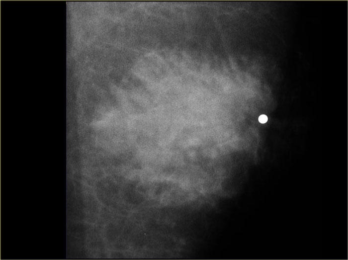 Gynecomastia: nodular glandular pattern.Notice how it blends into the surrounding fat.Vú to nam giới: mô hình tuyến nốt, chú ý nó hoà lẫn vào mỡ xung quanh như thế nào.
Imaging pattern
There are three imaging patterns:Hình ảnh mẫu
Có ba kiểu hình ảnh:
- Nodular glandular (acute florid phase)Nốt tuyến (giai đoạn hoa cấp tính)
- Dendritic (chronic fibrotic phase)Dendritic (giai đoạn xơ mãn tính)
- Diffuse glandular Tuỷ lan toả
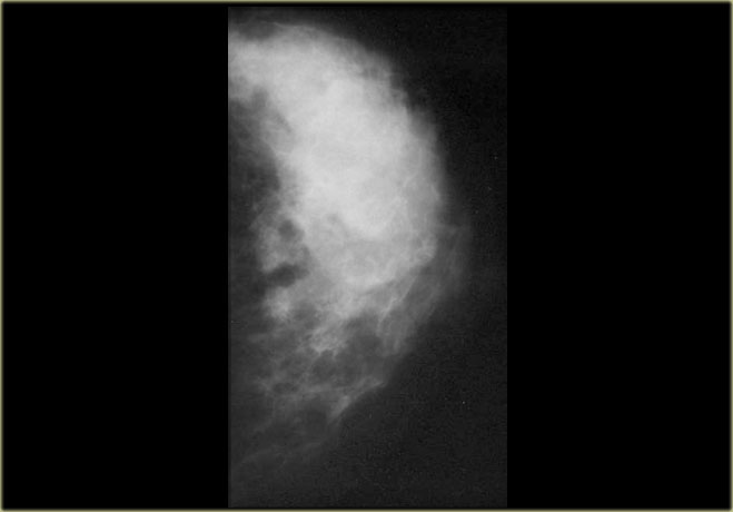
On the left a male breast with a nodular glandular pattern of gynecomastia.
There is a fan shaped density radiating from the nipple.
It may be more prominent in the upper outer quadrant and, more importantly, it blends into the surrounding fat.Bên trái là vú nam giới với mô hình tuyến vú dạng nốt của vú to nam giới.
Có một mật độ hình quạt phát ra từ núm vú.
Nó có thể nổi bật hơn ở góc phần tư trên ngoài và quan trọng hơn, nó trộn lẫn vào mỡ xung quanh.
If you think about the mammogram on the left as the breast of a woman instead of a man, than you might say that there is an ill-defined mass and you might conclude that this is a malignancy .
However, in a man this indistinct border is a sign of a gynecomastia.Nếu bạn nghĩ về chụp X-quang tuyến vú ở bên trái là vú của phụ nữ thay vì nam giới, thì bạn có thể nói rằng có một khối bờ không rõ và bạn có thể kết luận rằng đây là một khối ác tính.
Tuy nhiên, ở một người đàn ông, đường viền không rõ ràng này là dấu hiệu của chứng vú to ở nam giới.

Gynecomastia is simultaneous proliferation of ducts and stroma without encapsulation, so it must blend into the surrounding fat tissue.
There is no proliferation of lobuli, like there is in women.
So you will not see tumors that start in the lobuli, for example lactating adenomas, fibroadenomas, phyllodes tumors, and also invasive lobular carcinomas are extremely rare.Vú to nam giới là sự tăng sinh đồng thời của các ống dẫn và chất nền mà không có sự đóng gói, vì vậy nó phải hoà trộn vào mô mỡ xung quanh.
Không có sự tăng sinh của lobuli, giống như ở phụ nữ.
Vì vậy, bạn sẽ không thấy các khối u bắt đầu ở thuỳ, ví dụ như các u tuyến cho con bú, u xơ tuyến, u phyllodes và ung thư biểu mô thuỳ xâm lấn cũng cực kỳ hiếm gặp.
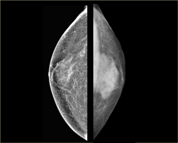 Left: gynecomastia in a young maleRight: Juvenile hypertrophy in an 8 year old femaleTrái: vú to ở nam giới trẻ tuổiPhải: phì đại vị thành niên ở nữ 8 tuổi
On the far left a mammogram of a male with gynecomastia and next to it a mammogram of an eight year old girl with juvenile hypertrophy.
Notice that they look very much the same.
Ở xa bên trái, một ảnh chụp tuyến vú của một người đàn ông bị vú to ở phụ nữ và bên cạnh đó là ảnh chụp tuyếnvú của một cô gái tám tuổi bị phì đại vị thành niên.
Lưu ý rằng chúng trông rất giống nhau.
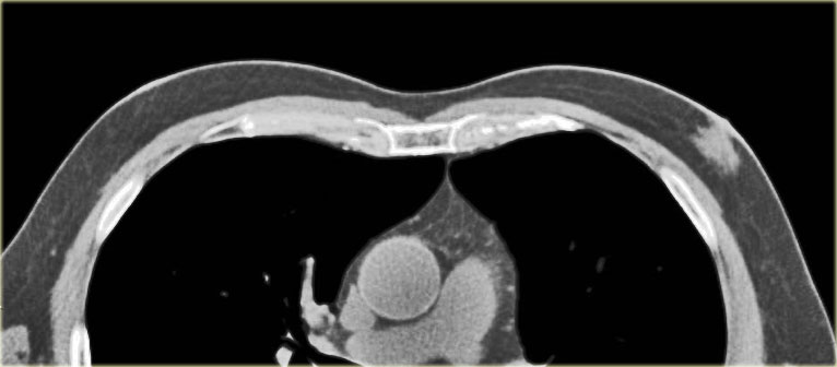 Gynecomastia nodular pattern: Incidental finding on CT-scanHình ảnh nốt vú to nam giới: phát hiện tình cờ trên CT-scan
On the left unilateral gynecomastia.
This was an incidental finding on a CT-scan done for some other reason.
Ở bên trái vú to nam giới.
Đây là một phát hiện tình cờ trên CT-scan được thực hiện vì một số lý do khác.
By definition gynecomastia is 2 cm or more of subareolar tissue in a non obese male.
It is a common 'normal' finding, that is seen in 55% of men at autopsy.
The peak incidence is 60 - 69 years.
It is significant if it is new or symptomatic.
In elderly males gynecomastia makes up 65% of all breast lesions.
25% is carcinoma and 10% are other lesions.Theo định nghĩa, vú to ở nam giới là vú to từ 2 cm trở lên so với mô dưới da ở nam giới không béo phì.
Đây là một phát hiện 'bình thường' phổ biến, được thấy ở 55% nam giới khi khám nghiệm tử thi.
Tỷ lệ mắc cao nhất là 60 - 69 tuổi.
Nó có ý nghĩa nếu nó là mới hoặc có triệu chứng.
Ở nam giới cao tuổi, vú to ở nam giới chiếm 65% tổng số các tổn thương vú.
25% là ung thư biểu mô và 10% là các tổn thương khác.
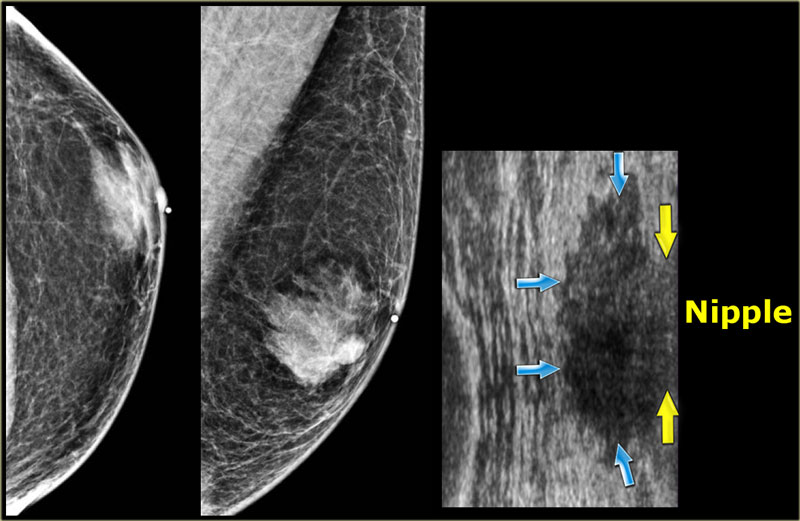
 Mammogran and rotated ultrasound imageHình ảnh siêu âm Mammogran và siêu âm xoay
Nodular patternHình dạng nốt
The nodular pattern of gynecomastia is seen in the florid early phase.
It begins as an increased number of ducts and epithelial proliferation with edema and cellular fibroblastic stroma.
This phase is reversible.
Dạng nốt của vú to ở giai đoạn sớm.
Nó bắt đầu như là sự gia tăng số lượng ống dẫn và tăng sinh biểu mô với phù nề và mô đệm sợi bào.
Giai đoạn này có thể đảo ngược.
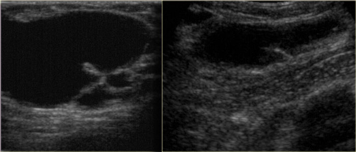
On the left a mammogram and an ultrasound image of a patient with a nodular glandular pattern of gynecomastia.
Notice that it is situated underneath the nipple.
The ultrasound image shows the typical appearance of gynecomastia: a hypoechoic mass with lobulation or even spiculation.
If this was seen in a woman, you would say that this is a mass with microlobulation and spiculation, i.e. Birads IV or V.
In a man this is typical for gynecomastia.
Bên trái là hình ảnh chụp nhũ ảnh và siêu âm của một bệnh nhân có mô hình tuyến vú dạng nốt.
Lưu ý rằng nó nằm bên dưới núm vú.
Hình ảnh siêu âm cho thấy sự xuất hiện điển hình của vú to nam giới: một khối giảm âm có thuỳ hoặc thậm chí có gai.
Nếu điều này đã được nhìn thấy ở một người phụ nữ, bạn sẽ nói rằng đây là một khối có vi nang và spiculation, tức là Birads IV hoặc V.
Ở nam giới, điều này là điển hình cho chứng vú to ở nam giới.
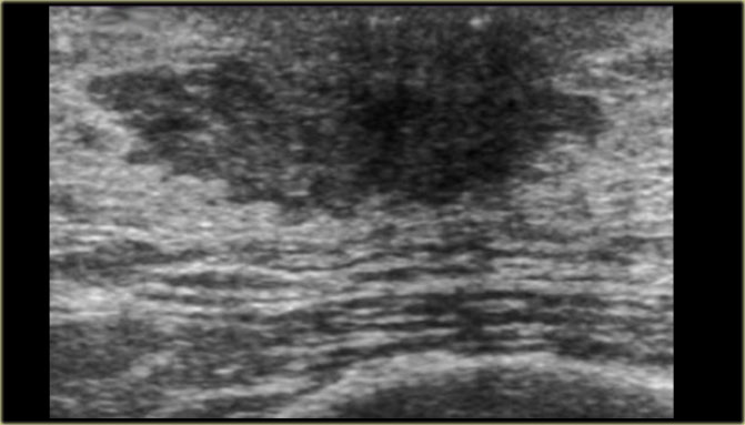
On the left the same ultrasound image, but now in the normal position.
Notice how 'malignant' it looks.Bên trái vẫn hình ảnh siêu âm, nhưng giờ ở vị trí bình thường.
Chú ý xem nó trông 'ác tính' như thế nào.
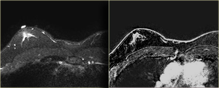 Gynecomastia nodular pattern: T2W-fatsat and T1WI+GdMô hình nốt vú to nam giới: T2W-fatsat và T1WI + Gd
On the left a T2W-image with fatsat and a T1W-image after Gadolinium with fatsat.
A radiologist who was not used to looking at 'male' mammograms ordered the MR for problem solving.
Obviously this MR was performed for the wrong reason.
MR should not be used to solve a problem that can be solved with mammography.
Anyhow the MR shows gynecomastia of the nodular pattern.
Bên trái là hình ảnh T2W với fatsat và hình ảnh T1W sau Gadolinium với fatsat.
Một bác sĩ X quang không quen nhìn vào ảnh chụp X-quang vú của nam giới đã yêu cầu chụp cộng hưởng từ để giải quyết vấn đề.
Rõ ràng là chụp cộng hưởng từ này được thực hiện không đúng lý do.
Không nên sử dụng MR để giải quyết vấn đề có thể giải quyết bằng chụp nhũ ảnh.
Tuy nhiên, MR cho thấy vú to ở dạng nốt.
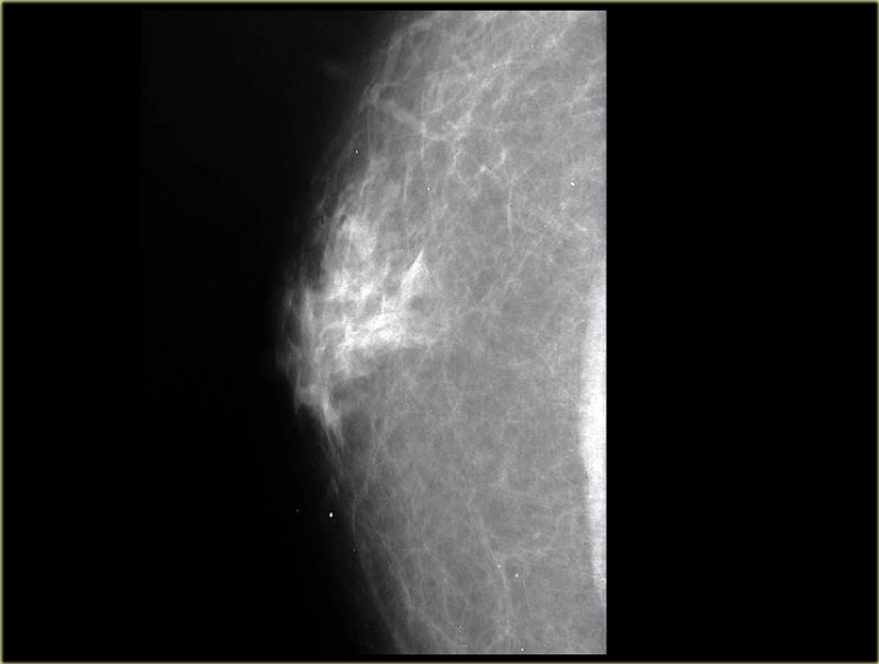 Dendritic pattern of gynecomastiaHình thái đuôi gai của vú to nam giới
Dendritic PatternMẫu hình đuôi gai
The dendritic pattern is seen in the fibrotic or late phase.
There are dilated ducts, moderate epithelial proliferation and fibrosis.
Dạng sợi nhánh được nhìn thấy trong giai đoạn xơ hóa hoặc giai đoạn muộn.
Có giãn ống dẫn, tăng sinh biểu mô vừa phải và xơ hóa.
On the left a mammogram showing a subareolar density with prominent extensions into the fat.
Usually the density is smaller than in the nodular pattern.Ở bên trái, một hình ảnh chụp nhũ ảnh cho thấy mật độ dưới xương cùng với các phần mở rộng nổi bật vào trong mỡ.
Thông thường, mật độ nhỏ hơn so với dạng nốt.
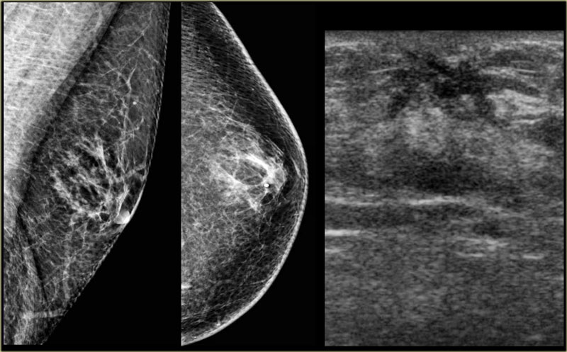 Classic benign chronic gynecomastiaVú to nam giới lành tính kinh điển
On the mammogram on the left we can imagine, that there is fibrosis with extension into the fat.
This is different from the glandular edema-like appearance in the acute phase of gynecomastia.
The ultrasound shows a spiculated appearance.Trên hình xạ hình vú bên trái, chúng ta có thể tưởng tượng, có sự xơ hóa với sự mở rộng vào trong mỡ.
Điều này khác với sự xuất hiện giống như phù nề tuyến trong giai đoạn cấp tính của vú to nam giới.
Siêu âm cho thấy hình dạng bờ tua gai.

These cases clearly demonstrate that gynecomastia can have an appearance which we would call malignant in a woman.
Unfortunately some of the malignant lesions in a man can look benign and we will show some examples in the next chapter.Những trường hợp này chứng minh rõ ràng rằng vú to ở nữ giới có thể có một ngoại hình mà chúng ta gọi là ác tính ở phụ nữ.
Thật không may, một số tổn thương ác tính ở người có thể trông lành tính và chúng tôi sẽ cho một số ví dụ trong chương tiếp theo.
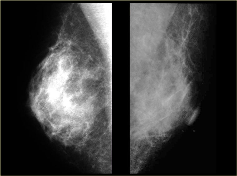
Diffuse glandular patternKiểu tuyến khuếch tán
This pattern is seen in males with very high estrogen levels.Mô hình này được thấy ở nam giới có nồng độ estrogen rất cao.
The images on the left simply look like small female breasts.
This was a patient who was on estrogen therapy for prostate carcinoma.Hình ảnh bên trái đơn giản trông giống như ngực phụ nữ nhỏ.
Đây là một bệnh nhân đang điều trị ung thư tuyến tiền liệt bằng estrogen.
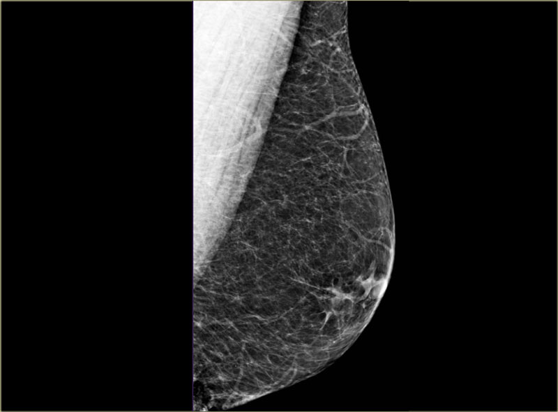 PseudogynecomastiaGiả nữ hóa tuyến vú
This is usually bilateral and there is no palpable mass.
Remember that gynecomastia presents clinically as a soft, mobile, tender, subareolar mass.
Pseudogynecomastia results from excessive fat deposition in the breast area.
It is seen as a normal variant, in obesity and in neurofibromatosis.Thường là hai bên và không có khối sờ thấy.
Hãy nhớ rằng vú to ở nam giới biểu hiện lâm sàng như một khối mềm, di động, mềm, ở dưới da.
Giả vú to do sự lắng đọng quá nhiều chất béo ở vùng vú.
Nó được xem như là một biến thể bình thường, trong béo phì và trong u sợi thần kinh.
Benign LesionsTổn thương lành tính
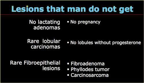
Let's first start with a list of lesions that should not be diagnosed in male patients, because they simply do not get these lesions.
A man does not get a lactating adenoma, because it is only seen in pregnancy.
Because there are very few lobules in a man, lobular tumors are extremely rare.
There are only a few invasive lobular carcinomas reported in men.
Fibroepithelial lesions are also extremely rare because they too start in the lobules.
So do not diagnose a fibroadenoma in a man, even if it looks like a fibroadenoma.
When you get a biopsy result that says fibroadenoma, get another pathologist.
Trước tiên, hãy bắt đầu với một danh sách các tổn thương không nên chẩn đoán ở bệnh nhân nam, bởi vì họ đơn giản là không có những tổn thương này.
Một người đàn ông không được cho con bú u tuyến, bởi vì nó chỉ được thấy trong thai kỳ.
Bởi vì có rất ít thuỳ ở nam giới, các khối u thuỳ là cực kỳ hiếm.
Chỉ có một vài trường hợp ung thư biểu mô thuỳ xâm lấn được báo cáo ở nam giới.
Các tổn thương dạng sợi cũng cực kỳ hiếm vì chúng cũng bắt đầu ở thuỳ.
Vì vậy, không chẩn đoán u xơ tuyến ở nam giới, ngay cả khi nó trông giống như u xơ.
Khi bạn nhận được kết quả sinh thiết cho biết u xơ tuyến, hãy tìm một nhà giải phẫu bệnh khác.
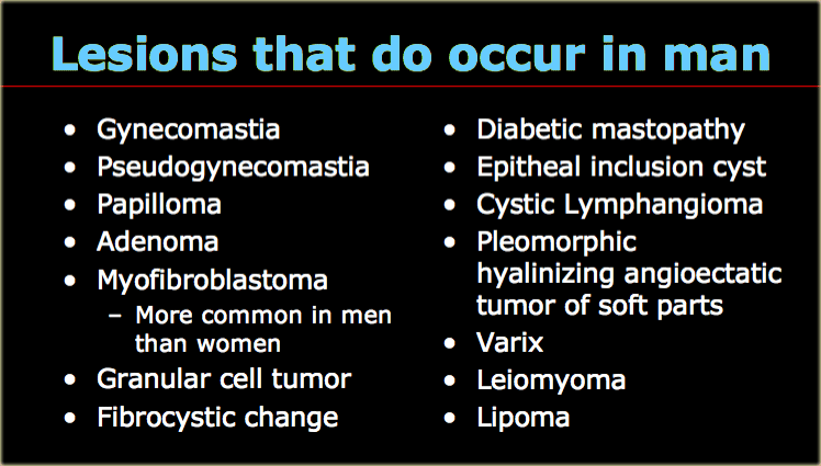
On the left lesions that do occur in males.
Exept for gynecomastia and pseudogynecomastia, in most of these lesions we will not get a diagnosis from imaging.
We just report that there is a Birads IV lesion and do a biopsy.
Ở bên trái các tổn thương xảy ra ở nam giới.
Đối với vú to nam giới và giả vú to nữ giới, trong hầu hết các tổn thương này chúng ta sẽ không có được chẩn đoán từ chẩn trị hình ảnh.
Chúng tôi chỉ báo cáo có tổn thương Birads IV và làm sinh thiết.
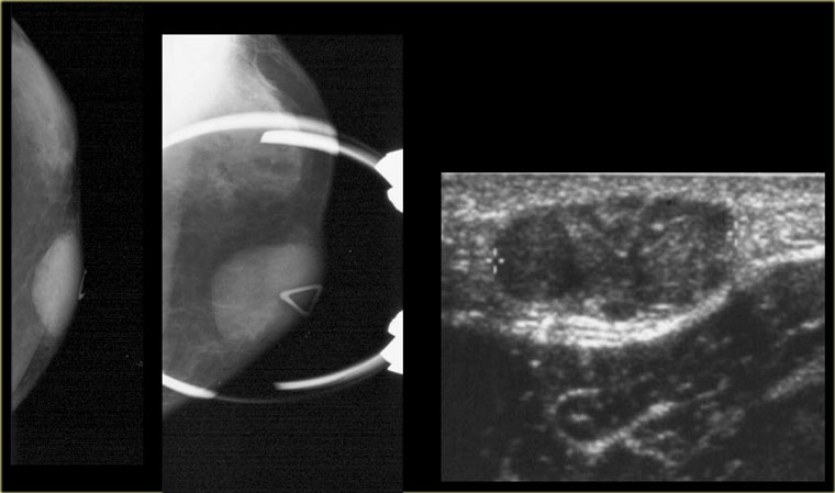 MyofibroblastomaU nguyên bào sợi cơ
Myofibroblastoma is an interesting lesion because it is the only one lesion that is more common in men than in women.
It presents as a freely moveable, solitary, palpable, firm mass.
There are no calcifications.
The mean age is in the late 50's.
U sợi cơ là một tổn thương thú vị vì nó là tổn thương duy nhất phổ biến ở nam giới hơn ở nữ giới.
Nó biểu hiện dưới dạng một khối vững chắc, có thể di chuyển tự do, đơn độc, sờ thấy được.
Không có vôi hóa.
Tuổi trung bình là cuối 50 tuổi.
On the left a large lesion, that looks like a fibroadenoma.
Now we know that men do not get fibroadenomas.
The pathology diagnosis was myofibroblastoma and the lesion was treated with local excision.Bên trái một tổn thương lớn, trông giống như một u xơ tuyến.
Bây giờ chúng ta biết rằng nam giới không bị u xơ tuyến.
Bệnh lý chẩn đoán là u nguyên bào sợi cơ và được điều trị bằng phẫu thuật cắt bỏ u tại chỗ.
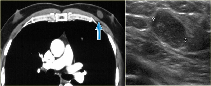 Myofibroblastoma eccentric to the nipple in a patient with a little bit of gynecomastia bilaterallyU sợi cơ lệch tâm đến núm vú ở bệnh nhân vú to nam giới hai bên
On the left another myofibroblastoma.
It presents as a circumscribed lobulated mass without calcification.
Notice that the lesion is eccentric to the nipple.
It is the lobulated mass that needs to be biopsied, not the retromamillary gynecomastia.
Bên trái là một u nguyên bào sợi cơ khác.
Nó biểu hiện dưới dạng khối có phân thuỳ không có vôi hóa. Lưu ý rằng tổn thương lệch tâm với núm vú, đó là khối có phân luồng cần được sinh thiết, không phải vú to nam giới sau vú.
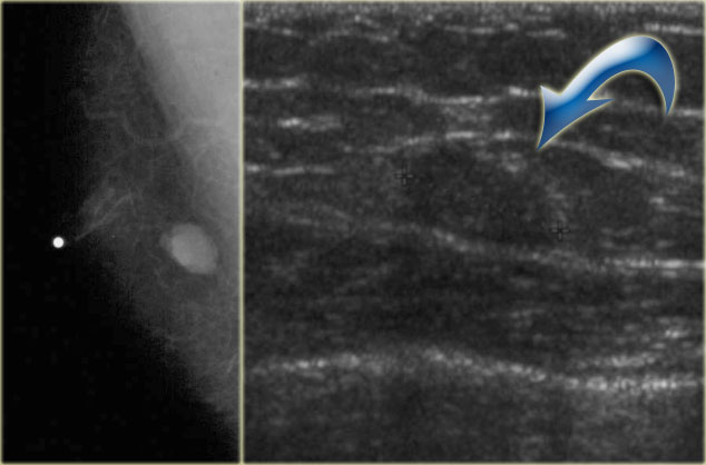 Myofibroblastoma eccentric to the nipple U nguyên bào sợi cơ lệch tâm đến núm vú
On the left another myofibroblastoma.
The nipple is marked and the lesion is not retromamillary.
On the ultrasound image the lesion is difficult to differentiate from the surrounding fat.
Bên trái là một u nguyên bào sợi cơ khác.
Núm vú được đánh dấu và tổn thương không phải là hậu môn.
Trên siêu âm, tổn thương khó phân biệt với mỡ xung quanh.
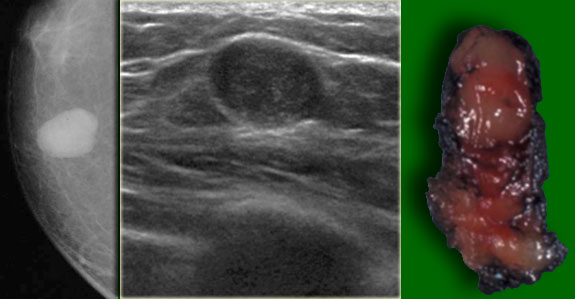 Myofibroblastoma presenting as a circumscribed lobulated mass without calcificationU nguyên bào sợi cơ biểu hiện dưới dạng khối phân thuỳ bao quanh không có vôi hóa
On the left another myofibroblastoma.
Even if this lesion was located behind the nipple, you would not diagnose this as gynecomastia, because it is a lobulated mass.Bên trái là một u nguyên bào sợi cơ khác.
Ngay cả khi tổn thương này nằm phía sau núm vú, bạn sẽ không chẩn đoán đây là vú to do nam giới, bởi vì nó là một khối có nhiều thuỳ.
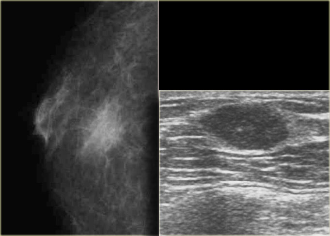 Granular Cell TumorU tế bào hạt
This is a benign tumor of neural origin.
They occur anywhere in the body.
6% occur in the breasts.
They are typically seen in men in their 30's.
Usually they present as circumscribed in males, but sometimes they have a spiculated appearance.
Đây là một khối u lành tính có nguồn gốc thần kinh.
Chúng xuất hiện ở bất cứ đâu trong cơ thể.
6% xảy ra ở vú.
Chúng thường thấy ở nam giới ở độ tuổi 30.
Thông thường chúng xuất hiện như được định nghĩa ở nam giới, nhưng đôi khi chúng có hình dạng bờ tua gai.
Notice that the lesion on the left has a indistinct border as is usually seen in gynecomastia, but on the mammogram it is not located directly under the skin.
So this is not gynecomastia and a biopsy is necessary.Lưu ý rằng tổn thương ở bên trái có đường viền không rõ ràng như thường thấy trong vú to ở phụ nữ, nhưng trên ảnh chụp X-quang tuyến vú, nó không nằm trực tiếp dưới da.
Vì vậy, đây không phải là vú to nam giới và sinh thiết là cần thiết.
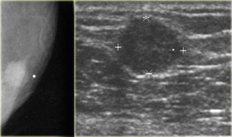 Granular Cell TumorU tế bào hạt
On the left another Granular cell tumor. Bên trái một khối u tế bào hạt khác.
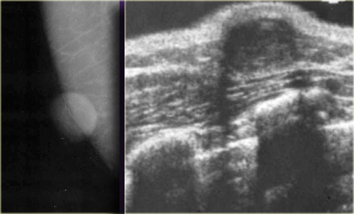 Epidermal inclusion cystNang bao biểu bì
Epidermal inclusion cyst is a skin lesion.
It presents as a round well circumscribed dense mass.
On the left a small epidermal inclusion cyst.
Notice how it raises the skin.Nang bao biểu bì là một tổn thương da, biểu hiện dưới dạng một khối tròn dày đặc bao quanh.
Bên trái là một nang biểu bì nhỏ, chú ý nó làm nổi da như thế nào.
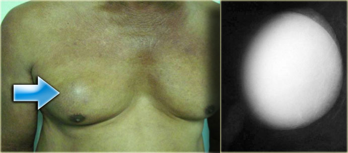 Epidermal inclusion cystNang bao biểu bì
On the left a large epidermal inclusion cyst.Bên trái là một nang biểu bì lớn.
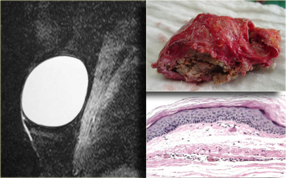 Epidermal inclusion cyst. T2WI with fat sat and pathologyNang biểu bì T2WI có sat mỡ và giải phẫu bệnh
On the left a T2W image demonstrating the cystic nature and the pathology specimen.Bên trái là hình ảnh T2W thể hiện bản chất nang và bệnh phẩm.
 Granulomatous MastitisViêm vú do u hạt
Most are idiopathic.
Specific causes must be excluded like TB, Sarcoid and fat necrosis.
On the left is a male breast that looks like the breast of a female.
These lesions can sometimes be spiculated.Hầu hết là vô căn.
Các nguyên nhân đặc hiệu phải được loại trừ như lao, Sarcoid và hoại tử mỡ.
Bên trái là vú nam trông giống như vú nữ.
Những tổn thương này đôi khi có thể là bờ tua gai.
 VarixViet Nam
On the left a lesion that looks like a cyst, but remember that cysts originate in the lobules and men do not have lobules, unless they take estrogen.
This is a varix and if you puncture it, you get a big red surprise.Ở bên trái, một tổn thương trông giống như một u nang, nhưng hãy nhớ rằng u nang bắt nguồn từ các tiểu thuỳ và nam giới không có tiểu thuỳ, trừ khi họ dùng estrogen.
Đây là một con varix và nếu bạn đâm nó, bạn sẽ có một bất ngờ lớn màu đỏ.
 LeiomyomaU cơ trơn
On the left a lesion, that looks like a fibroadenoma, but men do not get fibroadenomas.
It is a solid encapsulated mass and at biopsy it happened to be a leiomyoma.
If there are more than 2 mitoses per high power field the pathologist calls it a leiomyosarcoma.Ở bên trái, một tổn thương, trông giống như một u xơ tuyến, nhưng nam giới không bị u xơ.
Đó là một khối u đặc, khi sinh thiết nó là một u cơ trơn.
Nếu có hơn 2 ty thể trên một trường năng lượng cao, nhà giải phẫu bệnh gọi đó là sarcoma cơ trơn.
Male Breast CancerUng thư vú nam giới
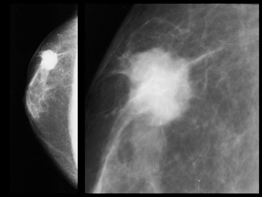 Invasive ductal carcinomaUng thư biểu mô ống xâm nhập
Malignant disease in men just looks like malignant disease in women.
In the USA there are about 1700 new cases each year, which is 1% of all breast cancer.
There is a higher incidence in people from China and Africa due to hyperestrogenism secondary to parasitic liver disease.
These cancers present as a unilateral painless subareolar mass.
This subareolar location is just like in gynecomastia, but usually it is eccentric to the nipple.
It sometimes presents with bloody nipple discharge, which is unlike gynecomastia.
Usually it is invasive ductal cancer.
As stated above invasive lobular cancer is extremely rare.
Also DCIS is rare because there is no screening program for men, so they will present when there is a palpable mass.Bệnh ác tính ở nam giới chỉ giống như bệnh ác tính trong phụ nữ.
Tại Hoa Kỳ có khoảng 1700 trường hợp mới mỗi năm, chiếm 1% tổng số ung thư vú.
Có tỷ lệ mắc bệnh cao hơn ở những người từ Trung Quốc và Châu Phi do chứng cường estrogen thứ phát do bệnh gan ký sinh trùng.
Những khối ung thư này biểu hiện dưới dạng một khối dưới da không đau một bên.
Vị trí dưới âm đạo này giống như trong vú to, nhưng thường lệch tâm so với núm vú.
Đôi khi xuất hiện tiết dịch núm vú có máu, không giống như vú to ở phụ nữ.
Thông thường đó là ung thư ống xâm lấn.
Như đã nêu ở trên, ung thư thuỳ xâm lấn là cực kỳ hiếm.
Ngoài ra, DCIS hiếm gặp vì không có chương trình sàng lọc cho nam giới, vì vậy họ sẽ xuất hiện khi có khối sờ thấy được.
On the left an eccentric irregular mass with spiculae.
If this was a women you would have no trouble in diagnosing this as a cancer.
In a man it is the same.Bên trái là một khối bất thường lệch tâm có gai.
Nếu đây là một phụ nữ bạn sẽ không gặp khó khăn trong việc chẩn đoán đây là một bệnh ung thư. Ở một người đàn ông thì cũng vậy.
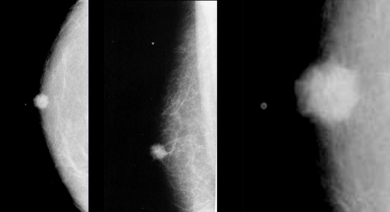 Invasive ductal carcinomaUng thư biểu mô ống xâm nhập
Male breast cancer presents as a round, oval or irregular mass.
Calcifications are rare, but when they occur they are coarser than in women.Ung thư vú ở nam giới có dạng khối tròn, bầu dục hoặc không đều.
vôi hóa rất hiếm, nhưng khi chúng xuất hiện, chúng thô hơn ở phụ nữ.
On the left a small invasive ductal carcinoma.
It is subareolar and central, but it is also encapsulated.
This is not gynecomastia.Bên trái là một ung thư biểu mô ống xâm nhập nhỏ.
Nó là subareolar và trung tâm, nhưng nó cũng được đóng gói.
Đây không phải là vú to nam giới.
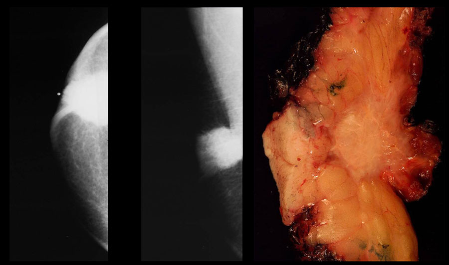 Invasive ductal carcinomaUng thư biểu mô ống xâm nhập
Paget's disease of the nipple and skin ulceration are more common than in women.Bệnh Paget ở núm vú và loét da thường gặp hơn ở phụ nữ.
On the left an invasive ductal carcinoma with skin retraction.Bên trái là ung thư biểu mô ống xâm nhập có da co rút.
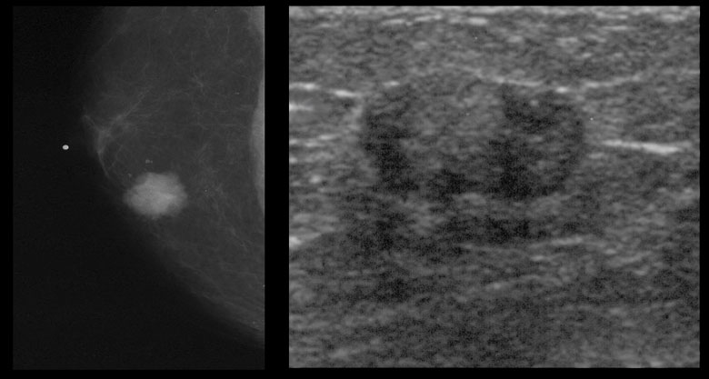 Invasive ductal carcinomaUng thư biểu mô ống xâm nhập
There is a long list of carcinoma risk factors and they are the same as in women: Có một danh sách dài các yếu tố nguy cơ ung thư biểu mô và chúng giống như ở phụ nữ:
- Advanced ageTuổi cao
- Family historyTiền sử gia đình
- Jewish heritageDi sản Do Thái
- Chest wall irradiationXạ trị thành ngực
- HyperestrogenismCường estrogen
- HyperthyroidismCường giáp
- Exposure to hepatotoxinsPhơi nhiễm với độc tố gan
- Occupational exposure to high heat (steel industry)Tiếp xúc nghề nghiệp với nhiệt độ cao (ngành thép)
- BRCA 2 is seen in 4 - 16% of male breast cancer patients (40% in Iceland)BRCA 2 gặp ở 4- 16% bệnh nhân ung thư vú nam (40% ở Iceland)
- Undescended testesTinh hoàn lạc chỗ
- Orchiectomy and orchitisCắt tinh hoàn và viêm tinh hoàn
- Klinefelter's syndrome (47, XXY - 6% of all male breast cancer is in Klinefelter - 3% lifetime risk)Hội chứng Klinefelter (47, XXY - 6% ung thư vú nam giới thuộc Klinefelte - 3% nguy cơ suốt đời)
On the left a small eccentric encasulated invasive ductal carcinoma.Bên trái là một ung thư biểu mô ống xâm nhập nhỏ, lập dị.
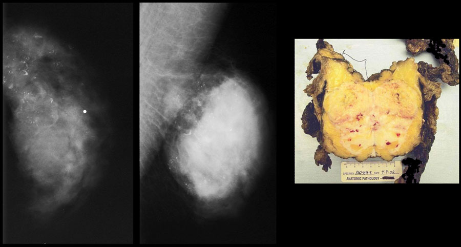 Invasive ductal carcinomaUng thư biểu mô ống xâm nhập
On the left a huge invasive ductal carcinoma with some coarse benign looking calcifications.Bên trái là một ung thư biểu mô ống xâm nhập lớn với một số vôi hóa lành tính thô.
Other malignancies of the male breastCác bệnh lý ác tính khác ở vú nam giới
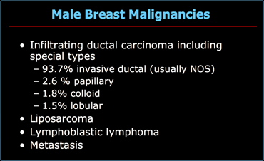
Malignancies other than ductal carcinoma are uncommon.
On the left a list of all malignancies in men.Các bệnh lý ác tính khác ngoài ung thư biểu mô ống thận không phổ biến.
Bên trái là danh sách tất cả các khối u ác tính ở nam giới.
MetastasesDi căn
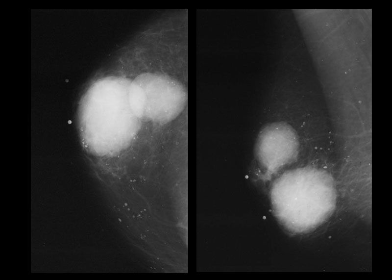
Metastases from prostate cancer are the most common metastases in males.
It results from hematogenous spread and is usually seen in patients with widespread disease.
It presents as round or lobulated non-calcified masses.Di căn từ ung thư tuyến tiền liệt là di căn phổ biến nhất ở nam giới.
Nó là kết quả của sự lây lan theo đường máu và thường thấy ở những bệnh nhân bị bệnh lan rộng.
Nó biểu hiện dưới dạng các khối tròn hoặc các khối không vôi hóa.
On the left a patient with two metastases of a small cell lung carcinoma.Bên trái là một bệnh nhân ung thư phổi tế bào nhỏ có hai di căn.
 LiposarcomaSarcôm mỡ
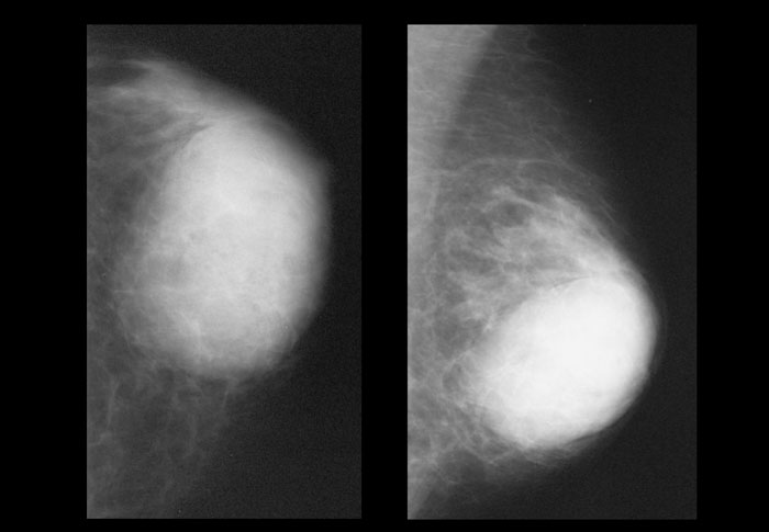
A liposarcoma is a rare sarcoma.
It presents as a slowly enlarging painful mass.
It is usually of water-density and is not typically fatty.Sarcom mỡ là một loại sarcom hiếm gặp.
Nó biểu hiện như một khối đau từ từ to lên.
Nó thường có mật độ nước và thường không béo.
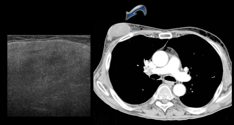
On the CT on the left you can see the density of the lesion that proved to be a liposarcoma.
Trên CT bên trái, bạn có thể thấy mật độ của tổn thương được chứng minh là một liposarcoma.
ConclusionKết luận
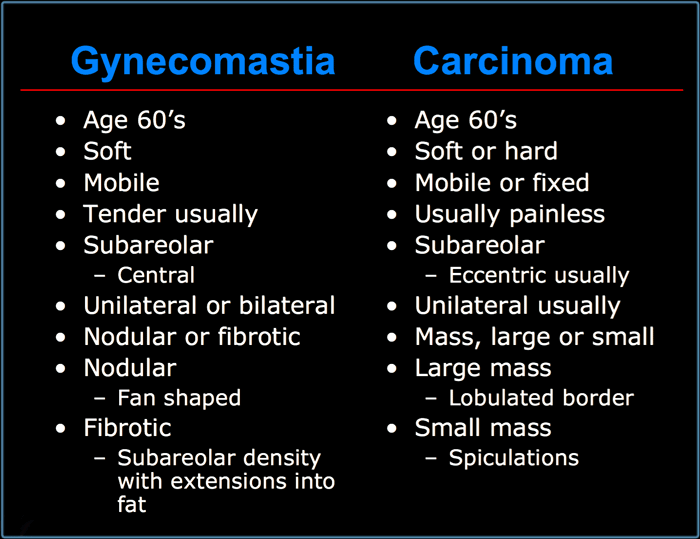
Gynecomastia versus CarcinomaVú to nam giới so với ung thư biểu mô
In conclusion we can say, that male breast disease either presents as mass, pain or nipple discharge.
Gynecomastia and invasive ductal cancer are the most common lesions in the male breast, but there are other rarer benign and malignant lesions.
Gynecomastia and carcinoma can usually be differentiated, but biopsy is sometimes necessary to separate them.
All lesions eccentric to the nipple need biopsy unless they are characteristically benign, i.e.contain fat or typical lymph node.
Kết luận: Bệnh vú ở nam giới có thể biểu hiện bằng khối u, đau hoặc tiết dịch núm vú.
Vú to nam giới và ung thư ống tuyến xâm lấn là những tổn thương phổ biến nhất ở vú nam giới, nhưng có những tổn thương lành tính và ác tính hiếm gặp khác.
Vú to nam giới và ung thư biểu mô thường có thể được phân biệt, nhưng đôi khi cần phải sinh thiết để tách chúng.
Tất cả các tổn thương lệch tâm đến núm vú cần sinh thiết trừ khi chúng đặc trưng lành tính, tức là chứa chất béo hoặc hạch bạch huyết điển hình.
On the left a list of characteristics of gynecomastia versus carcinoma.
Notice that there are many similarities.
Both gynecomastia and carcinoma occur mostly at the age of 60 and can be soft, mobile, subareolar and unilateral.
So that does not help.
Carcinoma is usually eccentric, while gynecomastia is never eccentric.
Gynecomastia has to have extensions into the surroundig fat.
Carcinoma sometimes may have spiculations, that can look the same.
Actually we call it extension into the fat, if we think it is gynecomastia and spiculation, if we think it is a carcinoma.
Bên trái là danh sách các đặc điểm của vú to nam giới so với ung thư biểu mô.
Lưu ý rằng có nhiều điểm tương đồng.
Cả vú to và ung thư biểu mô xảy ra chủ yếu ở tuổi 60 và có thể mềm, di động, dưới da và một bên.
Vì vậy, điều đó không giúp ích gì.
Ung thư biểu mô thường lệch tâm, trong khi vú to nam giới không bao giờ lệch tâm.
Vú to nam giới phải có phần mở rộng vào lớp mỡ bao quanh.
Ung thư biểu mô đôi khi có thể có các gai, có thể trông giống nhau.
Trên thực tế chúng tôi gọi nó là sự mở rộng vào trong mỡ, nếu chúng tôi nghĩ rằng đó là vú to ở phụ nữ và sự nhô ra, nếu chúng ta nghĩ rằng đó một ung thư biểu mô.
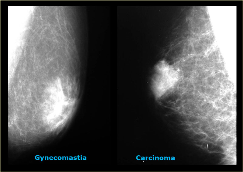

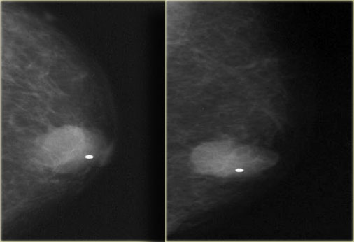
On the left two cases, that demonstrate, that it can be difficult to differentiate gynecomastia from carcinoma on a mammogram.
The carcinoma on the right is a little bit more encapsulated than the gynecomastia on the right.
In less than 10% of the cases a biopsy can be needed to make the differentiation.Ở bên trái hai trường hợp, chứng minh, rằng có thể khó phân biệt vú to nam giới với ung thư biểu mô trên chụp X-quang tuyến vú.
Ung thư biểu mô bên phải được bọc nhiều hơn một chút so với vú to ở bên phải.
Trong ít hơn 10% trường hợp, có thể cần sinh thiết để phân biệt.
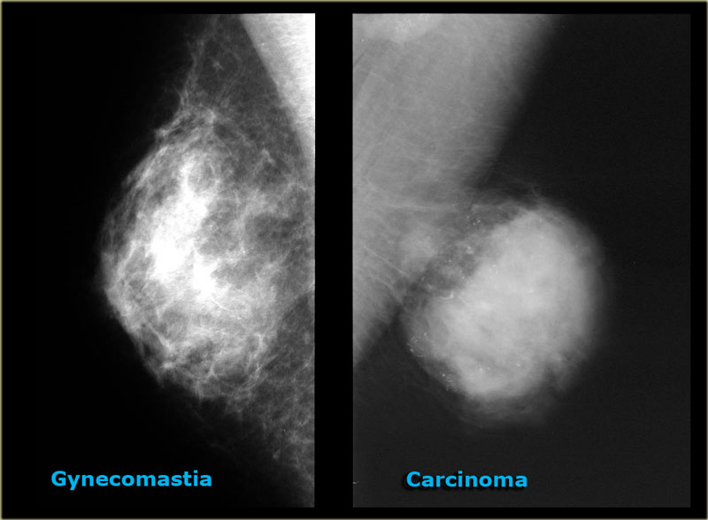

On the left two more cases.
On the far left there is diffuse gynecomastia.
On the right a huge cancer which is encapsulated.Bên trái hai trường hợp nữa.
Ở phía ngoài cùng bên trái có sự lan tỏa lan tỏa.
Bên phải một căn bệnh ung thư khổng lồ được gói gọn.
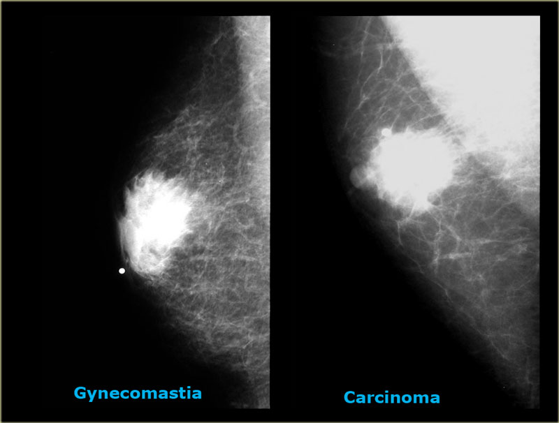

The last cases on the left look very similar to each other.
Based on the mammogram these two can not be differentiated.
In those rare instances a biopsy is needed.Các trường hợp cuối cùng bên trái nhìn rất giống nhau.
Dựa trên chụp X-quang tuyến vú, hai loại này không thể phân biệt được.
Trong những trường hợp hiếm gặp đó, cần phải sinh thiết.