
Pulmonary Hypertension and Thromboembolic diseaseTăng áp phổi và bệnh lý huyết khối thuyên tắc
¹University Medical Center Amsterdam and ²Alrijne Hospital in Leiden, the Netherlands1Trung tâm Y khoa Đại học Amsterdam và Bệnh viện 2Alrijne ở Leiden, Hà Lan
Ngày đăng / cập nhật: 20-07-2025
Bản gốc trên radiologyassistant.nl ↗ · tải về lúc 2026-11-19 20:19 · bản dịch máy có chuẩn hóa thuật ngữ chẩn đoán hình ảnh
Pulmonary hypertension (PH) is characterized by elevated blood pressure within the pulmonary arteries, which increases the workload for the right ventricle (RV).
RV failure is the leading cause of morbidity and mortality in these patients. Diagnosis can be challenging, as symptoms often develop gradually and are non-specific (dyspnea, fatigue,
syncope, chest pain, leg edema, or ascites).
Definitive diagnosis requires right heart catheterization—an invasive procedure that is not typically performed in the early stages of the disease.Tăng áp phổi (PH) được đặc trưng bởi tăng huyết áp trong động mạch phổi, làm tăng khối lượng công việc cho tâm thất phải (RV).
Suy thất phải là nguyên nhân hàng đầu gây bệnh tật và tử vong ở những bệnh nhân này. Chẩn đoán có thể là một thách thức, vì các triệu chứng thường phát triển dần dần và không đặc hiệu (khó thở, mệt mỏi, ngất, đau ngực, phù chân, hoặc cổ trướng).
Chẩn đoán xác định cần phải đặt ống thông tim phải - một thủ thuật xâm lấn thường không được thực hiện trong giai đoạn đầu của bệnh.
The radiologist can play a key role in identifying imaging signs of pulmonary hypertension.
Specifically, the radiologist should:Bác sĩ chẩn đoán hình ảnh có thể đóng vai trò quan trọng trong việc xác định các dấu hiệu hình ảnh của tăng áp phổi.
Cụ thể, bác sĩ X-quang nên:
- Recognize imaging features of PH and associated cardiac remodelling.Nhận biết các đặc điểm hình ảnh của PH và tái cấu trúc tim liên quan.
- Identify congenital cardiovascular anomalies and other thoracic pathologies that may suggest a specific underlying etiology of PH.Xác định các dị tật tim mạch bẩm sinh và các bệnh lý lồng ngực khác có thể gợi ý nguyên nhân cơ bản cụ thể của PH.
- Detect imaging signs of chronic thromboembolic pulmonary hypertension (CTEPH), the only potentially curable form of PH.Phát hiện các dấu hiệu hình ảnh của tăng áp phổi (CTEPH) thuyên tắc huyết khối mạn tính, dạng PH duy nhất có khả năng chữa khỏi.
We thank Prof.dr.
Harm Jan Bogaard for his comments on the manuscript draft.Chúng tôi xin cảm ơn GS.TS Harm Jan Bogaard đã góp ý kiến về bản thảo.
Role of RadiologyVai trò của X quang

Steps in diagnosing Pulmonary HypertensionCác bước chẩn đoán tăng áp phổi
- Evaluate imaging studies for signs suggestive of pulmonary hypertension (PH).
Do not disregard incidental imaging findings that may indicate the presence of PH.Đánh giá các nghiên cứu hình ảnh cho các dấu hiệu gợi ý tăng áp động mạch phổi (PH).
Không bỏ qua các phát hiện hình ảnh ngẫu nhiên có thể cho thấy sự hiện diện của pH. - Subsequently, assess for imaging features that may help determine the underlying etiology of PH. These may include cardiovascular abnormalities, pulmonary parenchymal disease, or evidence of chronic thromboembolic pulmonary hypertension (CTEPH), and can guide the selection of further diagnostic imaging modalities and referral pathways.Sau đó, đánh giá các đặc điểm hình ảnh có thể giúp xác định nguyên nhân cơ bản của PH. Chúng có thể bao gồm các bất thường về tim mạch, bệnh nhu mô phổi hoặc bằng chứng về huyết khối tăng áp phổi mạn tính (CTEPH) và có thể hướng dẫn lựa chọn các phương thức chẩn đoán hình ảnh và các con đường chuyển tuyến.
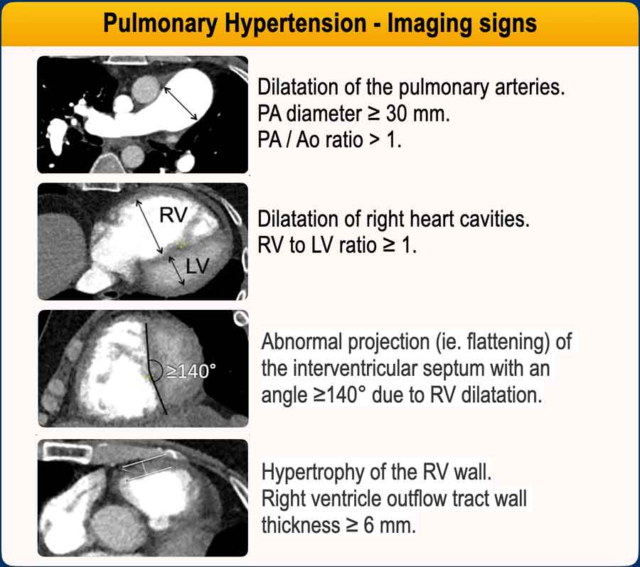
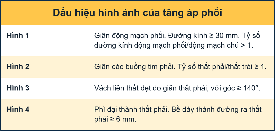
Imaging features of PHĐặc điểm hình ảnh của bệnh phình động mạch phổi
The following imaging findings on CT indicate a high resistance and high pressure in the pulmonary vascular bed, suggestive of PH:
Các dấu hiệu hình ảnh sau đây trên CT cho thấy sức cản và áp lực cao ở giường mạch phổi, gợi ý PH:
- Main pulmonary artery (MPA) enlargement
MPA diameter >30 mm.
MPA/Ao ratio >1.
Phì đại động mạch phổi chính (MPA)
Đường kính MPA > 30 mm.
Tỷ số MPA / Ao > 1. - Right ventricular dilatation
RV/LV ratio >1 on axial images.Giãn thất phải
Tỉ số RV / LV > 1 trên ảnh mặt phẳng ngang. - Flattened or bowed interventricular septum
Abnormal projection (ie. flattening) of the interventricular septum with an angle ≥140° on a short axis view.Vách liên thất dẹt hoặc lõm
Hình chiếu bất thường (tức là phẳng) của vách liên thất với góc ≥ 140 ° trên chế độ xem trục ngắn. - Hypertrophy of RV wall
Right ventricle outflow tract wall thickness ≥ 6 mm.
Phì đại thành thất phải
Độ dày thành đường ra thất phải ≥ 6 mm.
Additional findings include:Các phát hiện bổ sung bao gồm:
- Dilated right atrium, which is often seen in chronic pressure elevation.Tâm nhĩ phải giãn, thường thấy trong tăng áp lực mạn tính.
- Dilatation of the inferior vena cava and hepatic veins, due to elevated right-sided pressures.Giãn tĩnh mạch tĩnh mạch chủ dưới và tĩnh mạch gan, do tăng áp lực bên phải.
- Segmental artery-to-bronchus ratio >1, especially in the upper lobes.Tỷ lệ động mạch / phế quản đoạn > 1, đặc biệt ở thuỳ trên.
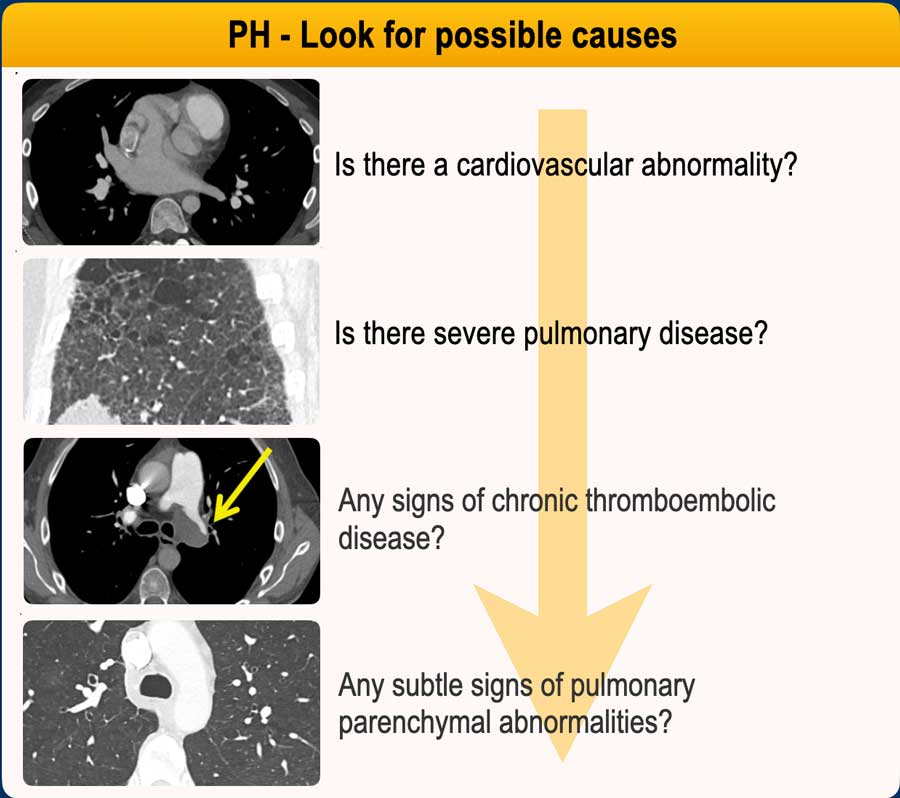

Possible causes of PHNguyên nhân có thể gây ra PH
When there is a suspicion of PH, the next step is to look for features that help identify the cause and guide further imaging.Khi có nghi ngờ về PH, bước tiếp theo là tìm kiếm các đặc điểm giúp xác định nguyên nhân và hướng dẫn chẩn đoán hình ảnh thêm.
Radiologic approach to identify PH etiology:X quang để xác định nguyên nhân PH:
- Cardiac causes?
Check for congenital defects (e.g., atrial septal defect (ASD), partial anomalous pulmonary venous return (PAPVR), valvular disease, or pericardial thickening or calcification).
Nguyên nhân tim?
Kiểm tra các khuyết tật bẩm sinh (ví dụ, thông liên nhĩ (ASD), bất thường tĩnh mạch phổi trở lại một phần (PAPVR), bệnh van tim, hoặc dày màng ngoài tim hoặc vôi hóa). - Severe lung disease?
Look for extensive fibrosis or emphysema.Bệnh phổi nặng?
Tìm dấu hiệu xơ hóa lan toả hoặc khí phế thũng. - Chronic thromboembolic disease?
Look for filling defects, vessel caliber changes, and/or mosaic attenuation.
PH results from unresolved blood clots that scar and block pulmonary arteries. Identification is important, because chronic emboli can be treated surgically (PEA) or with balloon angioplasty (BPA), making it potentially curable.Bệnh thuyên tắc huyết khối mạn tính?
Tìm các khuyết tật khi đổ đầy, thay đổi kích thước mạch máu và / hoặc tỷ trọng khảm.
PH là kết quả của cục máu đông chưa được giải quyết, sẹo và tắc nghẽn động mạch phổi. Xác định là rất quan trọng, bởi vì tắc mạch mạn tính có thể được điều trị bằng phẫu thuật (PEA) hoặc với nong bóng (BPA), làm cho nó có khả năng chữa khỏi. - Subtle parenchymal signs of pulmonary arterial hypertension (PAH) or pulmonary veno-occlusive disease (PVOD)?
Look for centrilobular ground-glass opacities, smooth interlobular septal thickening, or a dilated main pulmonary artery.Các dấu hiệu nhu mô tế bào của tăng áp động mạch phổi (PAH) hoặc bệnh tắc tĩnh mạch phổi?
Tìm trung tâm tiểu thùy đám mờ kính mờ, dày vách liên tiểu thùy nhẵn, hoặc động mạch phổi chính giãn.
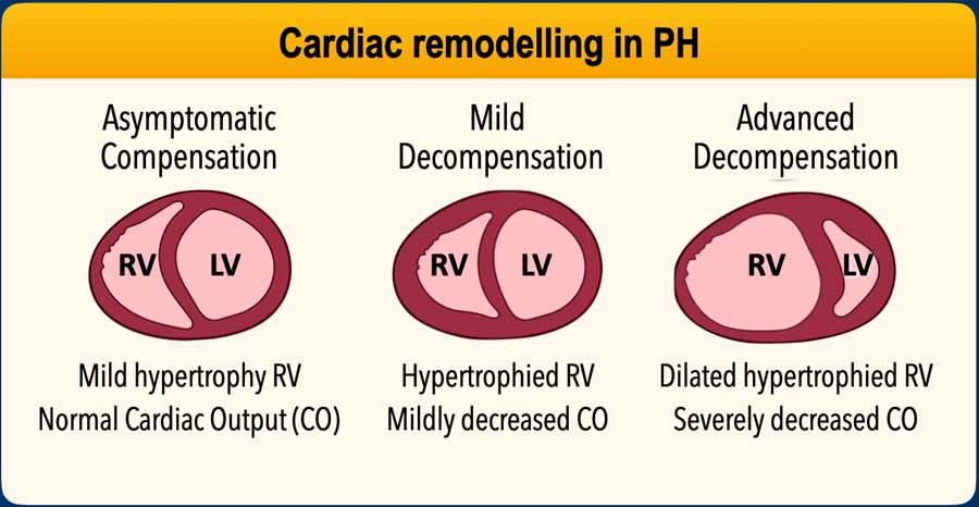
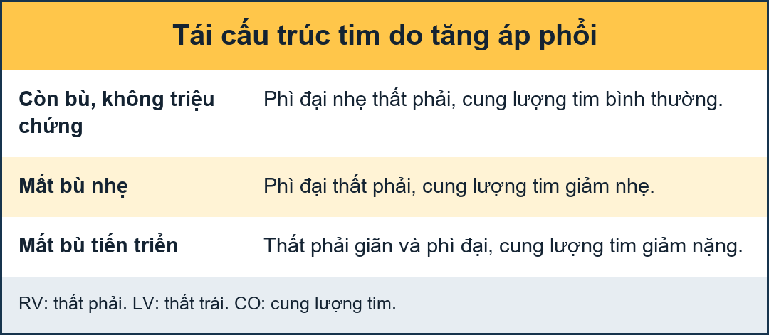
Cardiac remodelling in PHTái cấu trúc tim trong PH
1. Initial Compensation – Right Ventricular (RV) Adaptation1. Bù trừ ban đầu – Thích ứng thất phải (RV)
- Increased pressure in the pulmonary arteries will raise the RV afterload.Tăng áp lực động mạch phổi sẽ làm tăng hậu gánh thất phải.
- RV hypertrophy: The right ventricle adapts by increasing muscle mass (concentric hypertrophy) to generate higher pressure.Phì đại thất phải: Tâm thất phải thích nghi bằng cách tăng khối lượng cơ (phì đại đồng tâm) để tạo ra áp lực cao hơn.
- Preserved function: Initially, this adaptation helps maintain cardiac output.Chức năng được bảo tồn: Ban đầu, sự thích ứng này giúp duy trì cung lượng tim.
2. Progressive Strain – Maladaptation2. Chủng tiến bộ – Không thích nghi
- RV dilatation: Over time, the RV begins to dilate as it struggles to overcome resistance.Giãn thất phải: Theo thời gian, thất phải bắt đầu giãn ra khi nó phải vật lộn để vượt qua sự kháng cự.
- Decreased contractility: The hypertrophied RV eventually weakens, leading to reduced stroke volume.Giảm co bóp: Cơ thất phải phì đại cuối cùng sẽ yếu đi, dẫn đến giảm thể tích nhát bóp.
3. Right Heart Failure3. Suy tim phải
- RV failure: The ventricle can no longer pump effectively.Suy thất phải: Tâm thất không còn khả năng bơm hiệu quả.
- Systemic congestion: Leads to liver congestion, ascites, peripheral edema.Ứ máu toàn thân: Dẫn đến ứ máu gan, cổ trướng, phù ngoại vi.
- Decreased left heart preload: As the RV fails, less blood reaches the left heart, reducing systemic output.Giảm tiền gánh tim trái: Khi thất phải thất trái, máu đến tim trái ít hơn, làm giảm cung lượng hệ thống.
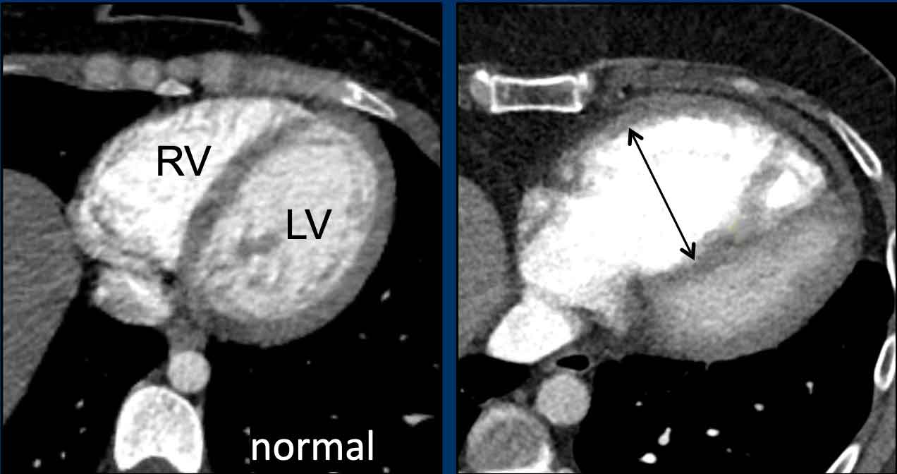

Dilatation of RVSự giãn của thất phải
The images show a normal right and left ventricle compared to a dilated right ventricle.Hình ảnh cho thấy tâm thất phải và tâm thất trái bình thường so với tâm thất phải giãn.
Proposed cut-off values for right heart dilatation on axial CT:Giá trị ngưỡng nong tim phải trên CT scan trục:
- Right atrium: ≥ 65mm (women) and ≥ 70mm (men)Nhĩ phải: ≥ 65 mm (phụ nữ) và ≥ 70 mm (nam giới)
- Right ventricle: ≥ 55mm (women) and ≥ 60mm (men)Tâm thất phải: ≥ 55 mm (phụ nữ) và ≥ 60 mm (nam giới)
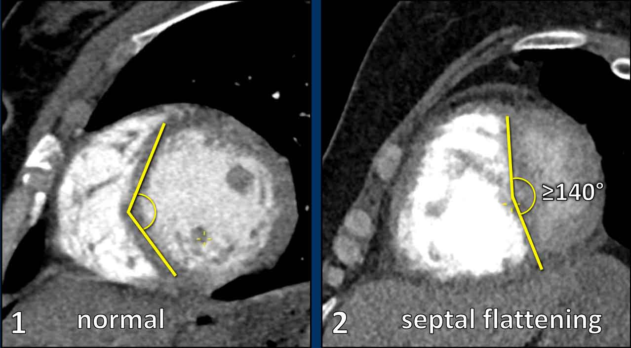

In the short-axis view, a normal left ventricle demonstrates a circular to elliptical configuration.
The interventricular septum contributes to the circular contour of the ventricular wall.Trong hình ảnh trục ngắn, tâm thất trái bình thường thể hiện cấu hình từ hình tròn đến hình elip.
Vách liên thất tạo thành đường viền tròn của thành thất.
Septal flattening or leftward bowing is indicative of pressure overload and right ventricular dilatation.Bẹp vách hoặc cúi trái là dấu hiệu của quá tải áp lực và giãn thất phải.
Pressure measurements in PHĐo áp suất trong PH
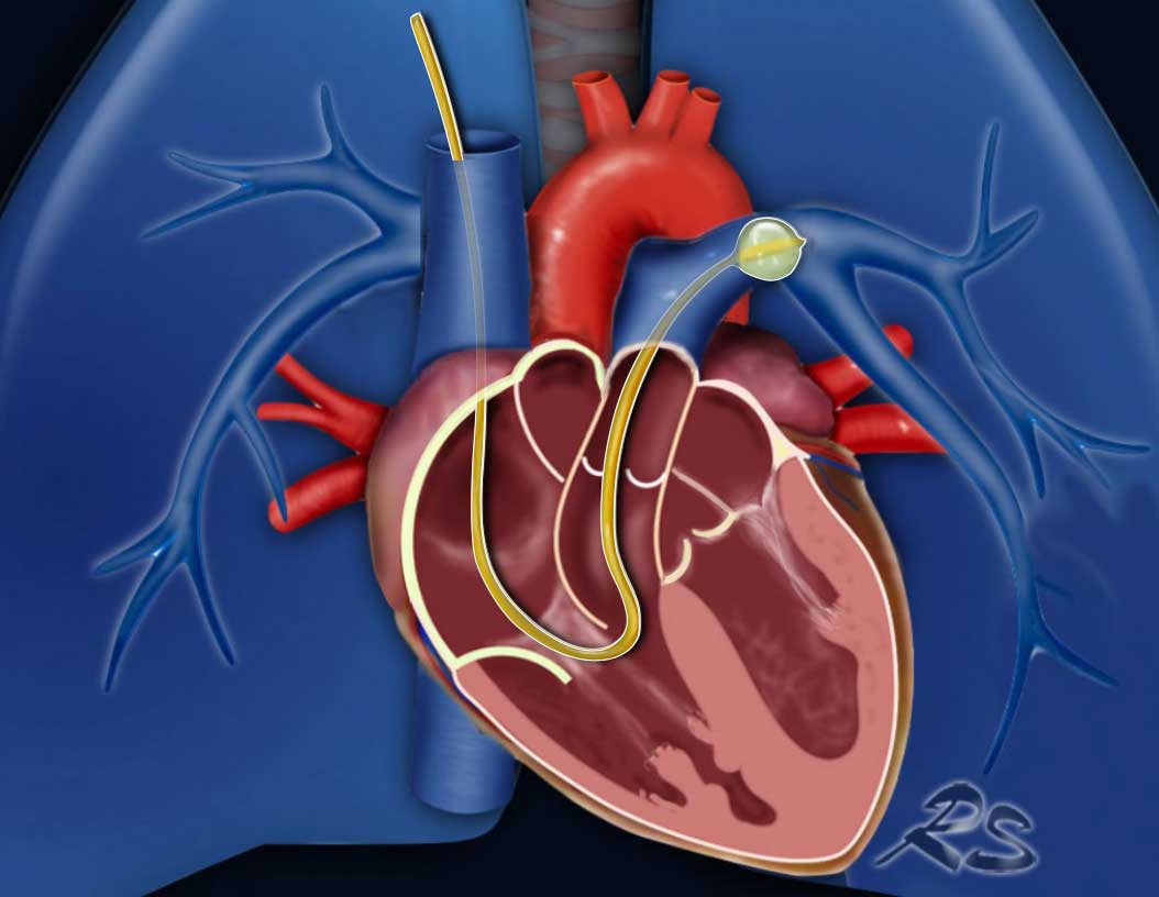 Swan-Ganz catheter inserted into the left pulmonary artery to measure the pulmonary arterial wedge pressure (PAWP).Ống thông Swan-Ganz được đưa vào động mạch phổi trái để đo áp lực động mạch phổI (PAWP).
Under normal conditions, the pulmonary vascular bed is characterized by low resistance and low pressure, and a high blood flow volume.
The definitive diagnosis of pulmonary hypertension is established by right heart catheterization, which is an invasive procedure to measure the pulmonary artery pressure.
PH is diagnosed when the mean pulmonary artery pressure (mPAP) is ≥ 20 mmHg at rest.Trong điều kiện bình thường, giường mạch phổi được đặc trưng bởi sức cản thấp và áp suất thấp, và lưu lượng máu cao.
Chẩn đoán xác định tăng áp phổi được xác định bằng cách đặt ống thông tim phải, đây là một thủ thuật xâm lấn để đo áp lực động mạch phổi.
PH được chẩn đoán khi áp lực động mạch phổi trung bình (mPAP) ≥ 20 mmHg khi nghỉ.
Elevated pulmonary arterial pressure can result from:Tăng áp lực động mạch phổi có thể là kết quả của:
- Precapillary PH
Increased pulmonary vascular resistance due to small vessel obstruction or loss of pulmonary vasculature as a result of chronic emboli.PH trước mao mạch
Tăng sức cản mạch phổi do tắc nghẽn mạch máu nhỏ hoặc mất mạch phổi là hậu quả của tắc mạch mạn tính. - Postcapillary PH
Elevated left heart pressures as a result of left-sided heart disease leading to increased pulmonary venous and capillary pressure.
PH sau mao mạch
Tăng áp lực tim trái do bệnh tim trái dẫn đến tăng áp lực tĩnh mạch phổi và mao mạch.
Pulmonary Arterial Wedge Pressure (PAWP)
Also known as wedge pressure—is a key hemodynamic measurement used to estimate left atrial pressure and helps distinguish between pre-capillary and post-capillary PH.
The wedge pressure is measured as follows:Áp lực động mạch phổi chêm (PAWP)
Còn được gọi là áp lực nêm – là một phép đo huyết động quan trọng được sử dụng để ước tính áp lực nhĩ trái và giúp phân biệt giữa PH trước và sau mao mạch.
Áp lực nêm được đo như sau:
- During right heart catheterization, a catheter (Swan-Ganz) is inserted into a pulmonary artery branch.Trong quá trình thông tim phải, một ống thông (Swan-Ganz) được đưa vào nhánh động mạch phổi.
- The balloon at the tip is inflated to temporarily occlude the vessel.Bóng ở đầu được bơm phồng để tạm thời làm tắc mạch.
- This isolates the pressure distal to the occlusion, which reflects left atrial pressure because there are no valves between the pulmonary capillaries and the left atrium. The wedge pressure will be high in left heart disease and low in other causes of PH.Điều này phân lập áp lực xa với chỗ tắc, phản ánh áp lực tâm nhĩ trái vì không có van giữa mao mạch phổi và tâm nhĩ Trái.Áp lực nêm sẽ cao trong bệnh tim trái và thấp trong các nguyên nhân khác của PH.
Pulmonary Vascular Resistance (PVR)
The pulmonary vascular resistance is expressed in Woods Units (mmHg x min/L) and is the resistance that must be overcome for blood to flow through the circulatory system.
It is a variable that reflects the size (diameter and length) of the vascular bed.
It can be calculated by the formula: PVR = (mPAP - PAWP) / CO (cardiac output).Phổi kháng mạch (PVR)
Sức cản mạch máu phổi được biểu thị bằng đơn vị Woods (mmHg x phút / L) và là sức cản phải được khắc phục để máu chảy qua hệ thống tuần hoàn.
Nó là một biến phản ánh kích thước (đường kính và chiều dài) của giường mạch máu.
Nó có thể được tính theo công thức: PVR = (mPAP - PAWP) / CO (sản lượng tim).
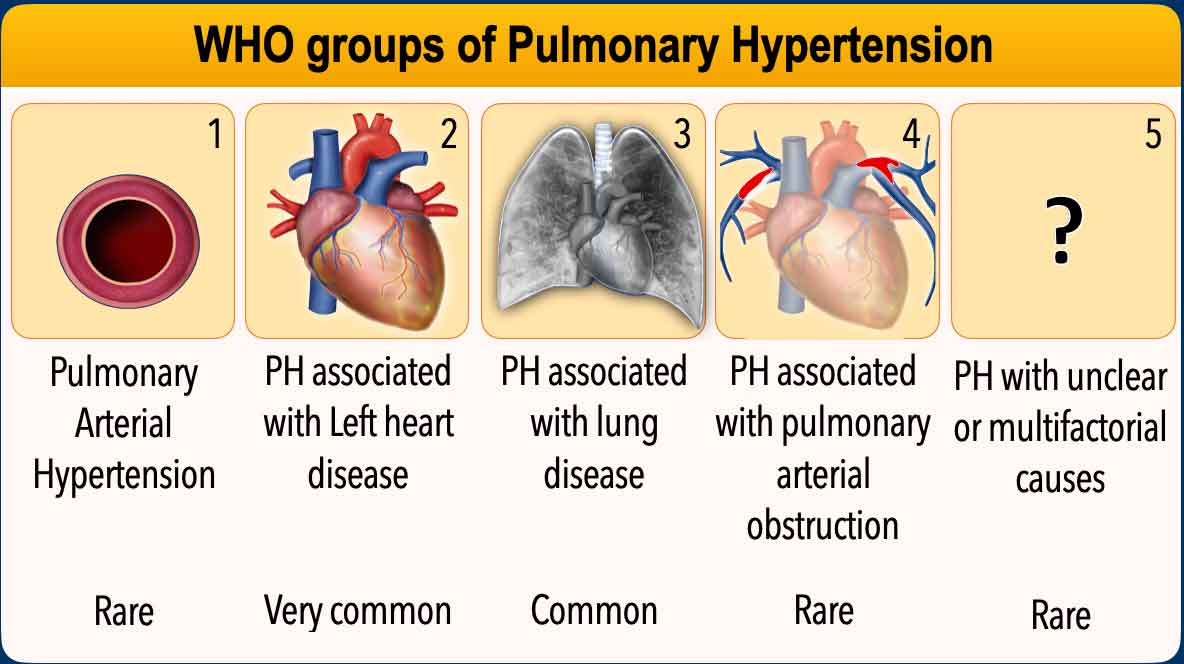
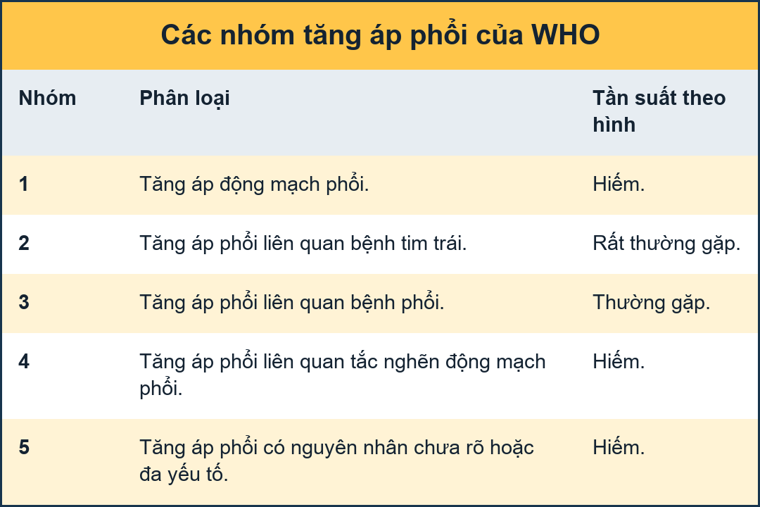
Intravascular pressure measurements through right-sided catherization, as well as clinical and imaging findings, help to subdivide PH into five different groups, as defined by the World Health Organization (WHO). Các phép đo áp lực nội mạch thông qua quá trình đốt bên phải, cũng như các dấu hiệu lâm sàng và hình ảnh, giúp chia PH thành năm nhóm khác nhau, theo định nghĩa của Tổ chức Y tế Thế giới (WHO).
WHO groupsNhóm WHO
The WHO classifies pulmonary hypertension (PH) into five groups based on their underlying causes and mechanisms. WHO phân loại tăng áp phổi (THA) thành năm nhóm dựa trên các nguyên nhân và cơ chế cơ bản của chúng.
Group 1 – Pulmonary Arterial Hypertension (PAH)
PH caused by pathology of the smallest pulmonary arteries, which become narrowed or stiff. This remodelling increases the resistance of the vascular bed, where the right ventricle has to work against, eventually leading to right heart failure. PAH can be idiopathic, inherited or related to conditions like HIV or systemic sclerosis.Nhóm 1 – Tăng áp động mạch phổi (PAH)
PH gây ra bởi bệnh lý của các động mạch phổi nhỏ nhất, trở nên hẹp hoặc cứng.Sự tái cấu trúc này làm tăng sức cản của giường mạch máu, nơi tâm thất phải phải hoạt động, cuối cùng dẫn đến suy tim phải.PAH có thể là vô căn, di truyền hoặc liên quan đến các tình trạng như HIV hoặc xơ cứng bì hệ thống.
Group 2 – PH due to Left Heart Disease
PH due to left heart dysfunction arises from an impaired ability to effectively handle pulmonary venous return. This results in elevated left atrial pressure, which is passively transmitted backward through the pulmonary veins and capillaries, leading to increased pressure in the pulmonary arteries. In advanced stages, secondary remodelling of the pulmonary arterial vasculature may occur, contributing a precapillary component to the disease process.Nhóm 2 – PH do bệnh tim trái
PH do rối loạn chức năng tim trái phát sinh từ sự suy giảm khả năng xử lý hiệu quả trở lại tĩnh mạch phổi, dẫn đến tăng áp lực tâm nhĩ trái, được truyền thụ động ngược qua các tĩnh mạch phổI và mao mạch, dẫn đến gia tăng áp lực trong động mạch phổi. Trong giai đoạn tiến triển, tái tạo thứ phát của mạch máu động mạch phổI có thể xảy ra, góp phần vào thành phần tiền mao mạch cho quá trình bệnh.
Group 3 – PH due to Lung Disease or Hypoxia
PH resulting from chronic lung diseases and/or hypoxia leads to narrowing or obliteration of the peripheral pulmonary arteries, thereby reducing the pulmonary vascular bed and consequently increasing vascular pressure.Nhóm 3 – PH do bệnh phổi hoặc thiếu oxy
PH do bệnh phổi mạn tính và / hoặc thiếu oxy dẫn đến hẹp hoặc tắc nghẽn các động mạch phổi ngoại biên, do đó làm giảm giường mạch phổi và do đó làm tăng áp lực mạch máu.
Group 4 – PH due to pulmonary arterial obstruction
The most common cause of arterial obstruction is chronic thromboembolic pulmonary disease. Although rare, this form is important because it is potentially curable.Nhóm 4 – PH do tắc động mạch phổi
Nguyên nhân phổ biến nhất gây tắc nghẽn động mạch là bệnh phổi tắc mạch huyết khối mạn tính, tuy hiếm gặp nhưng dạng tắc nghẹn này rất quan trọng vì có khả năng chữa khỏi.
Group 5 – PH with Unclear or Multifactorial Causes
PH linked to conditions with complex or unknown mechanisms, such as sarcoidosis, certain hematologic disorders, splenectomy, metabolic diseases, and complex congenital heart defects.Nhóm 5 – PH với nguyên nhân không rõ ràng hoặc đa yếu tố
PH liên quan đến các tình trạng có cơ chế phức tạp hoặc chưa biết, chẳng hạn như bệnh sarcoid, một số rối loạn huyết học, cắt lách, bệnh chuyển hóa và các khuyết tật tim bẩm sinh phức tạp.
Pressure measurementsĐo áp suất
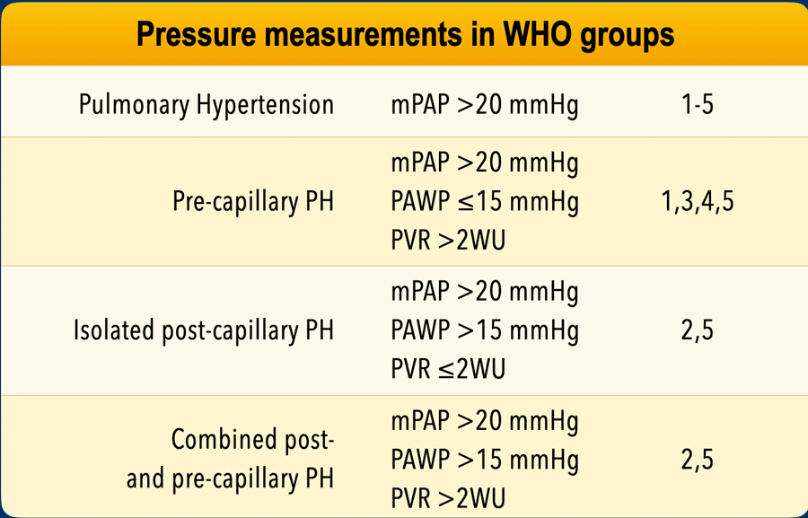
In all five WHO groups the mean pulmonary arterial pressure (mPAP) is above 20 mmHg.
In all groups except for PH resulting from left heart disease (group 2) the wedge pressure (PAWP) will be low and the pulmonary vascular resistance (PVR) will be high.
In isolated post-capillary PH resulting from left heart disease or unclear causes (group 2 and 5) the wedge pressure is high and the pulmonary vascular resistance is low.
Sometimes there is a combined pre- and post-capillary PH with a high wedge pressure and a high vascular resistance.Ở cả năm nhóm WHO, áp lực động mạch phổi trung bình (mPAP) đều trên 20 mmHg.
Ở tất cả các nhóm ngoại trừ nhóm bệnh tim trái (nhóm 2) áp lực chêm (PAWP) thấp và sức cản mạch phổi (PVR) cao.
Trong PH sau mao mạch đơn độc do bệnh tim trái hoặc nguyên nhân không rõ ràng (nhóm 2 và 5), áp lực nêm cao và sức cản mạch phổi thấp.
Đôi khi có sự kết hợp giữa PH trước và sau mao mạch với áp lực nêm cao và sức cản mạch máu cao.
Pulmonary arterial hypertensionTăng áp động mạch phổi

PAH
Pulmonary arterial hypertension (PAH) is classified under WHO group 1.
PAH, also known as primary pulmonary hypertension, is a form of pulmonary hypertension caused by pathological changes in the pulmonary arteries, which become thickened, stiffened, or narrowed.Tăng áp động mạch phổi (PAH) được phân loại theo WHO nhóm 1.
PAH, còn được gọi là tăng áp phổi nguyên phát, là một dạng tăng áp phổi gây ra bởi những thay đổi bệnh lý trong động mạch phổi, trở nên dày lên, cứng lại hoặc thu hẹp.
As a result of this arteriopathy, the right side of the heart must work against increased pulmonary vascular resistance. Over time, this leads to right ventricular dysfunction and an inability to maintain adequate pulmonary blood flow.
PAH may be idiopathic (IPAH), heritable (HPAH), or associated with other conditions such as HIV infection, systemic sclerosis, congenital heart disease or PAH with features of venous involvement (PVOD).Do bệnh động mạch này, tim phải hoạt động chống lại sự gia tăng sức cản mạch máu phổi, theo thời gian, điều này dẫn đến rối loạn chức năng thất phải và không có khả năng duy trì lưu lượng máu phổi đầy đủ.
PAH có thể là vô căn (IPAH), di truyền (HPAH), hoặc liên quan đến các tình trạng khác như nhiễm HIV, xơ cứng bì hệ thống, bệnh tim bẩm sinh hoặc PAH có các đặc điểm liên quan đến tĩnh mạch (PVOD).
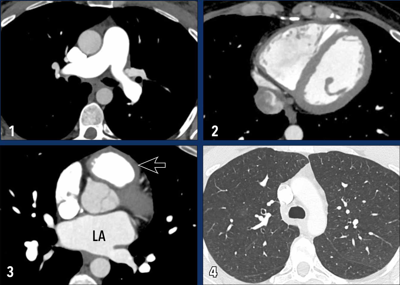
These images are of a young female with idiopathic PAH.Những hình ảnh này là của một phụ nữ trẻ bị tăng áp động mạch phổi vô căn.
Imaging findings
Chẩn đoán hình ảnh
- Mild dilatation of the main pulmonary arterySự giãn động mạch phổi chính mức độ nhẹ
- Mild dilatation of the right ventricle.Giãn nhẹ thất phải.
- Mild wall thickening of the right ventricle outflow tract (arrow).
The left atrium has a normal size.Dày thành nhẹ đường ra thất phải (mũi tên).
Tâm nhĩ trái có kích thước bình thường. - Normal lung parenchyma.Nhu mô phổi bình thường.
PVOD
Pulmonary veno-occlusive disease (PVOD) is a rare subtype of PAH.
It is caused by narrowing and obliteration of the small pulmonary veins (venules), as opposed to other causes of group 1 pulmonary hypertension in which the pre-capillary arteries are primarily involved.
The venous occlusion leads to post-capillary pressure build-up and subsequently pulmonary arterial hypertension.Bệnh tắc tĩnh mạch phổi (PVOD) là một phân nhóm hiếm gặp của tăng áp động mạch phổi.
Nguyên nhân là do thu hẹp và tắc nghẽn các tĩnh mạch phổi nhỏ (các tĩnh mạch), trái ngược với các nguyên nhân khác của tăng áp phổi nhóm 1, trong đó các động mạch trước mao mạch chủ yếu bị ảnh hưởng.
Tắc tĩnh mạch dẫn đến tăng áp lực sau mao mạch và sau đó là tăng áp động mạch phổi.
The classic CT triad in PVOD includes:Bộ ba CT cổ điển trong PVOD bao gồm:
- Smooth interlobular septal thickening.
Smooth dày vách liên tiểu thùy. - Centrilobular ground-glass opacities.
Trung tâm tiểu thùy đám mờ kính mờ. - Enlarged mediastinal and hilar lymph nodes resulting from chronic lymphatic congestion.Trung thất to và hạch rốn phổi do tắc nghẽn mạn tính.
PH due to left heart diseasePH do bệnh tim trái
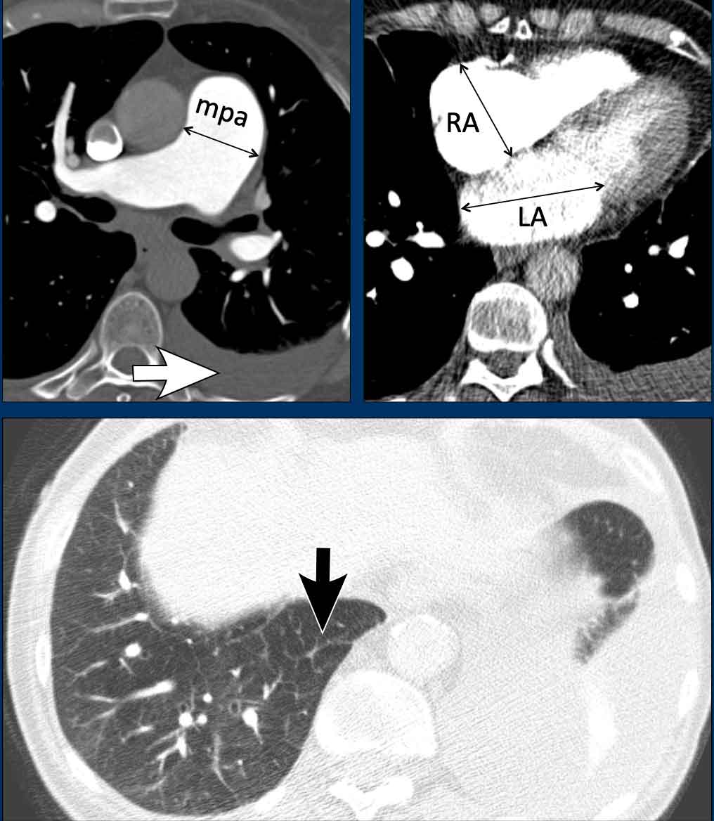

Pulmonary hypertension due to left heart disease is asseigned to WHO group 2.
It occurs when the left heart is unable to effectively accommodate or eject the blood returning from the lungs.
This results in a backward transmission of pressure into the pulmonary circulation, first leading to elevated pulmonary venous pressure and subsequently also elevated arterial pressure.Tăng áp phổi do bệnh tim trái được phân loại vào nhóm 2 của WHO.
Nó xảy ra khi tim trái không thể chứa hoặc đẩy máu trở lại từ phổi một cách hiệu quả.
Điều này dẫn đến việc truyền áp lực ngược vào tuần hoàn phổi, đầu tiên dẫn đến tăng áp lực tĩnh mạch phổi và sau đó cũng tăng áp lực động mạch.
It may result from:Nó có thể là kết quả của:
- Systolic dysfunction (i.e., impaired left ventricular contraction)Rối loạn chức năng tâm thu (tức là suy giảm co bóp thất trái)
- Diastolic dysfunction (i.e., reduced ventricular filling due to myocardial or pericardial stiffness)Rối loạn chức năng tâm trương (tức là giảm đổ đầy thất do cơ tim hoặc độ cứng màng ngoài tim)
- Severe valvular disease, particularly mitral or aortic valve pathology.Bệnh van tim nặng, đặc biệt là bệnh lý van hai lá hoặc động mạch chủ.
Given the high prevalence of cardiac disease, this is the most common form of pulmonary hypertension.Với tỷ lệ mắc bệnh tim cao, đây là dạng phổ biến nhất của tăng áp phổi.
Images
In this patient with left heart disease the findings are:Hình ảnh
Ở bệnh nhân này có bệnh tim trái, những phát hiện sau:
- Dilatation of the main pulmonary artery.Sự giãn của động mạch phổi chính.
- Dilatation of the right atrium and mild thickening of the myocardium of the right ventricle.Sự giãn nở của tâm nhĩ phải và dày lên nhẹ của cơ tim của tâm thất phải.
- Dilatation of the left atriumSự giãn nở của tâm nhĩ trái
- Pleural fluid (white arrow).Dịch màng phổi (mũi tên trắng).
- Smooth interlobular thickening (black arrow).Dày liên thuỳ (mũi tên đen).
PH due to cardiac anomaliesPH do dị tật tim
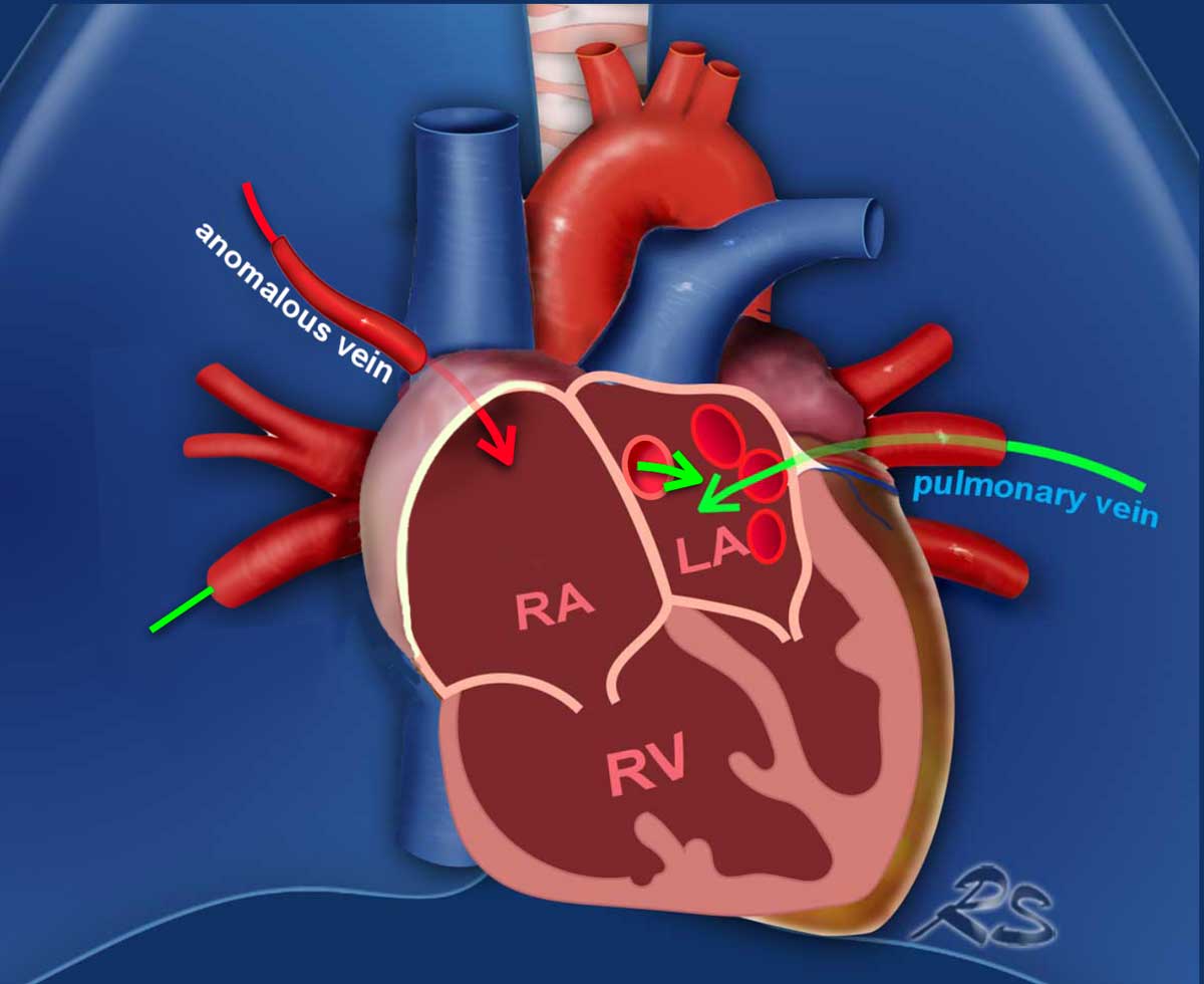
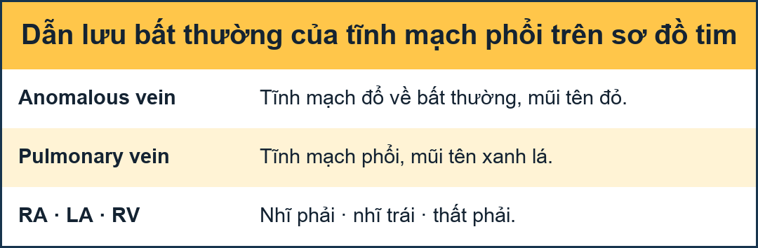
Partial anomalous pulmonary venous return (PAPVR)Bất thường tĩnh mạch phổi hồi lưu một phần (PAPVR)
In partial anomalous pulmonary venous return, one or more pulmonary veins abnormally drain into the systemic venous circulation (mainly into the superior vena cava on the right, and into the left brachiocephalic vein on the left side), instead of the normal drainage into the left atrium. Trong trường hợp bất thường tĩnh mạch phổi một phần, một hoặc nhiều tĩnh mạch phổI dẫn lưu bất thường vào tuần hoàn tĩnh mạch hệ thống (chủ yếu vào tĩnh mạch chủ trên ở bên phải, và vào tĩnh mạch cánh tay đầu bên trái), thay vì dẫn lưu bình thường vào tâm nhĩ trái.
Depending on the shunt volume, chronically increased pulmonary circulation from this shunt can result in endothelial injury, smooth muscle hypertrophy, and vascular fibrosis, ultimately leading to elevated pulmonary vascular resistance, and the development of pulmonary hypertension.Tuỳ thuộc vào thể tích shunt, tuần hoàn phổi tăng mạn tính từ shunt này có thể dẫn đến tổn thương nội mô, phì đại cơ trơn và xơ hóa mạch máu, cuối cùng dẫn đến tăng sức cản mạch máu phổi và phát triển tăng áp phổi.
Illustration
In this case, most pulmonary veins drain normally into the left atrium (green arrows).
However, the right upper lobe pulmonary vein is anomalous and drains via the superior vena cava into the right atrium rather than the left atrium (red arrow).
This anomalous venous return creates a left-to-right shunt, leading to volume overload of the right atrium and right ventricle.Minh hoạ
Trong trường hợp này, hầu hết các tĩnh mạch phổi đều chảy bình thường vào tâm nhĩ trái (các mũi tên màu xanh lá cây).
Tuy nhiên, tĩnh mạch phổi thuỳ trên bên phải là bất thường và chảy qua tĩnh mạch chủ trên vào tâm nhĩ phải hơn là tâm nhĩ trái (mũi tên màu đỏ).
Sự hồi lưu tĩnh mạch bất thường này tạo ra một luồng thông từ trái sang phải, dẫn đến quá tải thể tích của tâm nhĩ phải và tâm thất phải.
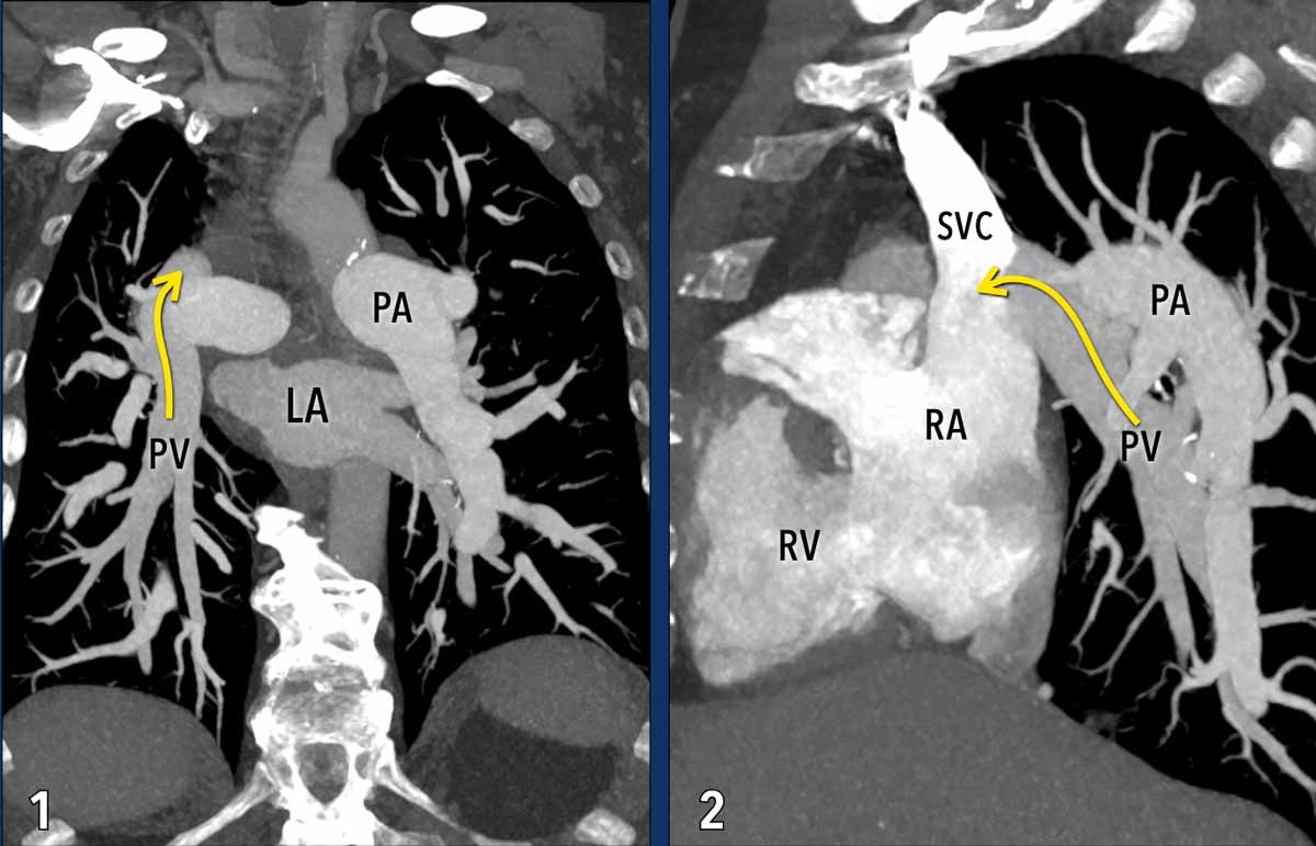
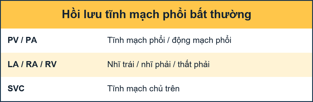
These images are of a patient with pulmonary hypertension secondary to partial anomalous pulmonary venous return (PAPVR).Những hình ảnh này là của một bệnh nhân bị tăng áp phổi thứ phát do bất thường tĩnh mạch phổi trở lại một phần (PAPVR).
Imaging Findings
The right lower lobe pulmonary veins are not connected to the left atrium (LA) but instead drain into the superior vena cava (arrows).
Note the bilateral dilatation of the pulmonary arteries, consistent with elevated pulmonary arterial pressure.Hình ảnh học
Các tĩnh mạch phổi thuỳ dưới bên phải không được kết nối với tâm nhĩ trái (LA) mà thay vào đó chảy vào tĩnh mạch chủ trên (các mũi tên).
Lưu ý sự giãn hai động mạch phổi, phù hợp với áp lực động mạch phổI tăng cao.
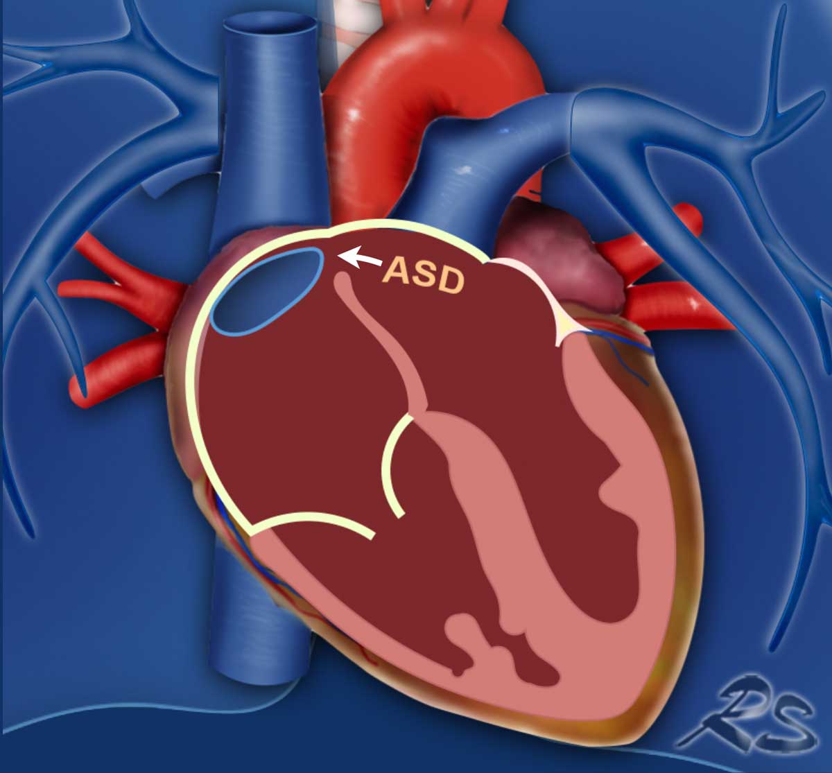

Atrial Septal Defect (ASD)Khiếm khuyết vách liên nhĩ (ASD)
In an Atrial Septal Defect (ASD), there is a communication between the left and right atrium.
Since left atrial pressure is higher, blood flows from the left atrium to the right atrium.
This increases blood volume in the right heart and pulmonary circulation.
The chronically elevated flow and pressure in the lung circulation causes endothelial damage, vascular remodelling and an increased pulmonary vascular resistance.Trong thông liên nhĩ (ASD), có sự giao tiếp giữa tâm nhĩ trái và tâm nhĩ phải.
Vì áp lực nhĩ trái cao hơn, máu chảy từ tâm nhĩ trái sang tâm nhĩ phải.
Điều này làm tăng thể tích máu ở tim phải và tuần hoàn phổi.
Dòng chảy và áp lực tăng lên mãn tính trong tuần hoàn phổi gây ra tổn thương nội mô, tái tạo mạch máu và tăng sức cản mạch máu phổi.
Illustration
Example of a sinus venosus superior defect.
This is a subtype of ASD, in which there
is an abnormal interatrial communication between the left atrium and the
superior cavo-atrial junction (arrow).
Since this subtype of ASD frequently coexists with PAPVR, always check for both conditions.Minh hoạ
Ví dụ về một lỗ thông xoang tĩnh mạch trên.
Đây là một phân nhóm của ASD, trong đó có sự giao tiếp bất thường giữa tâm nhĩ trái và điểm nối giữa tâm nhĨ trên (mũi tên).
Vì dạng phụ này của ASD thường cùng tồn tại với PAPVR, nên luôn kiểm tra cả hai tình trạng.
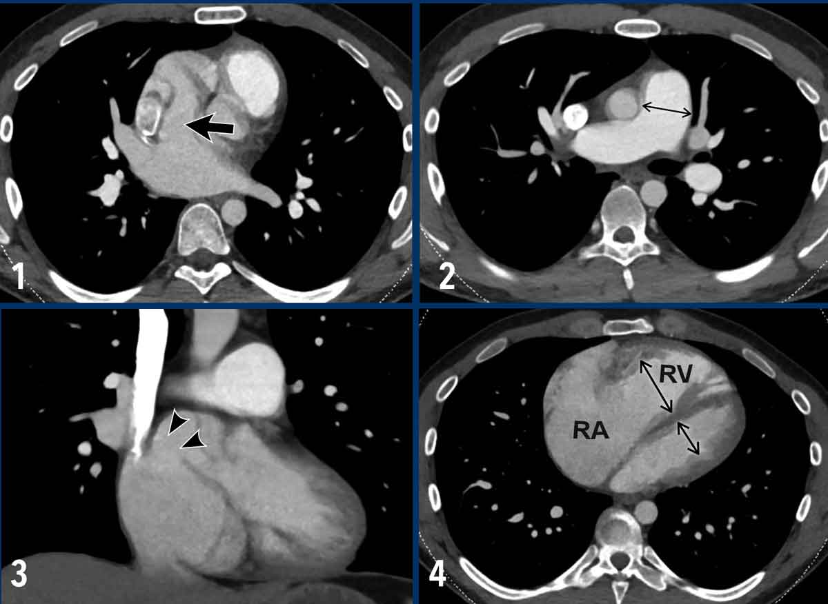

Example of a sinus venosus superior defect.Ví dụ về một lỗ thông xoang tĩnh mạch trên.
ImagesHình minh họa
- There is an abnormal communication between the left atrium and the superior cavo-atrial junction (arrow). Có sự giao tiếp bất thường giữa tâm nhĩ trái và điểm nối cavo-atrial trên (mũi tên).
- Dilatation of the pulmonary artery.Sự giãn nở của động mạch phổi.
- Coronal reconstruction demonstrating the abnormal communication between the left atrium and the superior cavo-atrial junction (arrowheads).Tái tạo vành cho thấy sự bất thường trong giao tiếp giữa nhĩ trái và điểm nối trên tâm nhĩ (các đầu mũi tên).
- Severe dilatation of the right atrium and ventricle due to the left-to-right shunt. Giãn nhĩ phải và tâm thất phải nặng do luồng thông từ trái sang phải.
PH due to lung diseasePH do bệnh phổi
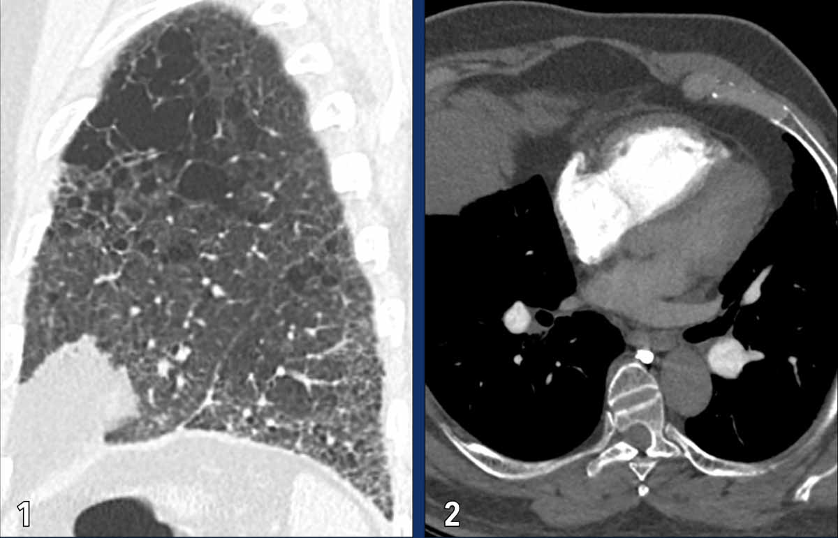
Pulmonary hypertension secondary to chronic lung diseases and/or hypoxia is categorized under WHO Group 3. It results from both structural and functional alterations in the pulmonary vasculature due to underlying pulmonary pathology. In this context, the small pulmonary vessels may be lost or undergo vasoconstriction and remodelling, leading to a reduced vascular bed and a subsequent increase in pulmonary arterial pressure.Tăng áp phổi thứ phát do bệnh phổi mạn tính và / hoặc thiếu oxy được phân loại theo Nhóm WHO 3. Nó là kết quả của cả sự thay đổi cấu trúc và chức năng trong mạch máu phổi do bệnh lý phổi tiềm ẩn. Trong bối cảnh này, các mạch máu phổI nhỏ có thể bị mất hoặc trải qua sự co mạch và tái tạo, dẫn đến giảm giường mạch và tăng áp lực động mạch phổi sau đó.
Common etiologies include chronic obstructive pulmonary disease (COPD) and pulmonary fibrosis. Obstructive sleep apnea and chronic exposure to hypoxic conditions at high altitudes may also play a role in its development.Các nguyên nhân thường gặp bao gồm bệnh phổi tắc nghẽn mạn tính (COPD) và xơ phổi. Ngưng thở khi ngủ do tắc nghẹt và tiếp xúc lâu dài với tình trạng thiếu oxy ở độ cao cũng có thể đóng một vai trò trong sự phát triển của nó.
Images
This patient has a combination of
emphysema and pulmonary fibrosis.
The dilated and hypertrophic right ventricle, and flattening of the
intraventricular septum are the result of PH.Hình ảnh
Bệnh nhân này có sự kết hợp của khí phế thũng và xơ phổi.
Tâm thất phải giãn và phì đại, và vách ngăn trong thất phẳng là kết quả của PH.
Chronic thromboembolic pulmonary hypertensionTăng áp phổi do huyết khối tắc mạch mạn tính
The most common disease of WHO group 4 (pulmonary arterial obstruction) is chronic thromboembolic pulmonary hypertension (CTEPH).
CTEPH occurs when thrombi fail to fully resolve following an acute pulmonary embolism.
This results in persistent thrombotic obstruction, vascular scarring, and arterial remodelling, which impair pulmonary blood flow and elevate pulmonary artery pressure.Bệnh lý thường gặp nhất của nhóm 4 theo WHO (tắc động mạch phổi) là thuyên tắc huyết khối mạn tính tăng áp phổi (CTEPH).
CTEPH xảy ra khi huyết khối không giải quyết hoàn toàn sau khi thuyên tắc phổi cấp tính.
Điều này dẫn đến tắc nghẽn huyết khối dai dẳng, sẹo mạch máu và tái tạo động mạch, làm giảm lưu lượng máu phổi và tăng áp lực động mạch phổi.
Because these obstructions can potentially be treated with pulmonary endarterectomy (PEA) or balloon pulmonary angioplasty (BPA), CTEPH is the only potentially curable form of pulmonary hypertension.Vì những tắc nghẽn này có khả năng được điều trị bằng phẫu thuật cắt bỏ nội mạc động mạch phổi (PEA) hoặc nong bóng động mạch phổI (BPA), CTEPH là dạng tăng áp phổi duy nhất có khả năng chữa khỏi.
Imaging findings
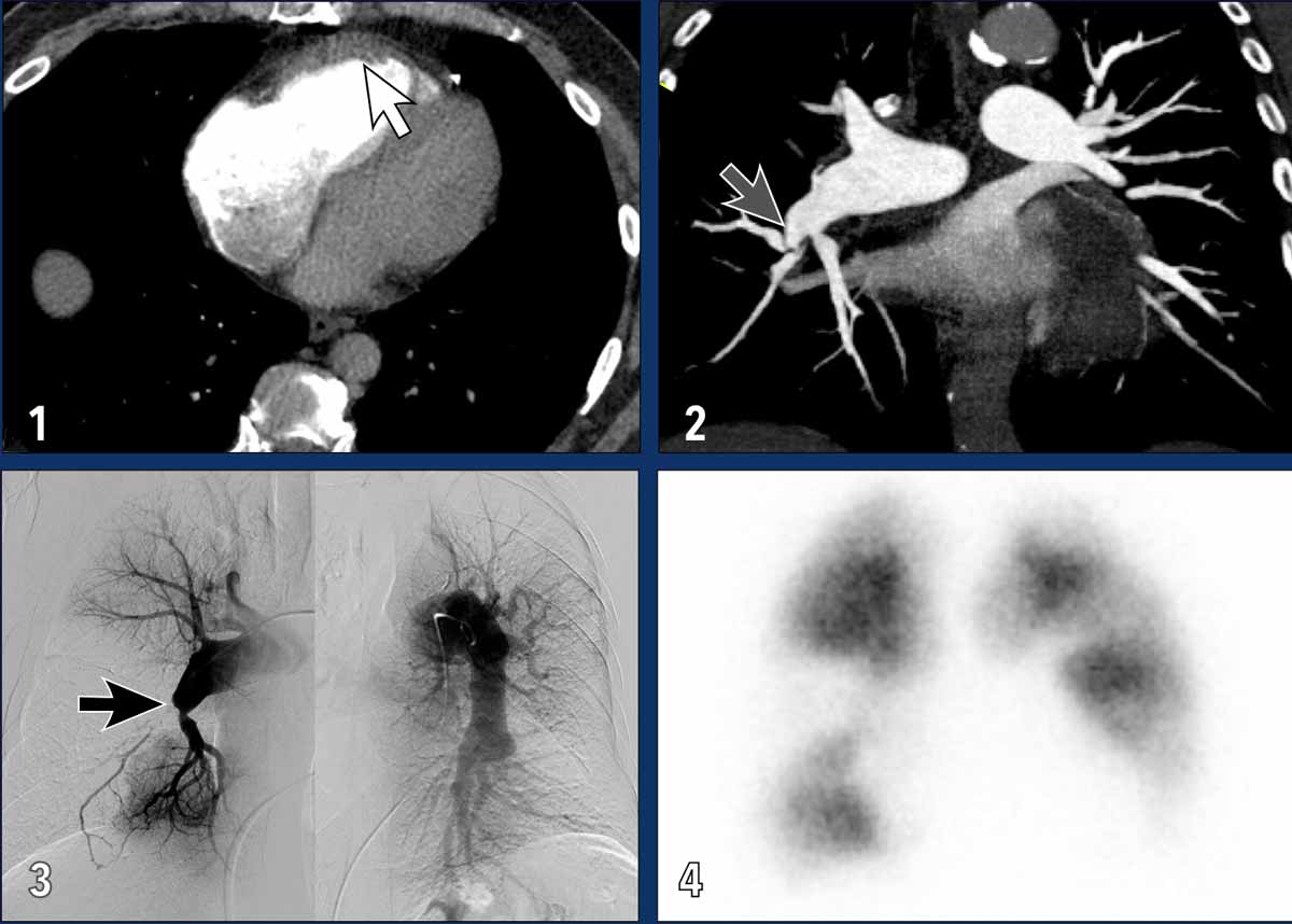
These images are of a patient with severe CTEPH.Chẩn đoán hình ảnh
Những hình ảnh này là của một bệnh nhân bị CTEPH nặng.
- Dilatation of RA and RV with muscular hypertrophy of RV (white arrow).Tình trạng giãn cơ và thất phải kèm theo phì đại cơ thất phải (mũi tên trắng).
- Web causing stenosis of this segmental pulmonary artery; one of the features of chronic thromboembolic disease (grey arrow).Web gây hẹp động mạch phổi đoạn này; một trong những đặc điểm của bệnh huyết khối tắc mạch mạn tính (mũi tên xám).
- Pulmonary angiogram showing total occlusion (black arrow) and stenosis of segmental pulmonary arteries, with large perfusion defects.Chụp động mạch phổi cho thấy tắc hoàn toàn (mũi tên màu đen) và hẹp các động mạch phổI phân đoạn, với các khiếm khuyết tưới máu lớn.
- Wedge shaped perfusion defects on a perfusion scan.Hình ảnh các khuyết hổng tưới máu hình nêm trên phim tưới máu.
Differentiation Acute vs Chronic EmboliPhân biệt tắc mạch cấp tính và mạn tính
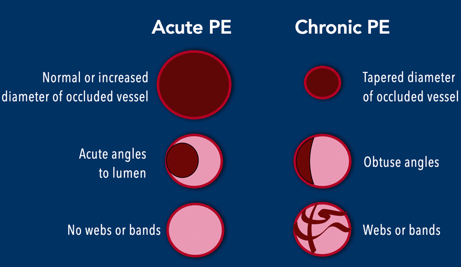
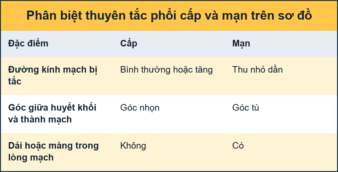
When pulmonary emboli are present, how do we differentiate acute from chronic disease?Khi có thuyên tắc phổi, làm thế nào để phân biệt bệnh cấp tính và mạn tính?
- The diameter of occluded vessels in acute PE is normal or slightly increased, while in chronic PE the occluded vessels are tapered.
Đường kính mạch bị tắc trong PE cấp tính bình thường hoặc tăng nhẹ, trong khi ở PE mạn tính, các mạch bị tắc sẽ giảm dần. - Partial filling defects in acute PE typically show acute angles to the vessel lumen, while obtuse angles are seen in chronic disease due to the wall adhering morphology.Các khuyết hổng một phần trong PE cấp tính thường cho thấy các góc cấp tính với lòng mạch, trong khi các góc tù được nhìn thấy trong bệnh mạn tính do hình thái bám dính vào thành.
- Intraluminal webs and bands may be seen in chronic disease due to partial recanalization of the prior occlusive thrombus.Các mạng nhện và dải trong lòng mạch có thể được nhìn thấy trong bệnh mạn tính do tái thông một phần của huyết khối tắc trước đó.
- Bronchial artery dilatation further points towards longstanding abnormalities in the pulmonary arterial flow, with recruitment of the systemic arterial blood flow as alternate source.Sự giãn nở động mạch phế quản tiếp tục hướng tới những bất thường lâu dài trong dòng chảy động mạch phổi, với việc thu nhận dòng máu động mạch hệ thống như là nguồn thay thế.

Please note that a combination of clot morphology may be present, which should point you towards the diagnosis of acute PE on top of pre-existing chronic thromboembolic disease.
This is an important finding that is often overlooked.Xin lưu ý rằng có thể có sự kết hợp của hình thái cục máu đông, điều này sẽ hướng bạn đến chẩn đoán PE cấp tính trên bệnh thuyên tắc huyết khối mạn tính đã có từ trước.
Đây là một phát hiện quan trọng thường bị bỏ qua.
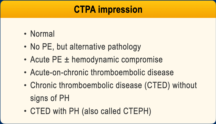
Computed tomography pulmonary angiography (CTPA) is the imaging modality of choice for the diagnosis of pulmonary embolism (PE).Chụp cắt lớp vi tính mạch phổi (CTPA) là phương pháp chẩn đoán hình ảnh được lựa chọn để chẩn định thuyên tắc phổi (PE).
When interpreting a CTPA, several outcomes should be evaluated:Khi diễn giải CTPA, một số kết quả cần được đánh giá:
- No abnormalities.Không có bất thường.
- No evidence of PE, but presence of alternative pathology (cardiovascular or non-cardiovascular) suggesting the underlying etiology of pulmonary hypertension.Không thấy dấu hiệu PE, nhưng có các bệnh lý khác (tim mạch hoặc không phải tim mạch) gợi ý nguyên nhân cơ bản của tăng áp phổi.
- Acute pulmonary embolism, with or without hemodynamic compromise due to flow obstruction.Thuyên tắc phổi cấp tính, có hoặc không có tổn thương huyết động do tắc nghẽn dòng chảy.
- Acute-on-chronic thromboembolic disease.Bệnh thuyên tắc huyết khối cấp tính trên mạn tính.
- Chronic thromboembolic disease (CTED) without radiologic signs of pulmonary hypertension.Bệnh thuyên tắc huyết khối mạn tính (CTED) không có dấu hiệu tăng áp phổi trên X quang.
- CTED with radiologic signs of pulmonary hypertension (suggestive of CTEPH).CTED có dấu hiệu tăng áp phổi (gợi ý CTEPH).
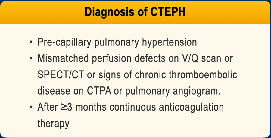
Diagnosis of CTEPHChẩn đoán CTEPH
For the definitive diagnosis of CTEPH, the following criteria are required:Để chẩn đoán xác định CTEPH, cần có các tiêu chuẩn sau:
- CT findings consistent with chronic thromboembolic disease.CT phù hợp với bệnh lý huyết khối mạn tính.
- Intravascular measurements confirming the presence of pre-capillary pulmonary hypertension.Các phép đo nội mạch xác nhận sự hiện diện của tăng áp phổi trước mao mạch.
- Mismatched perfusion defects on ventilation/perfusion (V/Q) scan or SPECT/CT perfusion imaging.Các thông số tưới máu không tương xứng trên phim thông khí/tưới máu (V/Q) hoặc SPECT/CT.
- A history of continuous anticoagulation therapy for at least 3 months.Tiền sử điều trị chống đông máu liên tục ít nhất 3 tháng.
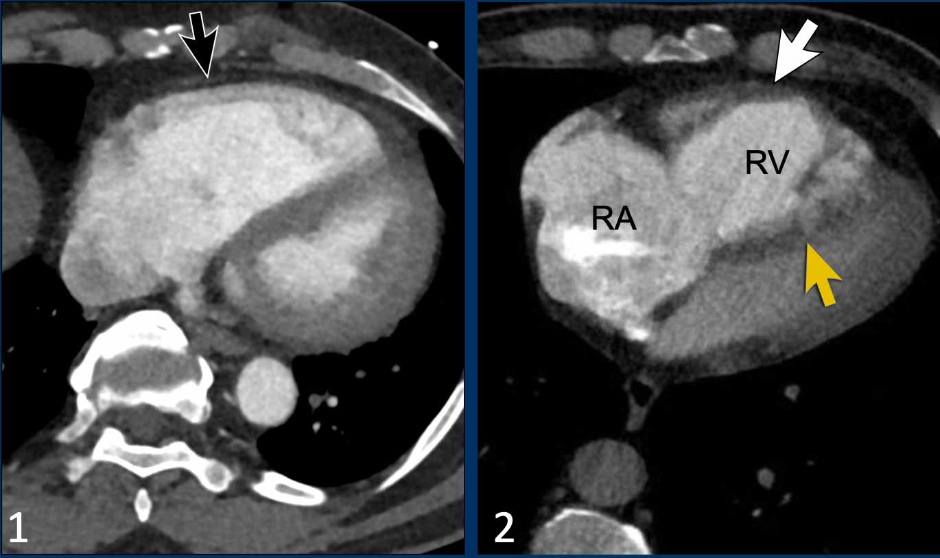

Cardiac remodelling in pulmonary emboliTái cấu trúc tim trong thuyên tắc phổi
Looking at remodelling of the right ventricle can help to differentiate between acute and chronic emboli.Việc xem xét sự tái cấu trúc của tâm thất phải có thể giúp phân biệt giữa thuyên tắc cấp tính và mạn tính.
Here we have two examples of patients with pulmonary emboli and cardiac remodelling.
Ở đây chúng tôi có hai ví dụ về bệnh nhân bị thuyên tắc phổi và tái tạo tim.
ImagesHình minh họa
- There is right ventricular dilatation with septal flattening, but no evidence of RV wall hypertrophy (black arrow), consistent with an acute increase in RV afterload.
This can be seen in patients with acute pulmonary embolism.Có giãn thất phải kèm theo phẳng vách liên thất, nhưng không có bằng chứng phì đại thành thất phải (mũi tên đen), phù hợp với sự gia tăng cấp tính của hậu gánh thất phải.
Điều này có thể thấy ở những bệnh nhân có thuyên tắc phổi cấp tính. - In contrast, in this patient there is dilatation of the right ventricle and atrium with interventricular septal flattening, accompanied by RV wall thickening (white arrow).
This suggests chronic pressure overload and is indicative of longstanding pulmonary hypertension.
This can be seen in patients with chronic thromboembolic pulmonary hypertension.Ngược lại, ở bệnh nhân này có giãn tâm thất phải và tâm nhĩ với vách ngăn liên thất phẳng, kèm theo dày thành thất phải (mũi tên trắng).
Điều này cho thấy tình trạng quá tải áp lực mạn tính và là dấu hiệu của tăng áp phổi kéo dài.
Điều này có thể thấy ở những bệnh nhân tăng áp phổi do huyết khối tắc mạch mạn tính.
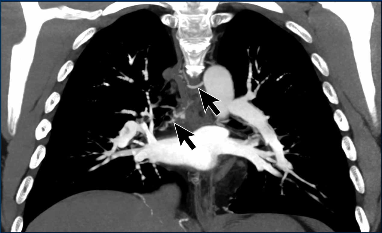
Bronchial artery hypertrophy in CTEPHPhì đại động mạch phế quản trong bệnh thận mạn tính có cuống
Bronchial artery dilatation further suggests longstanding abnormalities in pulmonary arterial flow, with recruitment of systemic arterial circulation as an alternative source of pulmonary perfusion.Sự giãn nở động mạch phế quản tiếp tục gợi ý những bất thường lâu dài trong dòng chảy động mạch phổi, với việc sử dụng tuần hoàn động mạch hệ thống như một nguồn tưới máu phổi thay thế.
Image
Dilatation of bronchial arteries (arrows) in a patient with PH.
Notice the widened pulmonary arteries.
Hình ảnh
Tình trạng giãn động mạch phế quản (các mũi tên) ở bệnh nhân có tăng áp động mạch phổi.
Chú ý các động mạch phổi giãn rộng.
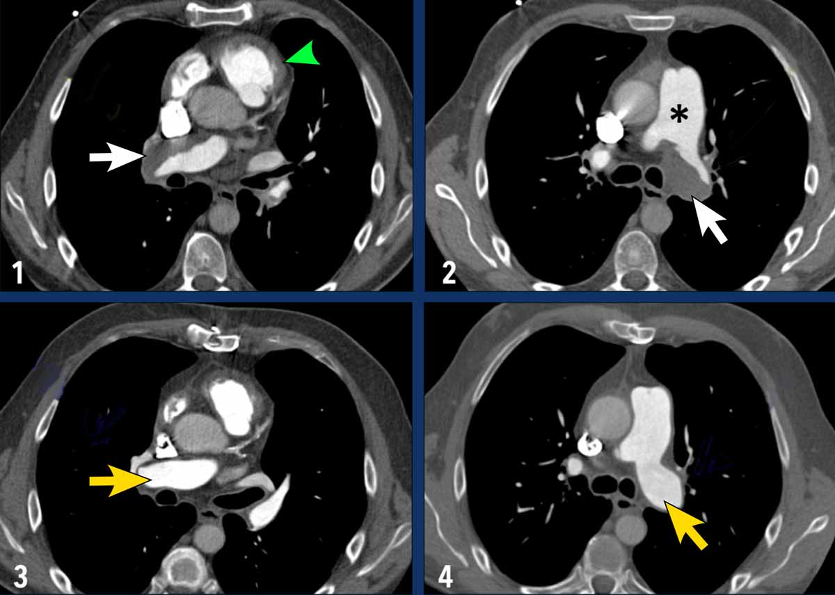
Pulmonary endarterectomy (PEA)Phẫu thuật bóc nội mạc động mạch phổi (PEA)
Pulmonary endarterectomy for CTEPH is a surgical and technically demanding procedure - performed under cardiopulmonary bypass and deep hypothermic circulatory arrest - to remove organized thromboembolic material from the pulmonary arteries.Phẫu thuật nội soi bóc lớp trong động mạch phổi điều trị bệnh lý tắc động mạch phổI do huyết khối tĩnh mạch là một thủ thuật phẫu thuật và đòi hỏi kỹ thuật cao - được thực hiện dưới tuần hoàn ngoài cơ thể và ngừng tuần hoàn do hạ thân nhiệt sâu - để loại bỏ các chất huyết khối có tổ chức khỏi động mạch phổ.
ImagingHình ảnh
- Pre-endarterectomy (Images 1-2): wall adhering thrombotic material is seen in both the right and left pulmonary arteries (indicated by white arrows). Notable dilatation of the main pulmonary artery (asterix) and hypertrophy of the wall of the right ventricular outflow tract (green arrowhead) is also present.Cắt bỏ trước nội mạc (Hình 1-2): có thể thấy huyết khối bám vào thành ở cả động mạch phổi phải và động mạch phổI trái (biểu hiện bằng mũi tên màu trắng). Có thể thấy giãn động mạch phổ biến chính (asterix) và phì đại thành đường ra thất phải (đầu mũi tên màu xanh lá cây).
- Post-endarterectomy (Images 3-4): The pulmonary arteries on both sides are cleared of thrombotic material.Sau khi cắt bỏ nội mạc (Hình 3-4): Các động mạch phổi ở cả hai bên đều được làm sạch các chất huyết khối.
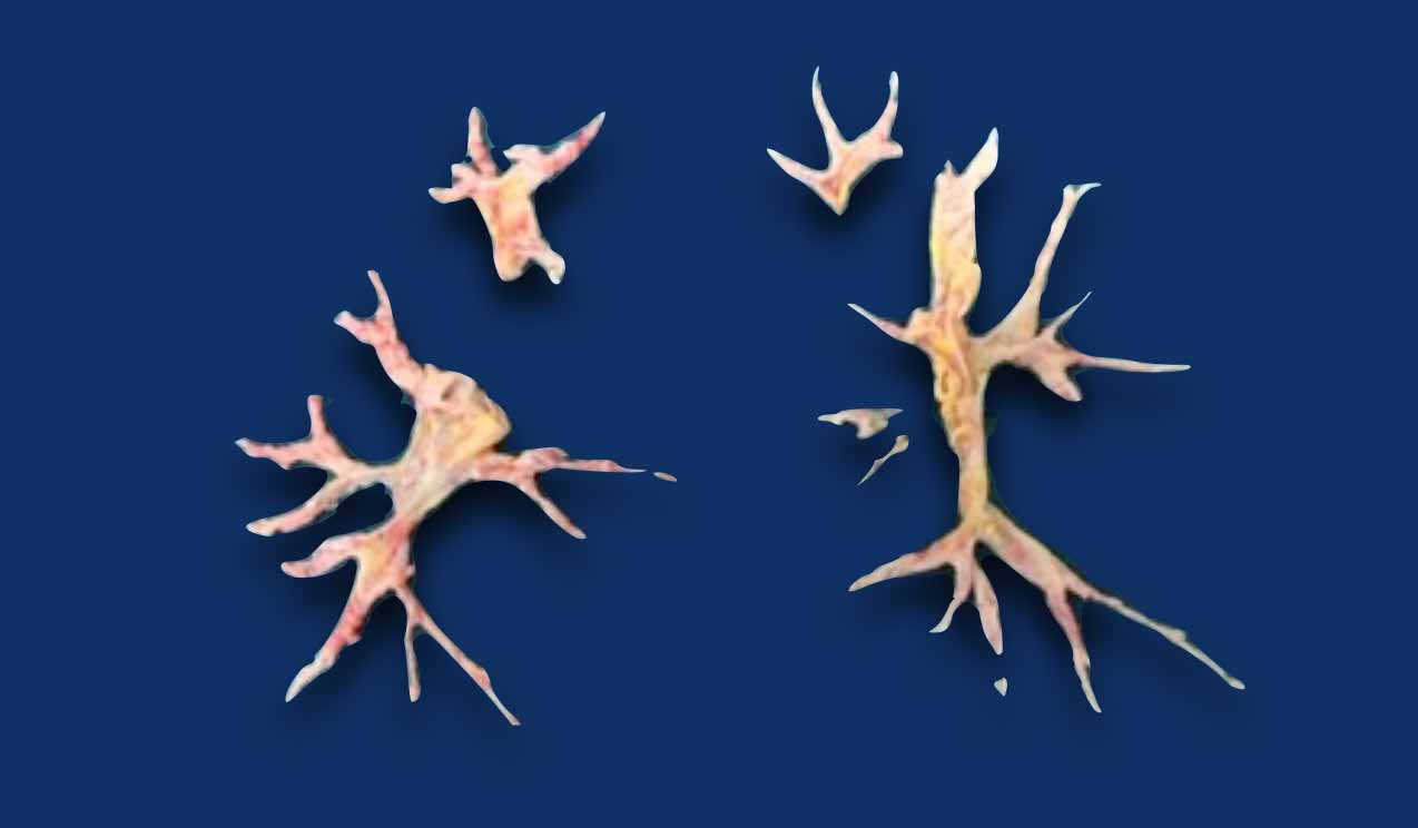
This is the organized thrombotic material that was excised from both the right and left pulmonary arteries, including the segmental branches.Đây là các chất huyết khối có tổ chức được cắt bỏ từ cả động mạch phổi phải và trái, bao gồm cả các nhánh phân đoạn.

Balloon Pulmonary Angioplasty (BPA)Tạo hình mạch phổi bằng bóng (BPA)
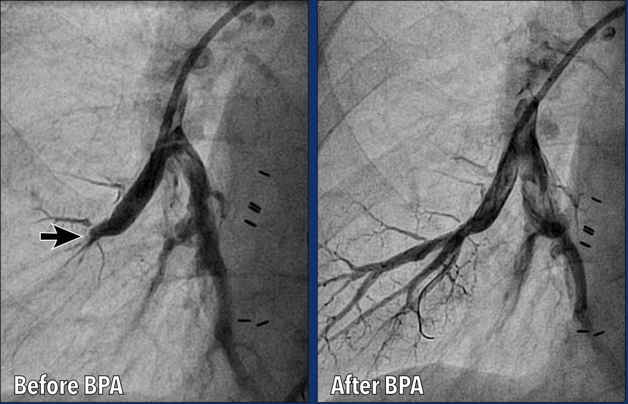
Balloon Pulmonary Angioplasty (BPA) is a catheter-based interventional procedure used to treat CTEPH. Tạo hình mạch phổi bằng bóng (BPA) là một thủ thuật can thiệp dựa trên catheter được sử dụng để điều trị CTEPH.
BPA is a minimally invasive alternative to surgery for patients with:BPA là một phương pháp thay thế xâm lấn tối thiểu cho phẫu thuật cho bệnh nhân:
- Inoperable CTEPH (e.g., distal disease not accessible by surgery).CTEPH không thể phẫu thuật được (ví dụ, bệnh ở đầu xa không thể tiếp cận được bằng phẫu thuật).
- High surgical risk.Nguy cơ phẫu thuật cao.
- Residual pulmonary hypertension after pulmonary endarterectomy (PEA).Tăng áp phổi tồn dư sau phẫu thuật bóc nội mạc động mạch phổi (PEA).
Images
Before BPA there is acute tapering and occlusion of a basal segmental pulmonary artery of the right lower lobe.
After BPA the artery is patent, with restored flow into the segmental arteries.
Hình ảnh
Trước khi có BPA, có sự giảm dần và tắc nghẽn cấp tính của động mạch phổi đoạn đáy của thuỳ dưới bên phải.
Sau khi BPA động mạch được bảo tồn, với dòng chảy được phục hồi vào các động mạch phân đoạn.
Pulmonary artery sarcomaSarcoma động mạch phổi
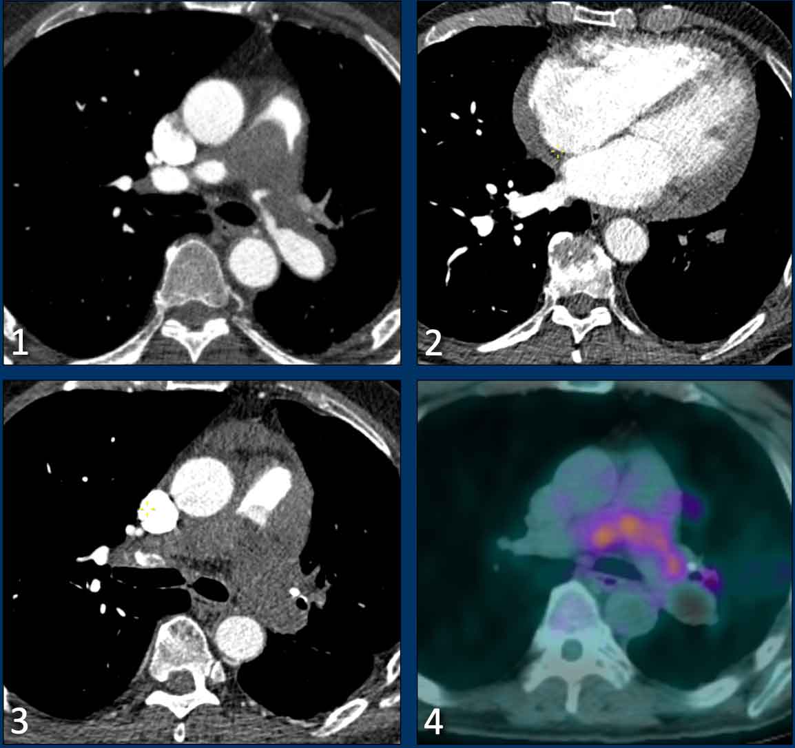
The most important mimicker of chronic thromboembolic disease is pulmonary artery sarcoma, a rare primary malignant tumor arising from the vessel wall.Biểu hiện quan trọng nhất của bệnh thuyên tắc huyết khối mạn tính là sarcoma động mạch phổi, một khối u ác tính nguyên phát hiếm gặp phát sinh từ thành mạch.
It typically presents as a central filling defect within the pulmonary artery and may initially be mistaken for thrombus.
Although signs of flow obstruction and right heart strain may be present, right ventricular hypertrophy is often absent due to the rapid progression of the tumor.Nó thường biểu hiện như một lỗ thông trung tâm trong động mạch phổi và ban đầu có thể bị nhầm lẫn với huyết khối.
Mặc dù có thể có các dấu hiệu tắc nghẽn dòng chảy và căng thẳng tim phải, nhưng phì đại thất phải thường không có do sự tiến triển nhanh chóng của khối u.
Imaging features suggestive of a pulmonary artery sarcoma include:Các đặc điểm hình ảnh gợi ý sarcoma động mạch phổi bao gồm:
- An expansile intraluminal mass, which may have an extravascular infiltrative component.Một khối trong lòng mạch giãn nở, có thể có thành phần thâm nhiễm ngoài mạch.
- Involvement of the pulmonary valve and/or right ventricular outflow tract.Sự tham gia của van động mạch phổi và / hoặc đường ra thất phải.
- Absence of ancillary signs of chronic thromboembolic disease.Không có các dấu hiệu phụ của bệnh thuyên tắc huyết khối mạn tính.
- Enhancement after contrast administration.Tăng cường sau khi dùng thuốc cản quang.
- FDG-PET avidity (frequent, but may be low or absent).Nhu cầu FDG-PET (thường xuyên, nhưng có thể thấp hoặc không có).
- Growth despite anticoagulant therapy.Tăng trưởng mặc dù đã điều trị kháng đông.
Images
Hình minh họa
- There is a large filling defect within the central pulmonary arteries.Có một lỗ thông lớn trong động mạch phổi trung tâm.
- Notably, there is no distinct right ventricular hypertrophy or dilatation.Đáng chú ý, không có phì đại hoặc giãn thất phải rõ rệt.
- Progressive size despite anticoagulation therapy.Kích thước tiến triển mặc dù đã điều trị chống đông.
- Metabolic activity on FDG-PET imaging.Hoạt tính chuyển hóa trên FDG-PET.
Conclusion
Pulmonary hypertension (PH) is a relative rare but very important disease. Since initial signs of PH may appear incidentally on imaging, radiologists play a vital role in recognizing the condition.Thrombus morphology in thromboembolic disease and cardiac remodelling are important imaging clues. Identifying these signs can help diagnose CTEPH, which is the only potentially curable form of PH.Kết luận
Tăng áp phổi (THA) là một bệnh tương đối hiếm gặp nhưng rất quan trọng. Vì các dấu hiệu ban đầu của THA có thể xuất hiện tình cờ trên chẩn đoán hình ảnh, các bác sĩ X-quang đóng một vai trò quan trọng trong việc nhận biết tình trạng này. Hình thái huyết khối trong bệnh huyết khối tắc mạch và tái tạo tim là những đầu mối hình ảnh quan trọng. Xác định các dấu hiệu này có thể giúp chẩn định CTEPH, đây là dạng THA duy nhất có khả năng chữa khỏi.

CharityHoạt động từ thiện
All the profits of the Radiology Assistant go to Medical Action Myanmar which is run by Dr. Nini Tun and Dr. Frank Smithuis sr, who is a professor at Oxford university and happens to be the brother of Robin Smithuis.Toàn bộ lợi nhuận của Radiology Assistant được chuyển cho Medical Action Myanmar, do bác sĩ Nini Tun và bác sĩ Frank Smithuis Sr. điều hành. Frank Smithuis Sr. là giáo sư tại Đại học Oxford và là anh em ruột của Robin Smithuis.
Click here to watch the video of Medical Action Myanmar and if you like the Radiology Assistant, please support Medical Action Myanmar with a gift.Nhấp vào đây để xem video về Medical Action Myanmar và nếu bạn thích Trợ lý X quang, vui lòng hỗ trợ Medical Action Myanmar bằng một món quà.
ReferencesTài liệu tham khảo
-
About Pulmonary Hypertension
Về tăng áp phổi
Pulmonary hypertension association Mối liên quan với tăng áp phổi
-
Revised Definition of Pulmonary Hypertension and Approach to Management: A Clinical Primer
Định nghĩa sửa đổi về tăng áp phổi và cách tiếp cận quản lý: Một cuốn sách lâm sàng
by Bradley A. Maron
Journal of the American Heart Association Volume 12, Number 8
Published 7 April 2023
bởi Bradley A. Maron -
Going beyond Cardiomegaly: Evaluation of Cardiac Chamber Enlargement at Non–ECG Gated Multidetector CT: Current Techniques, Limitations, and Clinical Implications.
Vượt ra ngoài bệnh tim to: Đánh giá sự mở rộng buồng tim tại CT đa dãy không có cổng ECG: Các kỹ thuật hiện tại, hạn chế và tác động lâm sàng.
Hota P, et al.
Radiology Cardiothoracic Imaging (2019) doi: 10.1148/ryct.2019180024
Hota P. và cộng sự. -
Imaging of pulmonary hypertension in adults: a position paper from the Fleischner Society.
Chẩn đoán hình ảnh tăng áp phổi ở người lớn: một bài báo của Hiệp hội Fleischner.
Remy-Jardin M, et al. Radiology (2021) doi: 10.1148/radiol.2020203108 Remy-Jardin M, và đồng nghiệp. X quang (2021) làm: 10.1148 / radiol.2020203108
-
ESC/ERS Guidelines for the diagnosis and treatment of pulmonary hypertension.
Hướng dẫn chẩn đoán và điều trị tăng áp phổi của ESC/ERS.
Humbert M, et al. Eur Respir J. (2023) doi: 10.1183/13993003.00879-2022 Người hâm mộ lựa chọn: Humbert M, et al Euro Respir J. (2023) làm: 10.1183/13993003.00879-2022
-
Treatment of pulmonary arterial hypertension: recent progress and a look to the future.
Điều trị tăng áp động mạch phổi: những tiến bộ gần đây và một số dự báo trong tương lai.
Humbert M, et al.
Lancet Respir Med (2023) doi: 10.1016/ S2213-2600(23)00264-3
Humbert M. và cộng sự. -
Pulmonary hypertension associated with lung disease: new insights into pathomechanisms, diagnosis, and management.
Tăng áp phổi liên quan đến bệnh phổi: những hiểu biết mới về cơ chế bệnh sinh, chẩn đoán và xử trí.
Olsson KM, et al.
Lancet Respir Med (2023) doi: 10.1016/ S2213-2600(23)00259-X
-
Chronic thromboembolic pulmonary hypertension: realising the potential of multimodal management.
Tăng áp phổi do thuyên tắc huyết khối mạn tính: nhận thấy tiềm năng của điều trị đa mô thức.
Delcroix M, et al.
Lancet Respir Med (2023) doi: 10.1016/ S2213-2600(23)00292-8
Delcroix M và cộng sự. -
Definition, classification and diagnosis of pulmonary hypertension.
Định nghĩa, phân loại và chẩn đoán tăng áp phổi.
Kovacs G. et al.
European Respiratory Journal (2024) doi: 10.1183/13993003.01324-2024
Kovacs G. và cộng sự. -
Diagnostic accuracy of CT pulmonary angiography in suspected pulmonary hypertension.
Chẩn đoán chính xác của chụp CT mạch phổi trong nghi ngờ tăng áp phổi.
Swift AJ, et al.
Eur Radiol (2020) doi: 10.1007/s00330-020-06846-1.
Swift AJ và cộng sự.