
Chest X-Ray - Heart FailureX-quang ngực - Suy tim
Radiology department of the Albert Schweitzer Hospital in Dordrecht and the Medical Centre Alkmaar, the Netherlands, the NetherlandsKhoa X quang của Bệnh viện Albert Schweitzer ở Dordrecht và Trung tâm Y tế Alkmaar, Hà Lan, Hà Lan
Ngày đăng / cập nhật: 2010-09-01
Bản gốc trên radiologyassistant.nl ↗ · tải về lúc 2026-11-19 20:19 · bản dịch máy có chuẩn hóa thuật ngữ chẩn đoán hình ảnh
In this article we will discuss the radiographic signs of congestive heart failure on the chest X-ray.Trong bài viết này chúng tôi sẽ bàn luận về các dấu hiệu X quang của suy tim sung huyết trên máy X-quang ngực.
IntroductionMở đầu
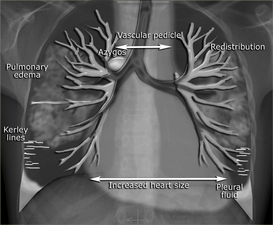

Congestive heart failure (CHF) is the result of insufficient output because of cardiac failure, high resistance in the circulation or fluid overload.
Left ventricle (LV) failure is the most common and results in decreased cardiac output and increased pulmonary venous pressure.
In the lungs LV failure will lead to dilatation of pulmonary vessels, leakage of fluid into the interstitium and the pleural space and finally into the alveoli resulting in pulmonary edema.
Suy tim sung huyết (Congestive Heart Suy tim cấp) là kết quả của sự thiếu cung lượng do suy tim, sức cản cao trong tuần hoàn hoặc quá tải dịch.
Suy thất trái (LV) là phổ biến nhất và dẫn đến giảm cung lượng tim và tăng áp lực tĩnh mạch phổi.
Trong phổi, thất trái sẽ dẫn đến giãn mạch phổi, rò rỉ dịch vào khoảng kẽ và khoang màng phổi và cuối cùng vào phế nang dẫn đến phù phổi.
Right ventricle (RV) failure is usually the result of long standing LV failure or pulmonary disease and causes increased systemic venous pressure resulting in edema in dependent tissues and abdominal viscera. Suy thất phải (RV) thường là kết quả của suy thất trái kéo dài hoặc bệnh phổi và gây tăng áp lực tĩnh mạch hệ thống dẫn đến phù nề ở các mô phụ thuộc và tạng bụng.
In the illustration on the left some of the features, that can be seen on a chest-film in a patient with CHF.Trong hình minh hoạ bên trái, một số đặc điểm, có thể được nhìn thấy trên phim ngực ở bệnh nhân CHF.
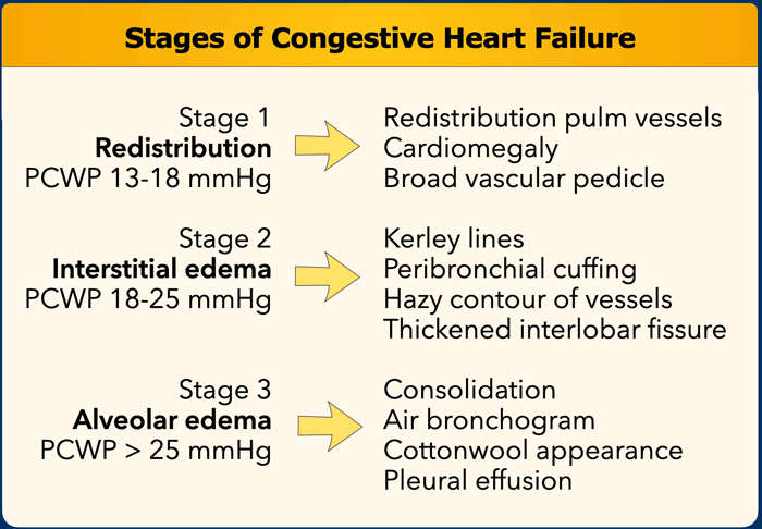
Increased pulmonary venous pressure is related to the pulmonary capillary wedge pressure (PCWP) and can be graded into stages, each with its own radiographic features on the chest film (Table).
This grading system provides a logical sequence of signs in congestive heart failure.
In daily clinical practice however some of these features are not seen in this sequence and sometimes may not be present at all.
This can be seen in patients with chronic heart failure, mitral valve disease and in chronic obstructive lung disease.Tăng áp lực tĩnh mạch phổi có liên quan đến áp lực mao mạch phổi (PCWP) và có thể được phân thành các giai đoạn, mỗi giai đoạn có đặc điểm X quang riêng trên bảng phim ngực (Bảng).
Hệ thống phân loại này cung cấp một chuỗi các dấu hiệu hợp lý trong suy tim sung huyết.
Tuy nhiên, trong thực hành lâm sàng hàng ngày, một số đặc điểm này không được nhìn thấy trong chuỗi này và đôi khi có thể không có mặt.
Điều này có thể thấy ở bệnh nhân suy tim mạn tính, bệnh van hai lá và bệnh phổi tắc nghẽn mạn tính.
Congestive Heart FailureSuy tim sung huyết
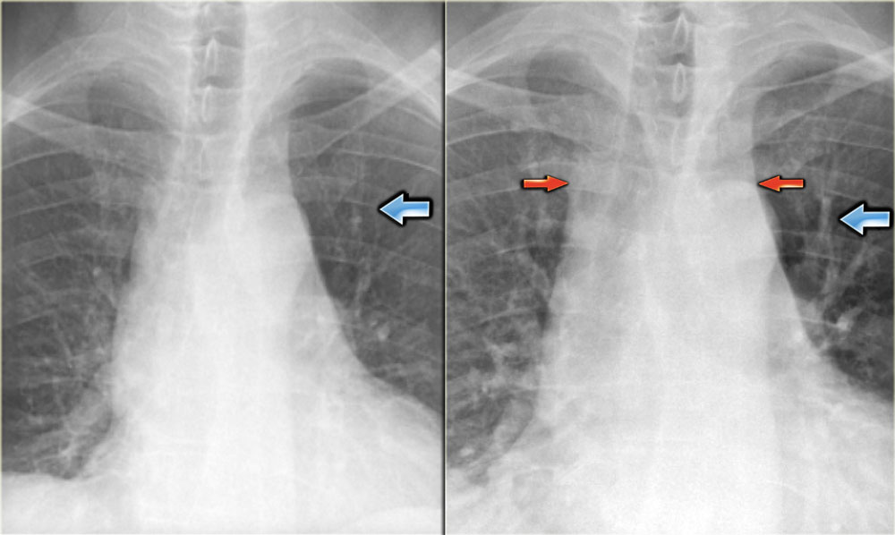 Views of the upper lobe vessels of a patient in good condition (left) and during a period of CHF (right). Notice also the increased width of the vascular pedicle (red arrows).Quan sát các mạch máu thuỳ trên của một bệnh nhân trong tình trạng tốt (trái) và trong thời gian CHF (phải) Chú ý cũng tăng chiều rộng của cuống mạch (các mũi tên màu đỏ).
Stage I - RedistributionGiai đoạn I - Tái phân phối
In a normal chest film with the patient standing erect, the pulmonary vessels supplying the upper lung fields are smaller and fewer in number than those supplying the lung bases.
The pulmonary vascular bed has a significant reserve capacity and recruitment may open previously non-perfused vessels and causes distension of already perfused vessels.
This results in redistribution of pulmonary blood flow.
First there is equalisation of blood flow and subsequently redistribution of flow from the lower to the upper lobes.Trong phim ngực bình thường với bệnh nhân đứng thẳng, các mạch máu phổi cung cấp cho các trường phổi trên nhỏ hơn và ít hơn về số lượng so với các mạch máu cung cấp hai đáy phổi.
Giường mạch phổi có khả năng dự trữ đáng kể và việc huy động có thể mở các mạch máu chưa được tưới máu trước đó và gây ra sự căng giãn của các mạch máu đã được tưới.
Điều này dẫn đến việc phân phối lại lưu lượng máu phổi.
Đầu tiên là cân bằng lưu lượng máu và sau đó phân phối lại dòng chảy từ thuỳ dưới lên thuỳ trên.
The term redistribution applies to chest x-rays taken in full inspiration in the erect position.
In daily clinical practice many chest films are taken in a supine or semi-erect position and the gravitational difference between the apex and the lung bases will be less.
In the supine position, there will be equalisation of blood flow, which may give the false impression of redistribution.
In these cases comparison with old fims can be helpful.Thuật ngữ phân phối lại áp dụng cho chụp X-quang ngực khi hít vào đầy đủ ở tư thế cương cứng.
Trong thực hành lâm sàng hàng ngày, nhiều phim chụp ngực được thực hiện ở tư thế nằm ngửa hoặc bán thẳng và sự khác biệt về trọng lực giữa đỉnh và hai đáy phổi sẽ ít hơn.
Ở tư thế nằm ngửa, sẽ có sự cân bằng lưu lượng máu, điều này có thể gây ấn tượng sai về sự phân phối lại.
Trong những trường hợp này, so sánh với phim cũ có thể hữu ích.
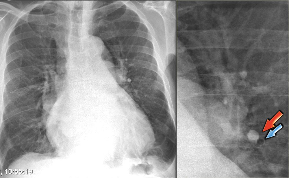 Increased artery-to-bronchus ratio in CHFTỉ lệ động mạch-phế quản tăng trong bệnh thận mạn giai đoạn cuối
Artery-to-bronchus ratio
Normally the vessels in the upper lobes are smaller than the accompanying bronchus with a ratio of 0.85 (3).
At the level of the hilum they are equal and in the lower lobes the arteries are larger with a ratio of 1.35.
When there is redistribution of pulmonary blood flow there will be an increased artery-to-bronchus ratio in the upper and middle lobes.
This is best visible in the perihilar region. Tỉ lệ động mạch-phế quản
Thông thường các mạch máu ở thuỳ trên nhỏ hơn so với phế quản đi kèm với tỷ lệ 0,85 (3).
Ở mức độ của rốn động mạch, chúng bằng nhau và ở thuỳ dưới, các động mạch lớn hơn với tỷ lệ 1,35.
Khi có sự phân phối lại lưu lượng máu phổi sẽ có sự gia tăng tỷ lệ động mạch trên phế quản ở thuỳ trên và thuỳ giữa.
Điều này có thể nhìn thấy rõ nhất ở vùng quanh rốn.
On the left a patient with cardiomegaly and redistribution.
The upper lobe vessels have a diameter > 3 mm (normal 1-2 mm).
Notice the increased artery-to-bronchus ratio at hilar level (arrows).
Bên trái là một bệnh nhân bị tim to và tái phát.
Các mạch máu thuỳ trên có đường kính > 3 mm (bình thường 1-2 mm).
Lưu ý tỷ lệ động mạch trên phế quản tăng ở mức rốn phổi (các mũi tên).
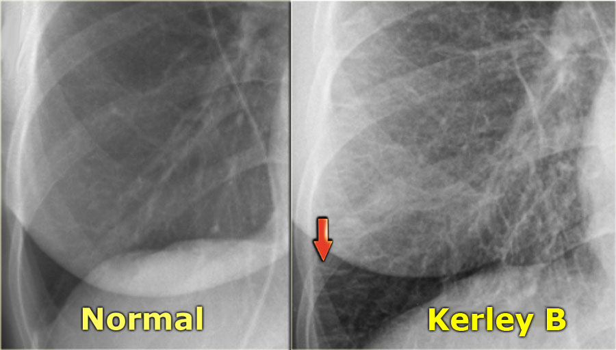
 LEFT: normal. RIGHT: CHF stage II with Kerley B-lines due to interstitial edemaTRÁI: bình thường. PHẢI: CHF giai đoạn II với Kerley B-lines do phù mô kẽ
Stage II - Interstitial edemaGiai đoạn II - Phù mô kẽ
Stage II of CHF is characterized by fluid leakage into the interlobular and peribronchial interstitium as a result of the increased pressure in the capillaries.
When fluid leaks into the peripheral interlobular septa it is seen as Kerley B or septal lines.
Kerley-B lines are seen as peripheral short 1-2 cm horizontal lines near the costophrenic angles.
These lines run perpendicular to the pleura.Giai đoạn II của CHF được đặc trưng bởi sự rò rỉ dịch vào khoảng kẽ giữa tiểu thuỳ và quanh phế quản do áp lực tăng lên trong các mao mạch.
Khi dịch rò rỉ vào vách liên tiểu thùy ngoại vi, nó được xem là Kerley B hoặc các vách ngăn.
Đường Kerley-B được xem là đường ngắn ngoại vi 1-2 cm nằm ngang gần góc sườn-đầu.
Những đường này chạy vuông góc với màng phổi.
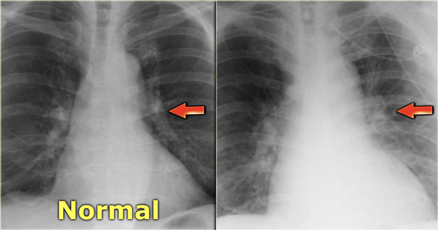
 Perihilar haze in interstitial stage of CHFMù quanh rốn ở bệnh nhân suy thận mạn giai đoạn kẽ
When fluid leaks into the peribronchovascular interstitium it is seen as thickening of the bronchial walls (peribronchial cuffing) and as loss of definition of these vessels (perihilar haze).
Khi dịch rò rỉ vào khoảng kẽ quanh phế quản, nó được xem là sự dày lên của thành phế quản (màng quanh phế nang) và mất định nghĩa của các mạch này (mù quanh rốn phổi).
On the left a patient with congestive heart failure.
There is an increase in the caliber of the pulmonary vessels and they have lost their definition because they are surrounded by edema. Bên trái là một bệnh nhân suy tim sung huyết.
Có sự gia tăng kích thước của các mạch máu phổi và chúng đã mất đi độ rõ của chúng vì chúng được bao quanh bởi phù nề.
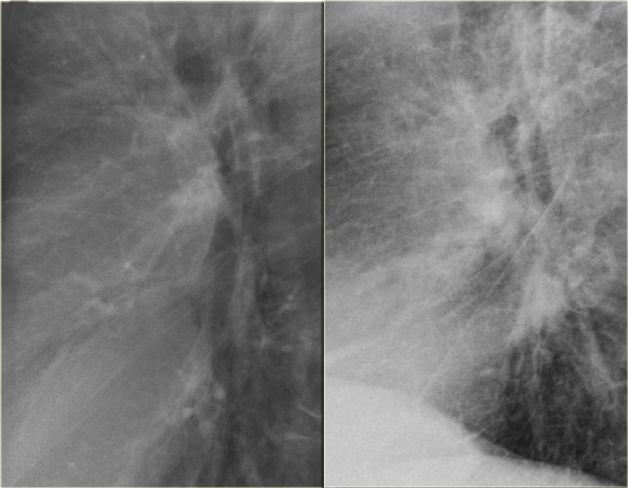 Previous normal chest x-ray (left) and CHF stage II with perihilar haze (right)X-quang ngực trước đây bình thường (trái) và CHF giai đoạn II với sương mù quanh răng (phải)
On the left another patient with congestive heart failure.
The lateral view nicely demonstrates the increased diameter of the pulmonary vessels and the hazy contours.
Notice also the septal lines and the accentuated interstitium.
Furthermore the fissura major is markedly thickened.Bên trái là một bệnh nhân khác bị suy tim sung huyết.
Mặt cắt ngang cho thấy rõ đường kính mạch phổi tăng lên và các đường viền mờ.
Chú ý đến các đường vách ngăn và các khoảng kẽ nổi bật.
Hơn nữa, phần lớn của cơ quan này dày lên rõ rệt.
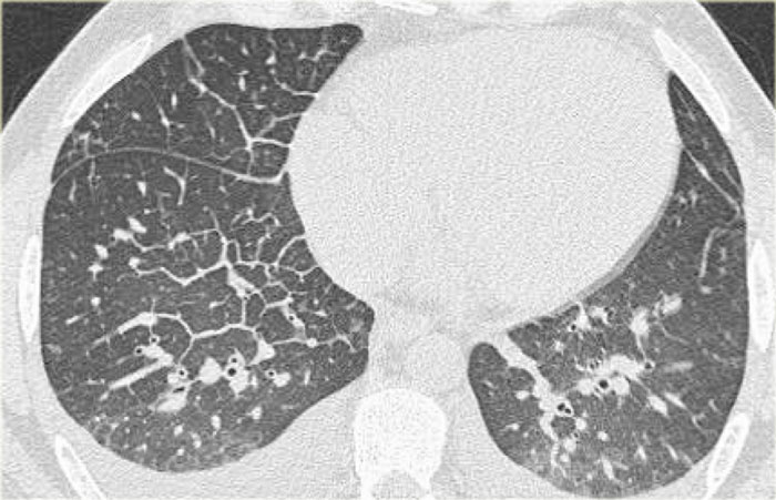 Thickened septal lines due to interstitial edema in CHFPhù mô kẽ vách liên thất dày trên bệnh nhân suy tim mạn tính
CT will also demonstrate signs of congestive heart failure. CT cũng sẽ cho thấy các dấu hiệu của suy tim sung huyết.
On the image on the left notice the following:Trên hình ảnh bên trái lưu ý những điều sau:
- Thickened septal lines due to interstitial edema Dày vách ngăn do phù kẽ
- Subtle ground glass opacity in the dependent part of the lungs (HU difference of 100-150 between the dependent and non-dependent part of the lung). Đám mờ kính mờ tinh tế ở phần phụ thuộc của phổi (sự khác biệt HU từ 100-150 giữa phần phụ thuộc và không phụ thuộc của phế quản).
- Bilateral pleural fluid. Dịch màng phổi hai bên.
In a patient with a known malignancy lymphangitic carcinomatosis would be high in the differential diagnostic list.
Ground glass opacity is the first presentation of alveolar edema and a precursor of consolidation.Ở bệnh nhân có carcinôm tế bào lymphô ác tính đã biết sẽ có tỷ lệ cao trong danh sách chẩn đoán phân biệt.
đám mờ kính mờ là biểu hiện đầu tiên của phù phế nang và là tiền thân của đông đặc.
Stage III - Alveolar edemaGiai đoạn III - Phù phế nang
This stage is characterized by continued fluid leakage into the interstitium, which cannot be compensated by lymphatic drainage.
This eventually leads to fluid leakage in the alveoli (alveolar edema) and to leakage into the pleural space (pleural effusion).
The distribution of the alveolar edema can be influenced by: Giai đoạn này được đặc trưng bởi sự rò rỉ dịch liên tục vào mô kẽ, không thể bù lại bằng dẫn lưu bạch huyết.
Điều này cuối cùng dẫn đến rò rỉ dịch trong phế nang (phù phế nang) và rò rĩ vào khoang màng phổi (tràn dịch màng phổi).
Sự phân bố phù phế nang có thể bị ảnh hưởng bởi:
- Gravity: supine or erect position and right or left decubitus position Trọng lực: tư thế nằm ngửa hoặc đứng thẳng và tư thế nằm nghiêng sang phải hoặc trái
- Obstructive lung disease, i.e. fluid leakage into the less severe diseased areas of the lung Bệnh phổi tắc nghẽn, tức là rò rỉ dịch vào các vùng bệnh ít nghiêm trọng hơn của phổi
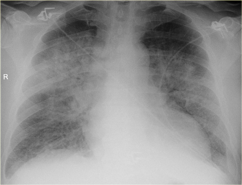
On the left a patient who was admitted with severe dyspnoe due to acute heart failure.
The following signs indicate heart failure: alveolar edema with perihilar consolidations and air bronchograms (yellow arrows); pleural fluid (blue arrow); prominent azygos vein and increased width of the vascular pedicle (red arrow) and an enlarged cardiac silhouette (arrow heads).
After treatment we can still see an enlarged cardiac silhouette, pleural fluid and redistribution of the pulmonary blood flow, but the edema has resolved.
Bên trái là một bệnh nhân nhập viện với triệu chứng khó thở nặng do suy tim cấp.
Các dấu hiệu sau đây cho thấy suy tim: phù phế nang với sự đông đặc quanh rốn phổi và khí phế quản đồ (mũi tên màu vàng); dịch màng phổi (mũI tên màu xanh); tĩnh mạch đơn nổi và tăng chiều rộng của cuống mạch (mũ I tên màu đỏ) và hình bóng tim to (đầu mũi tên).
Sau khi điều trị chúng ta vẫn có thể thấy hình bóng tim to, dịch màng phổi và phân phối lại lưu lượng máu phổi, nhưng phù đã hết.
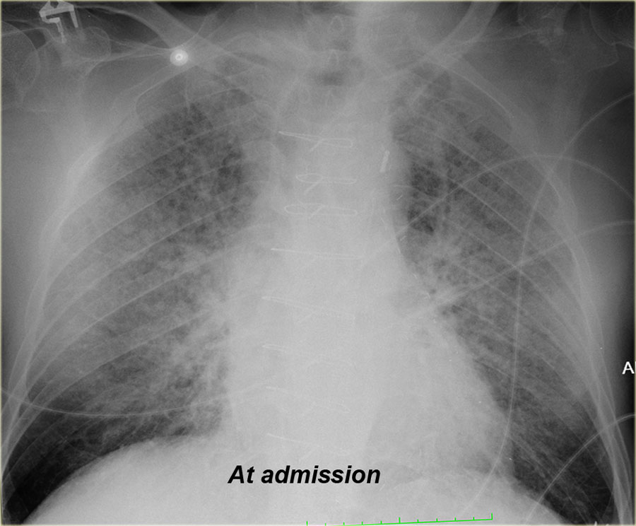
On the left another patient with alveolar edema at admission, which resolved after treatment.
When you scroll through the images and go back and forth, you will notice the difference in vascular pedicle width and distribution of pulmonary flow.Bên trái là một bệnh nhân khác có phù phế nang lúc nhập viện, khỏi sau điều trị.
Khi bạn cuộn qua các hình ảnh và đi tới đi lui, bạn sẽ nhận thấy sự khác biệt về chiều rộng cuống mạch và sự phân bố của dòng chảy phổi.
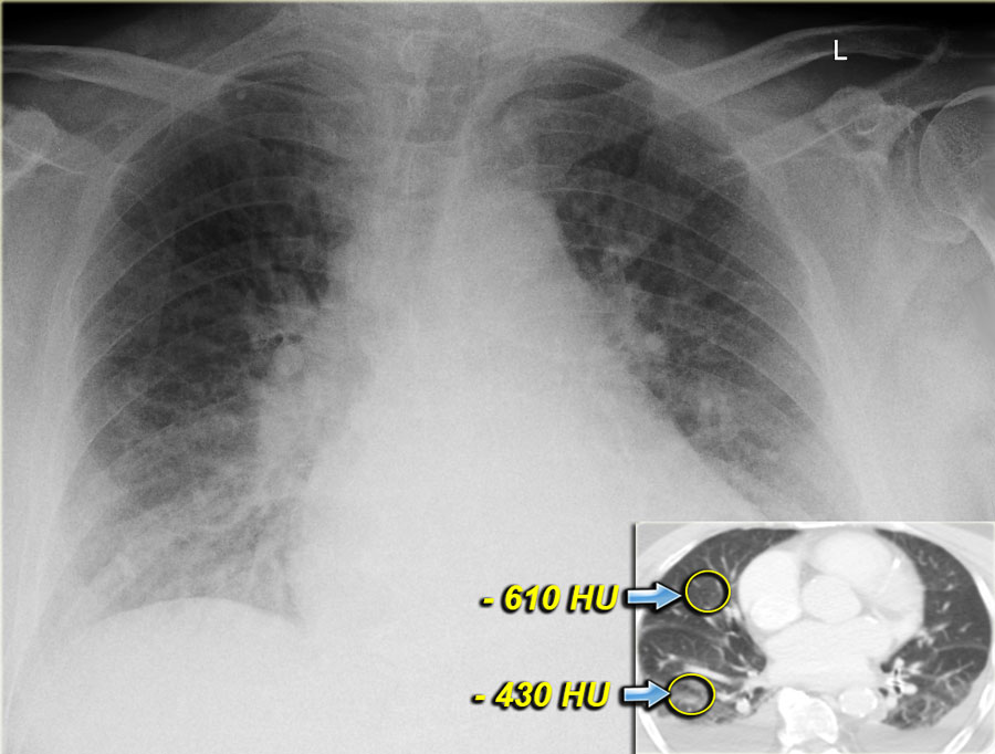

Both on the chest x-ray and on the CT the edema is gravity dependent and differences in density can be measured.Cả trên X-quang ngực và trên CT, phù nề phụ thuộc vào trọng lực và có thể đo được sự khác biệt về mật độ.
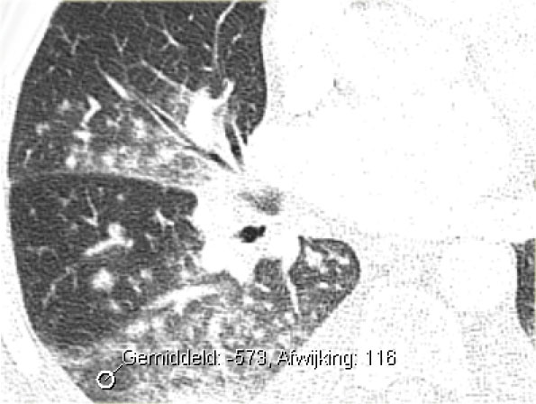

Notice that even within each lobe there is a gravity dependent difference in density.
This is only seen when the consolidations are the result of transudate like in CHF.
This is not seen when the consolidations are the result of exsudate due to infection, blood due to hemorrhage or when there is a capillary leak like in ARDS.Lưu ý rằng ngay cả trong mỗi thuỳ cũng có sự khác biệt về mật độ phụ thuộc vào trọng lực.
Điều này chỉ thấy khi các khối đông đặc là kết quả của dịch ngấm như trong CHF.
Điều này không thấy được khi đông đặc là kết quả của xuất tiết do nhiễm trùng, máu do xuất huyết hoặc khi có rò rỉ mao mạch như trong ARDS.
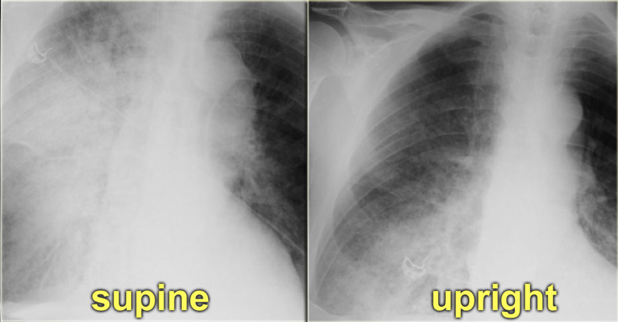

On the left a patient who first had a chest film in a supine position.
Notice the pulmonary edema, which is almost exclusively seen in the right lung.
A possible explanation for this phenomenon could be, that the patient had been lying on his right side for a while before the x-ray was taken.
Bên trái là bệnh nhân có phim ngực lần đầu tiên ở tư thế nằm ngửa.
Chú ý đến phù phổi, hầu như chỉ thấy ở phổi phải.
Một lời giải thích có thể cho hiện tượng này có thể là bệnh nhân đã nằm nghiêng bên phải một thời gian trước khi chụp X-quang.
Cardiothoracic ratioTỉ lệ tim phổi
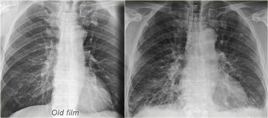
 Old film for comparison (left) CHF with redistribution, interstitial edema and some pleural fluidPhim cũ để so sánh (trái) CHF với phân bố lại, phù mô kẽ và một số dịch màng phổi
The cardiothoracic ratio (CTR) is the ratio of the transverse diameter of the heart to the internal diameter of the chest at its widest point just above the dome of the diaphragm as measured on a PA chest film.
An increased cardiac silhouette is almost always the result of cardiomegaly, but occasionally it is due to pericardial effusion or even fat deposition.
The heart size is considered too large when the CTR is > 50% on a PA chest x-ray.
A CTR of > 50% has a sensitivity of 50% for CHF and a specificity of 75-80%.
An increase in left ventricular volume of at least 66% is necessary before it is noticeable on a chest x-ray. Tỷ số tim-lồng ngực (CTR) là tỷ số giữa đường kính ngang của tim với đường kính trong của ngực tại điểm rộng nhất của tim ngay trên vòm cơ hoành được đo trên máy PA phim ngực.
Tăng bóng tim hầu như luôn là kết quả của bệnh tim to, nhưng đôi khi nó là do tràn dịch màng ngoài tim hoặc thậm chí lắng đọng mỡ.
Kích thước tim được coi là quá lớn khi CTR > 50% trên PA X-quang ngực.
CTR > 50% có độ nhạy 50% đối với CHF và độ đặc hiệu 75-80%.
Sự gia tăng thể tích thất trái ít nhất 66% là cần thiết trước khi có thể nhận thấy trên X-quang ngực.
On the left a patient with CHF.
There is an increase in heart size compared to the old film.
Other signs of CHF are visible, such as redistribution of pulmonary flow, interstitial edema and some pleural fluid. Bên trái là một bệnh nhân suy tim mạn tính.
Có sự gia tăng kích thước tim so với phim cũ.
Các dấu hiệu khác của CHF có thể nhìn thấy, chẳng hạn như sự phân bố lại lưu lượng phổi, phù nề mô kẽ và một số dịch màng phổi.
On a supine film the cardiac silhouette will be larger due to magnification and high position of the hemidiafragms.
Exact measurements are not that helpful, but comparison to old supine films can be of value.Trên phim ngửa, hình bóng tim sẽ lớn hơn do độ phóng đại và vị trí cao của các mảnh ghép.
Các phép đo chính xác không hữu ích, nhưng so sánh với các phim cũ nằm ngửa có thể có giá trị.
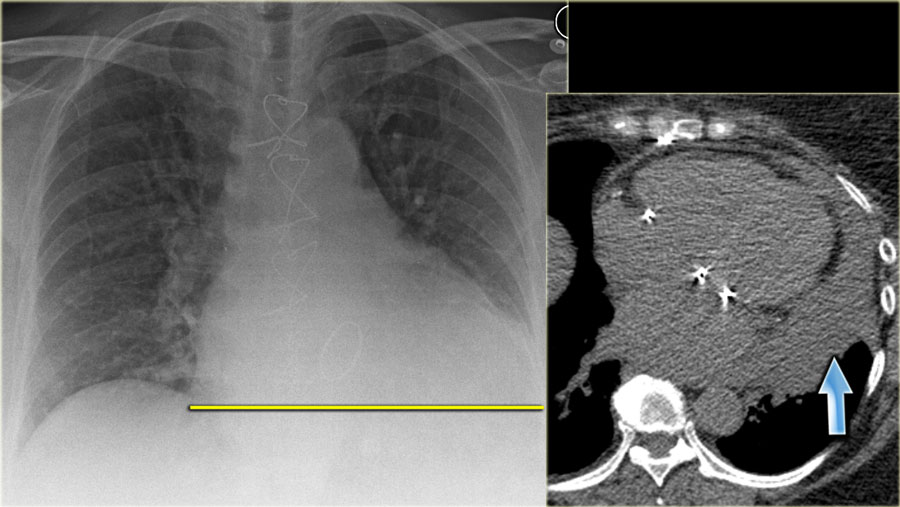 Increased CTR due to pericardial effusionTăng CTR do tràn dịch màng ngoài tim
On the left a patient, who recently underwent a valve replacement.
There is a large cardiac silhouette, which could be the result of cardiomegaly.
Because of the recent cardiac surgery, the possibility of pericardial effusion was taken into account, which is nicely demonstrated on the CT-image.Bên trái là một bệnh nhân, vừa trải qua thay van tim.
Có một hình bóng tim lớn, có thể là kết quả của bệnh tim to.
Do phẫu thuật tim gần đây, khả năng tràn dịch màng ngoài tim đã được tính đến, điều này được chứng minh rõ ràng trên hình ảnh CT.
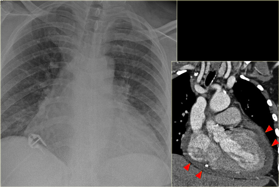
On the left another patient with a large cardiac silhouette on the chest x-ray due to pericardial effusion.
Pericardial effusion is demonstrated on the coronal CT-reconstruction.Bên trái một bệnh nhân khác có hình bóng tim lớn trên X-quang ngực do tràn dịch màng ngoài tim.
Tràn dịch màng ngoài tim được biểu hiện trên CT-reconstruction mạch vành.
Pleural effusionTràn dịch màng phổi
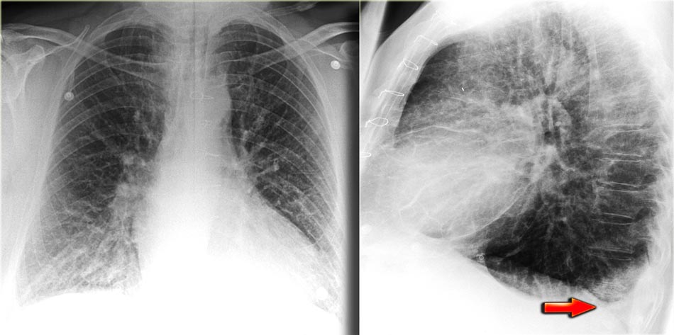 Pleural effusion more evident on lateral viewTràn dịch màng phổi rõ ràng hơn trên phim nghiêng
Pleural effusion is bilateral in 70% of cases of CHF.
When unilateral, it is slightly more often on the right side than on the left side.
There has to be at least 175 ml of pleural fluid, before it will be visible on a PA image as a meniscus in the costophrenic angle.
On a lateral image effusion of > 75 ml can be visible.
If pleural effusion is seen on a supine chest film, it means that there is at least 500 ml present.Tràn dịch màng phổi là hai bên trong 70% trường hợp CHF.
Khi ở một bên, nó thường ở bên phải hơn bên trái.
Phải có ít nhất 175 ml dịch màng phổi, trước khi nó có thể nhìn thấy trên hình ảnh PA như sụn chêm ở góc sườn-phổi.
Trên hình ảnh bên, tràn dịch > 75 ml có thể nhìn thấy được.
Nếu tràn dịch màng phổi được nhìn thấy trên phim ngực nằm ngửa, điều đó có nghĩa là có ít nhất 500 ml.
On the left images of a patient who has bilateral pleural effusions.
Notice that it is more evident on the lateral view.Trên hình ảnh bên trái của một bệnh nhân bị tràn dịch màng phổi hai bên.
Lưu ý rằng nó rõ ràng hơn ở góc nhìn bên.
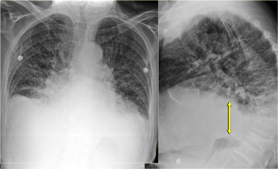 Subpulmonic pleural effusion with increased distance of the stomach air bubble to the lung base (arrow)Tràn dịch màng phổi dưới động mạch phổi với khoảng cách từ bóng khí dạ dày đến đáy phổi (mũi tên) tăng dần
Pleural effusion is not always visible as a meniscus in the costophrenic angle.
A subpulmonic effusion may follow the contour of the diaphragm making it tricky to discern.
In these cases, the only way to detect pleural effusion, is when you notice that there is an increased distance between the stomach bubble and the lung.
The stomach is normally located directly under the diaphragm, so, on an erect PA radiograph, the stomach bubble should always appear in close proximity to the diaphragm and the lung.
Tràn dịch màng phổi không phải lúc nào cũng nhìn thấy được như sụn chêm ở góc sườn-đầu-mắt.
Tràn dịch dưới động mạch phổi có thể đi theo đường viền của cơ hoành, khó phân biệt.
Trong những trường hợp này, cách duy nhất để phát hiện tràn dịch màng phổi, là khi bạn nhận thấy có sự gia tăng khoảng cách giữa bong bóng dạ dày và phổi.
Dạ dày thường nằm ngay dưới cơ hoành, vì vậy, trên phim X quang PA thẳng, bong bóng dạ dày phải luôn xuất hiện gần cơ hoành và phổi.
On the left images of a patient with signs of CHF.
At first glance you might get the impression that there is a high position of the diaphragm.
However when you notice the increased distance of the stomach air bubble to the lung base, you realize that there is a large amount of pleural fluid on both sides (arrow). Trên hình ảnh bên trái của một bệnh nhân có dấu hiệu CHF.
Thoạt nhìn bạn có thể có ấn tượng rằng có một vị trí cao của cơ hoành.
Tuy nhiên, khi bạn nhận thấy khoảng cách của bong bóng khí dạ dày với đáy phổi tăng lên, bạn nhận ra rằng có một lượng lớn dịch màng phổi ở cả hai bên (mũi tên).
Vascular pedicleCuống mạch
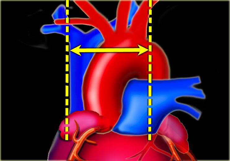
The vascular pedicle is bordered on the right by the superior vena cava and on the left by the left subclavian artery origin (6).
The vascular pedicle is an indicator of the intravascular volume.
A vascular pedicle width less than 60 mm on a PA chest radiograph is seen in 90% of normal chest x-rays.
A vascular pedicle width of more than 85 mm is pathologic in 80% of cases.
5 mm increase in diameter corresponds to 1 liter increase of intravascular fluid.
An increase in width of the vascular pedicle is accompanied by an increased width of the azygos vein.Cuống mạch được bao quanh bên phải bởi tĩnh mạch chủ trên và bên trái bởi gốc động mạch dưới đòn trái (6).
Cuống mạch là một chỉ số về thể tích nội mạch.
Chiều rộng cuống mạch nhỏ hơn 60 mm trên X quang phổi được thấy trong 90% các phim X-quang ngực bình thường.
Độ rộng cuống mạch trên 85 mm là bệnh lý trong 80% trường hợp.
Đường kính tăng 5 mm tương ứng với tăng 1 lít dịch nội mạch.
Sự gia tăng chiều rộng của cuống mạch đi kèm với sự gia tăng chiều ngang của tĩnh mạch đơn.
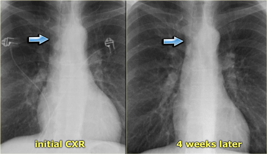
 Subtle increased width of vascular pedicle (left) and normalisation (right)Tăng chiều rộng của cuống mạch (trái) và bình thường hóa (phải)
There are three principal varieties of pulmonary edema: cardiac,
overhydration and increased capillary permeability (ARDS).
The vascular pedicle width (VPW) can help in differentiating these different forms of pulmonary edema (6):Có ba loại phù phổi chính: tim, quá mẫn nước và tăng tính thấm mao mạch (ARDS).
Chiều rộng cuống mạch (VPW) có thể giúp phân biệt các dạng phù phổi khác nhau này (6):
- Normal VPW: most common in capillary permeability or acute cardiac failure. VPW bình thường: thường gặp nhất trong rối loạn thẩm thấu mao mạch hoặc suy tim cấp.
- Widened VPW: most common in overhydration/renal failure and chronic cardiac failure. VPW giãn rộng: thường gặp nhất trong tình trạng thừa nước / suy thận và suy tim mạn tính.
- Narrowed VPW: most common in capillary permeability.VPW hẹp: thường gặp nhất trong tính thấm mao mạch.
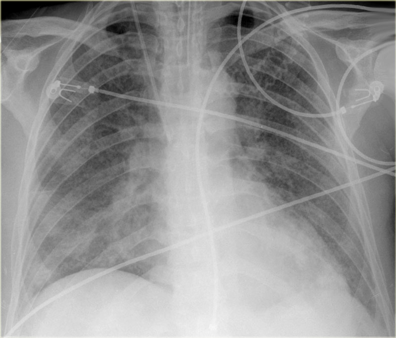 Normal VPW in a patient with ARDSVPW bình thường ở bệnh nhân ARDS
On the left a patient with ARDS.
There is alveolar edema in both lungs.
Notice that the VPW is normal.
The vessels in the upper lobes are not dilated and the cardiac silhouette is not enlarged.Bên trái là bệnh nhân ARDS.
Có phù phế nang ở cả hai phổi.
Lưu ý rằng VPW là bình thường.
Các mạch máu ở thuỳ trên không giãn và hình bóng tim không to ra.
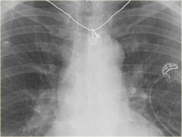
The VPW is best used as a measure to compare serial chest x-rays of the same patient, as there is a wide range of values for the VPW.
The VPW may increase due to rotation to the right.
On an AP-view the VPW will increase 20% compared to a PA-view.VPW được sử dụng tốt nhất như một thước đo để so sánh các phim X-quang ngực nối tiếp của cùng một bệnh nhân, vì có một loạt các giá trị cho VPW.
VPW có thể tăng do xoay sang phải.
Trên phim AP-view, VPW sẽ tăng 20% so với phim PA-view.
On the left a patient with subtle signs of congestive heart failure on the initial chest x-ray (image 1/2).
There is a slightly enlarged vascular pedicle, which becomes more obvious when you compare to the chest film after diuretic therapy (image 2/2).Bên trái một bệnh nhân có dấu hiệu suy tim sung huyết trên X-quang ngực ban đầu (hình 1/2).
Có một cuống mạch hơi to, trở nên rõ ràng hơn khi bạn so sánh với phim ngực sau khi điều trị bằng thuốc lợi tiểu (hình 2/2).
Dilatation of azygos veinTĩnh mạch đơn nhân giãn
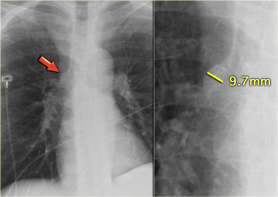

Dilation of the azygos vein is a sign of increased right atrial pressure and is usually seen when there is also an increase in the width of the vascular pedicle.
The diameter of the azygos vein varies according to the positioning.
In the standing position a diameter > 7 mm is most likely abnormal and a diameter > 10 mm is definitely abnormal.
In a supine patient > 15 mm is abnormal.
An increase of 3 mm in comparison to previous films is suggestive of fluid overload.
The difference of the azygos diameter on an inspiration film compared to an expiration film is only 1mm.
This means that the diameter of the azygos is a valuable tool whether or not there is good inspiration. Tĩnh mạch đơn tĩnh mạch giãn là dấu hiệu của tăng áp lực nhĩ phải và thường thấy khi có sự gia tăng chiều rộng của cuống mạch.
Đường kính của tĩnh mạch đơn thay đổi tuỳ theo vị trí.
Ở tư thế đứng, đường kính > 7 mm rất có thể là bất thường và đường kính > 10 mm chắc chắn là bất thường.
Ở bệnh nhân nằm ngửa > 15 mm là bất thường.
Sự gia tăng 3 mm so với các phim trước đó gợi ý tình trạng quá tải dịch.
Sự khác biệt về đường kính đơn nhân trên phim hít vào so với phim thở ra chỉ là 1 mm.
Điều này có nghĩa là đường kính của đơn âm là một công cụ có giá trị cho dù có cảm hứng tốt hay không.
Right ventricular failureSuy thất phải
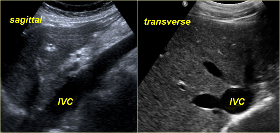
 Dilatation of IVC and hepatic veins on US images in a patient with RV failureNghiên cứu giãn tĩnh mạch trung tâm và tĩnh mạch gan trên hình ảnh siêu âm ở bệnh nhân suy thất phải
RV failure is most commonly caused by longstanding LV failure, which increases the pulmonary venous pressure and leads to pulmonary arterial hypertension, thus overloading the RV.
Other less common causes of RV failure are: Suy thất phải thường do suy thất trái kéo dài, làm tăng áp lực tĩnh mạch phổi và dẫn đến tăng áp động mạch phổi, do đó làm quá tải thất phải.
Các nguyên nhân khác ít phổ biến hơn của suy thất phải là:
- Severe lung disorder (cor pulmonale) Rối loạn phổi nặng (cor pulmonale)
- Multiple pulmonary emboliNhiều cục nghẽn động mạch phổi
- RV infarctionNhồi máu cơ tim
- Primary pulmonary hypertension Tăng áp phổi chính
- Tricuspid regurgitation or stenosis, mitral stenosis and pulmonary valve stenosis.Hở van ba lá hoặc hẹp van ba lá, hẹp van hai lá và hẹp van động mạch phổi.
Radiographic signs of RV failure: Các dấu hiệu X quang thất bại thất phải:
- Increased VPW due to dilatation of the superior vena cava Tăng áp lực tĩnh mạch cửa do giãn tĩnh mạch chủ trên
- Dilatation of azygos vein Tĩnh mạch đơn nhân giãn
- Dilatation of the right atrium Sự giãn nở của tâm nhĩ phải
- In many cases there will be both signs of RV and LV failure Trong nhiều trường hợp sẽ có cả hai dấu hiệu của suy thất phải và thất trái
Sonographic signs of RV failure: Các dấu hiệu siêu âm thất bại thất phải:
- Dilatation of the inferior vena cava (IVC) and hepatic veins Sự giãn tĩnh mạch chủ dưới (IVC) và tĩnh mạch gan
- Hepatomegaly Gan to
- AscitesCổ trướng
The indication for ultrasound examination in many of these patients is abnormal liver function tests.
It is therefore important to consider the possibility of RV failure when a patient presents with liver enzyme abnormalities.Chỉ định siêu âm ở nhiều bệnh nhân này là xét nghiệm chức năng gan bất thường.
Do đó, điều quan trọng là phải xem xét khả năng suy tim thất phải khi bệnh nhân có bất thường về men gan.
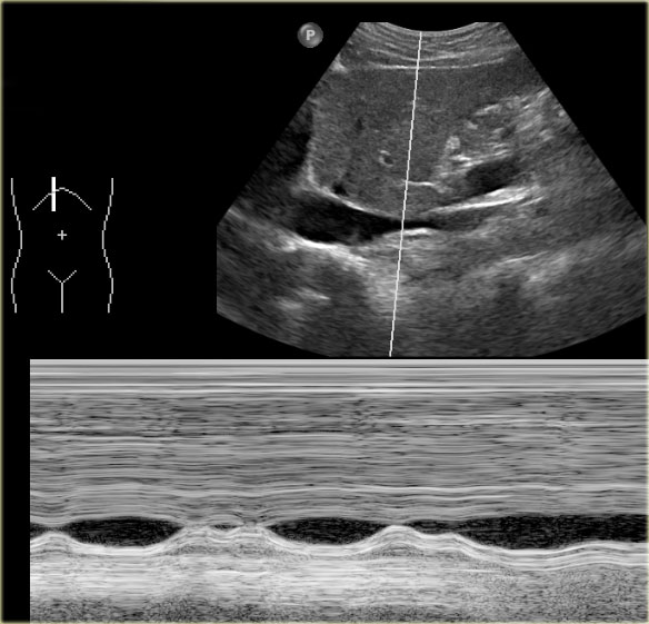
Under normal conditions dynamic ultrasound will demonstrate changes in caliber of the IVC.
These changes in caliber can be attributed to variations in blood flow in the IVC in accordance with the respiratory and cardiac cycles.
Trong điều kiện bình thường, siêu âm động sẽ cho thấy những thay đổi về kích thước của IVC.
Những thay đổi về tầm cỡ này có thể là do sự thay đổi lưu lượng máu trong IVC phù hợp với chu kỳ hô hấp và chu kỳ tim.
ReferencesTài liệu tham khảo
-
Heart Failure (HF) (Congestive Heart Failure)
Suy tim (HF) (Suy tim sung huyết)
in Merck manual trong Merck hướng dẫn sử dụng
-
Radiographic analysis of vascular distribution: a review (PDF)
Phân tích hình ảnh X quang về sự phân bố mạch máu: tổng quan (PDF)
by Carle Ravin
Presented as part of a Conference on Chest Radiology 1982
bởi Carle Ravin -
Pulmonary artery-bronchus ratios in patients with normal lungs, pulmonary vascular plethora, and congestive heart failure.
Tỷ lệ động mạch phổi-phế quản ở bệnh nhân có phổi bình thường, tràn dịch màng phổi nhiều và suy tim sung huyết.
by J H WoodringApril 1991 Radiology, 179, 115-122. của J H Woodringapril 1991 X quang, 179, 115-122.
- American College of Radiology ACR Appropriateness Criteria: Congestive Heart Failure Tiêu chí phù hợp của ACR của Trường Cao đẳng X quang Hoa Kỳ: Suy tim sung huyết
-
The Radiologic Distinction of Cardiogenic and Noncardiogenic Edema (PDF)
Phân biệt X quang của phù tim và không do tim (PDF)
by Eric Milne et al
American Journal of Roentgenology, Vol 144, Issue 5, 879-894
bởi Eric Milne và cộng sự -
The vascular pedicle of the heart and the vena azygos. Part II: Acquired heart disease.
Cuống mạch của tim và tĩnh mạch đơn nhân Phần II: Bệnh tim mắc phải.
by M Pistolesi, E N Milne, M Miniati and C Giuntini
July 1984 Radiology, 152, 9-17.
bởi M. Pistolesi, E. N. Milne, M. Miniati và C. Giuntini -
Pulmonary hypertension secondary to left-sided heart disease: a cause for ventilation-perfusion mismatch mimicking pulmonary embolism.
Tăng áp phổi thứ phát do bệnh tim trái: nguyên nhân gây ra sự không phù hợp giữa thông khí và tưới máu giống thuyên tắc phổi.
by Au VW, Jones DN, Slavotinek JP.
British Journal of Radiology. 2001 Jan; 74(877) 86-88.
bởi Au VW, Jones DN, Slavotinek JP. -
The pulmonary vessels in incipient left ventricular decompensation
Mạch máu phổi trong suy tim cấp do mất bù thất trái
by M. Simon
Circulation 1961; 24:185-190
bởi M. Simon -
Pulmonary vascular congestion in acute myocardial infarction: Hemodynamic and radiologic correlation
Tắc mạch phổi trong bệnh cơ tim cấp nhồi máu: Mối tương quan huyết động và X quang
by McHugh, T. J., Forrester, J. S., Adler, L., et al. Ann. Intern. Med.,
1972; 76:29-33
bởi McHugh, T. J., Forrester, J. S., Adler, L., và cộng sự Ann. Thực tập y khoa, -
Radiological detection of clinically occult cardiac failure following myocardial infarction
Phát hiện suy tim ẩn trên lâm sàng sau phẫu thuật cơ tim nhồi máu
Harrison M.O., Conte P.J., Heitzman E.R.
Br J Radiol. 1971; 44:265-272
-
Absolute lungdensity in experimental canine pulmonary edema
Đặc điểm phổi tuyệt đối trong phù phổi chó thực nghiệm
by Gamsu G,Kaufman K ,Swann S ,Brito A.
Invest Radiol 1979 ; 14: 261-269
Gamsu G.,Kaufman K.,Swann S.,Brito A. -
Aging and the respiratory system
Lão hóa và hệ hô hấp
Bonomo L., et al.
Radiologic Clinics of North America, vol. 46, nr 4, July 2008: 685-702
Bonomo L. và cộng sự.