
Sella Turcica and Parasellar RegionHố yên và vùng cạnh yên
Radiology department of the University of Toronto, Canada and the Radiology department the Medical Centre Alkmaar, the NetherlandsKhoa X quang của Đại học Toronto, Canada và khoa X quang của Trung tâm Y tế Alkmaar, Hà Lan
Ngày đăng / cập nhật: 2008-08-10
Bản gốc trên radiologyassistant.nl ↗ · tải về lúc 2026-11-19 20:19 · bản dịch máy có chuẩn hóa thuật ngữ chẩn đoán hình ảnh
This review is based on a presentation given by Walter Kucharczyka and was adapted for the Radiology Assistant by Marieke Hazewinkel.
In this review a systematic anatomic approach to differential diagnosis of a sellar or parasellar mass is described.
By clicking on one of the subjects in the list on the left, you will go directly to this item.
If you have printing problems with the margins of the document, you may have to adjust the margins in the page set up of your internet browser, which you will find in the top left of the menu bar.Bài tổng quan này dựa trên một bài thuyết trình của Walter Kucharczyka và được Marieke Hazewinkel chuyển thể cho Trợ lý Chẩn đoán hình ảnh.
Trong bài tổng quan này, chúng tôi trình bày cách tiếp cận giải phẫu hệ thống đối với chẩn đoán phân biệt của khối u xương bánh chè hoặc cạnh bánh chè.
Bằng cách nhấp vào một trong các đối tượng trong danh sách bên trái, bạn sẽ đi trực tiếp đến mục này.
Nếu bạn gặp vấn đề khi in lề tài liệu, bạn có thể phải điều chỉnh lề trong cài đặt trang của trình duyệt internet, bạn sẽ tìm thấy ở phía trên bên trái của thanh menu.
Anatomic Approach to Differential DiagnosisPhương pháp tiếp cận giải phẫu chẩn đoán phân biệt
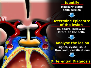

In order to analyze a sellar or parasellar mass on MRI we use the following anatomic approach: Để phân tích khối u xương bánh chè hoặc cạnh bánh chè trên MRI chúng tôi sử dụng phương pháp giải phẫu sau:
- First identify the pituitary gland and sella turcica. Đầu tiên xác định tuyến yên và hố yên.
- Then determine the epicenter of the lesion and whether it is in the sella or above, below or lateral to the sella. Sau đó xác định tâm chấn thương của tổn thương và liệu nó ở trong hố yên hay ở trên, dưới hay bên hố yên.
- If it is in the sella, determine whether or not the sella is enlarged. Nếu nó nằm trong hố yên, hãy xác định xem hố yên có to lên hay không.
- Once the location of the mass is clear, analyze the signal intensity patterns: is the lesion cystic or solid? Khi vị trí của khối u đã rõ ràng, phân tích các mẫu cường độ tín hiệu: là tổn thương dạng nang hay dạng đặc?
- Does it contain any abnormal vessels? Nó có chứa bất kỳ mạch máu bất thường nào không?
- Are there any calcifications? And so on.Có vôi hóa nào không? Và cứ thế.
- Finally establish a Differential Diagnosis.Cuối cùng thiết lập một chẩn đoán phân biệt.

Pituitary gland
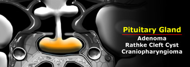
On a coronal section through the brain the reference structure is the pituitary gland which lies in the sella turcica.
It is usually larger in females than in males - in females the superior border tends to be convex, whereas in males it is usually concave.
The most common abnormalities that arise in the pituitary gland are pituitary adenoma, Rathke's cleft cyst and craniopharyngioma.
Tuyến yên
Trên một mặt cắt đứng ngang xuyên qua não, cấu trúc tham chiếu là tuyến yên nằm trong hố yên.
Nó thường lớn hơn ở nữ so với nam - ở nữ, bờ trên có xu hướng lồi, trong khi ở nam, nó thường lõm.
Các bất thường thường gặp nhất phát sinh ở tuyến yên là u tuyến tuyến yên, nang Rathke và u sọ hầu.
Pituitary stalk
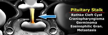
The next structure to identify is the pituitary stalk.
This is a vertically oriented structure which connects the pituitary gland to the brain.
It is thinner at the bottom and thicker at the top.
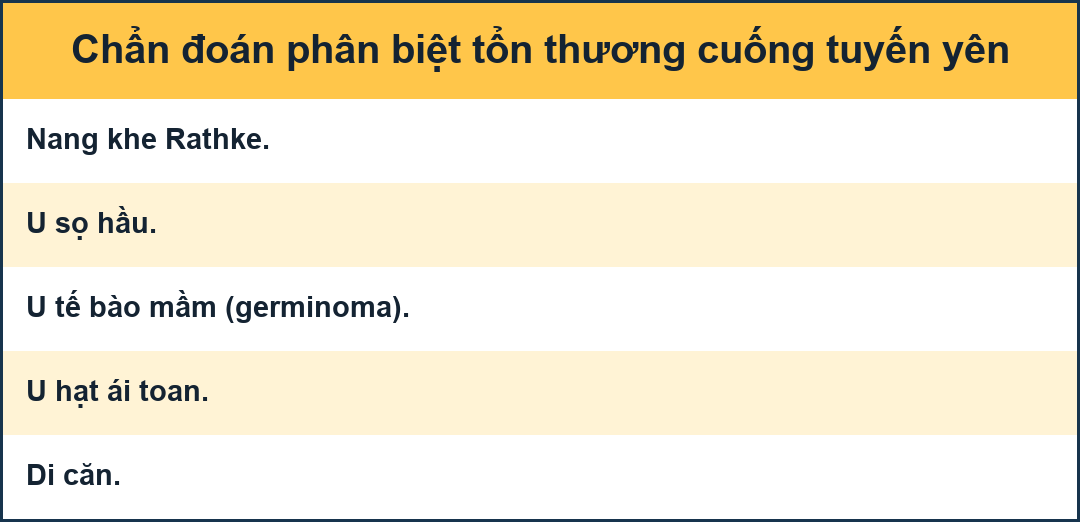
Embryologically, it is also derived from Rathke's cleft epithelium and therefore the pathologies, which can arise in the pituitary gland can also arise in the stalk.
There are a few unusual things to be considered in children, such as germinomas and eosinophilic granulomas.
In adults metastases and occasionally lymphoma can arise in the pituitary stalk.
Cuống tuyến yên
Cấu trúc tiếp theo cần xác định là cuống tuyến yên.
Đây là một cấu trúc theo chiều dọc kết nối tuyến yên với não.
Nó mỏng hơn ở phía dưới và dày hơn ở phía trên.
Về mặt phôi học, nó cũng có nguồn gốc từ biểu mô khe Rathke và do đó các bệnh lý, có thể phát sinh ở tuyến yên cũng có thể phát triển ở cuống.
Có một vài điều bất thường cần được xem xét ở trẻ em, chẳng hạn như u hạt và u hạt bạch cầu ái toan.
Ở người lớn, di căn và đôi khi u lympho có thể phát sinh ở cuống tuyến yên.
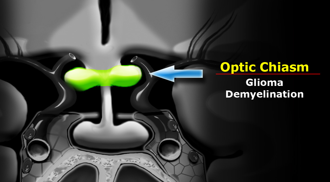

Optic chiasm
Another major structure in the suprasellar cistern is the optic chiasm.
It is an extension of the brain and looks like the number 8 lying on its side.
It is glial tissue - therefore the most common tumors to originate here are gliomas.
In the US and Europe another frequent pathology in this region is demyelinating disease - particularly multiple sclerosis.
This can also be associated with some swelling of the optic chiasm.
Giao thoa thị giác
Một cấu trúc chính khác trong bể chứa trên xương đòn là giao thoa thị giác.
Nó là một phần mở rộng của não và trông giống như số 8 nằm ở bên cạnh nó.
Đó là mô thần kinh đệm - do đó, khối u phổ biến nhất bắt nguồn từ đây là u thần kinh đệM.
Trong siêu âm và Châu Âu, một bệnh lý thường gặp khác ở khu vực này là bệnh mất myelin - đặc biệt là bệnh đa xơ cứng.
Điều này cũng có thể liên quan đến một số sưng nề của giao thoa thị giác.
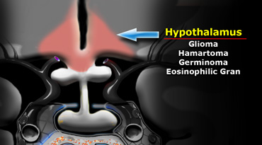

Hypothalamus
Further cephalad lies the base of the brain, which at this location is the hypothalamus.
Anatomically the hypothalamus forms the lateral walls and floor of the third ventricle.
The most common pathologies to arise here are gliomas - in children hamartomas, germinomas and eosinophilic granuloma.Vùng dưới đồi
Phần não dưới nằm ở đầu xa hơn, tại vị trí này là vùng dưới đồi.
Về mặt giải phẫu, vùng dưới đồi tạo thành các thành bên và sàn của tâm thất thứ ba.
Các bệnh lý thường gặp nhất là u thần kinh đệm - hamartomas ở trẻ em, u mầm và u hạt tăng bạch cầu ái toan.
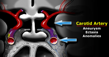

Carotid artery
A very important structure in this area is the internal carotid artery.
It runs a complex anatomic course as it passes through the skull base shaped like an S on lateral views.
It passes through the cavernous sinus.
The segment cranial to this is known as the supracavernous segment.
This bifurcates into the anterior cerebral artery, which passes cranially to the optic chiasm, and the middle cerebral artery, which runs laterally.
Aneurysms and ectasias are pathologies that can arise here.
One must also be aware of congenital variations in the course of the internal carotid
Sometimes it is very medially positioned and can actually lie in the midline.
Động mạch cảnh
Một cấu trúc rất quan trọng trong khu vực này là động mạch cảnh trong.
Nó chạy một đường giải phẫu phức tạp khi nó đi qua nền sọ có hình chữ S ở mặt bên.
Nó đi qua xoang hang.
Đoạn sọ này được gọi là đoạn trên hang.
Động mạch này chia đôi thành động mạch não trước, đi qua sọ đến giao thoa thị giác và động mạch não giữa, chạy theo chiều ngang.
Phình mạch và giãn mạch là những bệnh lý có thể phát sinh ở đây.
Cũng cần phải nhận thức được các biến thể bẩm sinh trong quá trình tiến triển của động mạch cảnh trong.
Đôi khi nó nằm ở vị trí rất trung gian và thực sự có thể nằm ở đường giữa.
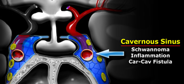

Cavernous sinus
The cavernous sinus is a paired complex of venous channels.
In the lateral wall of the sinus run nerve III (oculomotorius), IV (trochlearis), V1 and V2 (trigeminus).
The sixth cranial nerve (abducens) runs more medially and is located caudal to the carotid artery.
The most common pathologies occurring in the cavernous sinus include schwannomas arising from the cranial nerves and inflammation, which can lead to thrombosis.
This is known as cavernous sinus thrombophlebitis.
Carotid-cavernous fistulas are fistulous communications between the carotid artery and the veins of the cavernous sinus.
Xoang hang
Xoang hang là một phức hợp các kênh tĩnh mạch.
Trong thành bên của xoang chạy dây thần kinh III (oculomotorius), IV (trochlearis), V1 và V2 (trigeminus).
Dây thần kinh sọ thứ sáu (dây chằng chéo) chạy về phía trong và nằm ở đuôi động mạch cảnh.
Các bệnh lý phổ biến nhất xảy ra trong xoang hang bao gồm u dây thần kinh schwann phát sinh từ dây thần kinh sọ và viêm, có thể dẫn đến huyết khối.
Đây được gọi là viêm tắc tĩnh mạch xoang hang.
Rò động mạch cảnh-xoang hang là đường thông nối giữa động mạch cảnh và tĩnh mạch xoang hang.

Meninges
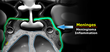
The meninges cover the cavernous sinus.
They are thicker laterally and superiorly than medially and inferiorly.
The most common tumor to arise from the meninges is of course the meningioma.
Dural metastasis is the second most common tumor to arise here.
Also inflammatory pathologies occur in the basal meninges - the most common infection being tuberculous meningitis.
Of the non-infectious inflammatory pathologies sarcoidosis is the commonest.
Trang chủ
Các màng não bao phủ xoang hang.
Chúng dày hơn ở bên và trên so với ở giữa và dưới.
U màng não là u thường gặp nhất.
Di căn màng cứng là khối u thường gặp thứ hai ở đây.
Ngoài ra các bệnh lý viêm xảy ra ở màng não đáy - nhiễm trùng phổ biến nhất là viêm màng não do lao.
Trong các bệnh lý viêm không nhiễm trùng, bệnh sarcoid là phổ biến nhất.
Sphenoid sinus
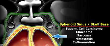
Inferior to the pituitary gland lies the sphenoid sinus.
This structure contains air and is lined by mucosa and bone.
Posterior to the sphenoid sinus lies the clivus (not shown on this coronal section through the brain).
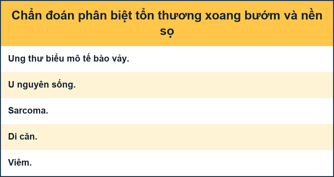
Pathology that arises in this area includes carcinomas arising from the mucosa of the sphenoid sinus - squamous cell carcinoma and adenoid cystic carcinoma are the most common.
Chordomas arise in the clivus and chondrosarcomas and osteosarcomas also occur in this area.
Metastases can occur anywhere.
Bacterial or fungal inflammatory processes in the sphenoid sinus can spread intracranially via the cavernous sinus.
Xoang bướm
Dưới tuyến yên là xoang bướm.
Cấu trúc này chứa không khí và được lót bởi niêm mạc và xương.
Phía sau xoang bướm là vùng âm hộ (không được hiển thị trên mặt cắt đứng ngang này qua não).
Bệnh lý phát sinh ở khu vực này bao gồm ung thư biểu mô phát sinh từ niêm mạc xoang bướm - ung thư biểu bì tế bào vảy và ung thư biểu Mô tuyến dạng nang là phổ biến nhất.
U sụn phát sinh ở vùng âm hộ và sarcom sụn và sarcom xương cũng xảy ra ở khu vực này.
Di căn có thể xảy ra ở bất cứ đâu.
Các quá trình viêm do vi khuẩn hoặc nấm trong xoang bướm có thể lan truyền trong nội sọ qua xoang hang.
Pituitary MicroadenomaVi u tuyến yên
 Pituitary MicroadenomaVi u tuyến yên
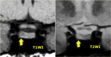
By definition, pituitary microadenomas are less than 10 mm in diameter and are located in the pituitary gland.
These images show a classic case: on T1 a lesion about 3-4 mm in diameter, slightly hypointense compared to normal pituitary tissue, located in the pituitary gland.
On T2, the lesion is slightly hyperintense.
The differential diagnosis: pituitary microadenoma or Rathke's cleft cyst (the two can be indistinguishable).
The sensitivity of an unenhanced MRI scan for detecting pituitary microadenomas is about 70%.
It is not always necessary to give intravenous contrast for detecting pituitary microadenomas as patients with a negative scan generally receive the same symptomatic treatment as patients with a microadenoma (usually these patients are women with symptoms of hyperprolactinemia).
The purpose of the scan is to rule out any large lesions.
In possible surgical candidates (for example patients with failed medical therapy or pituitary disease not amenable to medical therapy such as Cushing's disease) it is necessary to give contrast to localize the lesion as accurately as possible.
Theo định nghĩa, u tuyến yên nhỏ hơn 10 mm đường kính và nằm trong tuyến yên.
Hình ảnh này cho thấy một trường hợp kinh điển: trên T1 một tổn thương đường kính khoảng 3-4 mm, giảm tín hiệu nhẹ so với mô tuyến yên bình thường, nằm ở tuyến yên.
Ở T2, tổn thương có kích thước nhỏ tăng tín hiệu.
chẩn đoán phân biệt: u tuyến yên hoặc nang Rathke (hai loại này có thể không thể phân biệt được).
độ nhạy của chụp MRI không tăng cường để phát hiện các vi u tuyến yên là khoảng 70%.
Không phải lúc nào cũng cần phải tiêm thuốc cản quang tĩnh mạch để phát hiện u tuyến yên nhỏ vì bệnh nhân có kết quả chụp âm tính thường được điều trị triệu chứng giống như bệnh nhân có u tuyến yên (thường là những bệnh nhân này là phụ nữ có triệu chứng tăng prolactin máu).
Mục đích của việc chụp là để loại trừ bất kỳ tổn thương lớn nào.
Trong các trường hợp phẫu thuật có thể xảy ra (ví dụ bệnh nhân điều trị nội khoa thất bại hoặc bệnh tuyến yên không tuân thủ điều trị nội như bệnh Cushing), cần phải cho thuốc cản quang để xác định vị trí tổn thương càng chính xác càng tốt.
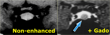
On an unenhanced scan, approximately 70% of all pituitary microadenomas can be detected.
If you give gadolinium, you can reduce the false-negative rate from 30% to 15%.
As mentioned earlier, this usually does not affect patient management.
Trên một lần chụp không tăng cường, có thể phát hiện khoảng 70% tất cả các u tuyến yên.
Nếu bạn cho gadolinium, bạn có thể giảm tỷ lệ âm tính giả từ 30% xuống 15%.
Như đã đề cập trước đó, điều này thường không ảnh hưởng đến việc quản lý bệnh nhân.
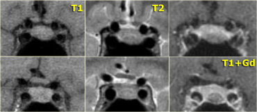
Coronal T1 and T2-weighted images and T1-weighted images before and after gadolinium.
In this patient the lesion in the pituitary gland is only detectable after the administration of intravenous contrast.
The differential diagnosis: pituitary microadenoma or Rathke's cleft cyst.
Hình ảnh T1 và T2 và T1 trước và sau khi tiêm gadolinium.
Ở bệnh nhân này, tổn thương ở tuyến yên chỉ có thể phát hiện được sau khi tiêm thuốc cản quang đường tĩnh mạch.
chẩn đoán phân biệt: u tuyến yên nhỏ hoặc nang Rathke.
Pituitary MacroadenomaU tuyến yên lớn
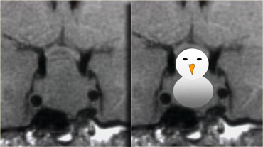
By definition, pituitary macroadenomas are adenomas over 10mm in size.
They tend to be soft, solid lesions, often with areas of necrosis or hemorrhage as they get bigger.
As they grow, they first expand the sella turcica and then grow upwards.
In this example of a pituitary macroadenoma there is suprasellar extension with elevation and compression of the optic chiasm.
Because they are soft tumors, they usually indent at the diaphragma sellae, giving them a 'snowman' configuration.
This is one feature that can help distinguish between a pituitary macroadenoma and a meningioma.
Another feature which can help differentiate them is enlargement of the sella turcica - this generally only occurs with pituitary macroadenomas that originate in the sella.
Theo định nghĩa, u tuyến yên lớn có kích thước trên 10 mm là các u tuyến.
Chúng có xu hướng là những tổn thương mềm, đặc, thường có các vùng hoại tử hoặc xuất huyết khi chúng lớn hơn.
Khi chúng lớn lên, đầu tiên chúng mở rộng hố yên và sau đó phát triển lên trên.
Trong ví dụ về u tuyến yên lớn này, có sự mở rộng trên yên với sự nâng cao và chèn ép của giao thoa thị giác.
Vì chúng là những khối u mềm, chúng thường thụt vào ở vùng yên, tạo cho chúng một cấu hình 'tuyết'.
Đây là một đặc điểm có thể giúp phân biệt giữa u tuyến yên và u màng não.
Một đặc điểm khác có thể giúp phân biệt chúng là sự mở rộng của hố yên - điều này thường chỉ xảy ra với các u tuyến yên lớn có nguồn gốc từ hố yên.
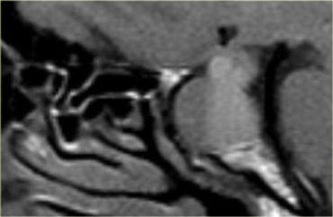
On the left another example of a pituitary macroadenoma.
The lesion starts in the sella, which is enlarged, and extends into the suprasellar cistern.
Note the classic 'snowman' configuration caused by constriction by the diaphragma sellae.
Notice the blood-fluid level, indicating hemorrhage.
Bên trái là một ví dụ khác về u tuyến yên lớn.
Tổn thương bắt đầu ở hố yên, được mở rộng, và kéo dài vào bể chứa trên hố yên.
Lưu ý cấu hình 'người tuyết' cổ điển gây ra bởi sự co thắt của cơ hoành yên.
Lưu ý mức độ dịch máu, cho thấy xuất huyết.
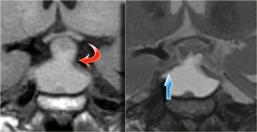
The usefulness of observing the inclination of the diaphragmatic leaflets was referred to earlier.
On the T2-weighted images on the right you can see that the leaflets are displaced upwards by this macroadenoma which started in the sella and is growing upwards.
A lesion originating above the sella and growing downwards would push the leaflets in the other direction (this can be seen with meningiomas for example).
Việc quan sát độ nghiêng của lá hoành đã được đề cập đến trước đó.
Trên hình ảnh xung T2W bên phải, bạn có thể thấy các lá rơi được di chuyển lên trên bởi khối u tuyến này bắt đầu ở hố yên và đang phát triển lên trên.
Một tổn thương bắt nguồn từ phía trên yên và phát triển xuống dưới sẽ đẩy các lá rơi theo hướng khác (điều này có thể được nhìn thấy với u màng não chẳng hạn).
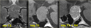
Usually the diagnosis of a macroadenoma is straightforward.
Sometimes a meningioma can give a similar appearance.
On the left an example of a meningioma.
Note there is no diaphragmatic constriction and there is uniform enhancement after the administration of intravenous gadolinium which is typical of meningioma.
Thông thường chẩn đoán u tuyến to rất đơn giản.
Đôi khi u màng não có thể cho hình ảnh tương tự.
Ở bên trái là một ví dụ về u màng não.
Lưu ý không có co thắt cơ hoành và có sự tăng cường đồng đều sau khi tiêm tĩnh mạch gadolinium, điển hình của u màng não.
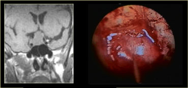
These images are of a transsphenoidal resection of a pituitary macroadenoma.
After the bony floor of the sella turcica has been removed, the dura is incised with a cruciate incision.
Những hình ảnh này là hình ảnh cắt bỏ u tuyến yên qua xoang bướm.
Sau khi sàn xương của hố yên đã được loại bỏ, màng cứng được rạch bằng một đường rạch chéo.
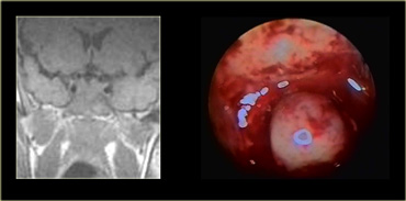
Because the pressure above the dura is larger than the pressure below, the macroadenoma then delivers itself into the sphenoid sinus.
Intra-operative MRI was performed in an experimental setting to determine whether the neurosurgeon had successfully removed all of the tumor.
Bởi vì áp lực trên màng cứng lớn hơn áp lực bên dưới, khối u lớn sau đó tự đưa vào xoang bướm.
MRI trong mổ được thực hiện trong một môi trường thực nghiệm để xác định xem phẫu thuật viên thần kinh đã lấy hết khối u thành công hay không.
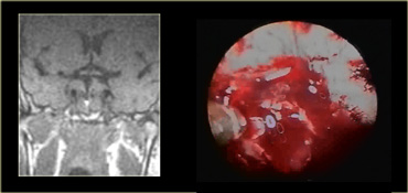
Because using this surgical approach means a limited field-of-view, it is important to know beforehand what it is you are operating on.
As we will see there are lesions you do not want to operate using this approach!Bởi vì sử dụng phương pháp phẫu thuật này có nghĩa là một lĩnh vực quan sát hạn chế, điều quan trọng là phải biết trước bạn đang phẫu thuật điều gì.
Như chúng ta sẽ thấy có những tổn thương mà bạn không muốn phẫu thuật bằng cách sử dụng phương pháp này!
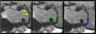
Another common pathway of extension is laterally into the cavernous sinus.
It is not always possible to tell if there is cavernous sinus invasion, but there are three signs to look out for:
-Is there more than 50% encirclement of the carotid artery? Note: meningiomas tend to constrict the carotid artery, macroadenomas do not.
-Is there lateral displacement of the lateral wall of the cavernous sinus compared to the opposite side?
-Is there an increased amount of tissue interposed between the carotid artery and the lateral wall of the cavernous sinus?
U cũng thường lan sang bên vào xoang hang. Đôi khi khó xác định u đã xâm lấn xoang hang hay chưa. Hãy tìm ba dấu hiệu sau: u bao quanh trên 50% chu vi động mạch cảnh, thành ngoài xoang hang bị đẩy ra ngoài so với bên đối diện, hoặc mô xen giữa động mạch cảnh và thành ngoài xoang hang tăng lên. U màng não thường làm hẹp động mạch cảnh, còn u tuyến yên lớn thường không.
At medical school they teach you that a rare manifestation of a common lesion is more likely than a rare abnormality.
Since pituitary adenomas are the most common lesions of the skull base, it is prudent to always include them in the differential diagnosis if you can not identify a normal pituitary gland when confronted with a mass in this region.
This patient presented with nasal obstruction.
She went to an ENT specialist who saw a large endonasal mass and she was referred to the neurosurgeon for planned major skull base resection.
The neurosurgeon had seen something similar before, and checked her prolactin-level.
This was 4000 (25 or less is normal). Endonasal biopsy revealed prolactinoma.
After treatment with bromocriptine the mass shrunk down and no surgery was necessary.
Ở trường y, họ dạy bạn rằng một biểu hiện hiếm gặp của một tổn thương thông thường có nhiều khả năng hơn là một bất thường hiếm gặp.
Vì các u tuyến của tuyến yên là tổn thương phổ biến nhất của nền sọ, nên thận trọng khi luôn đưa chúng vào chẩn đoán phân biệt nếu bạn không thể xác định được tuyến yên bình thường khi đối mặt với một khối ở khu vực này.
Bệnh nhân này có biểu hiện nghẹt mũi.
Cô đã đến gặp một chuyên gia tai mũi họng, người đã thấy một khối u lớn ở mũi và cô đã được chuyển đến bác sĩ phẫu thuật thần kinh để lên kế hoạch cắt bỏ nền sọ lớn.
Bác sĩ phẫu thuật thần kinh đã thấy một cái gì đó tương tự trước đây, và kiểm tra mức độ prolactin của cô ấy.
Đây là 4000 (25 hoặc thấp hơn là bình thường), sinh thiết mũi cho thấy prolactinoma.
Sau khi điều trị bằng bromocriptine khối u thu nhỏ lại và không cần phẫu thuật.
Rathke Cleft CystU nang khe hở Rathke
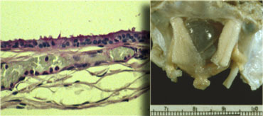
Rathke's cleft cyst is the second of three pathologies derived from Rathke's cleft epithelium.
The cyst is fluid-filled and has very thin walls with a thickness of only one or two cell layers.
This is illustrated by the microscopic image.
These walls can contain cells which secrete fluid, allowing the cyst to grow and compress adjacent structures.Nang Rathke là bệnh lý thứ hai trong ba bệnh lý có nguồn gốc từ biểu mô khe hở Rathke.
Nang chứa đầy dịch và có thành rất mỏng với độ dày chỉ một hoặc hai lớp tế bào.
Điều này được minh hoạ bằng hình ảnh hiển vi.
Những thành này có thể chứa các tế bào tiết dịch, cho phép nang phát triển và nén các cấu trúc lân cận.
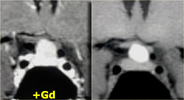
Rathke's cleft cysts can occur either in or above the sella turcica.
On the images above there is a normal pituitary gland, a normal optic chiasm and a normal carotid artery on each side.
The pituitary stalk is not identifiable, however, due to a round mass in this area.
The mass has a high signal intensity on the unenhanced T1-images.
Now the only two things that are this bright on unenhanced T1-weighted images are either fluid (blood or proteinacious fluid) or fat.
Solid masses are not this bright.
Therefore it is most likely a cystic structure originating from the pituitary stalk, probably a Rathke's cleft cyst.
A cystic craniopharyngioma is also in the differential diagnosis.
These images illustrate the importance of unenhanced T1 images.
They allow you to appreciate that the abnormality is located in the pituitary stalk alone.
If you were only presented with images after the administration of intravenous contrast, you might think the pituitary gland was abnormal as well.
Nang hở Rathke có thể xuất hiện trong hoặc trên hố yên.
Trên hình ảnh trên có tuyến yên bình thường, giao thoa thị giác bình thường và động mạch cảnh bình thường ở mỗi bên.
Tuy nhiên, không thể xác định được cuống tuyến yên do khối tròn ở khu vực này.
Khối u có cường độ tín hiệu cao trên hình ảnh T1 không tăng cường.
Bây giờ chỉ có hai thứ sáng như vậy trên hình ảnh xung T1W không được tăng cường là chất lỏng (máu hoặc chất lỏng có protein) hoặc chất béo.
Khối đặc không sáng như vậy.
Do đó, rất có thể là một cấu trúc nang có nguồn gốc từ cuống tuyến yên, có thể là nang Rathke.
U sọ hầu dạng nang cũng nằm trong chẩn đoán phân biệt Những hình ảnh này minh hoạ tầm quan trọng của hình ảnh T1 không được tăng cường.
Chúng cho phép bạn đánh giá rằng sự bất thường nằm ở cuống tuyến yên.
Nếu bạn chỉ được trình bày với hình ảnh sau khi tiêm thuốc cản quang đường tĩnh mạch, bạn có thể nghĩ rằng tuyến yên cũng bất thường.
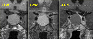
These T1, T2 and T1-weighted images after gadolinium demonstrate another Rathke's cleft cyst located in the pituitary gland.
Unlike the normal pituitary tissue and pituitary stalk it does not enhance after the administration of intravenous contrast.
The normal pituitary tissue is compressed and displaced far to the left. It is important to recognize this as it could be mistaken for an enhancing component of the cystic mass.
Các hình ảnh T1, T2 và xung T1W này sau khi lấy gadolinium cho thấy một nang khác của Rathke nằm trong tuyến yên.
Không giống như mô tuyến yên bình thường và cuống tuyến yên, nó không tăng cường sau khi tiêm thuốc cản quang đường tĩnh mạch.
Mô tuyến yên bình thường bị chèn ép và di lệch xa về bên trái Điều quan trọng là phải nhận ra điều này vì nó có thể bị nhầm lẫn với một thành phần tăng cường của khối u nang.
In general, all extra-axial masses , i.e. masses outside of the brain like the pituitary gland and stalk, will enhance because they do not have a blood-brain barrier.
If you have a non-enhancing extra-axial mass, there are three possibilities: Nói chung, tất cả các khối ngoài trục, tức là các khối bên ngoài não như tuyến yên và thân cây, sẽ tăng cường vì chúng không có hàng rào máu não.
Nếu bạn có một khối ngoài trục không tăng cường, có ba khả năng:
- Rapid arterial flow (eg. large blood vessel). Dòng chảy động mạch nhanh (ví dụ mạch máu lớn).
- No cellular tissue (eg. cyst). Không có mô tế bào (ví dụ nang).
- No blood supply (eg. infarcted mass). Không có nguồn cung cấp máu (ví dụ khối nhồi máu).
CraniopharyngiomaU sọ hầu
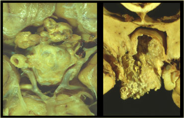
Craniopharyngioma is the third of the three pathologies derived from Rathke's cleft epithelium.
Technically these are benign tumors, but unlike Rathke's cleft cysts, they have thick walls and are locally invasive.
Macroscopically, it is a complex mass with multiple nodules at the base of the brain, sinuating along the fissures.
Often, it can not be completely resected.
The picture on the right shows a thick-walled cyst as part of the craniopharyngioma.
U sọ hầu là bệnh lý thứ ba trong ba bệnh lý có nguồn gốc từ biểu mô khe hở Rathke.
Về mặt kỹ thuật, đây là những khối u lành tính, nhưng không giống như nang Rathke, chúng có thành dày và xâm lấn tại chỗ.
Trên đại thể, đây là một khối phức tạp với nhiều nốt sần ở đáy não, nằm dọc theo các vết nứt.
Thông thường, nó không thể được cắt bỏ hoàn toàn.
Hình bên phải cho thấy một nang có thành dày như một phần của u sọ hầu.
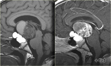
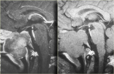
In over 50% of cases craniopharyngiomas have a pathognomonic appearance.
On these unenhanced and enhanced T1-weighted sagittal images, a compressed pituitary gland can be identified.
There is a large intrasellar and suprasellar mass with cystic and enhancing components as well as calcifications.
These findings in a child are virtually pathognomonic for craniopharyngioma (perhaps with only a dermoid in the differential diagnosis).
Trong hơn 50% trường hợp u sọ hầu có biểu hiện bệnh lý.
Trên các xung T1W ảnh mặt phẳng đứng dọc không tăng cường và tăng cường này, có thể xác định được một tuyến yên bị chèn ép.
Có một khối u lớn trong và trên xương cùng với các thành phần dạng nang và tăng cường cũng như vôi hóa.
Những phát hiện này ở trẻ em hầu như là đặc trưng của u sọ hầu (có lẽ chỉ có một lớp hạ bì trong chẩn đoán phân biệt).
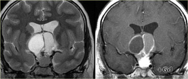
Coronal images of the same mass. Ảnh mặt phẳng đứng ngang có cùng khối lượng.
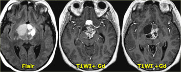
And axial images. Và ảnh mặt phẳng ngang.
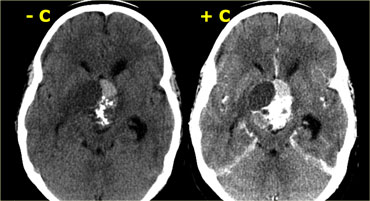
Unenhanced CT shows the calcifications more clearly.
After intravenous contrast the total extent of the lesion and its cystic components are much less evident.
CT không tiêm thuốc cản quang cho thấy vôi hóa rõ hơn.
Sau khi tiêm thuốc cản quang tĩnh mạch, toàn bộ tổn thương và các thành phần dạng nang của nó ít rõ ràng hơn nhiều.
MeningiomaU màng não
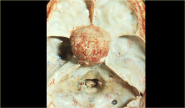
The most common intracranial tumor in adults is the meningioma with 20% of occurring at the skull base.
This is an autopsy specimen with the brain removed, showing a meningioma sitting on the diaphragma sellae.
Meningiomas are almost always solid lesions, sometimes with a cyst on the edge.
They can lift up the arachnoid a little bit and enhance uniformly as a general rule.
U nội sọ phổ biến nhất ở người lớn là u màng não với 20% xảy ra ở nền sọ.
Đây là một mẫu vật khám nghiệm tử thi với bộ não được lấy ra, cho thấy một u màng não nằm trên cơ hoành yên.
U màng não hầu như luôn là tổn thương đặc, đôi khi có một nang ở rìa.
Họ có thể nâng màng nhện lên một chút và tăng cường đồng đều như một quy luật chung.
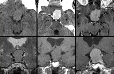
On the top-left unenhanced and enhanced CT-images, the main differential diagnosis of the enhancing mass would include meningioma, pituitary adenoma and an aneurysm.
The post-constrast MR-image on the top-right rules out an aneurysm as a possible diagnosis (no flow void), but on axial images a pituitary adenoma and meningioma are still difficult to differentiate.
Trên CT trước và sau tiêm ở phía trên bên trái, chẩn đoán phân biệt khối ngấm thuốc gồm u màng não, u tuyến yên và phình động mạch. MRI sau tiêm ở phía trên bên phải giúp loại trừ phình động mạch vì không có dấu hiệu mất tín hiệu dòng chảy. Tuy nhiên, trên ảnh mặt phẳng ngang vẫn khó phân biệt u tuyến yên với u màng não.
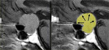
Notice the spread of the lesion along the meninges.
The epicentre of the lesion is above the sella.Chú ý sự lan rộng của tổn thương dọc theo màng não.
Tâm chấn của tổn thương nằm trên yên.
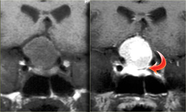 Meningioma. T1WI and T1WI-postcontrastU màng não. T1WI và T1WI sau tiêm thuốc cản quang
On the coronal images (T1 and T1-postcontrast), a compressed pituitary gland can be identified at the bottom of the sella turcica.
Above it lies a large mass, partially intrasellar and partially suprasellar.
Although the diaphragma sellae can not be identified on these images, it is probably a suprasellar mass growing downwards.
When pituitary macroadenomas get this size they usually have areas of hemorrhage or necrosis - in mengiomas this is less often the case.Trên ảnh mặt phẳng đứng ngang (T 1 và T 1 sau tiêm thuốc cản quang), có thể xác định được một tuyến yên bị chèn ép ở đáy hố yên.
Phía trên nó là một khối lớn, một phần trong và một phần trên xương ức.
Mặc dù không thể xác định được hố yên trên các hình ảnh này, nhưng có lẽ đó là một khối trên yên phát triển xuống dưới.
Khi u tuyến yên có kích thước như vậy, chúng thường có các vùng xuất huyết hoặc hoại tử - trong u tuyến yên, điều này ít gặp hơn.
AneurysmPhình mạch
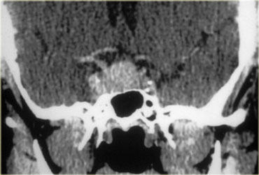
This is an important case to keep in mind.
This patient is a woman in her late forties, who presented to her family doctor with galactorrhea.
The family doctor did a number of tests, including a determination of her prolactin level.
This was about 150 (25 or less is normal).
Thinking the patient had a pituitary adenoma, the family doctor ordered this CT scan.
It is easy to get tunnel vision when reporting on a scan like this as a radiologist when the clinical information includes hyperprolactinemia and galactorrhea.
Of course your first thought is a pituitary adenoma.
If you look at the location of the lesion however (partially in the sella turcica and partially in the cavernous sinus), there are other possibilities, including a meningioma or an aneurysm.
The radiologist reported this as a pituitary adenoma, and the patient was treated with bromocriptine.Đây là một trường hợp quan trọng cần lưu ý.
Bệnh nhân này là một phụ nữ ở độ tuổi bốn mươi, đã trình bày với bác sĩ gia đình về chứng tiết sữa.
Bác sĩ gia đình đã làm một số xét nghiệm, bao gồm xác định mức độ prolactin của cô.
Con số này là khoảng 150 (25 hoặc ít hơn là bình thường).
Nghĩ rằng bệnh nhân có một tuyến yên u tuyến, bác sĩ gia đình đã yêu cầu chụp CT này.
Thật dễ dàng để có được thị lực đường hầm khi báo cáo về một lần chụp như thế này với tư cách là bác sĩ X quang khi thông tin lâm sàng bao gồm tăng prolactin máu và tiết sữa.
Tất nhiên suy nghĩ đầu tiên của bạn là tuyến yên u tuyến.
Tuy nhiên, nếu bạn nhìn vào vị trí của tổn thương (một phần ở hố yên và một phần ở xoang hang), có những khả năng khác, bao gồm u màng não hoặc phình mạch.
Bác sĩ X quang báo cáo bệnh nhân có u tuyến ở tuyến yên, và bệnh nhân được điều trị bằng bromocriptine.
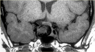
The bromocriptine had no effect, and the patient went to a neurosurgeon for a surgical opinion.
The neurosurgeon ordered this MRI.
The lesion partly in the right cavernous sinus and partly in the sella turcica is predominantly black on this T1-weighted image.
In general there are three things that are black on MRI: air, bone and rapid blood flow. In this case it is black due to rapid blood flow in a carotid aneurysm.Bromocriptine không có tác dụng, và bệnh nhân đã đến một bác sĩ phẫu thuật thần kinh để lấy ý kiến phẫu thuật.
Bác sĩ phẫu thuật thần kinh đã ra lệnh chụp MRI này.
Tổn thương một phần ở xoang hang bên phải và một phần ở Sella Turcica chủ yếu là màu đen trên hình ảnh xung T1W này.
Nói chung có ba thứ có màu đen trên MRI: không khí, xương và lưu lượng máu nhanh. Trong trường hợp này, nó có màu đen do lưu lượng máu nhanh trong chứng phình động mạch cảnh.
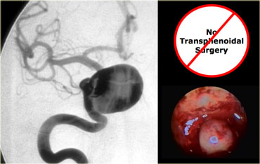

This is the corresponding angiogram.
Obviously, this is not a lesion to be operated on transsphenoidally!
Đây là chụp mạch tương ứng.
Rõ ràng, đây không phải là một tổn thương cần được phẫu thuật trên qua xoang bướm!
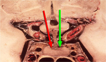 Hypothalamus hormones either stimulate (green arrow) or inhibit (red arrow) the production of pituitary hormones.Hormone vùng dưới đồi kích thích (mũi tên xanh) hoặc ức chế (mũI tên đỏ) sản xuất hormone tuyến yên.
Why did the aneurysm cause hyperprolactinemia and galactorrhea in this patient?
It was caused by compression of the pituitary stalk.
The pituitary stalk connects the hypothalamus to the pituitary gland and hormones produced in the hypothalamus are transported to the anterior lobe of the pituitary gland via portal veins running along the stalk.
Most of these hormones stimulate the production of other hormones in the pituitary gland (such as TRH, GnRH, GHRH and CRH), but the release of dopamine inhibits the production of prolactin by the anterior lobe of the pituitary.
Therefore when the stalk is compressed by a mass or is transected, the level of prolactin rises while all the other hormone levels decrease.
This is known as the 'Stalk Section Effect'.
It is the reason why masses other than adenomas can cause hyperprolactinemia.
This is also why an unenhanced MRI scan suffices in a patient with hyperprolactinemia: it is not the size of the microadenoma, but ruling out other pathology that matters.
Tại sao phình mạch gây tăng prolactin máu và tiết sữa ở bệnh nhân này?
Nguyên nhân là do chèn ép cuống tuyến yên.
Cuống tuyến yên kết nối vùng dưới đồi với tuyến yên và các hormone được sản xuất ở vùng dưới đồI được vận chuyển đến thuỳ trước của tuyến yên thông qua các tĩnh mạch cửa chạy dọc theo cuống.
Hầu hết các hormone này kích thích sản xuất các hormone khác trong tuyến yên (như TRH, GnRH, GHRH và CRH), nhưng sự giải phóng dopamine ức chế sản xuất prolactin bởi thuỳ trước của tuyến yên.
Do đó, khi cuống bị nén bởi một khối hoặc bị cắt ngang, mức độ prolactin tăng lên trong khi tất cả các mức hormone khác giảm.
Đây được gọi là “Hiệu ứng phần thân ”, đó là lý do tại sao các khối khác ngoài các u tuyến có thể gây tăng prolactin máu.
Đây cũng là lý do tại sao chụp MRI không tiêm thuốc ở bệnh nhân tăng prolactin máu: nó không phải là kích thước của u tuyến nhỏ, nhưng loại trừ các bệnh lý khác có vấn đề.
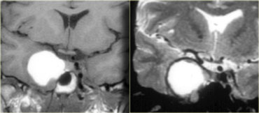
On the left the T1-weighted image of a thrombosed aneurysm with high signal intensity on the unenhanced scan.
It originates in the intracavernous segment of the right internal carotid artery.
On the right the T2-weighted images: the thrombosed aneurysm has a dark rim.
Bên trái hình ảnh xung T1W của túi phình động mạch bị huyết khối có cường độ tín hiệu cao trên phim chụp không tiêm thuốc cản quang.
Phình động mạch cảnh trong có nguyên uỷ từ đoạn trong hang động mạch cảnh bên phải, bên phải hình ảnh xung T2W: phình động mạch bị huyết khối có bờ tối.
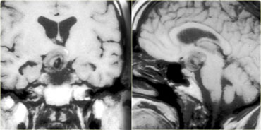
This is an example of a partially thrombosed aneurysm in the suprasellar cistern.
The patent lumen is black on these T1-weighted images.
It is surrounded by clot of different ages arranged in layers reaching from the lumen to the wall.
It resembles an onion cut in half.
Đây là một ví dụ về phình động mạch bị huyết khối một phần trong bể chứa trên yên.
Lumen sáng chế có màu đen trên các hình ảnh xung T1W này.
Huyết khối được bao quanh bởi các cục máu đông ở các độ tuổi khác nhau được sắp xếp thành các lớp từ lòng mạch đến thành mạch.
Nó giống như một củ hành tây cắt làm đôi.
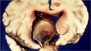
On the left an autopsy specimen.
You can see that this patient suffered a massive intraventricular and subarachnoid hemorrhage.
The layers of bloodclot are very nicely reflected in the MR images.
Bên trái là mẫu vật khám nghiệm tử thi.
Bệnh nhân này có một khối lượng lớn trong não thất và xuất huyết dưới nhện.
Các lớp máu đông được phản chiếu rất rõ ràng trong hình ảnh cộng hưởng từ.
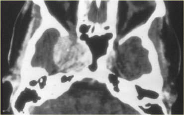
Aneurysm vs MeningiomaPhình động mạch và u màng não
One of the most difficult differential diagnoses on CT is aneurysm versus meningioma.
In this patient there is a large mass on the right hand side, possibly originating from the meninges or cavernous sinus.
On CT it is impossible to tell whether this mass is an aneurysm or a meningioma.
Một trong những chẩn đoán phân biệt khó khăn nhất trên CT là phình mạch so với u màng não.
Ở bệnh nhân này có một khối lớn ở bên phải, có thể bắt nguồn từ màng não hoặc xoang hang.
Trên CT không thể nói khối này là phình mạch hay u màng não.
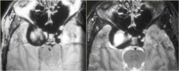
This is an MRI of the same patient.
The mass is predominantly black and there is a large flow artefact running in the phase-encoding direction.
These findings correspond to rapid blood flow, and the mass must therefore be an aneurysm.
Đây là hình ảnh chụp cộng hưởng từ của cùng một bệnh nhân.
Khối u chủ yếu là màu đen và có một dòng chảy lớn chạy theo hướng mã hóa pha.
Những phát hiện này tương ứng với lưu lượng máu nhanh, và khối đó phải là một phình mạch.
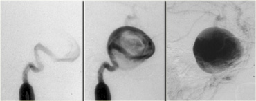
Angiogram of the same patient.
It demonstrates that the flow in the aneurysm is not laminar, but that it swirls, gradually filling the lumen with contrast.
Chụp mạch máu của cùng một bệnh nhân.
Nó chứng minh rằng dòng chảy trong túi phình không phải là tầng, mà nó xoáy, dần dần lấp đầy lòng mạch bằng chất cản quang.
HamartomaPhổi
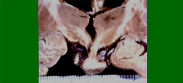
Hamartomas are masses of dysplastic tissue found almost exclusively in young children.
One of the most common locations is the floor of the third ventricle.
This is a pathology specimen showing a small nodule hanging in the suprasellar cistern.
They are benign lesions, but patients do succumb to them because of the bad location.U mô liên kết là khối mô loạn sản được tìm thấy hầu như chỉ ở trẻ nhỏ.
Một trong những vị trí phổ biến nhất là sàn của tâm thất thứ ba.
Đây là một bệnh phẩm cho thấy một nốt nhỏ treo trong bể chứa trên xương cùng.
Chúng là những tổn thương lành tính, nhưng bệnh nhân không chống chọi được với chúng vì vị trí xấu.
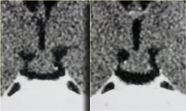
These are CT images of a hamartoma suspended from the floor of the third ventricle.
It does not enhance after the administration of intravenous contrast.
Đây là những hình ảnh CT của một hamartoma lơ lửng trên sàn của tâm thất thứ ba.
Nó không tăng cường sau khi dùng thuốc cản quang tiêm tĩnh mạch.
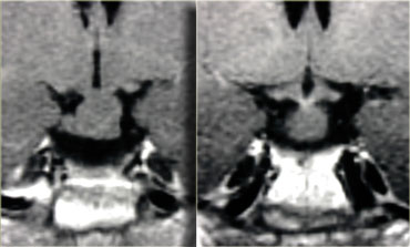
MR images of a similar small nodule suspended from the floor of the third ventricle. Hình ảnh cộng hưởng từ của một nốt nhỏ tương tự lơ lửng trên sàn của tâm thất thứ ba.
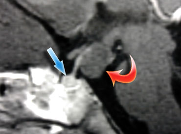 Hamartoma (red arrow) posterior to the enhancing pituitary gland and stalk.Hamartoma (mũi tên màu đỏ) phía sau tuyến yên tăng cường và cuống.
The best images to see hamartomas on are enhanced sagittal T1-weighted MR images.
Here you can see the non-enhancing hamartoma attached to the tuber cinereum between the pituitary stalk and mamillary body.
There really is no differential diagnosis.
Hình ảnh phù hợp nhất để phát hiện hamartomas là hình ảnh cộng hưởng từ xung T1W ngắt quãng dọc.
Ở đây bạn có thể thấy hamartoma không tăng cường gắn liền với củ cinereum giữa cuống tuyến yên và thân nhũ hoa.
Thực sự không có chẩn đoán phân biệt.
Hypothalamic and Chiasm GliomaU Glioma vùng dưới đồi và Chiasm
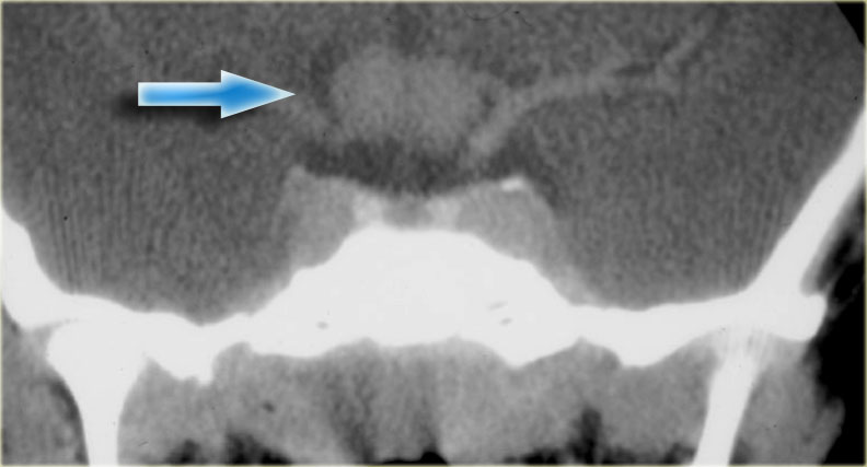 Optic nerve glioma in a patient with neurofibromatosisU thần kinh đệm thị giác ở bệnh nhân u sợi thần kinh
Gliomas can occur in any part of the brain and the optic chiasm is a common location, particularly in patients with neurofibromatosis type 1.
This enhanced CT shows an example of an optic nerve glioma in a patient with neurofibromatosis.
There is a suprasellar mass which is indistinguishable from the optic chiasm.
U thần kinh đệm có thể xảy ra ở bất kỳ phần nào của não và giao thoa thị giác là một vị trí phổ biến, đặc biệt ở những bệnh nhân bị u sợi thần kinh loại 1.
Phim chụp CT tăng cường này cho thấy một ví dụ về u thần kinh thị giác ở bệnh nhân bị u sợi thần kinh.
Có một khối trên xương cùng không thể phân biệt được với giao thoa thị giác.
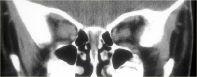
Further forward at the level of the orbits the optic nerve is abnormal on both sides. Dây thần kinh thị giác bất thường ở cả hai bên.
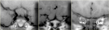
These consecutive coronal MR-images show the mass at the optic chiasm and the swollen optic nerves. Những hình ảnh cộng hưởng từ tiếp theo này cho thấy khối u ở giao thoa thị giác và các dây thần kinh thị giác bị sưng.
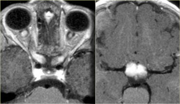
On these axial images you can see the optic nerves and chiasm enhance after the administration of intravenous gadolinium.
The way a patient is normally positioned, slices through the nerves themselves are not obtained.
These slices can be used to make oblique images along the axis of the nerves.
Trên ảnh mặt phẳng ngang này, bạn có thể thấy các dây thần kinh thị giác và chiasm tăng cường sau khi tiêm tĩnh mạch gadolinium.
Cách bệnh nhân được định vị bình thường, không có các lát cắt xuyên qua các dây thần kinh.
Những lát cắt này có thể được sử dụng để tạo ra hình ảnh xiên dọc theo trục của dây thần kinh.
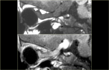
With these images as a result.
Note the enhancement of the nerve after intravenous contrast with sparing of the meninges.
Approximately 25% of optic nerve gliomas do not enhance, so a lack of enhancement should not prevent you from making the diagnosis.
Với những hình ảnh này là kết quả.
Lưu ý sự tăng cường của dây thần kinh sau khi tiêm thuốc cản quang tĩnh mạch với sự tiết kiệm màng não.
Khoảng 25% của u thần kinh thị giác không tăng cường, vì vậy việc thiếu sự tăng cường không ngăn cản bạn đưa ra chẩn đoán.
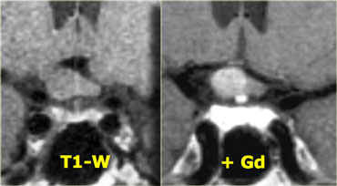
This is another example of a right-sided optic nerve glioma with enhancement after gadolinium.
Note the normal pituitary gland and stalk.
Đây là một ví dụ khác về u thần kinh đệm thị giác bên phải có ngấm thuốc sau khi tiêm gadolinium.
Lưu ý tuyến yên và cuống bình thường.
GerminomaVi khuẩn
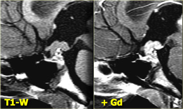 Germinoma (Courtesy of Dr. Susan Blaser)Germinoma (Lịch sự của Tiến sĩ Susan Blaser)
The following case concerns a 9-year-old male with a history of headache, nausea and vomiting.
Sagittal T1 images before and after intravenous contrast show a mass in the midline, on the floor of the third ventricle.
The mass enhances after gadolinium.
Continue with next images.
Ca bệnh sau đây liên quan đến một trẻ nam 9 tuổi có tiền sử đau đầu, buồn nôn và nôn.
Hình ảnh T1 dọc trước và sau khi tiêm thuốc cản quang cho thấy một khối ở đường giữa, trên sàn của tâm thất thứ ba.
Khối tăng cường sau khi gadolinium.
Tiếp tục với những hình ảnh tiếp theo.
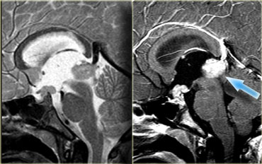
T2- and T1 weighted sagittal images of the same patient show a similar mass in the epiphysial area.
This is a germinoma - an intracranial germ cell tumor that occurs primarily in children and adolescents.
These are typical localisations.
These lesions crawl along the floor of the 3rd ventricle.
T2 và T1 ảnh mặt phẳng đứng dọc của cùng một bệnh nhân cho thấy một khối tương tự ở vùng đầu chi.
Đây là một u mầm - một khối u tế bào mầm trong sọ xảy ra chủ yếu ở trẻ em và thanh thiếu niên.
Đây là những khu vực điển hình.
Những tổn thương này bò dọc theo sàn của tâm thất thứ 3.
ChordomaU nguyên sống
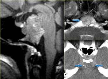
Chordomas are the most common lesions of the clivus, also a favored location for metastases and chondrosarcomas.
This patient has a normal pituitary gland.
Posterior to this is a large, fungating mass positioned at the level of the clivus.
The CT shows some calcifications in this area.
The differential diagnosis for this mass would be chordoma or chondrosarcoma.
Chordomas tend to occur in the midline, whereas chondrosarcomas tend to occur off the midline.U sụn là tổn thương phổ biến nhất của sụn viền, cũng là vị trí ưa thích cho di căn và sarcom sụn.
Bệnh nhân này có tuyến yên bình thường.
Phía sau đó là một khối nấm lớn, nằm ở mức của âm vật.
CT cho thấy một số vôi hóa ở vùng này.
chẩn đoán phân biệt cho khối lượng này sẽ là chordoma hoặc sarcôm sụn.
U sụn có xu hướng xuất hiện ở đường giữa, trong khi sarcom sụn xuất hiện ở ngoài đường giữa.
MetastasesDi căn
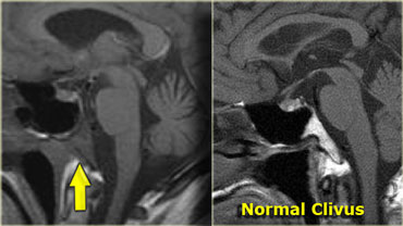

The patient on the left is a patient with lung cancer who presented with a sixth cranial nerve palsy.
The abnormality is in the clivus, which should have a high signal intensity on this sagittal T1-weighted image (as in the image on the left).
A low signal intensity means the normal fatty marrow has been replaced by some other tissue.
In this case by tumor metastasis.
Also lymphomas, myelomas or diffuse bone abnormalities can give this appearance.
Therefore always take a minute to look at the clivus.
Bệnh nhân bên trái là một bệnh nhân bị ung thư phổi bị liệt dây thần kinh sọ thứ sáu.
Sự bất thường là ở Clivus, nên có cường độ tín hiệu cao trên hình ảnh xung T1W sagittal này (như trong hình ảnh bên trái).
cường độ tín hiệu thấp có nghĩa là tủy mỡ bình thường đã được thay thế bằng một số mô khác.
Trong trường hợp này do di căn khối u.
Ngoài ra u lympho, u tủy hoặc các bất thường về xương lan tỏa có thể mang lại sự xuất hiện này.
Do đó, luôn dành một phút để nhìn vào Clivus.
So again in order to analyse a sellar or parasellar mass on MRI we use the following anatomic approach: Vì vậy, một lần nữa để phân tích khối u xương bánh chè hoặc cạnh bánh chè trên MRI chúng tôi sử dụng phương pháp giải phẫu sau:
- First identify the pituitary gland and sella turcica. Đầu tiên xác định tuyến yên và hố yên.
- Then determine the epicenter of the lesion and whether it is in the sella or above, below or lateral to the sella. Sau đó xác định tâm chấn thương của tổn thương và liệu nó ở trong hố yên hay ở trên, dưới hay bên hố yên.
- If it is in the sella, determine whether or not the sella is enlarged. Nếu nó nằm trong hố yên, hãy xác định xem hố yên có to lên hay không.
- Once the location of the mass is clear, analyze the signal intensity patterns: is the lesion cystic or solid? Khi vị trí của khối u đã rõ ràng, phân tích các mẫu cường độ tín hiệu: là tổn thương dạng nang hay dạng đặc?
- Does it contain any abnormal vessels? Nó có chứa bất kỳ mạch máu bất thường nào không?
- Are there any calcifications? And so on.Có vôi hóa nào không? Và cứ thế.
- Finally establish a Differential Diagnosis.Cuối cùng thiết lập một chẩn đoán phân biệt.