
Developmental Dysplasia of the HipLoạn sản phát triển khớp háng
Ultrasound examinationKhám siêu âm
Radiology Departement of the Maastricht University Hospital and the Alrijne hospital in the NetherlandsKhoa Chẩn đoán hình ảnh của Bệnh viện Đại học Maastricht và Bệnh viện Alrijne ở Hà Lan
Ngày đăng / cập nhật: 2017-12-01
Bản gốc trên radiologyassistant.nl ↗ · tải về lúc 2026-11-19 20:19 · bản dịch máy có chuẩn hóa thuật ngữ chẩn đoán hình ảnh
Developmental dysplasia of the hip is a common musculoskeletal disorder in newborns.Loạn sản phát triển xương hông là một rối loạn cơ xương phổ biến ở trẻ sơ sinh.
In this article we will discuss the ultrasound examination technique according to Graf.Trong bài viết này chúng ta sẽ bàn về kỹ thuật siêu âm theo Graf.
IntroductionMở đầu
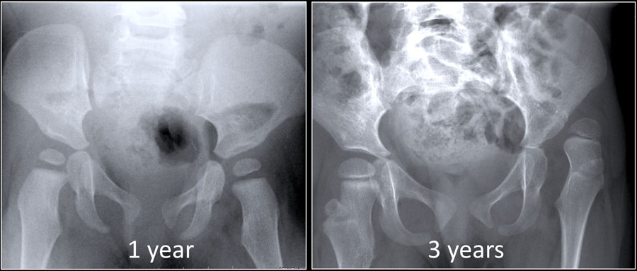

Developmental dysplasia of the hip (DDH) is one of the most common musculoskeletal problems in newborns.
It is also known as congenital hip dysplasia, but actually this is a misnomer.
It is a developmental disease.
There are children who are born with normal hips who develop dysplasia (figure).Loạn sản xương hông (DDH) là một trong những vấn đề về cơ xương khớp thường gặp ở trẻ sơ sinh.
Nó còn được gọi là loạn sản xương hông bẩm sinh, nhưng thực sự đây là một cách gọi sai.
Đây là một bệnh lý tiến triển.
Có những trẻ sinh ra với hông bình thường bị loạn sản (hình).
In this case the child had a neuromuscular disorder which caused the hip to become dysplastic and dislocate.Trong trường hợp này trẻ bị rối loạn thần kinh cơ khiến hông trở nên loạn sản và trật khớp.
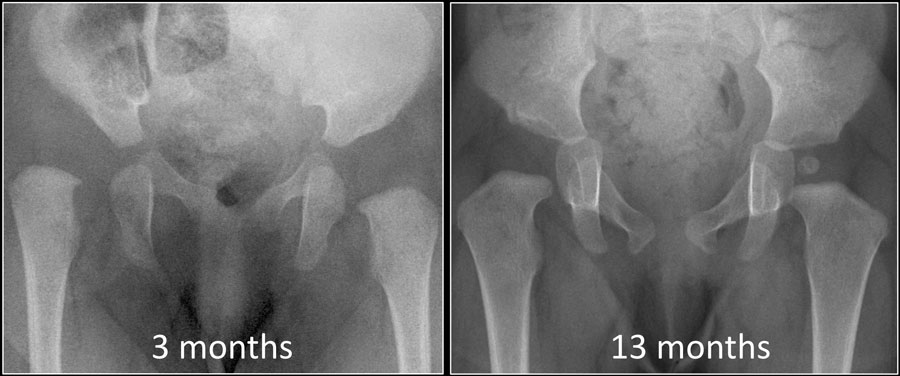

On the other hand there are children who are born with dysplasia of the hip that resolves spontaneously or after relatively simple treatment, e.g. a Pavlik harness (figure).Mặt khác, có những trẻ em bị loạn sản khớp háng tự khỏi hoặc sau khi điều trị tương đối đơn giản, ví dụ như dây nịt Pavlik (hình).
So we have to realize that DDH is a dynamic disease and it is not always present at birth as suggested by the name congenital hip dysplasia.
Treatment is easier and complications are less likely to occur when DDH is diagnosed early.Vì vậy, chúng tôi phải nhận ra rằng DDH là một bệnh động và không phải lúc nào cũng xuất hiện khi sinh như được gợi ý bởi tên gọi loạn sản xương hông bẩm sinh.
Điều trị dễ dàng hơn và ít biến chứng xảy ra khi DDH được chẩn đoán sớm.
In this case at 13 months the only sign of the former dysplasia is the delay of the femoral head to ossify.Trong trường hợp này, ở tháng thứ 13 dấu hiệu duy nhất của loạn sản trước đây là sự chậm trễ của xương đùi để cốt hóa.
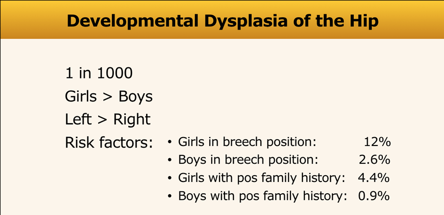
Developmental dysplasia of the hip is more common in girls especially if there has been a breech presentation or when they have a positive family history.
These risk factors play a lesser role in boys.Loạn sản phát triển của hông là phổ biến hơn ở trẻ gái đặc biệt là nếu có một phần mông hoặc khi họ có tiền sử gia đình tích cực.
Những yếu tố nguy cơ này đóng vai trò ít hơn ở trẻ trai.
Clubfoot was thought to be a risk factor, but this no longer holds true.Chân khoèo được cho là một yếu tố nguy cơ, nhưng điều này không còn đúng nữa.
Ultrasound examinationKhám siêu âm

Ultrasound orientationĐịnh hướng siêu âm
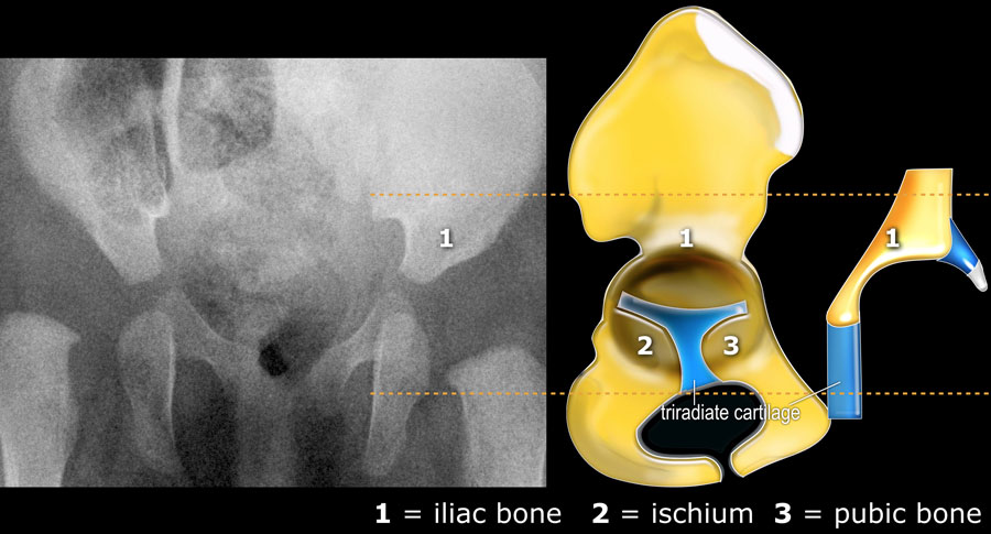
With ultrasound we are looking at the same anatomic structures as on the x-ray.
The ultrasound images are in the coronal plane (figure).Với siêu âm, chúng ta đang nhìn vào các cấu trúc giải phẫu giống như trên phim X-quang.
Hình ảnh siêu âm nằm trong mặt phẳng đứng ngang (hình).
As a consequence the Graf classification of hip dysplasia is mainly based on the morphology of the iliac bone, where we look at the shape of the acetabulum, the bony and cartilaginous acetabular rim, labrum and position of the femoral head.Kết quả phân loại loạn sản khớp háng theo Graf chủ yếu dựa vào hình thái xương chậu, trong đó chúng ta xem xét hình dạng ổ cối, vành ổ cốI xương sụn, xương ổ cốy và vị trí chỏm xương đùi.
Since ultrasound has the advantage of also displaying the cartilagenous structures, we can also look at the coverage of the femoral head by the cartilage of the acetabulum and the labrum.Vì siêu âm có ưu điểm là cũng hiển thị các cấu trúc sụn, chúng ta cũng có thể xem xét sự bao phủ của đầu xương đùi bởi sụn của ổ cối và xương ức.
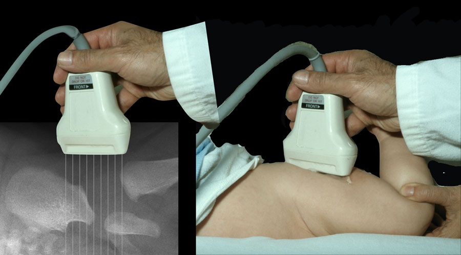
Because the infant is lying on its side the anatomy is displayed in a horizontal fashion instead of vertical (figure)Bởi vì trẻ sơ sinh nằm nghiêng, giải phẫu được hiển thị theo chiều ngang thay vì chiều dọc (hình)
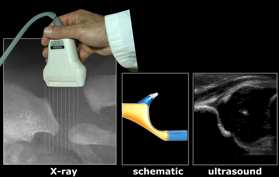
And this is the way the ultrasound image is displayed on the screen of the ultrasound machineVà đây là cách mà hình ảnh siêu âm được hiển thị trên màn hình của máy siêu âm
In this video the ultrasound anatomy is shown.Trong video này giải phẫu siêu âm được hiển thị.
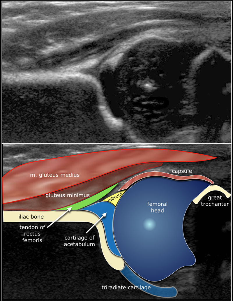
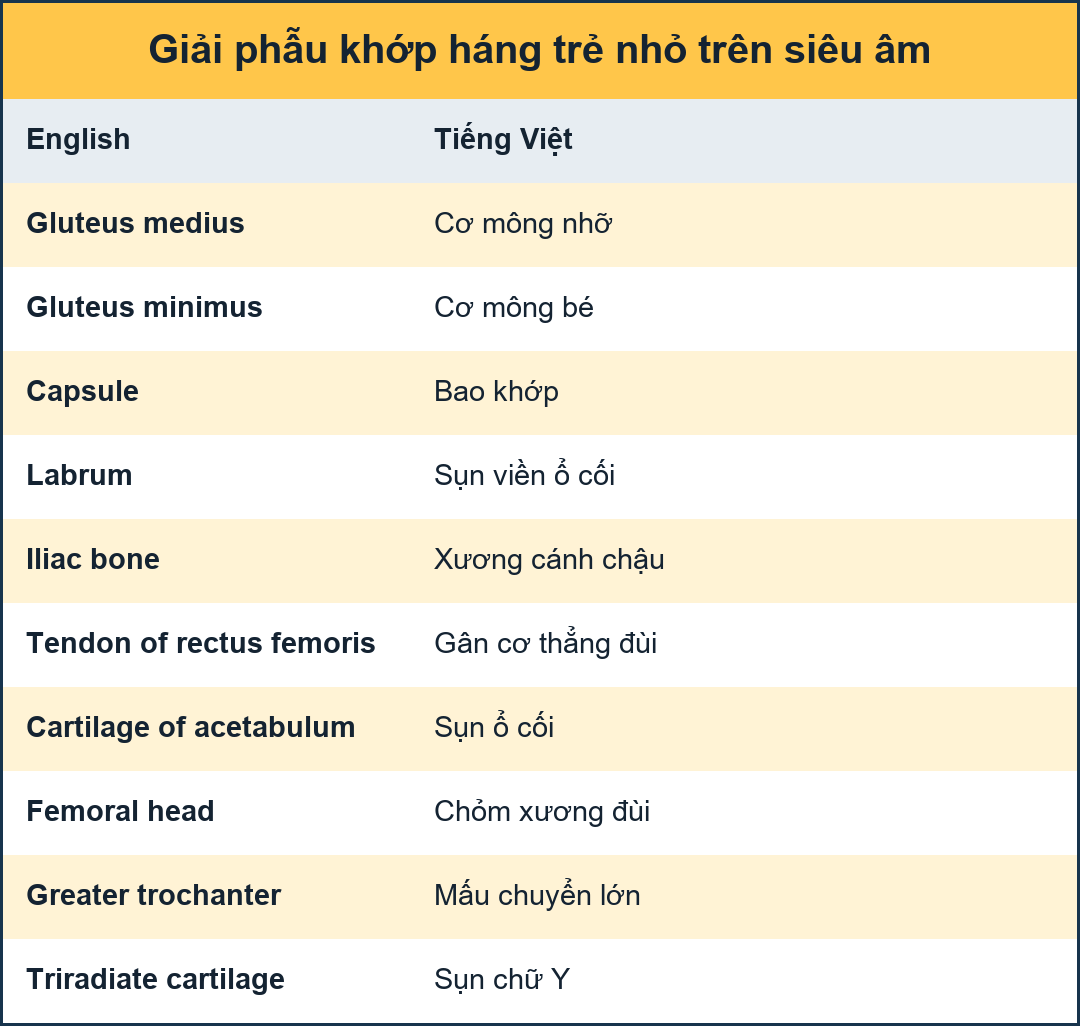
Here the anatomy in the coronal plane.Đây là giải phẫu trong mặt phẳng đứng ngang.
Examination techniqueKỹ thuật khám
A lineair, high frequency probe is used.
The focus is set at the acetabular edge.Một đầu dò lineair, tần số cao được sử dụng.
Trọng tâm được đặt ở mép ổ cối.
It is important to display an image in the coronal plane at the level of the triradiate cartilage, which is the synchondrosis between the iliac, ischial and pubic bones which form the acetabulum.Điều quan trọng là hiển thị hình ảnh trong mặt phẳng đứng ngang ở mức của sụn ba mảnh, đó là khớp sụn giữa xương chậu, xương đùi và xương mu tạo thành ổ cối.
This is shown in the video.Điều này được thể hiện trong video.
InstabilityMất ổn định
Sometimes in very displastic hips the use of a convex transducer can be of help.Đôi khi ở những khớp háng rất chênh lệch, việc sử dụng đầu dò lồi có thể giúp ích.
MeasurementsĐo lường
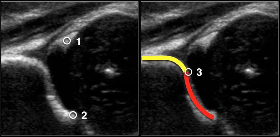
First three points of interest need to be indentified in the image:
Ba điểm quan tâm đầu tiên cần được xác định trong hình ảnh:
- Centre of labrumTrung tâm labrum
- Lower limb of the iliac bone (=Undersurface of the medial border of the acetabulum where the iliac bone meets the triradiate cartilage)Chi dưới của xương chậu (=Bề mặt của bờ trong ổ cối nơi xương chậu gặp sụn ba lá)
- Bony rim of acetabulum. When the bony rim is angular this point is easily recognized. When rounded this point is defined as the point where the concavity of the bony acetabular roof changes into the convexity of the iliumVành xương ổ cối Khi viền xương có góc thì dễ nhận biết điểm này Khi làm tròn thì điểm này được định nghĩa là điểm mà chỗ lõm của nóc xương ổ đĩa biến đổi thành chỗ lồi của chậu
In this video you'll see proper measurement after placement of the three points of interest.Trong video này, bạn sẽ thấy phép đo thích hợp sau khi đặt ba điểm quan tâm.
When you perform the ultrasound examination, make sure that these three points can be identified on the image.Khi bạn thực hiện kiểm tra siêu âm, hãy chắc chắn rằng ba điểm này có thể được xác định trên hình ảnh.
Graf's classificationPhân loại theo Graf
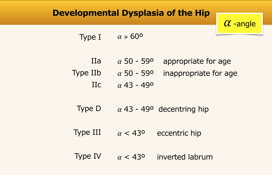
This is the Graf classification - short version.Đây là phân loại Graf - phiên bản ngắn.
The alpha-angle, which is a measurement of the bony roof of the acetabulum, mainly determines the hip type.Góc alpha, là thước đo phần mái xương của ổ cối, chủ yếu xác định loại hông.
Actually, for classification purposes the beta angle is only used to differentiate between type Ia and Ib (both normal hips) and between type IIc and type D)Trên thực tế, để phân loại, góc beta chỉ được sử dụng để phân biệt giữa loại Ia và Ib (cả hai khớp háng bình thường) và giữa loại IIc và loại D).
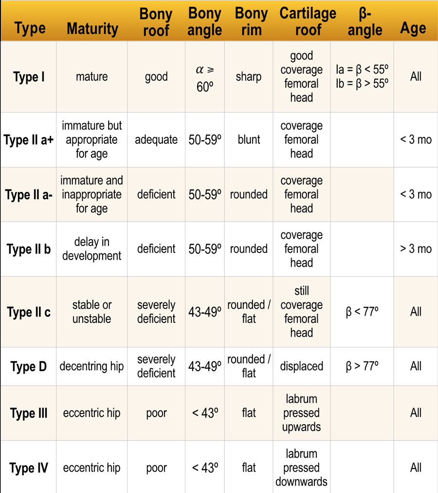
This is the Graf classification - long version.Đây là phân loại Graf - phiên bản dài.
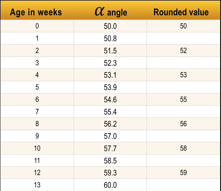
Another important factor is the age of the child.
Up to the age of 3 months (13 weeks) an alpha angle below 60 degrees is acceptable.
A neonate can even start with an alpha angle of 50 degrees provided that the angle gradually reaches the 60 degrees by the age of 12 weeks.Một yếu tố quan trọng khác là tuổi của trẻ.
Cho đến 3 tháng tuổi (13 tuần) góc alpha dưới 60 độ là chấp nhận được.
Trẻ sơ sinh thậm chí có thể bắt đầu với một góc alpha 50 độ với điều kiện là góc dần dần đạt đến 60 độ khi được 12 tuần tuổi.
At the age of 3 months the decision has to be made whether the hip is normal or not.Ở tuổi 3 tháng, quyết định phải được đưa ra cho dù hông có bình thường hay không.
Evidently if a neonate starts with an alpha angle of 60 degrees than everything is o.k. and no follow up is necessary.Rõ ràng nếu một trẻ sơ sinh bắt đầu với một góc alpha 60 độ thì mọi thứ đều ổn và không cần theo dõi.
Type ILoại I
Type I hips have an alpha angle of more than 60 degrees and are normal.
Although there is a distinction between type Ia and Ib this is not clinically relevant.Hông loại I có góc alpha trên 60 độ và bình thường.
Mặc dù có sự phân biệt giữa type Ia và Ib nhưng điều này không liên quan đến lâm sàng.
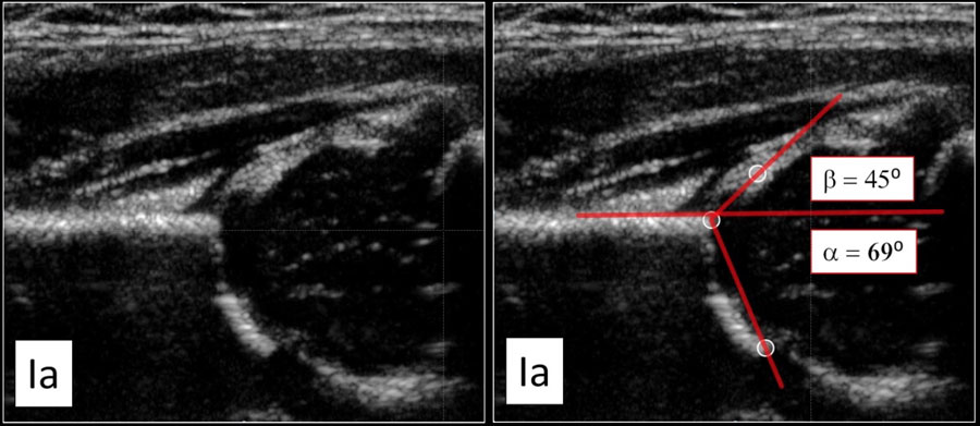
Type Ia Loại Ia
This examination shows a good morphology of the bony acetabular roof with a sharp angular bony rim.
No problem here in depicting the lateral rim of the bony roof of the acetabulum.Kết quả này cho thấy hình thái tốt của mái ổ cối xương với vành xương góc nhọn.
Không có vấn đề gì khi mô tả bờ ngoài của nóc xương ổ cối.
There is nice coverage of the femoral head by the cartilaginous roof and the labrum.Có sự bao phủ tốt của đầu xương đùi bởi mái sụn và xương ức.
The alpha angle is above the 60 degrees and the beta-angle is way below 55 degrees.Góc alpha nằm trên góc 60 độ và góc beta nằm dưới 55 độ.
Type Ib Loại Ib
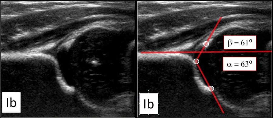
This is also a normal hip.
There is good coverage of the femoral head.
The only difference with the type Ia-hip is a blunted bony rim.
As a result the alpha-angle will be a little bit steeper than in type Ia, but still within a normal range.
The beta angle is 61º, i.e more than 55º.Đây cũng là một khớp háng bình thường.
Có sự che phủ tốt của đầu xương đùi.
Sự khác biệt duy nhất với loại Ia-hip là vành xương bị cụt.
Kết quả là góc alpha sẽ dốc hơn một chút so với loại Ia, nhưng vẫn nằm trong phạm vi bình thường.
Góc beta là 61o, tức là hơn 55o.
These hips are normal and follow up is not needed.Những khớp háng này bình thường và không cần theo dõi.
Type IILoại II
Type IIa
If a child is less than 3 months old, then an alpha angle of 50-59 degrees is considered an immature hip.
At the age of 6-13 weeks a distinction is made whether it is thought that the immature hip develops apropriate according to age (IIa+) or inappropriate (IIa-)Loại IIa
Nếu một đứa trẻ dưới 3 tháng tuổi, thì góc alpha từ 50-59 độ được coi là khớp háng chưa trưởng thành.
Ở tuổi 6-13 tuần, một sự phân biệt được đưa ra cho dù người ta cho rằng hông chưa trưởng thành phát triển không phù hợp theo tuổi (IIa +) hay không phù hợp (IIa-)
Type IIa+
The maturation process of type IIa+ hip is still within acceptable limits for age according to the table.Loại IIa +
Quá trình trưởng thành của khớp háng type IIa + vẫn nằm trong giới hạn cho phép theo tuổi theo bảng.
Type IIa-
A type IIa- hip is at risk to develop dysplasia.
So an alpha angle of 56 degrees at the age of 7 weeks is called type IIa+, while at the age of 10 weeks it is called a type IIa-.Loại IIa-
Khớp háng loại IIa có nguy cơ phát triển loạn sản.
Góc alpha 56 độ ở tuổi 7 tuần được gọi là loại IIa +, trong khi ở tuổi 10 tuần được gọi loại IIa-.
Type IIb
If a child is older than 3 months or 13 weeks, then an alpha angle of 50-59 degrees is considered a sign of dysplasia, i.e type IIb.Loại IIb
Nếu một đứa trẻ lớn hơn 3 tháng hoặc 13 tuần tuổi, thì góc alpha từ 50-59 độ được coi là dấu hiệu của loạn sản, tức là loại IIb.

Type IIa Loại IIa
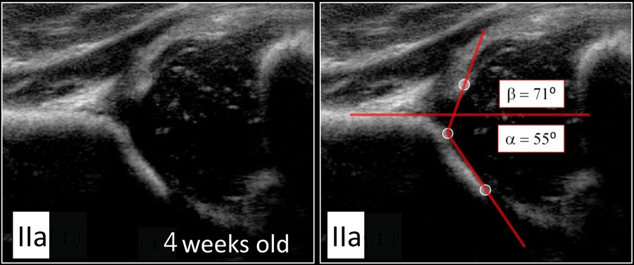
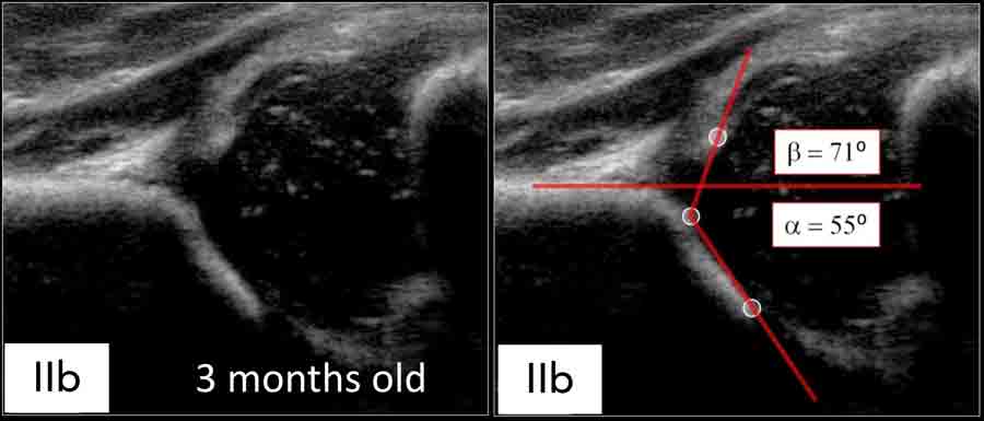
Here we see a hip with an alpha-angle of 55º.
The bony acetabular roof is less well-formed and there is a rounded acetabular bony rim.Ở đây chúng ta thấy một khớp háng có góc alpha là 55o.
Mái xương ổ cối ít được tạo hình tốt hơn và có vành xương ổ bụng tròn.
The age of the child is 4 weeks, so we call this a type IIa.Tuổi của trẻ là 4 tuần, vì vậy chúng tôi gọi đây là loại IIa.
About 90 % of newborns with Graf type IIa hips do not develop DDH.Khoảng 90% trẻ sơ sinh có khớp háng Graf loại IIa không phát triển DDH.

Type IIa(+) Loại IIa (+)
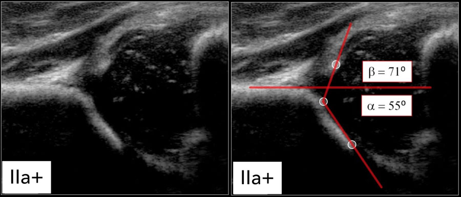
At the age of 6 weeks the same findings result in a type IIa(+).Ở tuổi 6 tuần, những phát hiện tương tự dẫn đến loại IIa (+).

Type IIb Loại IIb
At the age of 3 months or 13 weeks the same findings result in a type IIb-hip.Ở tuổi 3 tháng hoặc 13 tuần, những phát hiện tương tự dẫn đến hông loại IIb.
Type IIc Loại IIc
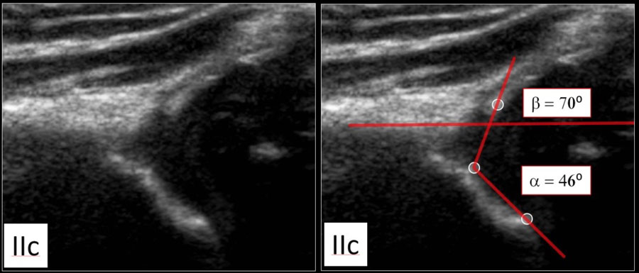
Here a type IIc hip.
The bony acetabular roof is severely deficient with a rounded to almost flat bony rim.
The alpha angle is 46 degrees.Đây là một khớp háng loại IIc.
Mái ổ cối xương bị thiếu hụt nghiêm trọng với vành xương tròn đến gần như phẳng.
Góc alpha là 46 độ.
The femoral head is still covered by the cartilaginous roof and the labrum.Đầu xương đùi vẫn được bao phủ bởi lớp sụn và xương ức.
Type DLoại D
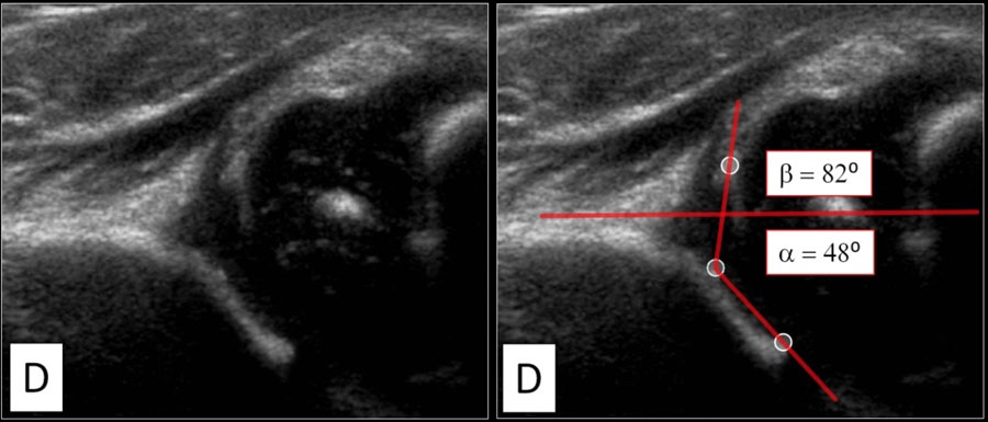
A type D hip is much like a type IIC hip, but the main difference is a decentring hip with a displaced cartilage roof.Hông loại D giống với loại IIC, nhưng sự khác biệt chính là khớp háng lệch tâm với phần sụn di lệch.
Type IIILoại III
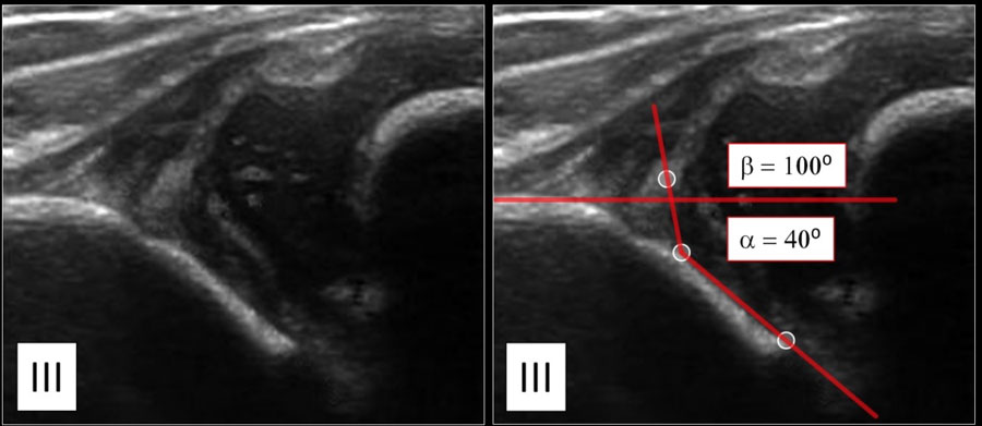
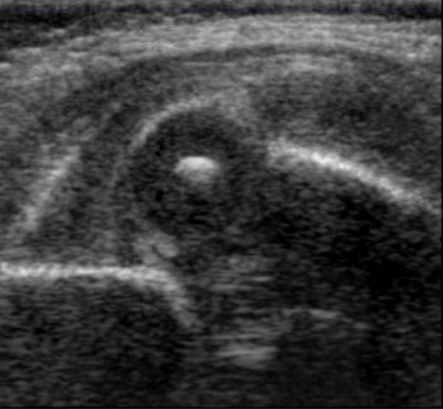
In type III hips the femoral head dislocated.
The labrum is moved upwards.Ở khớp háng loại III, đầu xương đùi bị trật.
Xương ức được di chuyển lên trên.
Type IVLoại IV
In Graf type IV there is a severe dislocation of the femoral head which obscures most of the bony roof.Trong Graf loại IV có trật khớp nặng đầu xương đùi che khuất phần lớn xương.
The cartilaginous roof is compressed between the femoral head and the bony acetabular rim.Mái sụn được nén giữa đầu xương đùi và vành xương ổ cối.
The labrum is dislocated downwards and interposed between the femoral head and the lateral acetabular edge.Xương ổ chảo bị trật xuống dưới và nằm giữa đầu xương đùi và cạnh ổ cối bên.
ReportingBáo cáo
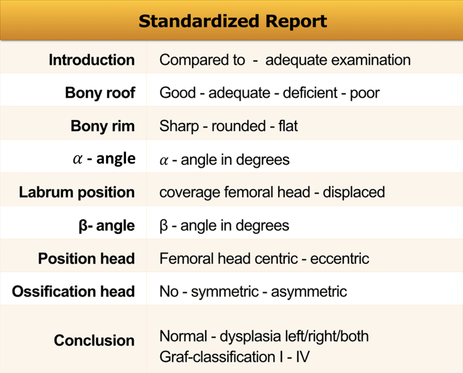
In the table a list of things that should be mentioned in your report.Trong bảng một danh sách những điều cần được đề cập trong báo cáo của bạn.
ReferencesTài liệu tham khảo
-
Hip sonography. Diagnosis and management of infant hip dysplasia.
Siêu âm hông. Chẩn đoán và điều trị chứng loạn sản xương hông ở trẻ sơ sinh.
by Graf R.
Berlin: Springer; 2006.
bởi Graf R. -
Ultrasonographic Graf type IIa hip needs more consideration in newborn girls
Siêu âm Graf type IIa hông cần được quan tâm hơn ở trẻ sơ sinh
by Hakan Ömeroğlu et al.
J Child Orthop. 2013 Mar; 7(2): 95-98.
bởi Hakan Ömeroğlu và cộng sự. -
Observational studies on ultrasound screening for developmental dysplasia of the hip in newborns - a systematic review.
Các nghiên cứu quan sát về sàng lọc siêu âm cho chứng loạn sản xương hông phát triển ở trẻ sơ sinh - một tổng quan hệ thống.
by Puhan MA, Woolacott N, Kleijnen J, Steurer.
JUltraschall Med. 2003 Dec; 24(6):377-82.
của Puhan MA, Woolacott N, Kleijnen J, Steurer. -
Transinguinal sonographic determination of the position of the femoral head after reposition and follow-up in a spica cast (2010) Beek FJ, Nievelstein RJ, Pruijs HE, de Jong PA, Sakkers RJ. Pediatr Radiol 40(11):1794-9. Xác định vị trí chỏm xương đùi qua siêu âm bẹn sau tái định vị và theo dõi trong bó bột spica (2010) Beek FJ, Nievelstein RJ, Pruijs HE, de Jong PA, Sakkers RJ. Pediatr Radiol 40(11):1794-9.