
Cervical Lymph Node MapBản đồ hạch vùng cổ
modified from RobbinsSửa đổi từ Robbins
Institut de Cancérologie de Lorraine in Nancy, France and the Alrijne hospital in Leiderdorp, the NetherlandsViện Cancérologie de Lorraine ở Nancy, Pháp và bệnh viện Alrijne ở Leiderdorp, Hà Lan
Ngày đăng / cập nhật: 2022-07-11
Bản gốc trên radiologyassistant.nl ↗ · tải về lúc 2026-11-19 20:19 · bản dịch máy có chuẩn hóa thuật ngữ chẩn đoán hình ảnh
This article is based on the nomenclature proposed by the American Head and Neck Society and the American Academy of Otolaryngology-Head and Neck Surgery.Bài viết này dựa trên danh pháp do Hiệp hội Đầu cổ Hoa Kỳ và Học viện Tai mũi họng Hoa Kỳ - Phẫu thuật Đầu cổ đề xuất.
10 node groups are defined with a concise description of their main anatomic boundaries, the normal structures juxtaposed to these nodes, and the main tumor sites at risk for harboring metastases in those levels [1]. 10 nhóm nút được xác định bằng cách mô tả ngắn gọn về ranh giới giải phẫu chính của chúng, các cấu trúc bình thường nằm cạnh các nút này và các vị trí khối u chính có nguy cơ chứa di căn ở các mức đó [1].
OverviewTổng quan
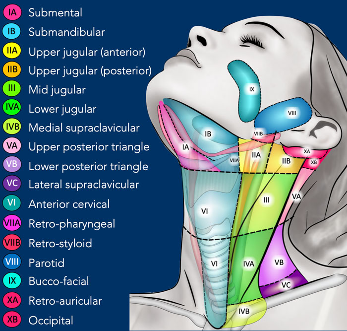
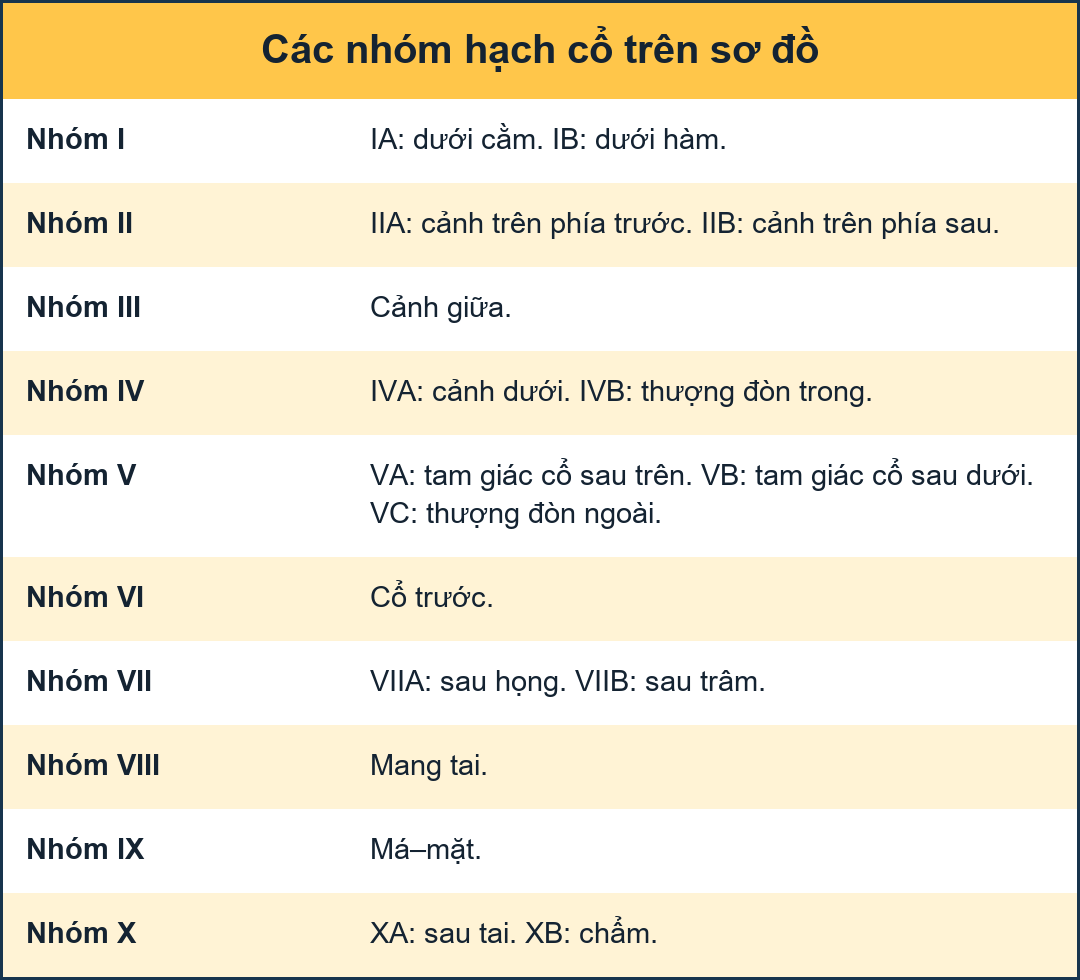
In this cervical lymph node map the levels were extended to 10.Trong bản đồ hạch bạch huyết cổ tử cung này, các mức độ đã được mở rộng đến 10.
Some of these are being divided into sub-levels to correspond more completely with the TNM atlas.Một số trong số này đang được chia thành các cấp độ phụ để tương ứng hoàn toàn hơn với bản đồ TNM.
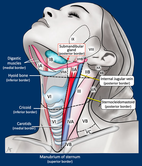
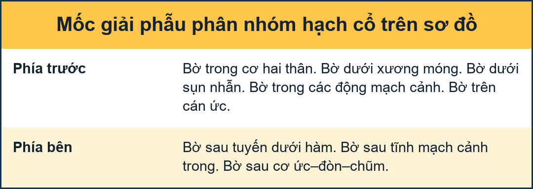
BordersBiên giới
Important landmarks are:Các mốc quan trọng là:
- Hyoid boneXương móng
- Cricoid
- Carotids
- Sternocleidomastoid muscleCơ ức đòn chũm
- Manubrium of sternumManubrium xương ức
Axial CTCT mặt cắt
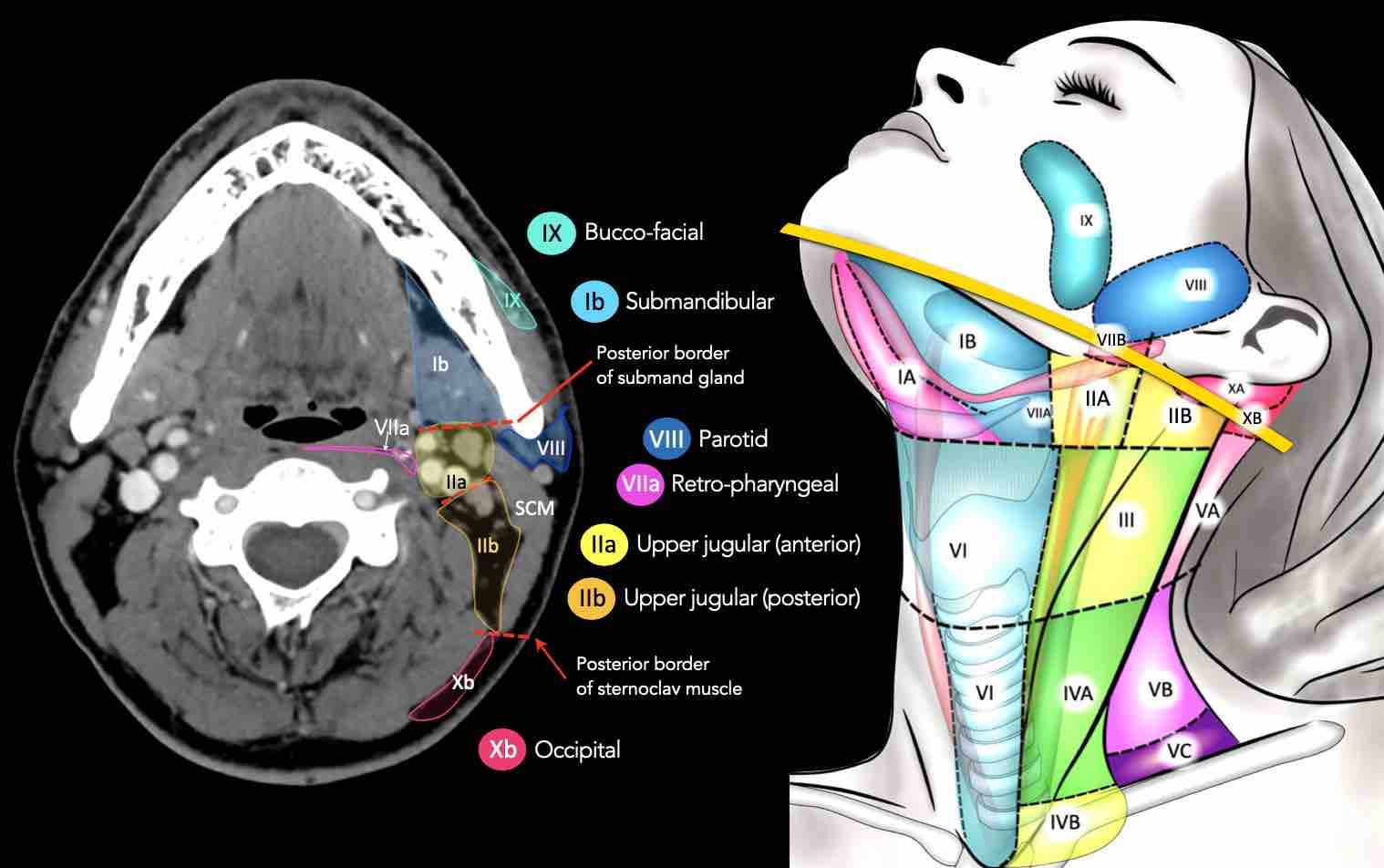
Axial CT slices in correlation to overview illustration.Các lát cắt CT hướng trục tương quan với hình ảnh tổng quan.
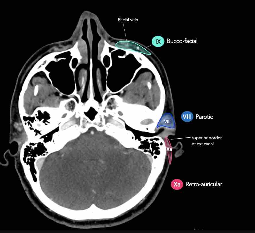
Axial CT slices in more detail.
Enlarge images by clicking on them.Cắt lớp vi tính (CLVT) cắt dọc trục chi tiết hơn.
Phóng to hình ảnh bằng cách nhấp vào chúng.
LevelsCấp độ
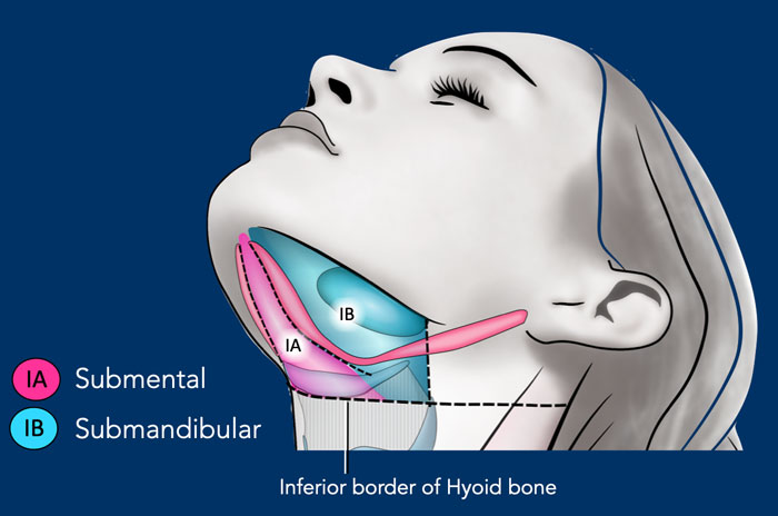
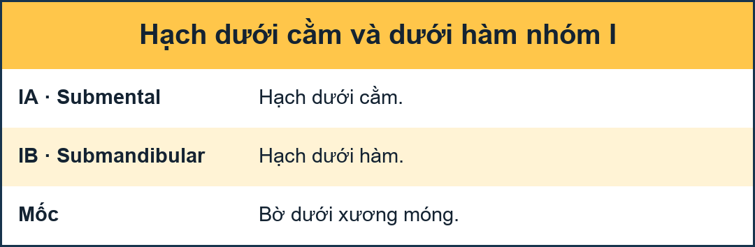
I - Submental and submandibularI - Cận dưới và dưới hàm
Nodes in level I are at risk of developing metastases from cancers of the oral cavity, anterior nasal cavity and the soft tissues of the mid-face and the submandibular gland.Các hạch ở mức độ I có nguy cơ phát triển di căn từ ung thư khoang miệng, khoang mũi trước và mô mềm của mặt giữa và tuyến dưới hàm.
Level Ia
is a median region located between the anterior belly of the digastric muscles, which contains the submental nodes.Cấp độ Ia
là vùng giữa nằm giữa bụng trước của cơ hai bụng, chứa các hạch dưới cằm.
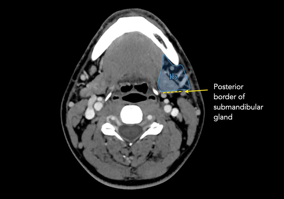
Level Ib
contains the submandibular nodes located in the space between the inner side of the mandible laterally and the digastric muscle medially, from the symphysis menti anteriorly to the submandibular gland posteriorly.Cấp độ Ib
chứa các hạch dưới hàm nằm ở khoảng giữa phía trong của hàm dưới và cơ hai hàm ở giữa, từ phía trước của khớp cắn đến phía sau của tuyến dưới hàm.
II - Upper jugularII - Cổ trên
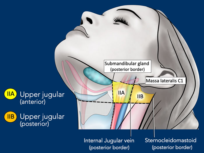
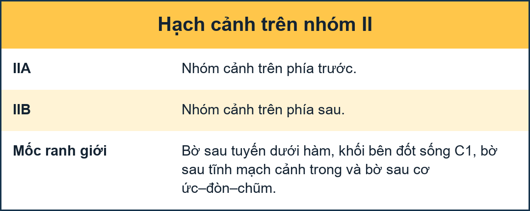
Level II receives lymphatics from the face, the parotid gland, and the submandibular, submental and retropharyngeal nodes.
Level II also directly receives the collecting lymphatics from the nasal cavity, the pharynx, the larynx, the external auditory canal, the middle ear, and the sublingual and submandibular glands [1]. Cấp II nhận được bạch huyết từ mặt, tuyến mang tai, và các hạch dưới hàm, dưới màng cứng và sau họng.
Cấp II cũng trực tiếp tiếp nhận các bạch huyết từ khoang mũi, họng, thanh quản, ống tai ngoài, tai giữa, và các tuyến dưới lưỡi và dưới hàm [1].
Level II can be divided into level IIa and level IIb by drawing a line at the posterior edge of the internal jugular vein.Mức II có thể được chia thành mức IIa và mức IIb bằng cách vẽ một đường ở mép sau của tĩnh mạch cảnh trong.
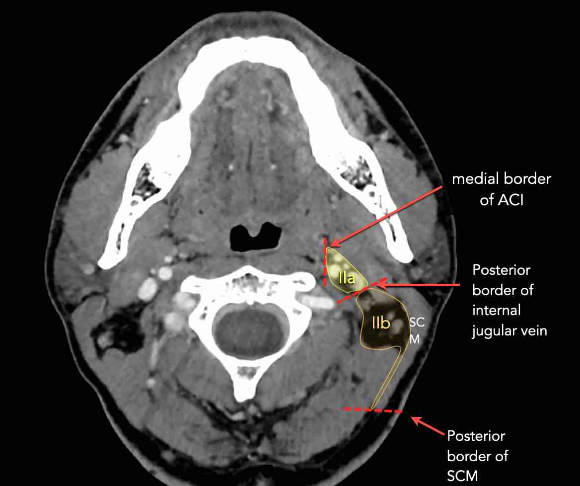
The nodes in level IIa and IIb are at risk of harboring metastases from cancers of the nasal and oral cavity, nasopharynx, oropharynx, hypopharynx, larynx and major salivary glands.Hạch độ IIa và IIb có nguy cơ ẩn chứa di căn từ ung thư khoang mũi miệng, vòm họng, hầu họng, hạ họng, thanh quản và tuyến nước bọt chính.
Level IIb is more likely associated with primary tumors of the oropharynx or nasopharynx, and less frequently with tumors of the oral cavity, larynx or hypopharynx [1].Mức độ IIb có nhiều khả năng liên quan đến các khối u nguyên phát của hầu họng hoặc vòm họng, và ít gặp hơn với các khối u của khoang miệng, thanh quản hoặc hạ họng [1].
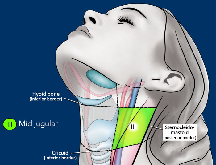
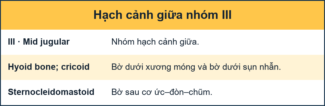
III - Mid jugularIII - Giữa tĩnh mạch cảnh
Level III receives efferent lymphatics from levels II and V, and some efferent lymphatics from the retropharyngeal, pretracheal and recurrent laryngeal nodes.
It collects the lymphatics from the base of the tongue, tonsils, larynx, hypopharynx and thyroid gland.Mức III nhận được các bạch huyết di chuyển từ mức II và V, và một số bạch huyết di động từ các hạch sau họng, trước khí quản và các hạch thanh quản tái phát.
Nó thu thập các bạch huyết từ gốc lưỡi, amidan, thanh quản, hạ họng và tuyến giáp.
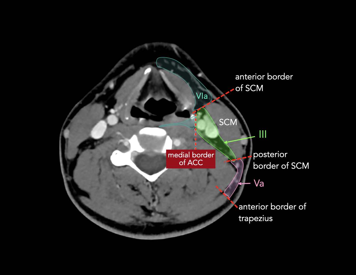
The inferior border of the cricoid is the border between level III and IVA.Ranh giới dưới của u là ranh giới giữa độ III và IVA.
Nodes in level III are at risk of harboring metastases from cancers of the oral cavity, nasopharynx, oropharynx, hypopharynx and larynx.Hạch độ III có nguy cơ ẩn chứa di căn từ ung thư khoang miệng, vòm họng, hầu họng, hạ họng và thanh quản.
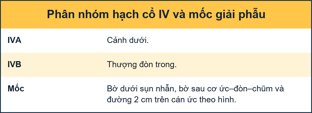
IV - Lower jugular and medial supraclavicularIV - Cổ tĩnh mạch cảnh dưới và trên đòn trong
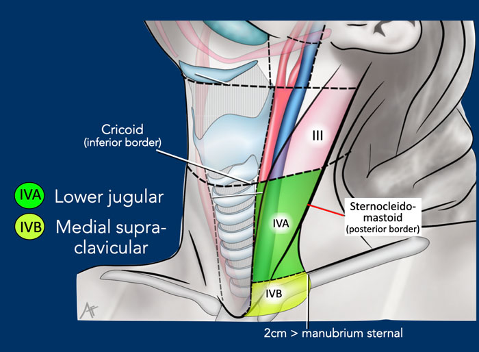
The border between level IVa and IVb is set arbitrarily 2 cm cranial to the sterno-clavicular joint.Đường viền giữa mức IVa và IVb được đặt tuỳ ý 2 cm từ sọ đến khớp ức-đòn.
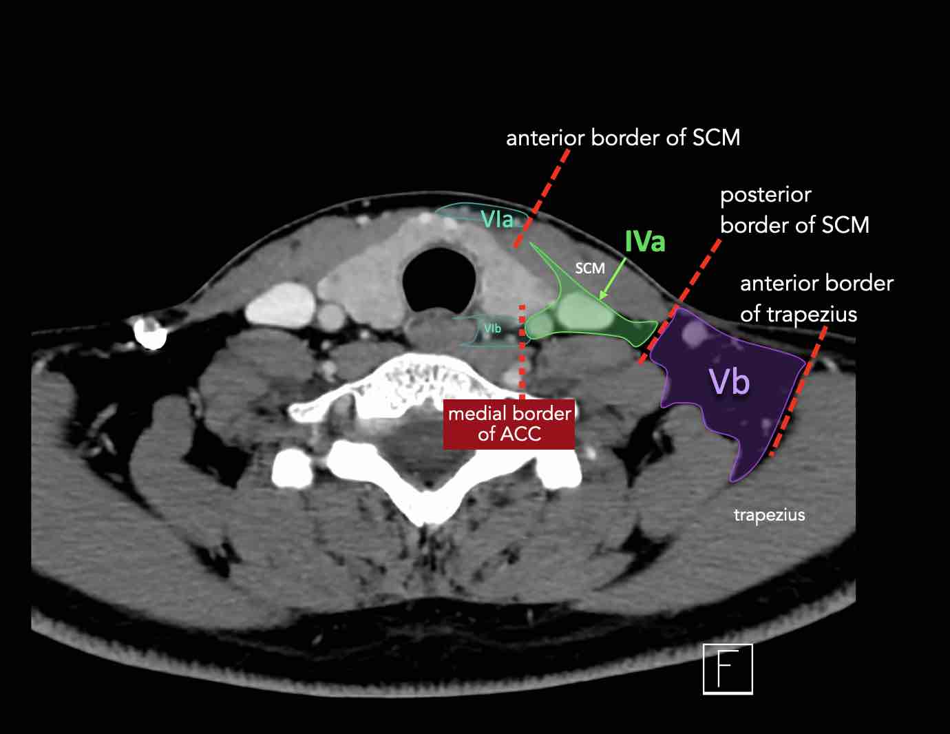
Level IVa
These nodes are at risk for harboring metastases from cancers of the hypopharynx, larynx, thyroid and cervical esophagus.
Rarely metastases from the anterior oral cavity may manifest in this location with minimal or no proximal nodal disease.Mức độ IVa
Những hạch này có nguy cơ chứa di căn từ ung thư hạ họng, thanh quản, tuyến giáp và thực quản cổ.
Hiếm khi di căn từ khoang miệng trước có thể biểu hiện ở vị trí này với bệnh hạch đầu gần tối thiểu hoặc không có.
Level IVb
These nodes are at risk for harboring metastases from cancers of the hypopharynx, subglottic larynx, trachea, thyroid and cervical esophagus.Cấp độ IVb
Những hạch này có nguy cơ chứa di căn từ ung thư hạ họng, thanh quản dưới thanh môn, khí quản, tuyến giáp và thực quản cổ.
V - Posterior triangle and SupraclavicularV - Tam giác sau và trên đòn
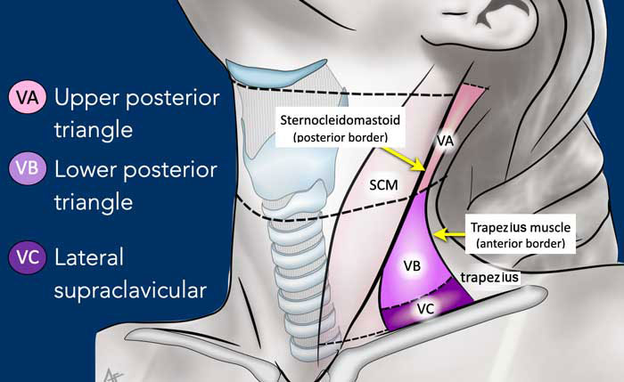
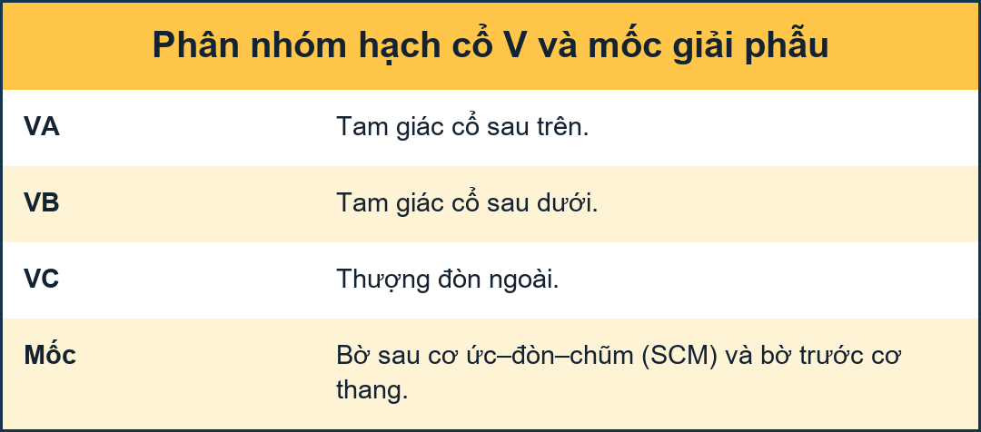
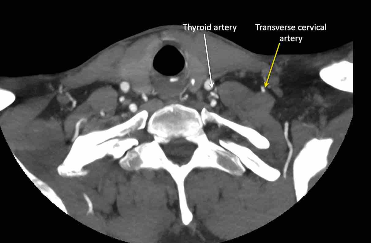
Level V contains the nodes of the posterior triangle group located posteriorly to the sternocleidomastoid muscle around the lower part of the spinal accessory nerve and the transverse cervical vessels.
Nồng độ V chứa các nút của nhóm tam giác sau nằm phía sau cơ ức đòn chũm xung quanh phần dưới của dây thần kinh phụ cột sống và các mạch máu cổ ngang.
Nodes in level V are most often associated with primary cancers of the nasopharynx, the oropharynx, the cutaneous structures of the posterior scalp, and the thyroid gland.Các hạch ở mức V thường liên quan đến ung thư nguyên phát của vòm họng, hầu họng, cấu trúc da của da đầu sau và tuyến giáp.
Level Vc - Supraclavicular
Mức Vc - Siêu đòn
This level contains the lateral supraclavicular nodes located in the continuation of the posterior triangle nodes (level Va and Vb) from the cervical transverse vessels down to a limit set arbitrarily 2 cm cranial to the sternal manubrium.
It corresponds partly to the area known as the supraclavicular fossa.Mức này chứa các hạch trên xương đòn bên nằm ở phần tiếp theo của các hạch tam giác sau (mức Va và Vb) từ các mạch máu ngang cổ xuống đến giới hạn đặt tuỳ ý 2 cm trong sọ đến ức ức.
Nó tương ứng một phần với khu vực được gọi là hố trên xương đòn.
Level Vc receives efferent lymphatics from the posterior triangle nodes (level Va and Vb) and is more commonly associated with nasopharyngeal tumors [1]. Mức Vc nhận được các bạch huyết di chuyển từ các hạch tam giác sau (mức Va và Vb) và thường liên quan đến các khối u vòm họng [1].
Transverse cervical artery
Scroll through the images for the anatomy of the transverse cervical artery.Động mạch cổ ngang
Cuộn qua các hình ảnh giải phẫu động mạch cổ ngang.
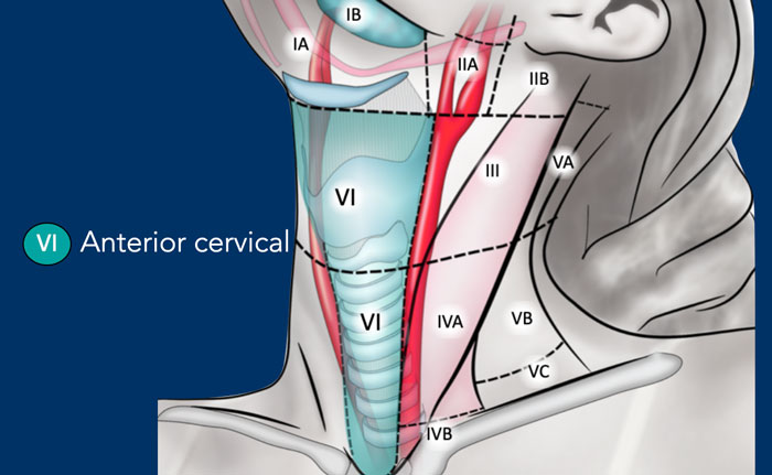
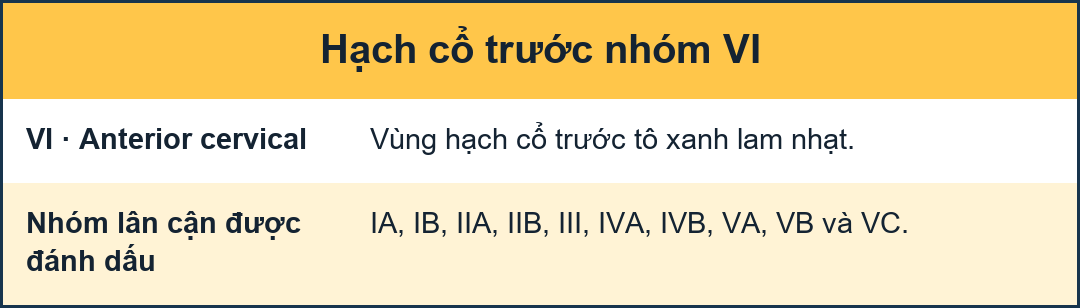
VI - Anterior cervicalVI - Cột sống cổ trước
This level contains the superficial anterior jugular nodes (level VIa) and the deeper prelaryngeal, pretracheal, paratracheal and recurrent laryngeal nerve nodes (level VIb).Nơi này chứa các hạch tĩnh mạch cảnh nông (nơi VIa) và các hạch thần kinh thanh quản trước trán, trước khí quản, cạnh khí quản và thanh quản (nơI VIb).
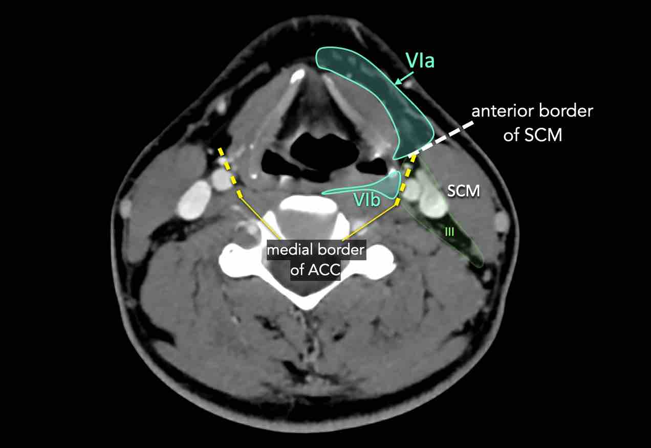
Level VIa
This level contains the superficially located anterior jugular nodes.Cấp độ VIa
Mức này chứa các hạch ở vùng nông của tĩnh mạch cảnh trước.
Level VIb
This level is contained between the medial borders of the common carotid arteries.
The nodes in this area are:
Cấp độ VIb
Mức này được chứa giữa các bờ trong của động mạch cảnh chung.
Các hạch trong khu vực này là:
- pre-laryngeal nodes in front of the larynx and cricoidcác hạch trước thanh quản ở phía trước thanh quản và u sần
- pre-tracheal nodes in front of the trachea các hạch trước khí quản ở phía trước khí quản
- paratracheal nodes also called recurrent laryngeal nerve nodesCác hạch cạnh khí quản còn được gọi là các hạch thần kinh thanh quản quặt ngược.
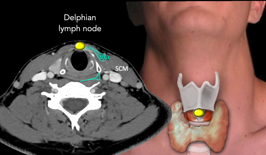
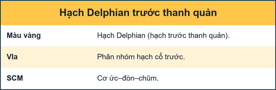
Delphian lymph nodeDelphian hạch bạch huyết
The Delphian lymph node derived its name from the oracle of Delphi, whose prophecy would be a death secondary to laryngeal cancer.Hạch bạch huyết của Delphian có nguồn gốc từ lời tiên tri của Delphi, người có lời tiên tri sẽ là một cái chết thứ phát do ung thư thanh quản.
It is a pretracheal node in level VIa located anterior to the cricoid and in between the cricothyroid muscles.Nó là một nút trước khí quản ở mức VIa nằm phía trước của cricoid và ở giữa các cơ nhẫn giáp.
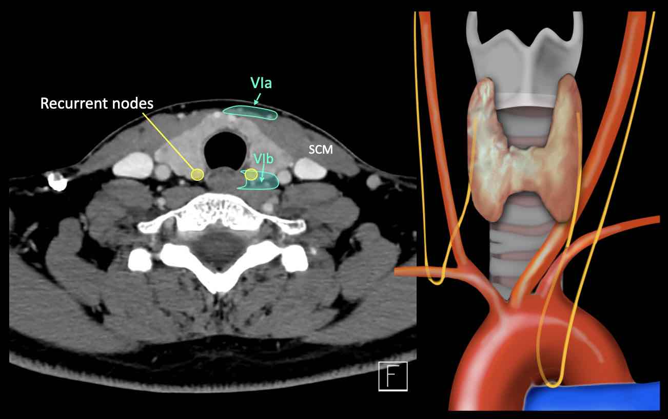
The recurrent laryngeal nerves branch off the vagus, the left at the aortic arch, and the right at the right subclavian artery.
Các dây thần kinh quặt ngược thanh quản phân nhánh từ phế vị, bên trái ở cung động mạch chủ, và bên phải ở động mạch dưới đòn phải.
The left laryngeal nerve can be compressed by subaortic lymph node metastases in the aorto-pulmonary window as seen in patients with lung cancer.Dây thần kinh thanh quản trái có thể bị chèn ép bởi di căn hạch bạch huyết dưới động mạch chủ ở cửa sổ chủ-phổi như đã thấy ở bệnh nhân ung thư phổi.

VII - Retropharyngeal and retrostyloidVII - Lồi họng và sau họng
Retropharyngeal nodes receive lymphatics from the mucosa of the nasopharynx, the Eustachian tube and the soft palate. Các hạch sau họng nhận được bạch huyết từ niêm mạc vòm họng, ống Eustachian và vòm miệng mềm.
These nodes are at risk of harboring metastases from cancers of the nasopharynx, the posterior pharyngeal wall and the oropharynx (mainly the tonsillar fossa and the soft palate).Những hạch này có nguy cơ chứa di căn từ ung thư vòm họng, thành sau họng và hầu họng (chủ yếu là hố amidan và vòm miệng mềm).
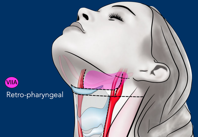
Level VIIa - retropharyngealMức VIIa - sau họng
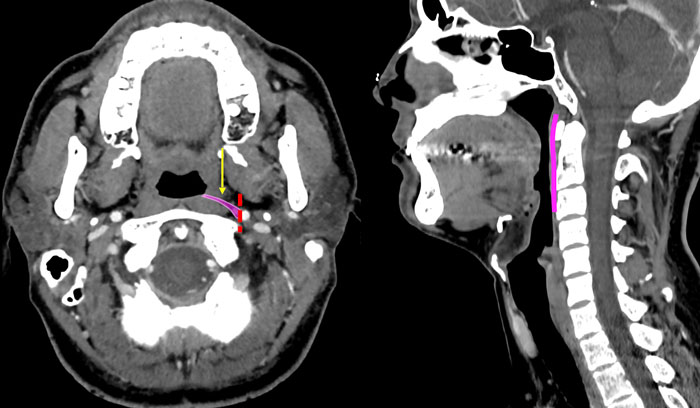
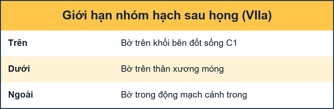
These nodes lie within the retropharyngeal space, extending cranially from the upper edge of the first cervical vertebrae (massa lateralis) to the cranial edge of the body of the hyoid bone caudally (figure).Các hạch này nằm trong khoang sau họng, kéo dài từ rìa trên của đốt sống cổ đầu tiên (massa lateralis) đến rìa sọ của thân xương móng (hình).
This space is bounded anteriorly by the pharyngeal constrictor muscles and posteriorly by the longus capitis and longus colli muscles.
Laterally, the retropharyngeal nodes are limited by the medial edge of the internal carotid artery.Khoảng trống này được giới hạn phía trước bởi các cơ co thắt họng và phía sau bởi cơ đầu dài và cơ ống dài.
Về phía sau, các hạch sau họng bị giới hạn bởi bờ trong của động mạch cảnh trong.
Retropharyngeal nodes receive efferent lymphatics from the mucosa of the nasopharynx, the Eustachian tube and the soft palate.
These nodes are at risk of harboring metastases from cancers of the nasopharynx, the posterior pharyngeal wall and the oropharynx (mainly the tonsillar fossa and the soft palate).Các hạch sau họng nhận được các bạch huyết từ niêm mạc của vòm họng, ống Eustachian và vòm miệng mềm.
Những hạch này có nguy cơ chứa di căn từ ung thư vòm họng, thành sau họng và hầu họng (chủ yếu là hố amidan và vòm miệng mềm).
Level VIIb - retrostyloidMức độ VIIb - retrostyloid
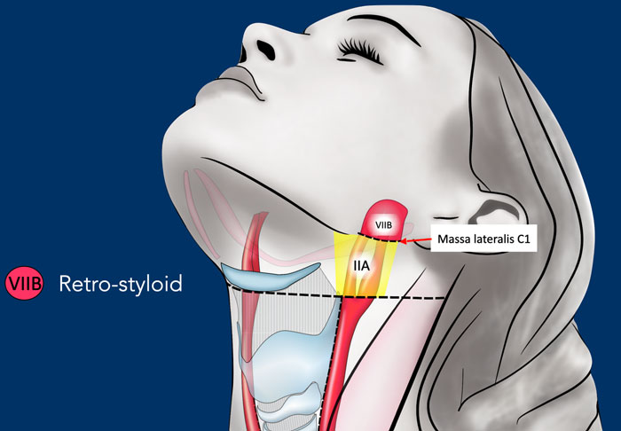
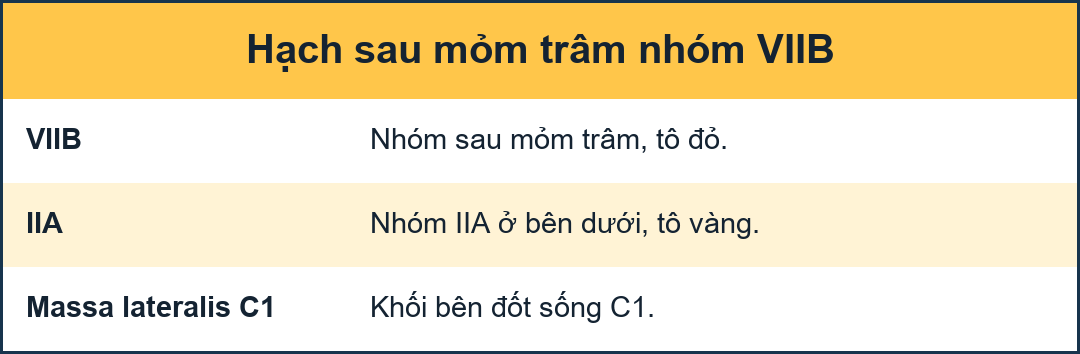
The retro-styloid nodes are the cranial continuation of the level II nodes.
They are located in the fatty space around the jugulo-carotid vessels up to the base of skull at the jugular foramen. Các hạch sau - thân mềm là sự tiếp nối của các hạch cấp II.
Chúng nằm trong khoảng mỡ xung quanh các mạch cảnh-cảnh đến đáy hộp sọ ở lỗ tĩnh mạch cảnh.
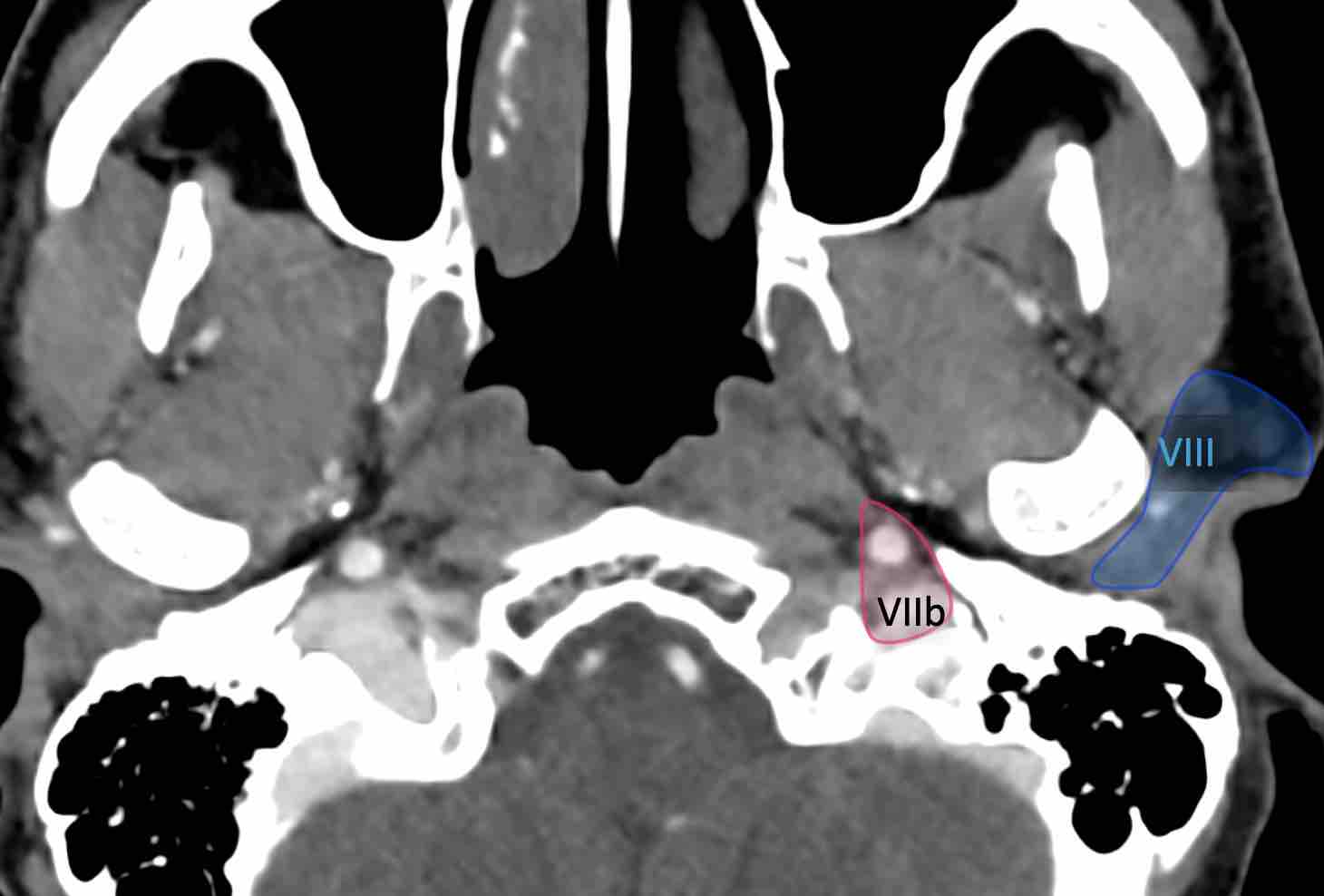
The retro-styloid space is delineated by the internal carotid artery medially, by the styloid process and the deep parotid lobe laterally, by the vertebral body of C1 and the base of skull posteriorly and by the pre-styloid para-pharyngeal space anteriorly.
Khoang sau xoang trâm được phân định bởi động mạch cảnh trong ở phía trong, bởi xoang trân và thuỳ mang tai sâu ở phía ngoài, bởi thân đốt sống C1 và nền sọ ở phía sau và khoang trước xoang trẩu cạnh họng ở phía trước.

VIII - ParotidVIII - Động mạch cảnh
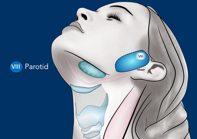
This level contains the parotid node group, which includes the subcutaneous pre-auricular nodes, the superficial and deep intraparotid nodes and the subparotid nodes. Nơi này chứa nhóm hạch mang tai, bao gồm các hạch trước tai dưới da, các hạch trong ổ bụng nông và sâu và các hạch dưới ổ bụng.
These nodes extend from the zygomatic arch and the external auditory canal down to the mandible.Các nút này kéo dài từ cung gò má và ống tai ngoài xuống đến xương hàm dưới.
They extend from the subcutaneous tissue laterally to the styloid process medially, and from the posterior edge of the masseter and the pterygoid muscles anteriorly to the anterior edge of the sternocleidomastoid muscle and the posterior belly of the digastric muscle posteriorly [1].Chúng kéo dài từ mô dưới da theo chiều ngang đến mỏm trâm ở trong, và từ mép sau của cơ cắn và cơ chân bướm về phía trước đến mép trước của cơ ức đòn chũm và bụng sau của cơ hai bụng về phía sau [1].
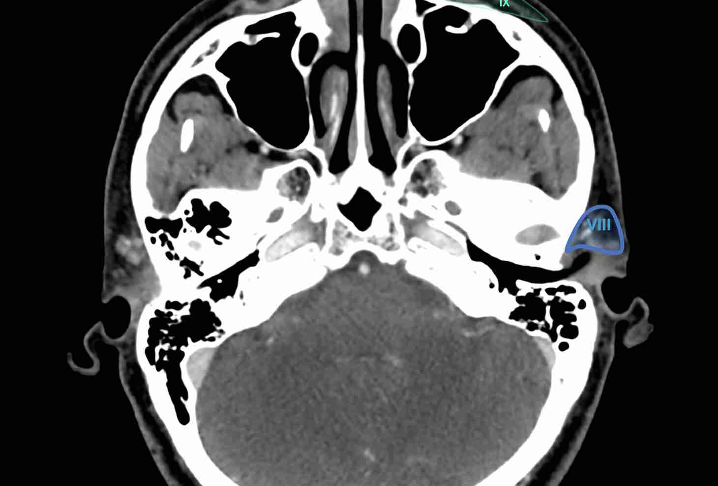
The parotid group receive lymphatic from the frontal and temporal skin, the eyelids, the conjunctiva, the auricle, the external acoustic meatus, the tympanum, the nasal cavities, the root of the nose, the nasopharynx, and the Eustachian tube. Nhóm mang tai nhận được bạch huyết từ da trán và thái dương, mí mắt, kết mạc, tai, lỗ tai ngoài, nhĩ, hốc mũi, chân mũi, vòm họng và vòi Eustachian.
They are at risk of harboring metastases from cancers of the frontal and temporal skin, orbit, external auditory canal, nasal cavities and parotid gland.Họ có nguy cơ bị di căn từ ung thư vùng trán và thái dương, hốc mắt, ống tai ngoài, hốc mũi và tuyến mang tai.
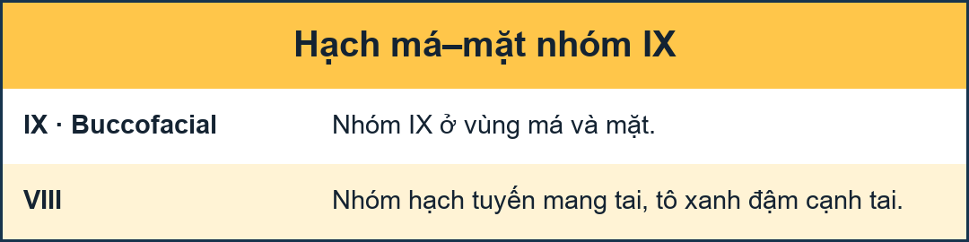
IX - BuccofacialIX - Vùng mặt
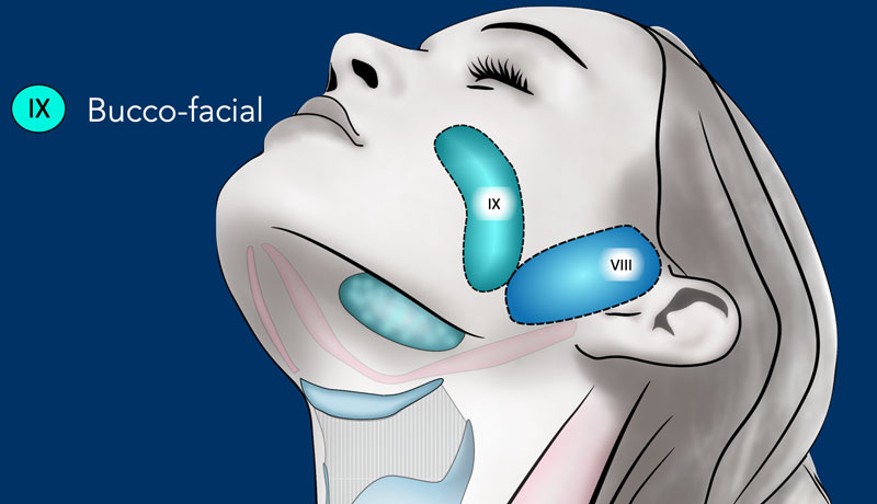
Level IX contains the malar and bucco-facial node group, which includes inconsistent superficial lymph nodes around the facial vessels on the external surface of the buccinator muscle.
These nodes extend from the caudal edge of the orbit (cranially) down to the caudal edge of the mandible (caudally) where they reached level Ib. Mức IX chứa nhóm nút malar và bucco-facial, bao gồm hạch bạch huyết bề mặt không nhất quán xung quanh các mạch máu mặt trên bề mặt ngoài của cơ mút.
Các hạch này kéo dài từ rìa đuôi của ổ mắt (xuống sọ) xuống rìa hàm dưới (xuống đuôi), nơi chúng đạt đến mức Ib.
They lay on the buccinators muscle (medially) in the sub-cutaneous tissue, from the anterior edge of the masseter muscle and the Bichat’s fat pad (posteriorly) to the anterior sub-cutaneous tissue of the face.Chúng nằm trên cơ túi mủ (ở giữa) trong mô dưới da, từ mép trước của cơ cắn và tấm mỡ của Bichat (ở phía sau) đến mô dưới da phía trước của khuôn mặt.
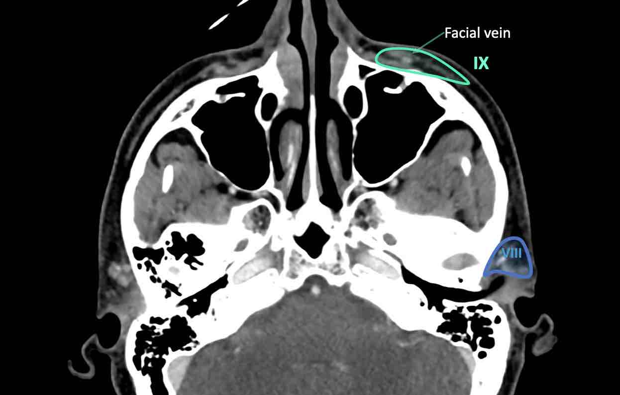
The bucco-facial nodes receive efferent vessels from the nose, the eyelids, and the cheek. Các hạch mặt vú nhận được các mạch máu từ mũi, mí mắt và má.
They are at risk of harboring metastases from cancers of the skin of the face, the nose, the maxillary sinus (infiltrating the soft tissue of the cheek) and the buccal mucosa.Họ có nguy cơ bị di căn từ ung thư da mặt, mũi, xoang hàm trên (xâm nhập mô mềm của má) và niêm mạc má.
X - Retroauricular and occipitalX - Sau tai và chẩm
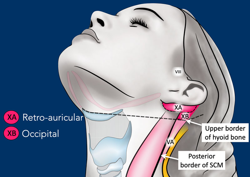
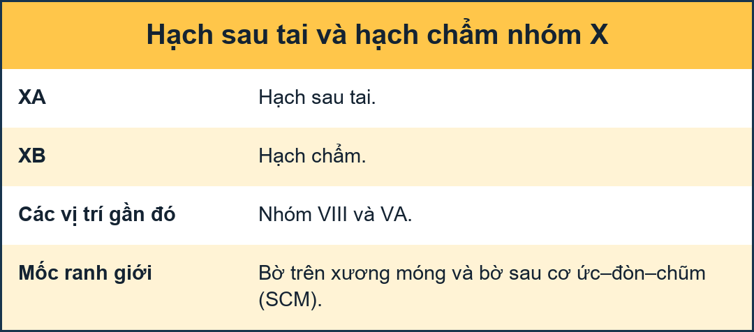
Level Xa contains the retroauricular (also called mastoid) and subauricular nodes, which includes superficial nodes lying on the mastoid process from the cranial edge of the external auditory canal cranially to the tip of the mastoid caudally. Mức Xa chứa các hạch sau tai (còn gọi là xương chũm) và hạch dưới tai, bao gồm các hạch nông nằm trên xương chũMức Xa chứa hạch sau và hạch sau, bao gồm hạch nông từ rìa sọ của ống tai ngoài đến đầu xương chũ.
Level Xb contains the occipital lymph nodes, which are the cranial and superficial continuation of the level Va nodes up to the cranial protuberance. They lie from the posterior edge of the sternocleidomastoid muscle to the anterior (lateral) edge of the trapezius muscle.Nồng độ Xb chứa hạch bạch huyết chẩm, là sự tiếp nối của các nút Va đến phần nhô ra của sọ, nằm từ mép sau của cơ ức đòn chũm đến mép trước (bên) của cơ thang.
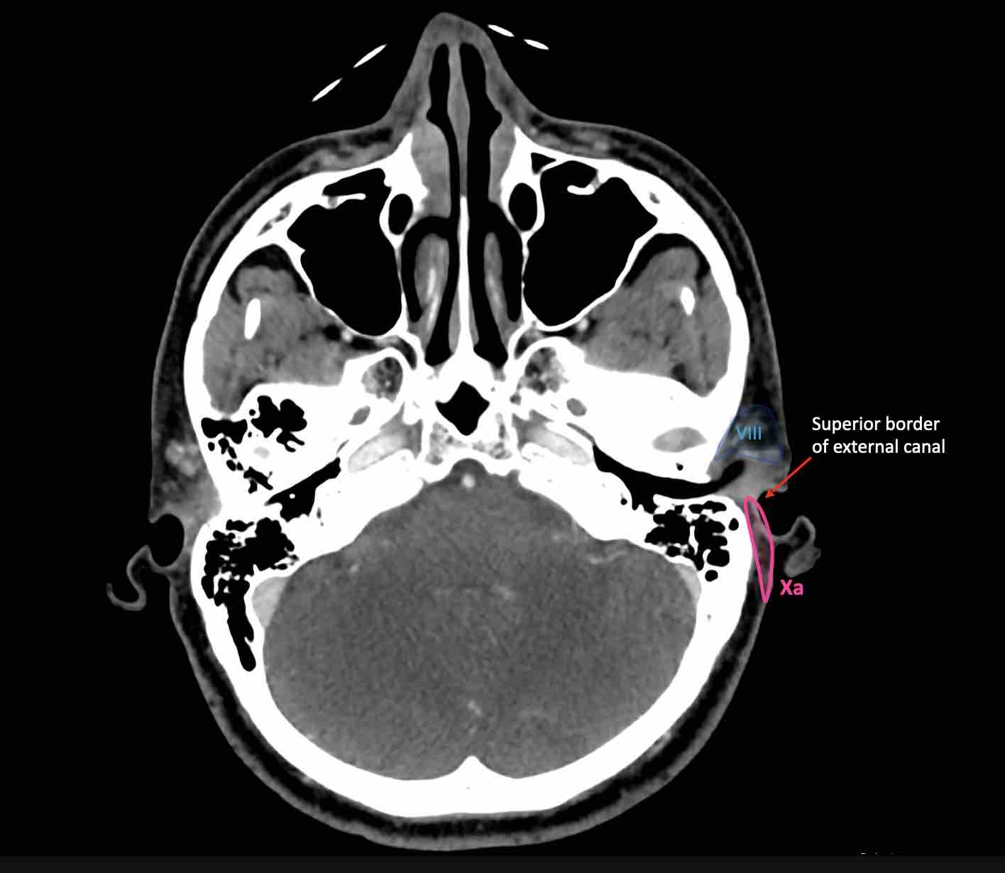
Lymph node metastases in level X are from skin cancers of the retro-auricular area (Xa) and skin cancers of the occipital area (Xb).Di căn hạch bạch huyết ở mức X là do ung thư da vùng sau tai (Xa) và ung thư da của vùng chẩm (Xb).
Click on the image below to get more information about Medical Action Myanmar, a medical organization run by Nini Tun and Frank Smithuis, who happens to be the brother of Robin Smithuis.Nhấp vào hình ảnh dưới đây để biết thêm thông tin về Medical Action Myanmar, một tổ chức y tế do Nini Tun và Frank Smithuis điều hành, người tình cờ là anh trai của Robin Smithuis.
ReferencesTài liệu tham khảo
-
Delineation of the neck node levels for head and neck tumors: A 2013 update. DAHANCA, EORTC, HKNPCSG, NCIC CTG, NCRI, RTOG, TROG consensus guidelines
Mô tả mức độ hạch cổ trong u vùng đầu cổ: Cập nhật năm 2013 với các hướng dẫn đồng thuận của DAHANCA, EORTC, HKNPCSG, NCIC CTG, NCRI, RTOG, TROG
DAHANCA, EORTC, HKNPCSG,NCIC CTG, NCRI, RTOG, TROG consensus guidelines.
by V Grégoire et al.
Radiother Oncol 2014 ;110:172–81.
Các hướng dẫn đồng thuận DAHANCA, EORTC, HKNPCSG, NCIC CTG, NCRI, RTOG, TROG. -
Integrating radiological criteria into the classification of cervical lymph node disease.
by Robbins KT.
Arch Otolaryngol Head Neck Surg 1999 ;125:385–7.
Lồng ghép các tiêu chuẩn chẩn đoán hình ảnh vào phân loại bệnh hạch bạch huyết cổ tử cung. -
International association for the study of lung cancer (IASLC) lymph node map: radiologic review with CT illustration.
Hiệp hội quốc tế về nghiên cứu ung thư phổi (IASLC) Bản đồ hạch bạch huyết: tổng quan X quang có hình minh hoạ CT.
by El-Sherief AH, Lau CT, Wu CC, Drake RL, Abbott GF, Rice TW.
Radiographics. 2014 Oct;34(6):1680-91.
bởi El-Sherief AH, Lau CT, Wu CC, Drake RL, Abbott GF, Rice TW.