
Swallowing disorders updateCập nhật rối loạn nuốt
Radiology department of the Alrijne Hospital in Leiderdorp, the NetherlandsKhoa Chẩn đoán hình ảnh của Bệnh viện Alrijne ở Leiderdorp, Hà Lan
Ngày đăng / cập nhật: 2018-08-13
Bản gốc trên radiologyassistant.nl ↗ · tải về lúc 2026-11-19 20:19 · bản dịch máy có chuẩn hóa thuật ngữ chẩn đoán hình ảnh
Swallowing is a complex movement.
It requires the coordination of nerves and muscles in the buccolabial area, the tongue, the palate, the pharynx, the larynx and finally the esophagus.
Radiographic studies of patients with swallowing disorders can help to analyse the problem.
In this article a systematic approach on how to analyse these videos is presented by focussing on four basic findings:Nuốt là một cử động phức tạp.
Nó đòi hỏi sự phối hợp của các dây thần kinh và cơ ở vùng gò má, lưỡi, vòm miệng, họng, thanh quản và cuối cùng là thực quản.
Các nghiên cứu X quang của bệnh nhân rối loạn nuốt có thể giúp phân tích vấn đề.
Trong bài viết này, một cách tiếp cận có hệ thống về cách phân tích các video này được trình bày bằng cách tập trung vào bốn kết quả cơ bản:
- Asymmetric swallowingNuốt không đối xứng
- Stasis of contrastNgưng thuốc cản quang
- Cricopharyngeal dysfunctionRối loạn chức năng thanh quản
- AspirationKhát vọng
Normal SwallowingNuốt bình thường
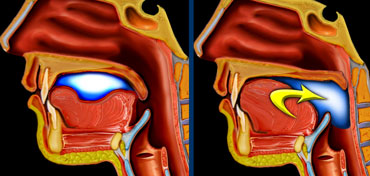
 LEFT: Oral or preparatory phase. RIGHT: Transport to pharynx and subsequent triggering of the actual swallowing reflex.Bên trái: Giai đoạn bằng miệng hoặc chuẩn bị. Phải: vận chuyển đến hầu họng và sau đó kích hoạt phản xạ nuốt thực tế.
Oral phaseGiai đoạn uống
In the oral phase food is prepared for swallowing and then transported to the pharynx.
This is a preparatory phase in which the food is held within the mouth while the base of the tongue and the soft palate close the oral cavity posteriorly to prevent food spilling into the open larynx and trachea. Trong giai đoạn uống, thức ăn được chuẩn bị để nuốt và sau đó được vận chuyển đến họng.
Đây là giai đoạn chuẩn bị trong đó thức ăn được giữ trong miệng trong khi phần lưỡi và vòm miệng mềm đóng lại phía sau để ngăn thức ăn tràn vào thanh quản và khí quản.
A bolus is formed in the central portion of the tongue and then pushed posteriorly toward the pharynx with an anterior-to-posterior tongue elevation.
As the bolus enters the pharynx the actual swallow or pharyngeal reflex is triggered.
Một bolus được hình thành ở phần trung tâm của lưỡi và sau đó đẩy về phía sau về phía họng với một lưỡi nâng lên phía trước.
Khi bolus đi vào họng, phản xạ nuốt hoặc phản xạ họng thực sự được kích hoạt.
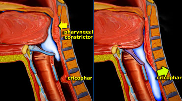
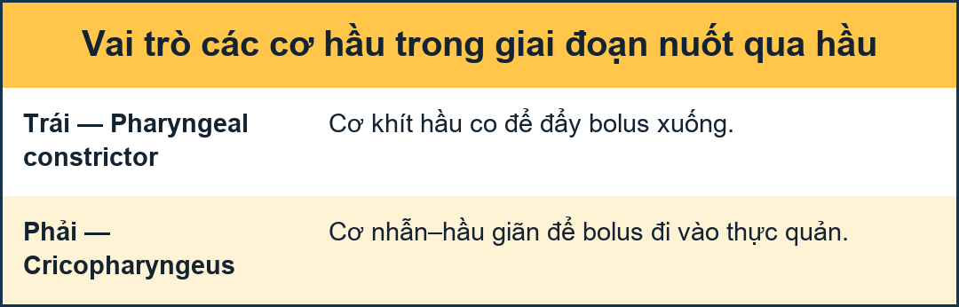 LEFT: Pharyngeal constrictors push the bolus down. RIGHT: Together with the contraction of the inferior constrictor, the cricopharyngeus relaxes.Trái: Các cơ khít hầu đẩy viên thức ăn xuống. Phải: Khi cơ khít hầu dưới co, cơ nhẫn–hầu giãn.
Pharyngeal phaseGiai đoạn hầu họng
This phase is a reflex action.
The bolus passes through the pharynx quickly and then enters the esophagus.
This takes place in less than a second.
The initiation of this process starts when the bolus passes the anterior faucial arch and reaches the posterior pharyngeal wall.
Elevation of the soft palate prevents material from entering the nasal cavity.
This stage is followed by the pharyngeal constrictor muscles pushing the bolus further into the pharynx, toward the cricopharyngeal sphincter.
The larynx prevents material from entering the trachea by respectively closing the true vocal cords, false vocal folds, and aryepiglottic folds.
Contraction of the lower pharyngeal constrictor is followed by relaxation of the cricopharyngeal muscle, allowing the bolus to pass into the esophagus.Giai đoạn này là một phản xạ.
Lỗ thông đi qua họng nhanh chóng và sau đó đi vào thực quản.
Điều này diễn ra trong vòng chưa đầy một giây.
Quá trình này bắt đầu khi bolus đi qua cung má trước và đến thành sau họng.
Nâng cao vòm miệng mềm ngăn không cho vật liệu đi vào khoang mũi.
Giai đoạn này được theo sau bởi các cơ co thắt họng đẩy bolus vào sâu hơn vào họng, về phía cơ thắt của hầu họng.
Thanh quản ngăn không cho vật liệu xâm nhập vào khí quản bằng cách đóng dây thanh âm thật, dây thanh âm giả và dây thanh âm.
Co thắt cơ thắt họng dưới được theo sau bởi sự giãn cơ nhẫn hầu, cho phép bolus đi vào thực quản.
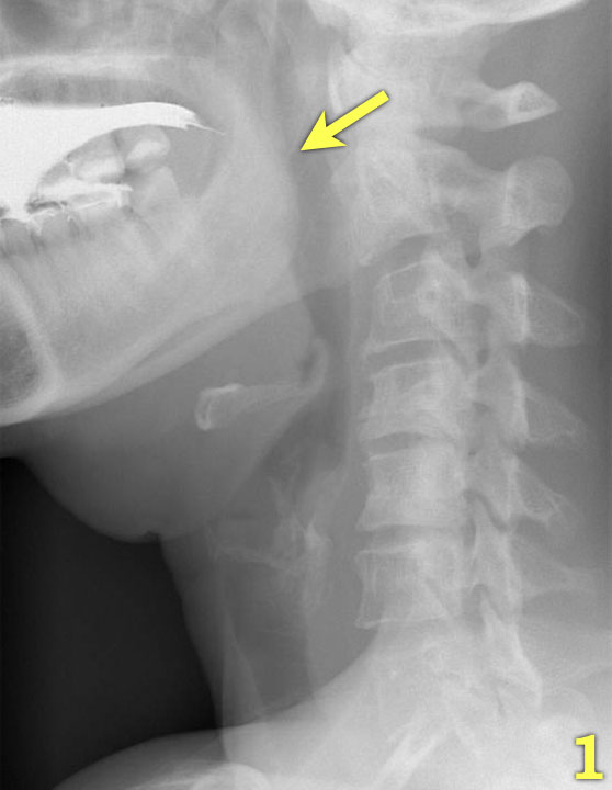
Fluoroscopic imagingHình ảnh học huỳnh quang
The most important images of the swallowing study are those taken of the lateral view.
Click through the images 1-7 on the left.Hình ảnh quan trọng nhất của nghiên cứu nuốt là những hình ảnh được chụp từ mặt bên.
Nhấp qua các hình ảnh 1-7 bên trái.
- The base of the tongue and the soft palate close the oral cavity posteriorly (arrow) to prevent spill of food into the pharynx and open larynx.Đế lưỡi và vòm miệng mềm đóng khoang miệng phía sau (mũi tên) để ngăn thức ăn tràn vào họng và thanh quản mở.
- The tongue start to transport the food towards the pharynx (yellow arrow).
The larynx is still open and in its normal position (green arrow).Lưỡi bắt đầu vận chuyển thức ăn về phía họng (mũi tên màu vàng).
Thanh quản vẫn mở và ở vị trí bình thường (mũi tên màu xanh lá cây). - the soft palate elevates to prevent spill into the nasopharynx (green arrow) and the tongue pushes the food further backwards (yellow arrow)khẩu cái mềm nâng lên để tránh tràn vào vòm họng (mũi tên màu xanh lá cây) và lưỡi đẩy thức ăn ra sau (mũI tên màu vàng)
- The hyoid bone elevates and the larynx closes (green arrow). The tongue pushes the food downwards, while the upper esophageal constrictor contracts.Xương móng nâng cao và thanh quản đóng lại (mũi tên màu xanh lá cây), lưỡi đẩy thức ăn xuống dưới, trong khi co thắt thực quản trên co lại.
- Contraction of the middle pharyngeal constrictor (yellow arrow), while the cricopharyngeus is already fully relaxed (green arrow).Co thắt của cơ thắt họng giữa (mũi tên màu vàng), trong khi xương ức đã được thư giãn hoàn toàn (mũI tên màu xanh lá cây).
- Contraction of the lower pharyngeal constrictor emptying the pharynx.Co thắt họng dưới làm rỗng họng.
- Epiglottis elevates to regain its resting position and the larynx opens (arrow).Nắp thanh môn nâng lên để lấy lại vị trí nghỉ ngơi và thanh quản mở ra (mũi tên).
Video of lateral viewVideo chụp tư thế nghiêng
Watch in HD.Xem ở chế độ HD.
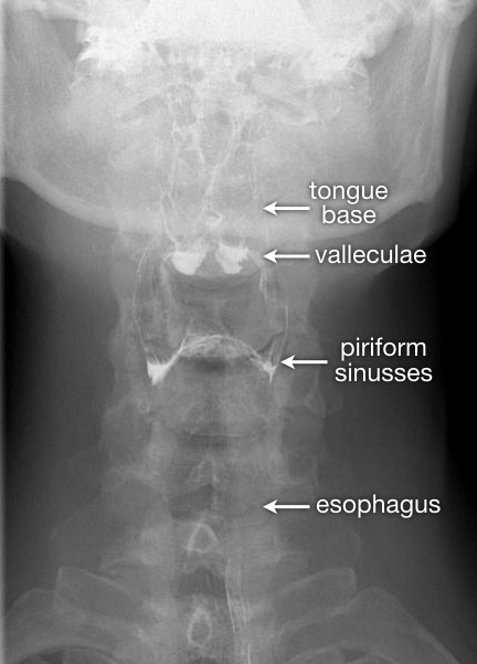
The AP-view is important to look for asymmetry.
Once the series of the pharyngeal phase has been acquired, follow the contrast bolus all the way down to the gastroesophageal junction.Quan sát AP là quan trọng để tìm sự bất đối xứng.
Khi đã có một loạt các giai đoạn của họng, hãy theo dõi thuốc cản quang đến tận chỗ nối dạ dày thực quản.
Indications for a studyChỉ định nghiên cứu
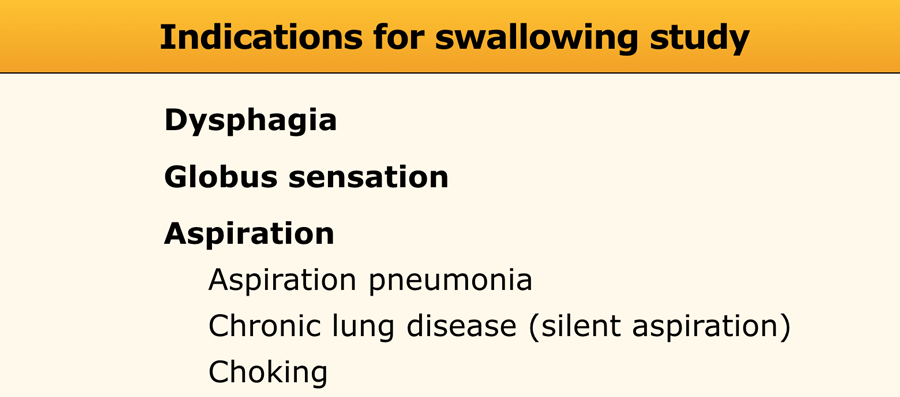
Indications for a swallowing study are dysphagia, globus sensation and aspiration.Chỉ định cho một nghiên cứu nuốt là khó nuốt, cảm giác hình cầu và hít phải.
Dysphagia is a general term used to describe the inability to transport the food properly from the mouth to the stomach.
Globus sensation is a term to describe the feeling, that there is something in the throat, that is in the way or needs to be swallowed.
Aspiration is the most severe form of swallowing disorders and can result in aspiration pneumonia, chronic lung disease or choking.
In some patients with chronic lung disease a swallowing study can be performed to look for 'silent aspiration'.Khó nuốt là một thuật ngữ chung được sử dụng để mô tả việc không có khả năng vận chuyển thức ăn đúng cách từ miệng đến dạ dày.
Cảm giác hình cầu là một thuật ngữ để mô tả cảm giác, rằng có thứ gì đó trong cổ họng, đang cản trở hoặc cần phải nuốt.
Hít phải là dạng rối loạn nuốt nặng nhất và có thể dẫn đến viêm phổi hít phải, bệnh phổi mạn tính hoặc nghẹt thở.
Ở một số bệnh nhân mắc bệnh phổi mạn tính, có thể thực hiện một nghiên cứu về nuốt để tìm kiếm 'hít vào thầm lặng'.
Study of SwallowingNghiên cứu về nuốt
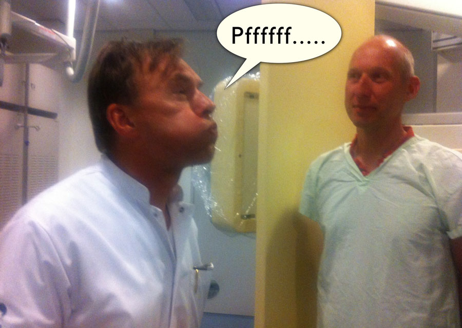 The radiologist is explaining how to perform the modified valsalvaBác sĩ chẩn đoán hình ảnh giải thích cách thực hiện phẫu thuật sửa van tim
First try to find out exactly what the patients problem is, so you can customize the series.
Is there a risk of aspiration, i.e. wet voice, recurrent pneumonia or aspiration?
If so, do not start the examination with barium contrast, but instead use non-ionic water-soluble contrast.
If during the first few swallows there is no aspiration, you can switch to barium, as this gives better quality images.Đầu tiên hãy cố gắng tìm hiểu chính xác vấn đề của bệnh nhân là gì, để bạn có thể tuỳ chỉnh loạt bài.
Có nguy cơ hít phải, tức là giọng nói ướt, viêm phổi tái phát hoặc hít phải không?
Nếu có, không bắt đầu khám bằng thuốc cản quang barium, mà thay vào đó sử dụng thuốc cản âm không tan trong nước.
Nếu trong vài lần nuốt đầu tiên không có hiện tượng hít phải, bạn có thể chuyển sang dùng barium, vì điều này cho hình ảnh chất lượng tốt hơn.
Before we start the examination, the procedure is explained to the patient and we practise certain manoeuvres like the modified Valsalva (figure).Trước khi bắt đầu khám, chúng tôi giải thích cho bệnh nhân về thủ thuật và thực hiện một số thao tác nhất định như Valsalva cải biên (hình).
When solid food is the problem, you may want to add a solid substance to the barium - for instance biscuits or bread with bariumpaste.Khi vấn đề là thức ăn đặc, bạn có thể muốn thêm một chất rắn vào barium - ví dụ như bánh quy hoặc bánh mì với bariumpaste.
The patient in the video only has problems with solid food.Bệnh nhân trong video chỉ gặp vấn đề với thức ăn đặc.
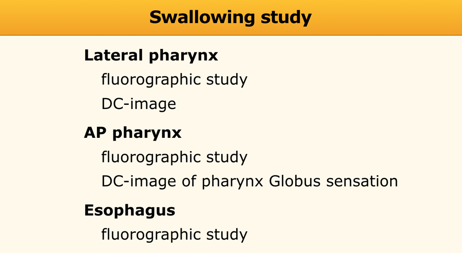
The examination of patients with a possible swallowing disorder consists of:Khám bệnh nhân có rối loạn nuốt có thể bao gồm:
- Fluorographic study of the actual swallowing.Nghiên cứu hình ảnh nuốt thực tế trên phim X quang.
- Double-contrast images of the pharynx.Hình ảnh hai mặt của họng.
- Examination of the esophagus.Khám thực quản.
Start with one or two lateral swallows followed by a lateral double-contrast view of the pharynx (see later).
Then an AP-swallow is recorded followed by an AP double-contrast view of the pharynx.
Next the passage through the esophagus is recorded, sometimes followed by double-contrast views of the gastroesophageal junction.Bắt đầu bằng một hoặc hai lần nuốt bên, sau đó là nhìn hai mặt đối xứng bên của họng (xem phần sau).
Sau đó, ghi lại nuốt AP, tiếp theo là nhìn họng với hai lớp thuốc cản quang.
Tiếp theo, đường đi qua thực quản được ghi lại, đôi khi theo sau là hình ảnh đối chiếu hai chiều của đường nối dạ dày thực quản.
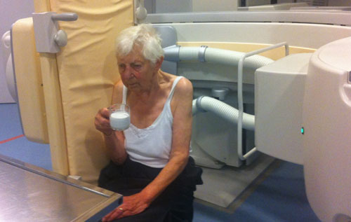 Always start with the lateral view first!Luôn bắt đầu với tư thế nghiêng trước!
Fluorographic study of the actual swallowingNghiên cứu đặc điểm nuốt thực tế bằng chụp huỳnh quang
The act of swallowing is recorded on video or some sort of digital recording.
We use grabs from the fluographic images and store them into the PACS-system, where we can play the images back and forth in slow motion.Hành động nuốt được ghi lại trên video hoặc một số loại ghi âm kỹ thuật số.
Chúng tôi sử dụng các hình ảnh chụp từ máy chụp và lưu trữ chúng vào hệ thống PACS, nơi chúng tôi có thể phát lại các hình ảnh qua lại trong chuyển động chậm.
It is very important to always start with the lateral view first, because if the patient aspirates, the lateral view will tell you how and why it happened.
If you have to stop the examination, the most important images will already have been recorded.
If you start with an AP-view and the patient aspirates, you may have to stop the examination and you will never know why aspiration occurred and what the problem is.Điều rất quan trọng là luôn bắt đầu với mặt bên trước, bởi vì nếu bệnh nhân hít phải, mặt bên sẽ cho bạn biết làm thế nào và tại sao nó xảy ra.
Nếu bạn phải dừng khám, những hình ảnh quan trọng nhất sẽ được ghi lại.
Nếu bạn bắt đầu với góc nhìn AP và bệnh nhân hít phải, bạn có thể phải dừng khám và bạn sẽ không bao giờ biết tại sao lại xảy ra hiện tượng hít phải và vấn đề là gì.
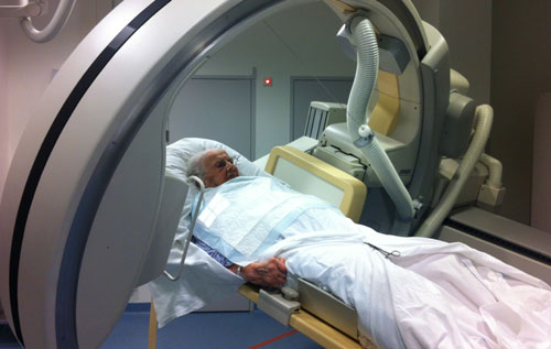
If a patient is unable to stand or sit, you can still do a lateral view.Nếu bệnh nhân không thể đứng hoặc ngồi, bạn vẫn có thể thực hiện tư thế nghiêng.
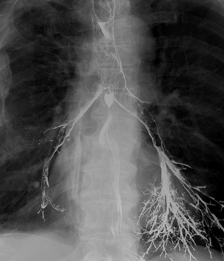
Use only a small amount of barium for the first swallow and if the patient is doing fine, coninue with larger portions.
Aspiration of a small amount of barium is usually not a big problem, but you don't want a lot of barium filling the bronchi.Chỉ sử dụng một lượng nhỏ barium cho lần nuốt đầu tiên và nếu bệnh nhân đang làm tốt, hãy nuốt với phần lớn hơn.
Hút một lượng nhỏ bari thường không phải là vấn đề lớn, nhưng bạn không muốn nhiều bari lấp đầy phế quản.
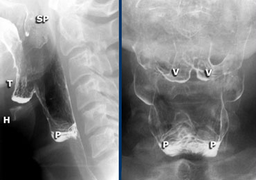
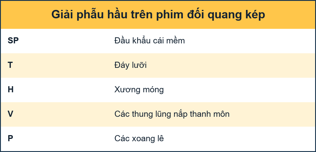 LEFT: Lateral view during singing aaa. Hyoid (H) and tongue base (T) move anteriorly. Left and right pyriform sinuses are projected on top of each other. Tip of soft palate (SP) is seen. RIGHT: Valleculae (V) and pyriform sinuses (P).TRÁI: Nhìn bên trong khi hát aaa. Sụn (H) và nền lưỡi (T) di chuyển về phía trước. Các xoang hình chóp trái và phải được chiếu lên nhau. Đầu vòm miệng mềm (SP) được nhìn thấy. QUYỀN: Valleculae (V) và xoang pyriform (P).
Double contrast images of the pharynxHình ảnh hai lớp thuốc cản quang của hầu họng
For the lateral view, ask the patient to sing an aaa, as this will move the tongue in an anterior position and give a better view on the oro- and hypopharynx.
In Dutch this will be the letter eee, as it is pronounced the same as the english aaa.Đối với chế độ xem bên, yêu cầu bệnh nhân hát aaa, vì điều này sẽ di chuyển lưỡi ở vị trí phía trước và cho cái nhìn tốt hơn về miệng và hạ họng.
Trong tiếng Hà Lan, đây sẽ là chữ eee, vì nó được phát âm giống như aaa trong tiếng Anh.
For the AP-view the modified Valsalva manoeuvre is performed.
The patients has to blow air through the tightened lips as in trumpet-playing, while relaxing the neck region.
Always practise this manoeuvre prior to the examination, so the patient knows what to do.Đối với chế độ xem AP, thực hiện thao tác Valsalva đã sửa đổi.
Bệnh nhân phải thổi khí qua môi bị siết chặt như trong thổi kèn, đồng thời thư giãn vùng cổ.
Luôn thực hành động tác này trước khi khám, để bệnh nhân biết phải làm gì.
On the left DC views of the pharynx.
Outpounching of the lateral wall of the pharynx is normal and can be quite severe (Dizzy Gillespie).
These are called 'lateral pharyngeal ears'.Hình ảnh DC bên trái của họng.
Miệng họng bên bị bật ra là bình thường và có thể khá nặng (Dizzy Gillespie).
Đây được gọi là 'tai họng bên'.
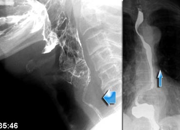 Cricopharyngeal hypertrophy in a patient with reflux.Phì đại hầu họng ở bệnh nhân trào ngược.
Examination of the esophagusKhám thực quản
Always follow the passage of barium through the esophagus until it enters the stomach.
Disorders of the gastroesophageal junction are often experienced as a problem within the throat.
The rationale for this is that in patients with a distal obstruction, gastroesophageal reflux or a motility disorder, the cricopharyngeal muscle has to work very hard to prevent foodspillage back into the pharynx - along with the risk of aspiration.
This increased muscle tone gives the patient the sensation that there is something in the throat.Luôn theo dõi đường đi của bari qua thực quản cho đến khi nó đi vào dạ dày.
Rối loạn của chỗ nối dạ dày thực quản thường gặp như một vấn đề trong cổ họng.
Lý do cho điều này là ở những bệnh nhân bị tắc nghẽn đầu xa, trào ngược dạ dày thực quản hoặc rối loạn nhu động, cơ nhẫn hầu phải làm việc rất chăm chỉ để ngăn thức ăn tràn ngược vào họng - cùng với nguy cơ hít phải.
Sự gia tăng trương lực cơ này cho bệnh nhân cảm giác có gì đó trong cổ họng.
The images are of a patient with globus sensation.
This was due to severe reflux and subsequent increased tone of the cricopharyngeal muscle.
A large paraesophageal hernia with reflux is the cause of the complaints.Hình ảnh là của một bệnh nhân có cảm giác như có hình cầu.
Nguyên nhân là do trào ngược nặng và sau đó là tăng trương lực cơ nhẫn hầu.
Thóat vị cạnh thực quản lớn kèm theo trào ngược là nguyên nhân gây ra các triệu chứng.
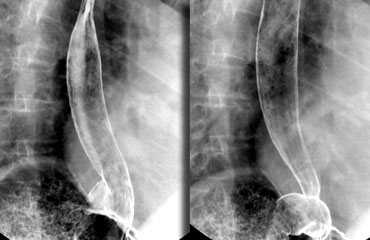 Optimal views of gastro-esophageal junction when air regurgitates from the stomach into the coated esophagus in a patient with a sliding hiatus hernia. Hình ảnh tối ưu của chỗ nối dạ dày-thực quản khi không khí trào ngược từ dạ dày vào thực quản được bao bọc ở bệnh nhân thóat vị qua khe trượt.
Excellent views of the gastroesophageal junction can be achieved by doing the following:Quan điểm tuyệt vời về chỗ nối dạ dày thực quản có thể đạt được bằng cách làm như sau:
- Ask the patient to swallow zoru-granules (or coca cola) for optimal gas filling of the stomach.Yêu cầu bệnh nhân nuốt các hạt zoru (hoặc coca cola) để làm đầy dạ dày tối ưu.
- Tell the patient not to belch, but to keep the air in the stomach until the moment of swallowing.Yêu cầu bệnh nhân không ợ hơi, nhưng phải giữ không khí trong dạ dày cho đến khi nuốt.
- Place the patient in the left anterior oblique position.Đặt bệnh nhân ở tư thế xiên trước bên trái.
- Lift the table top 45 degrees.Nâng mặt bàn lên 45 độ.
- Swallow high density barium for optimal esophageal coating.Nuốt barium mật độ cao để có lớp phủ thực quản tối ưu.
- Images are taken when air regurgitates from the stomach into the barium-coated esophagus.Hình ảnh được chụp khi không khí trào ngược từ dạ dày vào thực quản có phủ bari.
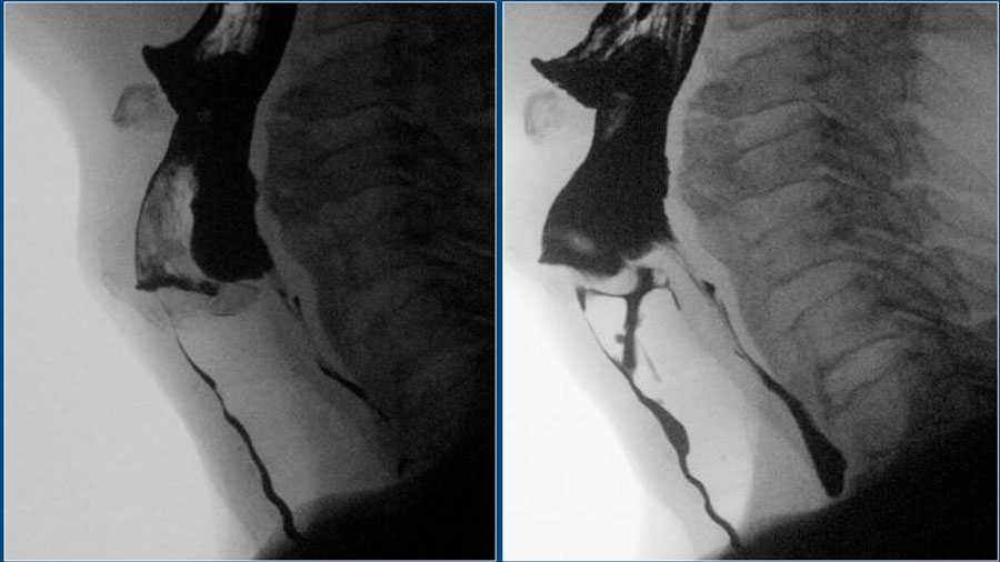
You get the best lighting, when the anterior part of the cervical spine is in the middle of the image.
Do not put the contrast bolus in the center of the image, because on the video you'll get a constant shift of images that are too dark to images that are too bright.Bạn sẽ có được ánh sáng tốt nhất, khi phần trước của cột sống cổ nằm ở giữa hình ảnh.
Không đặt bolus tương phản ở trung tâm của hình ảnh, bởi vì trên video, bạn sẽ nhận được một sự thay đổi liên tục của hình ảnh quá tối so với hình ảnh quá sáng.
Analyzing swallowing studiesPhân tích các nghiên cứu nuốt
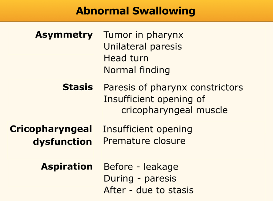
A simple way to analyse a swallowing study is to concentrate on four easily detectable findings:Một cách đơn giản để phân tích một nghiên cứu nuốt là tập trung vào bốn phát hiện dễ phát hiện:
- AsymmetryBất đối xứng
- StasisNgưng trệ
- Cricopharyngeal dysfunctionRối loạn chức năng thanh quản
- AspirationKhát vọng
These findings are mostly already apparent during the examination, but analysis of all the images will clarify the mechanism that causes these abnormal findings.Những phát hiện này hầu hết đã rõ ràng trong quá trình khám, nhưng phân tích tất cả các hình ảnh sẽ làm rõ cơ chế gây ra những phát hiện bất thường này.
These imaging findings may be isolated findings or may be related to each other.Những dấu hiệu hình ảnh này có thể là những dấu hiệu riêng biệt hoặc có thể liên quan đến nhau.
For instance, premature closure of the cricopharyngeal muscle may lead to stasis of contrast in the pharynx, which may result in aspiration as the larynx opens at the end of swallowing.Ví dụ, đóng sớm cơ nhẫn hầu có thể dẫn đến ứ đọng thuốc cản quang trong họng, có thể dẫn tới hít phải khi thanh quản mở ra khi nuốt xong.
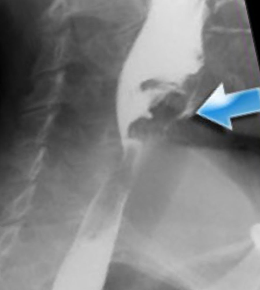 Asymmetric swallowing due to head turn. The head is turned to the left and contrast is only seen in the right food channel.Nuốt không đối xứng do quay đầu Đầu quay sang trái và độ tương phản chỉ thấy ở kênh thức ăn bên phải.
AsymmetryBất đối xứng
Asymmetric swallowing on an AP-view is usually the result of an asymmetric tilting of the epiglottis.
Sometimes it is caused by rotation of the head, but in many cases no real explanation is found.
Even when the head is not rotated, the epiglottis can tilt asymmetrically when it hits the posterior pharyngeal wall.
This is more likely to occur when only a small bolus is given, as the pharynx will not fully distend.
A larger bolus will result in a symmetric swallow.Nuốt không đối xứng trên góc nhìn của vòm họng thường là kết quả của sự nghiêng không đối xứng của nắp thanh môn.
Đôi khi nguyên nhân là do xoay đầu, nhưng trong nhiều trường hợp không tìm thấy lời giải thích thực sự.
Ngay cả khi đầu không xoay, nắp thanh môn có thể nghiêng không đối xứng khi chạm vào thành sau họng.
Điều này có nhiều khả năng xảy ra khi chỉ được tiêm một lượng nhỏ, vì hầu họng sẽ không bị căng hoàn toàn.
Một bolus lớn hơn sẽ dẫn đến nuốt đối xứng.
In the case on the left rotation of the head closes the side to which the head is turned (Figure).Trong trường hợp xoay đầu bên trái, đầu sẽ đóng lại ở bên mà đầu được xoay (Hình).
If a patient has a unilateral pharyngeal paresis, turning of the head towards the affected side will help the patient in preventing aspiration.
By turning the head towards the affected side, this side will be closed preventing stasis on this side and possible secondary aspiration.Nếu bệnh nhân bị liệt họng một bên, việc quay đầu về phía bị ảnh hưởng sẽ giúp bệnh nhân ngăn ngừa hít phải.
Bằng cách xoay đầu về phía bên bị ảnh hưởng, bên này sẽ được đóng lại để ngăn ngừa tình trạng ứ đọng ở bên này và có thể có hiện tượng hít phải thứ phát.
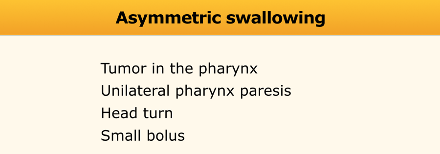
However before you decide that it is a normal finding you have to exclude a pharyngeal tumour or unilateral paresis.
The double-contrast views can be helpful in these cases.
In unilateral paresis the paralysed side will protrude during the modified Valsalva.
When a tumour is present in the pharynx, it is usually visible on the DC views.
Sometimes endoscopy is necessary to solve the problem of an asymmetric swallow.
Tuy nhiên, trước khi quyết định rằng đó là một phát hiện bình thường, bạn phải loại trừ một khối u họng hoặc liệt một bên.
Quan điểm đối chiếu đôi có thể hữu ích trong những trường hợp này.
Trong liệt một bên, bên liệt sẽ nhô ra trong quá trình Valsalva cải biên.
Khi một khối u có mặt trong họng, nó thường có thể nhìn thấy trên chế độ xem DC.
Đôi khi nội soi là cần thiết để giải quyết vấn đề nuốt không đối xứng.
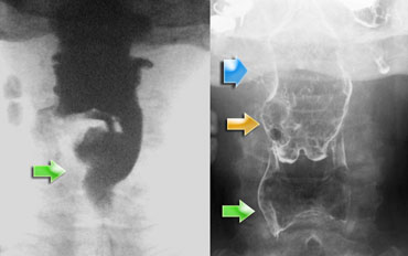
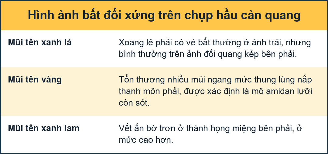
Asymmetry (2)
The case on the left is an odd case, but it nicely demonstrates the difficulty that sometimes exists in determining the cause of asymmetry.
On the far left asymmetry is seen on the fluorographic study (green arrow).
It looks as if there is something in the right pyriform sinus.
On the double contrast view on the right the pyriform sinus is normal (green arrow), but at the level of the vallecula on the right a lobulated proces is seen (yellow arrow).
At a higher level a smooth indentation of the oropharynx is seen (blue arrow).
The lobulated tumor at the level of the valleculae proved to be remnants of the lingual tonsil, which is a common finding and sometimes difficult to differentiate from cancer of the tongue base.
Continue with the CT of this patient.Bất đối xứng (2)
Trường hợp bên trái là một trường hợp kỳ lạ, nhưng nó chứng minh một cách độc đáo sự khó khăn đôi khi tồn tại trong việc xác định nguyên nhân của sự bất đối xứng.
Trên hình ảnh bất đối xứng bên trái được nhìn thấy trên nghiên cứu huỳnh quang (mũi tên màu xanh lá cây).
Có vẻ như có gì đó trong xoang mủ phải.
Trên chế độ xem tương phản kép ở bên phải, xoang pyriform bình thường (mũi tên màu xanh lá cây), nhưng ở ngang mức của thung lũng bên phải, có thể thấy một xoang hình thuỳ (mũa tên màu vàng).
Ở mức cao hơn, có thể thấy một vết lõm nhẵn của hầu họng (mũi tên màu xanh).
Khối u phân thuỳ ở thung lũng được chứng minh là tàn dư của amidan lưỡi, đây là một phát hiện phổ biến và đôi khi khó phân biệt với ung thư nền lưỡi.
Tiếp tục với CT của bệnh nhân này.
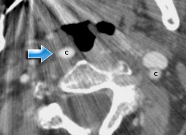
 Impression of the oropharynx by an elongated internal carotid artery (blue arrow.). Compare to the position of the carotid artery on the leftẤn tượng vùng hầu họng bởi động mạch cảnh trong thon dài (mũi tên.) So sánh với vị trí động mạch cảnh bên trái
The CT image shows that the smooth indentation of the oropharynx on the right was caused by an elongated internal carotid artery.Hình ảnh CT cho thấy vết lõm nhẵn của hầu họng bên phải là do động mạch cảnh trong bị kéo dài.
So indeed this is an uncommon case in which on the fluorographic study a tumor was suspected in the pyriform sinus.
Finally a proces was found within the oropharynx and a proces compressing the wall from outside at a higher level.
Due to these processes there was an asymmetric passage of contrast in the hypopharynx simulating a proces in the pyriform sinus.Vì vậy, thực sự đây là một trường hợp hiếm gặp trong đó trên phim huỳnh quang, một khối u được nghi ngờ trong xoang pyriform.
Cuối cùng, một quy trình được tìm thấy trong miệng họng và một quy trình nén thành từ bên ngoài ở mức cao hơn.
Do các quá trình này, có một đường đi thuốc cản quang không đối xứng ở vùng hạ họng mô phỏng một quá trình trong xoang hình chóp.
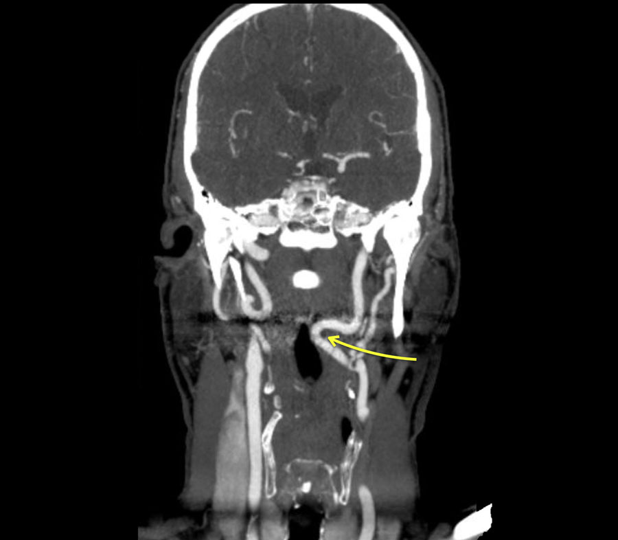
Here another patient with an elongated internal carotid artery.
This patient had swallowing problems and at inspection a pulsating structure was seen in the oropharynx.Đây là một bệnh nhân khác với động mạch cảnh trong bị kéo dài.
Bệnh nhân này có vấn đề về nuốt và khi kiểm tra, một cấu trúc dao động được nhìn thấy trong hầu họng.
This is not un uncommon finding.
So before you perform a biopsy in this area make sure that you are not dealing with the carotid artery.Đây không phải là một phát hiện hiếm gặp.
Vì vậy, trước khi bạn thực hiện sinh thiết ở khu vực này, hãy chắc chắn rằng bạn không làm việc với động mạch cảnh.
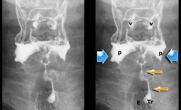
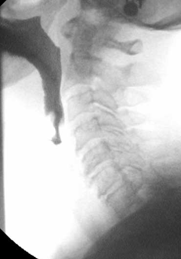
 Stasis of contrast at the level of the pyriform sinuses (blue arrows) with subsequent aspiration (yellow arrows)Ngừng tương phản ở mức độ của các xoang pyriform (mũi tên màu xanh) với lần hút tiếp theo (mũI tên màu vàng)
StasisNgưng trệ
Stasis is the result of insufficient cleansing of the pharynx, either due to an obstruction- i.e. dysfunction of the cricopharyngeus or tumor or due to insufficient contraction of the pharyngeal constrictors.
Insufficient contraction is the result of pharyngeal paresis resulting from a neuromuscular disorder.
Excessive movements of the tongue base and larynx are sometimes seen on lateral fluorographic studies to compensate for the loss of function of the pharyngeal constrictors.
When the patient resumes breathing, aspiration can occur (Figure).Ứ máu là kết quả của việc làm sạch họng không đủ, hoặc do tắc nghẽn - tức là rối loạn chức năng của vùng cổ họng hoặc khối u hoặc do sự co thắt không đủ của các cơ quan họng.
Co thắt không đủ là kết quả của liệt họng do rối loạn thần kinh cơ.
Đôi khi có thể thấy sự di chuyển quá mức của nền lưỡi và thanh quản trên các nghiên cứu huỳnh quang bên để bù đắp cho sự mất chức năng của các cơ thắt họng.
Khi bệnh nhân thở trở lại, có thể xảy ra hiện tượng hít phải (Hình).
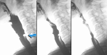
Stasis (2)
Here a set of images demonstrating a patient with a paresis of the pharynxconstrictors.
This is usually associated with insufficient relaxation of the cricopharyngeal muscle.
In this example we can see how the patient tries to compensate for the loss of pharyngeal constriction by excessive movement of the tongue and the head.
This patient is in extreme stress, because he knows that when he starts breathing and the throat is not empty, he will aspirate.Ngưng trệ (2)
Đây là một tập hợp các hình ảnh cho thấy một bệnh nhân bị liệt các thuốc co thắt họng.
Điều này thường liên quan đến việc thư giãn không đủ của cơ nhẫn hầu.
Trong ví dụ này, chúng ta có thể thấy bệnh nhân cố gắng bù đắp cho sự mất đi sự co thắt của họng bằng cách di chuyển quá mức của lưỡi và đầu.
Bệnh nhân này đang trong tình trạng cực kỳ căng thẳng, bởi vì anh ta biết rằng khi anh ta bắt đầu thở và cổ họng không trống rỗng, anh ta sẽ hít vào.
- 1-3. No contraction of the posterior wall of the pharynx is seen. Cricopharyngeus does not open properly.1-3. Không thấy sự co lại của thành sau họng. Cricopharyngeus không mở đúng cách.
- 4-7. Contrast enters the larynx, but does not enter the trachea.4-7. Thuốc cản quang đi vào thanh quản, nhưng không đi vào khí quản.
- 8-10. Excessive movements of the head in order to squeeze the bolus into the esophagus.8-10. Chuyển động quá mức của đầu để ép bolus vào thực quản.
In some of these cases cricopharyngeotomy is the only solution to facilitate the passage of food to the esophagus.Trong một số trường hợp, mở họng-hầu là giải pháp duy nhất để tạo điều kiện cho thức ăn đi vào thực quản.
 Small impression of the cricopharyngeus. No obstruction.Dấu ấn nhỏ của xương ức, không có tắc nghẽn.
Cricopharyngeal DysfunctionRối loạn chức năng thanh quản
Insufficient opening and premature closure are the most common problems of the cricopharyngeal muscle.
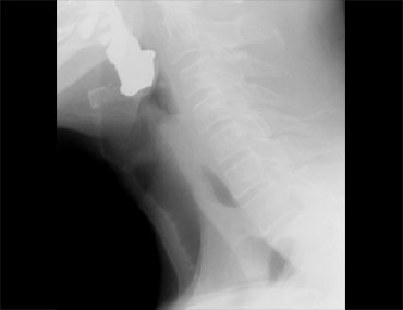
Normally you should not see an impression of the cricopharyngeus during passage of the bolus, but a small non-obstructive indentation is sometimes seen and is not clinically significant (Figure).
It can however sometimes explain the symptoms of the patient.
It is assumed that the passage of food irritates the mucosa that covers the cricopharyngeal muscle resulting in a globus sensation.
Mở không đủ và đóng sớm là những vấn đề phổ biến nhất của cơ nhẫn hầu.
Thông thường bạn không nên nhìn thấy dấu ấn của vùng cổ họng trong quá trình đi qua bolus, nhưng đôi khi có thể thấy một vết lõm nhỏ không tắc nghẽn và không có ý nghĩa lâm sàng (Hình).
Tuy nhiên, đôi khi nó có thể giải thích các triệu chứng của bệnh nhân.
Người ta cho rằng việc đi qua thức ăn gây kích thích niêm mạc bao phủ cơ nhẫn hầu dẫn đến cảm giác như có hình cầu.
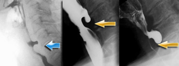 LEFT: small pouch. MIDDLE and RIGHT: true Zenker's diverticulum due to premature closure of the cricopharyngeus (yellow arrow)TRÁI: túi nhỏ. TRUNG TÂM VÀ PHẢI: túi thừa Zenker thật do đóng sớm xương ức (mũi tên màu vàng)
Zenker's diverticulumTúi thừa Zenker
A Zenker's diverticulum is always the result of cricopharyngeal dysfunction.Túi thừa Zenker luôn là kết quả của rối loạn chức năng vùng cổ họng.
Premature closure of the cricopharyngeus results in an increased pressure in the hypopharynx, just above the cricopharyngeus, as the pressurewave of the pharyngeal constrictors pushes the bolus downwards.Việc đóng sớm của xương sọ hầu dẫn đến tăng áp lực ở vùng hạ họng, ngay trên xương sọ, vì sóng áp lực của các cơ thắt họng đẩy bolus xuống dưới.
This increased pressure can result in an outpouching at a weak spot in the posterior pharyngeal wall (Killian's dehiscence).
First this will result in a small pouch, that in time can increase and form a true Zenker's diverticulum (Figure).
Áp lực tăng lên này có thể dẫn đến một chỗ yếu ở thành sau họng (tắc mạch của Killian).
Đầu tiên, điều này sẽ dẫn đến một túi nhỏ, theo thời gian có thể tăng lên và tạo thành một túi thừa Zenker thực sự (Hình).
Cricopharyngeal dysfunction (3)
On the left a patient who complained of globus sensation in the throat and difficulty of swallowing with regurgitation of undigested food.Rối loạn chức năng thanh quản (3)
Bên trái, một bệnh nhân than phiền về cảm giác nghẹt họng và khó nuốt kèm theo trào ngược thức ăn khó tiêu.
The digital recordings nicely demonstrate the filling of a large Zenker's diverticle.
The contraction of the lower pharyngeal constrictors is indicated by the blue arrow.Các bản ghi kỹ thuật số độc đáo cho thấy sự lấp đầy của một túi thừa Zenker lớn.
Sự co thắt của các cơ thắt hầu dưới được biểu thị bằng mũi tên màu xanh.
The contraction of the lower pharyngeal muscle against the closed cricopharyngeal muscle causes the posterior outpouching, just above the contracted cricopharyngeal muscle (yellow arrow).Sự co lại của cơ họng dưới chống lại cơ nhẫn hầu đóng gây ra sự ra ngoài phía sau, ngay phía trên cơ nhẫn hầu bị co lại (mũi tên màu vàng).
Cricopharyngeal dysfunction (4) Rối loạn chức năng thanh quản (4)
Here a video of a patient with a Zenker diverticulum.Đây là video của một bệnh nhân có túi thừa Zenker.
Notice that on the AP-view the patient keeps on swallowing because of the globus sensation.Lưu ý rằng trên chế độ xem AP, bệnh nhân tiếp tục nuốt vì cảm giác như có hình cầu.
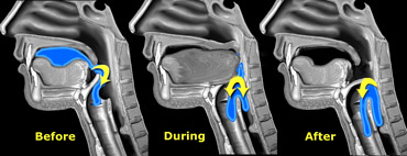
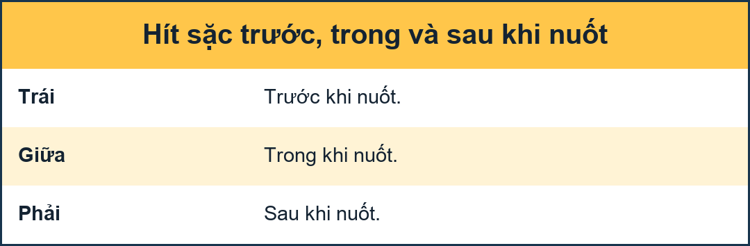 Aspiration before, during or after the actual swallow.Hít phải trước, trong hoặc sau khi nuốt thực sự.
AspirationKhát vọng
There are three instances when aspiration can occur: before, during or after the actual swallow.
Có ba trường hợp có thể xảy ra khi hít phải: trước, trong hoặc sau khi nuốt thực sự.
- Aspiration before swallowing is either the result of insufficient closure of the oral cavity during the preparatory phase or inability to start the swallow reflex when contrast enters the pharynx.
Hút thuốc trước khi nuốt hoặc là kết quả của việc đóng không đủ khoang miệng trong giai đoạn chuẩn bị hoặc không có khả năng bắt đầu phản xạ nuốt khi thuốc cản quang đi vào họng. - Aspiration during swallowing is due to insufficient closure of the larynx.Hít phải trong khi nuốt là do thanh quản đóng không đủ.
- Aspiration after swallowing is the result of stasis of contrast in the pharynx - when the larynx opens the contrast leaks into the trachea.Hít phải sau khi nuốt là kết quả của sự ứ đọng thuốc cản quang trong họng - khi thanh quản mở ra, thuốc cản Quang sẽ rò rỉ vào khí quản.
Aspiration before swallowing Hút trước khi nuốt
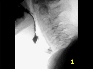
When tongue or soft palate are unable to prevent spillage of food into the pharynx, aspiration may occur since the larynx is still open.
Weakness of these muscles in the mouth and the throat is due to paralysis or myopathy.Khi lưỡi hoặc vòm miệng mềm không thể ngăn chặn thức ăn tràn vào họng, có thể xảy ra hiện tượng hít phải vì thanh quản vẫn còn mở.
Yếu các cơ này trong miệng và cổ họng là do liệt hoặc bệnh cơ.
- Notice that contrast enters the pharynx, but does not trigger a swallowing reflex.Lưu ý rằng thuốc cản quang đi vào họng, nhưng không kích hoạt phản xạ nuốt.
- No swallowing reflexKhông có phản xạ nuốt
- Contrast reaches the hypopharynx, but still no swallowing reflex.Tấm thuốc cản quang chạm đến hạ họng, nhưng vẫn không có phản xạ nuốt.
- Contrast enters the larynx, which is still open and not yet elevatedTấm thuốc cản quang đi vào thanh quản, thanh quản vẫn còn mở và chưa nâng cao
- At this moment the swallowing reflex starts and the larynx elevatesTại thời điểm này, phản xạ nuốt bắt đầu và thanh quản nâng lên
- Contrast is transported to the esophagus and the larynx closes, but there is already contrast in the trachea.Thuốc cản quang được vận chuyển đến thực quản và thanh quản đóng lại, nhưng đã có thuốc cản quang trong khí quản.
- Proper relaxation of the cricopharyngeus and finally there is good closure of the larynx.Thư giãn đúng cách của họng và cuối cùng là đóng thanh quản tốt.
- Notice that there is no stasis at the end of the swallow .Lưu ý rằng không có ứ đọng ở cuối nuốt.
We have to assume that a failure of the sensory nerves in the pharynx is the problem.
Notice also that there is no coughing reflex.
This patient probably has silent aspiration.Chúng ta phải giả định rằng sự thất bại của các dây thần kinh cảm giác ở họng là vấn đề.
Cũng lưu ý rằng không có phản xạ ho.
Bệnh nhân này có thể có hít vào thầm lặng.
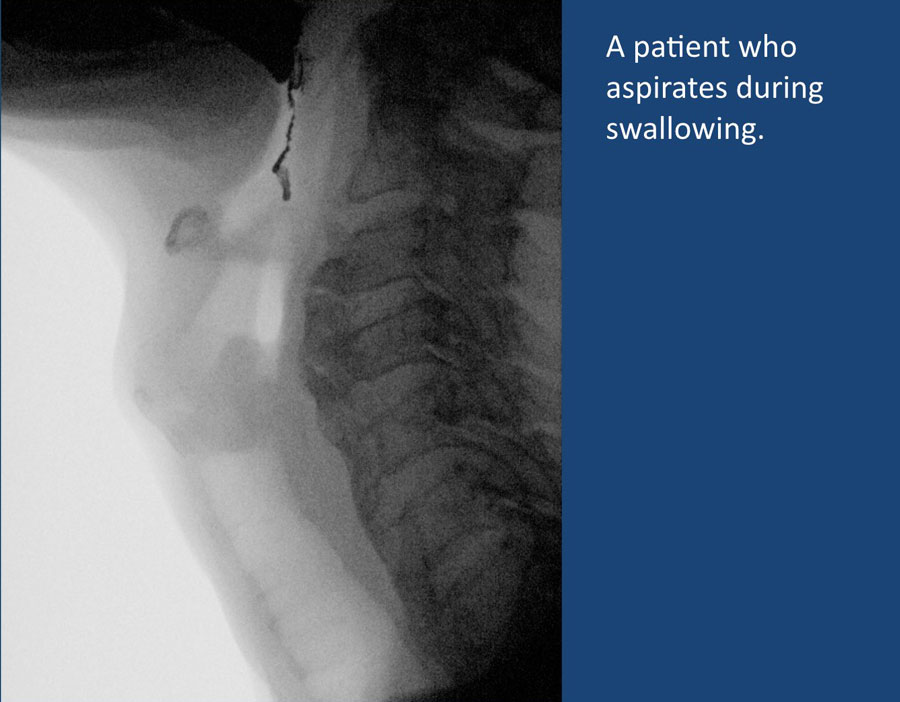
Aspiration during swallowing Hít phải trong khi nuốt
This is due to an insufficient closure of the larynx when it should be closed.
Closure of the larynx is a result of anterosuperior lifting of the larynx which allows the true cords, false cords and finally, the aryepiglottic folds to contract, followed by a backwards folding of the epiglottis over the closed larynx.
The aryepiglottic folds are the main gatekeepers, while the epiglottis plays only a minor role in preventing aspiration.Điều này là do thanh quản đóng không đủ khi cần đóng.
Đóng thanh quản là kết quả của việc nâng thanh quản lên trước trên cho phép các dây thật, dây giả và cuối cùng, các nếp gấp nắp thanh quản co lại, tiếp theo là nếp gấp của nắp gò thanh quản ngược lại thanh quản đóng.
Các nếp gấp nắp thanh môn là những cửa chính, trong khi nếp thanh môn chỉ đóng một vai trò nhỏ trong việc ngăn ngừa hít phải.
Both failure of these intrinsic muscles of the larynx as well as failure of the extrinsic muscles (i.e. muscles that lift the larynx) may lead to aspiration during swallowing.
Weakness of the extrinsic muscles is seen after radiotherapy, in neurologic disorders and in recurrens nerve paralysis (i.e. neuromuscular dysfunction).Cả hai sự thất bại của các cơ nội tại của thanh quản cũng như sự thất bại các cơ bên ngoài (tức là các cơ nâng thanh quản) có thể dẫn đến hít phải trong quá trình nuốt.
Sự yếu của các cơ bên ngoài được thấy sau khi xạ trị, trong các rối loạn thần kinh và trong liệt dây thần kinh tái phát (tức là rối loạn chức năng thần kinh cơ).
Notice that this patient has diffuse idiopathic skeletal hyperostosis (DISH) with syndesmofytes at many levels on the anterior side of the cervical spine.
However they are not the cause of the swallowing disorder.Lưu ý rằng bệnh nhân này có tăng sinh xương vô căn lan toả (DISH) với dây chằng dính ở nhiều mức độ ở phía trước của cột sống cổ.
Tuy nhiên, chúng không phải là nguyên nhân gây ra rối loạn nuốt.
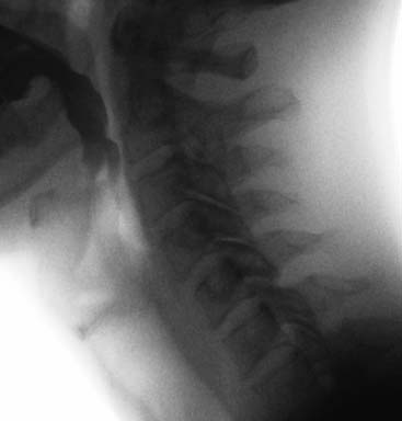
Aspiration during swallowing (2) Hút trong khi nuốt (2)
On the left a digital recording of a patient who complained of a sore throat and a wet voice.
Notice that when the contrast enters the throat, the swallowing reflex is not triggered immediately.
This allows for barium contrast to enter the larynx, where it sits on the vocals cords.
Once the swallowing reflex is initiated, the larynx closes properly, but contrast is already in.Bên trái là một bản ghi âm kỹ thuật số của một bệnh nhân phàn nàn về đau họng và giọng nói ẩm ướt.
Lưu ý rằng khi thuốc cản quang đi vào cổ họng, phản xạ nuốt không được kích hoạt ngay lập tức.
Điều này cho phép chất cản quang barium đi vào thanh quản, nơi nó nằm trên dây thanh âm.
Khi bắt đầu phản xạ nuốt, thanh quản đóng đúng cách, nhưng thuốc cản quang đã ở trong đó.
Notice also that while the contrast enters the larynx, it does not initiate a cough reflex.
Althoug the digital recording perfectly explains the complaints of the patient, it is difficult to say what causes this problem.Cũng lưu ý rằng trong khi thuốc cản quang đi vào thanh quản, nó không bắt đầu phản xạ ho.
Ngoài ra, khi ghi hình kỹ thuật số giải thích một cách hoàn hảo những phàn nàn của bệnh nhân, rất khó để nói được nguyên nhân gây ra vấn đề này.
Usually when there is aspiration during swallowing, the problem is at the level of the larynx.
Mostly it is the larynx that is unable to close either due to weakness of the intrinsic laryngeal muscles or weakness of the extrinsic muscles that lift the larynx and allow it to contract (for instance after neck radiation or surgery).Thông thường khi có hít phải trong khi nuốt, vấn đề nằm ở thanh quản.
Hầu hết là thanh quản không thể đóng lại do yếu cơ thanh quản bên trong hoặc yếu cơ bên ngoài nâng thanh quản và cho phép nó co lại (ví dụ sau khi xạ trị cổ hoặc phẫu thuật).
In this case the problem is probably at the level of the sensory nerves in the pharynx, who are not triggered properly.
You could also argue, that this maybe is aspiration before swallowing, because it happens before the actual swallowing reflex.
Maybe a more forcefull push of the bolus posteriorly from the mouth into the oropharynx could help in triggering these nerves instead of allowing the bolus to slide over the back of the tongue.Trong trường hợp này, vấn đề có thể là ở cấp độ của các dây thần kinh cảm giác ở họng, những dây thần kinh này không được kích hoạt đúng cách.
Bạn cũng có thể tranh luận, rằng đây có thể là hít vào trước khi nuốt, bởi vì nó xảy ra trước khi phản xạ nuốt thực sự.
Có thể một lực đẩy mạnh hơn của bolus từ miệng vào hầu họng có thể giúp kích hoạt các dây thần kinh này thay vì cho phép bolus trượt qua mặt sau của lưỡi.
Aspiration after swallowing Hút sau khi nuốt
This is the most common cause of aspiration.
It is the result of stasis of contrast in the pharynx due to insufficient contraction of the pharyngeal constrictors or insufficient opening of the cricopharyngeal muscle or an obstructing web or tumor.
When the larynx opens the contrast may leak into the trachea.Đây là nguyên nhân phổ biến nhất gây hít phải.
Đó là kết quả của sự ứ đọng thuốc cản quang trong họng do sự co thắt không đủ của các cơ thắt họng hoặc không đủ mở của cơ nhẫn hầu hoặc một mạng lưới hoặc khối u cản trở.
Khi thanh quản mở, thuốc cản quang có thể rò rỉ vào khí quản.
The patient in the video aspirates due to stasis in the hypopharynx.Bệnh nhân trong video hít phải do ứ đọng ở hạ họng.
In this video there is massive aspiration due to stasis as a result of insufficient contraction of the pharyngeal constrictors and insufficient opening of the cricopharyngeal muscle.Trong video này, có một sự hít vào lớn do sự ứ đọng do sự co thắt không đủ của các cơ thắt hầu họng và không đủ mở của cơ nhẫn hầu.
WebTrang chủ
Webs are mucosa folds which are usually located anteriorly in the hypopharynx or upper esophagus.Mạng nhện là các nếp gấp niêm mạc thường nằm ở phía trước ở hạ họng hoặc thực quản trên.
Liquids usually pass well, but in many cases a 'jet' is seen.
The passage of solid food may produce irritation or damage to the mucosa, resulting in a globus feeling.
They are best diagnosed on the lateral projection of the barium swallow. Chất lỏng thường đi qua tốt, nhưng trong nhiều trường hợp có thể nhìn thấy một 'tia lửa'.
Việc đi qua thức ăn đặc có thể gây kích ứng hoặc tổn thương niêm mạc, dẫn đến cảm giác như có khối cầu.
Chẩn đoán tốt nhất dựa vào hình chiếu bên của nuốt bari.
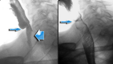
Here another example of a web.Đây là một ví dụ khác về một mạng nhện.
Webs are frequently overlooked at esophagoscopy unless special attention is given to this area.
During esophagoscopy they may rupture, but in many cases recurrence is seen.Các mạng nhện thường bị bỏ qua khi soi thực quản trừ khi đặc biệt chú ý đến khu vực này.
Trong quá trình nội soi thực quản, chúng có thể vỡ, nhưng trong nhiều trường hợp có thể thấy tái phát.
Pharyngeal earsTai họng
Outpounching of the lateral wall of the pharynx are called 'lateral pharyngeal ears'.Mở thành bên của họng được gọi là 'tai họng bên'.
Sometimes patients complain of globus sensation or that small food particles or pills get stuck in their throat, but usually it is an incidental finding.Đôi khi bệnh nhân phàn nàn về cảm giác có khối hoặc các hạt thức ăn nhỏ hoặc thuốc bị kẹt trong cổ họng, nhưng thường là phát hiện tình cờ.
They can be quite prominent as in Dizzy Gillespie the famous trumpet player.
Chúng có thể khá nổi bật như trong Dizzy Gillespie, người chơi kèn trômpet nổi tiếng.
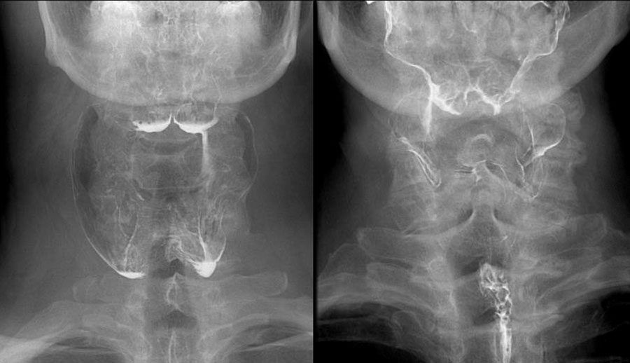
Two cases of pharyngeal ears.Hai trường hợp tai họng.
OsteofytesXương hóa
Although osteofytes can be quite big, they hardly ever cause swallowing problems.Mặc dù các xương hóa có thể khá lớn, nhưng chúng hầu như không gây ra vấn đề nuốt.
Esophagus pathologyBệnh lý thực quản
As mentioned earlier problems in the esophagus can give the sensation of a problem in the throat.Như đã đề cập trước đó các vấn đề trong thực quản có thể gây cảm giác có vấn đề trong cổ họng.
In the video there is stasis of contrast in the proximal esophagus due to a large carcinoma.Trong video có sự ứ đọng thuốc cản quang ở đầu gần thực quản do ung thư biểu mô lớn.
A common cause of swallowing problems is reflux which irritates the cricipharyngeal muscle and results in hypertrophy.Một nguyên nhân phổ biến của các vấn đề về nuốt là trào ngược gây kích thích cơ nhẫn hầu và dẫn đến phì đại.
The patient in the video has a large hiatal hernia with reflux and almost no peristatic movements in the esophagus.Bệnh nhân trong video có thóat vị khe lớn kèm theo trào ngược và hầu như không có chuyển động quanh thực quản.
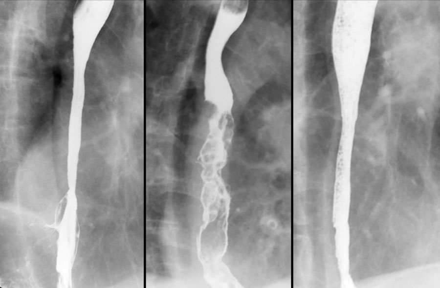
Here three patients with swallowing problems as a result of esophageal pathology.Ở đây ba bệnh nhân có vấn đề về nuốt là kết quả của bệnh lý thực quản.
The patient on the left has reflux esophagitis with a Barrett's esophagus.Bệnh nhân bên trái bị viêm thực quản trào ngược với thực quản Barrett.
The patient in the middle has a esophageal cancer.Bệnh nhân ở giữa có ung thư thực quản.
The patient on the right has a stricture due to ingestion of caustic material.Bệnh nhân bên phải bị hẹp do ăn phải chất ăn mòn.
Lingual tonsilsAmidan lưỡi
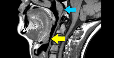
The lingual tonsils are nodules of lymphatic tissue at the back of the base of the tongue (yellow arrow).Amidan lưỡi là các nốt của mô bạch huyết ở mặt sau của lưỡi (mũi tên màu vàng).
They are similar to the adenoid or nasopharyngeal tonsil (blue arrow).Chúng tương tự như amidan hoặc amidan mũi họng (mũi tên màu xanh).
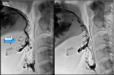
Large lingual tonsils can be a cause of dysphagia or globus sensation especially if the tonsils compress the epiglottis.
In many cases however they are incidental findings.
Endoscopy is sometimes needed to differentiate large tonsils from a carcinoma at the tongue base.Amidan lưỡi lớn có thể là nguyên nhân gây khó nuốt hoặc cảm giác như có hình cầu đặc biệt nếu amidan chèn ép nắp thanh môn.
Tuy nhiên, trong nhiều trường hợp, chúng là những phát hiện tình cờ.
Nội soi đôi khi cần thiết để phân biệt amidan lớn với ung thư biểu mô ở đáy lưỡi.
Notice that you have to look carefully to detect the large lingual tonsil.Lưu ý rằng bạn phải nhìn kỹ để phát hiện amidan lớn.
DiagnosisChẩn đoán
The results of the swallowing examination help in establishing a final diagnosis.
Based on this examination alone however, a specific diagnosis usually cannot be made, since most severe swallowing disorders are the result of a complex neuromuscular dysfunction.
Hence the swallowing study should be regarded as part of the total evaluation of the patient by gastroenterologist, neurologist and speech therapist.
The strength of the fluoroscopic examination is, that it is the only examination that can show us, what is really going on during swallowing and can therefore lead to a rehabilitation plan.
Kết quả của khám nuốt giúp thiết lập chẩn đoán cuối cùng.
Tuy nhiên, chỉ dựa trên khám này, thường không thể đưa ra chẩn đoán cụ thể, vì hầu hết các rối loạn nuốt nghiêm trọng là kết quả của rối loạn chức năng thần kinh cơ phức tạp.
Do đó, nghiên cứu nuốt nên được coi là một phần của đánh giá tổng thể của bệnh nhân bởi bác sĩ tiêu hóa, bác sĩ thần kinh và nhà trị liệu ngôn ngữ.
Sức mạnh của việc kiểm tra bằng kính hiển vi là, đó là cuộc kiểm tra duy nhất có thể cho chúng ta thấy, những gì thực sự đang xảy ra trong quá trình nuốt và do đó có thể dẫn đến một kế hoạch phục hồi chức năng.
Swallowing RehabilitationPhục hồi chức năng nuốt
Swallowing rehabilitation is a specialty on its own.
Here we will make some brief comments on rehabilitation as it may help you to better understand the dynamics of swallowing.Phục hồi chức năng nuốt là một chuyên khoa riêng.
Ở đây chúng tôi sẽ đưa ra một số nhận xét ngắn gọn về phục hồi chức năng vì nó có thể giúp bạn hiểu rõ hơn về động lực của việc nuốt.
In unilateral pharyngeal paralysis stasis can be prevented by closing down one of the lateral food channels by turning the head towards the affected side or by manually compressing it.Trong liệt hầu họng một bên, có thể ngăn ngừa tình trạng ứ đọng bằng cách đóng một trong các kênh thức ăn bên bằng cách xoay đầu về phía bị ảnh hưởng hoặc bằng cách ép thủ công.
Patients with aspiration before swallowing due to insufficient closure of the mouth, can be helped by flexing their head during chewing and thus holding the food in the anterior part of the oral cavity.Bệnh nhân bị hít phải trước khi nuốt do không đóng miệng đủ, có thể được giúp đỡ bằng cách gập đầu trong khi nhai và do đó giữ thức ăn ở phần trước của khoang miệng.
In patients with aspiration during or after swallowing the 'supraglottic swallow' may help.
Before swallowing a deep breath is taken.
Air is prevented to leak out of the airways by compressing the vocal cords.
Immediately after swallowing the patient has to cough forcefully in order to clear the airways and the throat from any residual food.Ở những bệnh nhân bị hít phải trong hoặc sau khi nuốt, có thể giúp đỡ.
Trước khi nuốt, hít một hơi sâu.
Không khí được ngăn chặn rò rỉ ra khỏi đường thở bằng cách nén dây thanh âm.
Ngay sau khi nuốt, bệnh nhân phải ho mạnh để làm sạch đường thở và cổ họng khỏi bất kỳ thức ăn còn sót lại nào.
Some patients only aspirate when they ingest fluids.
These patients can be helped by changing their fluid intake into jelly-like liquids.Một số bệnh nhân chỉ hít phải khi họ ăn dịch.
Những bệnh nhân này có thể được giúp đỡ bằng cách thay đổi lượng chất lỏng vào các chất lỏng giống như thạch.
ReferencesTài liệu tham khảo
-
Logemann JA: In: Manual for the Videofluorographic Study of Swallowing. 2nd ed. Austin, TX: Pro-Ed Inc; 1993.
Logemann JA: Trong: Sách hướng dẫn cho Nghiên cứu Videofluorographic về nuốt. Lần xuất bản thứ 2. Austin, TX: Pro-Ed Inc; 1993. -
Dysphagia in emedicine
Khó nuốt trong y học điện tử
by Nam-Jong Paik
bởi Nam-Jong Paik -
Radiologic assessment of abnormal oral and pharyngeal phases of swallowing (PDF-file)
Đánh giá các giai đoạn nuốt bất thường ở miệng và hầu họng (PDF-file)
by WJ Dodds, JA Logemann, ET Stewart
American Journal of Roentgenology, 1990 - Am Roentgen Ray Soc
Page 1. 965 Review Article
bởi WJ Dodds, JA Logemann, et Stewart
Tạp chí Roentgenology của Mỹ, 1990 - AM Roentgen Ray Soc
Trang 1. 965 Đánh giá bài báo