
Pulsatile and non-pulsatile tinnitusÙ tai theo nhịp mạch và không theo nhịp mạch
Department of radiology and nuclear medicine, Radboudumc, Nijmegen, the NetherlandsKhoa X quang và Y học hạt nhân, Radboudumc, Nijmegen, Hà Lan
Ngày đăng / cập nhật: 12-6-2020
Bản gốc trên radiologyassistant.nl ↗ · tải về lúc 2026-11-19 20:19 · bản dịch máy có chuẩn hóa thuật ngữ chẩn đoán hình ảnh
This article is based on a review by Pegge et al. describing a systematic approach for the diagnostic work-up of tinnitus [1].
The differential diagnosis is related to findings on different imaging modalities.
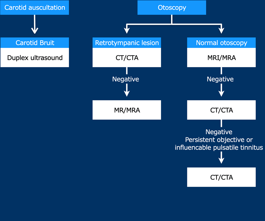
A flowchart for choosing the appropriate imaging modality in pulsatile tinnitus is provided.Bài viết này dựa trên một bài tổng quan của Pegge và cộng sự mô tả một cách tiếp cận có hệ thống để chẩn đoán bệnh ù tai [1].
chẩn đoán phân biệt có liên quan đến các phát hiện trên các phương tiện chẩn đoán hình ảnh khác nhau.
Một sơ đồ để lựa chọn phương thức hình ảnh thích hợp trong ù tai do rung được cung cấp.
Introduction and flowchartGiới thiệu và sơ đồ
Tinnitus is defined as an auditory perception of internal origin, and can have a significant influence on the wellbeing and performance in daily activities of affected subjects. Ù tai được định nghĩa là một nhận thức thính giác về nguồn gốc bên trong, và có thể có ảnh hưởng đáng kể đến sức khoẻ và hiệu suất trong các hoạt động hàng ngày của các đối tượng bị ảnh hưởng.
In pulsatile tinnitus, the auditory perception is repetitively synchronous to the patient’s heartbeat. Trong ù tai rung, nhận thức thính giác đồng bộ lặp đi lặp lại với nhịp tim của bệnh nhân.
All other auditory perceptions are considered non-pulsatile. Tất cả các cảm nhận thính giác khác được coi là không mạch.
Less than 10% of patients presenting with tinnitus have pulsatile tinnitus. Dưới 10% bệnh nhân có biểu hiện ù tai có ù tai kiểu xung.
In about 70% of the cases with pulsatile tinnitus, an underlying cause can be identified by adequate diagnostic work-up as shown on the flow chart. Trong khoảng 70% trường hợp ù tai theo nhịp đập, nguyên nhân cơ bản có thể được xác định bằng cách làm việc chẩn đoán đầy đủ như thể hiện trên biểu đồ dòng chảy.
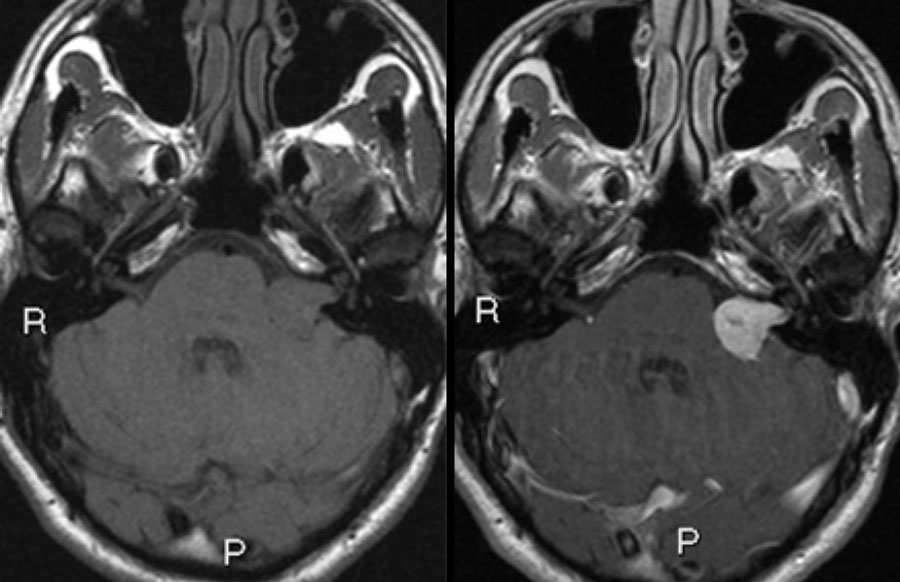 Left sided vestibular schwannoma. Axial non contrast-enhanced T1-W (left) and contrast-enhanced images (right).U tế bào Schwann tiền đình bên trái. Hình ảnh T 1 - W không tăng cường tương phản trục (trái) và hình ảnh tăng cường tương tác (phải).
Non-pulsatile tinnitusÙ tai không do va chạm
Non-pulsatile tinnitus is almost always subjective.
Different underlying conditions relate to the development of non-pulsatile tinnitus, including cerumen impaction, middle ear infection, medications, noise-induced hearing loss, presbycusis or chronic bilateral hearing loss, hemorrhage, neurodegeneration, and spontaneous intracranial hypotension [2]. Ù tai không do va chạm hầu như luôn chủ quan.
Các điều kiện cơ bản khác nhau liên quan đến sự phát triển của chứng ù tai không do xung, bao gồm rách ráy tai, nhiễm trùng tai giữa, thuốc, nghe kém do tiếng ồn, nghe kém trước hoặc nghe kém mạn tính hai bên, xuất huyết, thóai hóa thần kinh và hạ áp lực nội sọ tự phát [2].
MRI is only recommended in patients with unilateral (non-pulsatile) tinnitus with focal neurological abnormalities, or asymmetric hearing loss [4]. MRI chỉ được khuyến cáo ở những bệnh nhân bị ù tai một bên (không rung) có bất thường thần kinh khu trú, hoặc nghe kém không đối xứng [4].
The main purpose of diagnostic imaging is to identify or rule out a lesion in the cerebellopontine angle cistern, (e.g. vestibular schwannoma).
Labyrinthine abnormalities can be identified on MRI supporting the diagnosis of Ménière’s disease [3].
Mục đích chính của chẩn đoán hình ảnh là xác định hoặc loại trừ một tổn thương ở bể chứa góc cầu tiểu não, (ví dụ: u dây thần kinh Schwann tiền đình).
Các bất thường mê nhĩ có thể được xác định trên MRI hỗ trợ chẩn đoán bệnh Ménière [3].
Pulsatile tinnitusÙ tai xung động
 Xem hình gốc (EN)
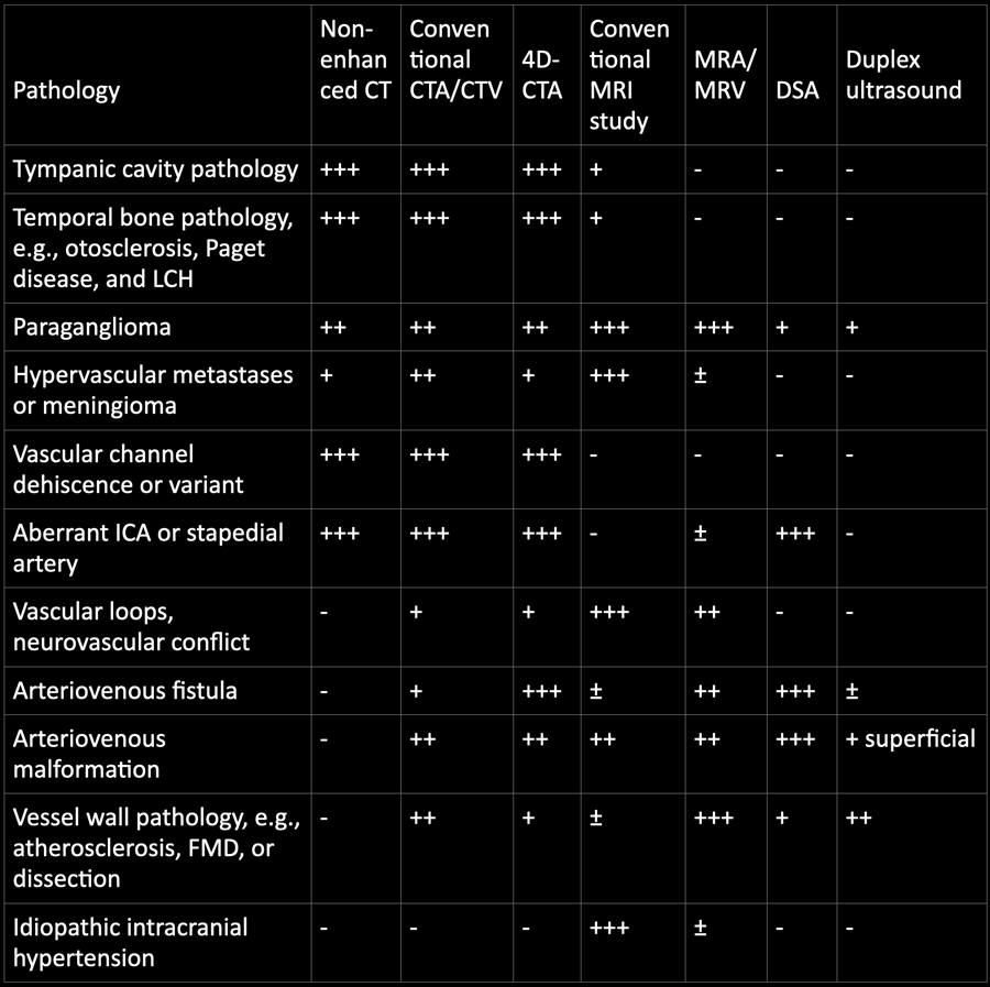
+++ Most optimal, ++ good, + moderate, ± indirect signs, – not suitable, LCH Langerhans cell histiocytosis, ICA internal carotid artery, FMD fibromuscular dysplasia+++ Tối ưu nhất, ++ tốt, + vừa, ± dấu hiệu gián tiếp, – không phù hợp, LCH Langerhans, ICA động mạch cảnh trong, loạn sản xơ cơ FMD
Pathology and imagingGiải phẫu bệnh và chẩn đoán hình ảnh
You can click on the table for a large view.Bạn có thể nhấp vào bảng để xem lớn hơn.
For screening for underlying pathology and for the evaluation of a possible soft tissue mass or intracranial pathology, initial evaluation with MRI and MR angiography (MRA) is recommended with reported high diagnostic accuracy. Để sàng lọc bệnh lý cơ bản và để đánh giá khối mô mềm hoặc bệnh lý nội sọ, đánh giá ban đầu bằng chụp MRI và chụp mạch MR (MRA) được khuyến nghị với độ chính xác chẩn đoán cao.
For the evaluation of osseous pathology of the temporal bone, a limited scanning range of thin-sliced (submillimetric) CT is sufficient.Để đánh giá bệnh lý xương của xương thái dương, một phạm vi quét giới hạn của CT lát mỏng (dưới milimét) là đủ.
Multi-detector CTA or CT venography (CTV) of the head and neck region can be performed for the evaluation of vascular pathology.Chụp CTA hoặc CT tĩnh mạch (CTV) nhiều đầu dò của vùng đầu và cổ có thể được thực hiện để đánh giá bệnh lý mạch máu.
Dynamic CTA, also referred to as 4D-CTA, is a technique that combines the non-invasive nature of CTA with the dynamic acquisition of digital subtraction angiography (DSA). CTA động, còn được gọi là 4D-CTA, là kỹ thuật kết hợp bản chất không xâm lấn của CTA với việc thu nhận động của chụp mạch số hóa (DSA).
The role of conventional angiography (DSA) in the diagnostic work-up of pulsatile tinnitus has been minimized, and should be reserved for the indication to rule out vascular pathology in case MRI/MRA and CT/(4D-)CTA have not revealed the cause of pulsatile tinnitus.Vai trò của chụp mạch vành thường quy (DSA) trong việc chẩn đoán ù tai do rung đã được giảm thiểu và nên được dành riêng cho chỉ định để loại trừ bệnh lý mạch máu trong trường hợp MRI / MRA và CT / (4 D-) CTA không tiết lộ nguyên nhân gây ù tai.
The role of duplex ultrasound in the diagnostic work-up of pulsatile tinnitus is limited, although duplex ultrasound is an effective screening tool for the evaluation of vessel wall pathology of the carotid arteries, e.g. stenosis and occlusion.
During ultrasound, manual compression can be performed to investigate the influence of compression on tinnitus.Vai trò của siêu âm hai chiều trong chẩn đoán ù tai rung động còn hạn chế, mặc dù siêu âm hai mặt là một công cụ sàng lọc hiệu quả để đánh giá bệnh lý thành mạch của động mạch cảnh, ví dụ như hẹp và tắc.
Trong quá trình siêu âm, có thể thực hiện ép bằng tay để khảo sát ảnh hưởng của việc ép lên ù tai.
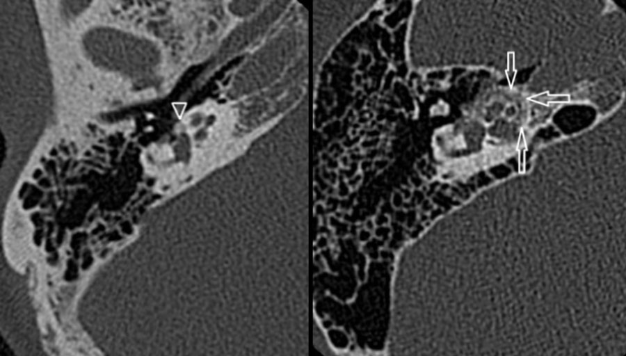 Otosclerosis: hypoattenuated bone in the region of the fissula ante fenestram in fenestral otosclerosis (left). Cochlear otosclerosis appears as a hypoattenuated halo surrounding the cochlea on CT (right).Xơ cứng tai: xương giảm âm ở vùng fissula ante fenestram trong xơ cứng tai giữa (trái) Xơ cứng ốc tai xuất hiện dưới dạng một quầng sáng giảm âm xung quanh ốc tai trên CT (phải).
Temporal bone pathologyBệnh lý xương thái dương
Temporal bone pathology like otosclerosis, Paget disease, and LCH can cause pulsatile tinnitus.Bệnh lý xương thái dương như xơ cứng tai, bệnh Paget và LCH có thể gây ù tai.
Otosclerosis
Is also known as otospongiosis, is an idiopathic infiltrative process of the petrous bone.
It causes both sensorineural and conductive hearing loss, and can be the cause of pulsatile tinnitus. Xơ cứng tai
Còn được gọi là xốp tai, là một quá trình thâm nhiễm tự phát của xương đá.
Nó gây ra cả nghe kém cảm giác và dẫn truyền, và có thể là nguyên nhân gây ra ù tai.
High-resolution, thin-sliced CT typically shows abnormal hypoattenuated bone in the region of the fissula ante fenestram in fenestral otosclerosis (left).CT lát mỏng có độ phân giải cao thường cho thấy xương giảm sắc tố bất thường ở vùng fissula ante fenestram trong xơ cứng tai giữa (trái).
Cochlear otosclerosis
This appears as a hypoattenuated halo surrounding the cochlea on CT (right).Xơ cứng ốc tai
Hình ảnh này xuất hiện dưới dạng một quầng sáng giảm dần bao quanh ốc tai trên CT (bên phải).
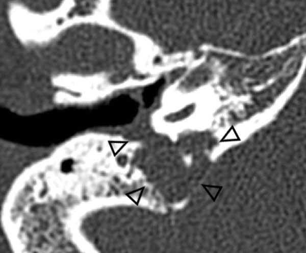 Langerhans cell histiocytosis arising from the jugular foramenBệnh mô bào Langerhans phát sinh từ lỗ tĩnh mạch cảnh
Langerhans cell histiocytosis
Langerhans cell histiocytosis (LCH) is a rare benign disorder of clonal histiocyte proliferation.
Clinical symptoms in LCH depend on the extent of bone and extraskeletal involvement.Bệnh mô bào Langerhans
Bệnh mô bào Langerhans (LCH) là một rối loạn lành tính hiếm gặp của sự tăng sinh mô bào vô tính.
Các triệu chứng lâm sàng trong LCH phụ thuộc vào mức độ tổn thương xương và ngoài xương.
Imaging typically reveals an aggressive lytic osseous lesion with associated soft tissue masses without surrounding sclerosis.
The image shows an aggressive lytic osseous lesion with soft tissue mass arising from the jugular foramen, which proved to be LCH.. Hình ảnh thường cho thấy một tổn thương xương với các khối mô mềm liên quan mà không có xơ cứng xung quanh.
Hình ảnh cho thấy một tổn thương xương lytic tích cực với khối mô mềm phát sinh từ lỗ tĩnh mạch cảnh, được chứng minh là LCH.
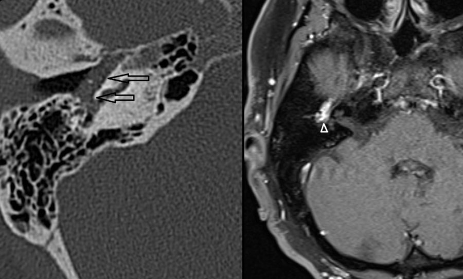 Paraganglioma: soft tissue mass in the middle ear (left), strong enhancement of the lesion (right)Paraganglioma: Khối mô mềm ở tai giữa (trái), tổn thương ngấm thuốc mạnh (phải)
ParagangliomaU cận hạch
Both CT and MRI can be used for the detection and evaluation of a paraganglioma.
The majority of tympanic paragangliomas are located on the promontory as a small well-defined tympanic soft tissue mass.
Usually, there is no or little surrounding bone erosion.
These small tumours are best evaluated using thin-sliced CT with a bone algorithm.Cả CT và MRI đều có thể được sử dụng để phát hiện và đánh giá u cận hạch.
Phần lớn các paragangliomas nhĩ nằm trên vùng tiền đình như một khối mô mềm nhĩ nhỏ được xác định rõ.
Thông thường, không có hoặc ít có sự xói mòn xương xung quanh.
Những khối u nhỏ này được đánh giá tốt nhất bằng cách sử dụng CT cắt lát mỏng với thuật toán xương.
Left Axial CT shows a soft tissue mass in the middle ear (arrows).
No visible bony erosion.
Right Axial contrast enhanced T1-W with fat suppression demonstrates strong enhancement of this lesion(arrowhead).CT trục trái cho thấy khối mô mềm ở tai giữa (các mũi tên).
Không có tổn thương xương.
T1 - W có tiêm thuốc cản quang trục phải với xóa mỡ cho thấy sự tăng cường mạnh mẽ của tổn thương này (đầu mũi tên).
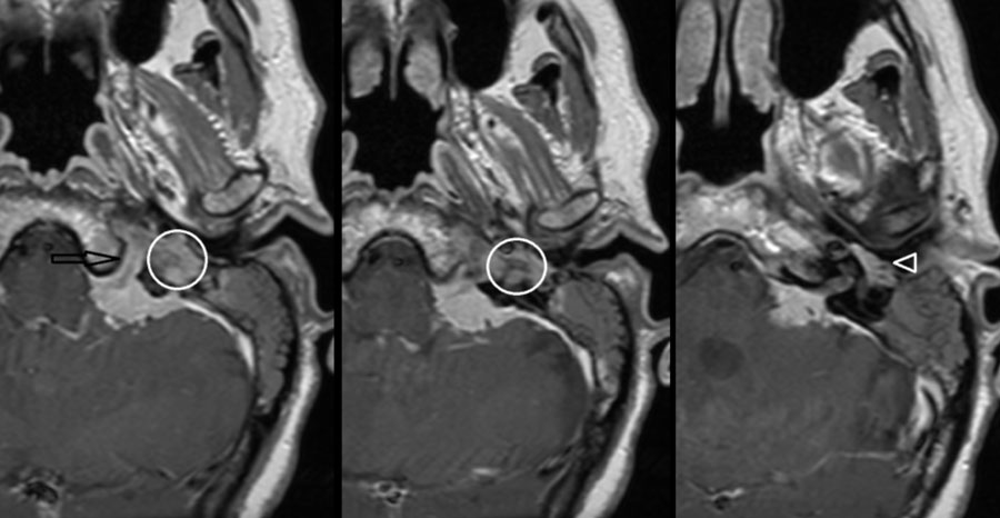 MeningiomaU màng não
Hypervascular metastases or meningiomaDi căn tăng sinh mạch hoặc u màng não
Highly vascularized bone lesions, like osseous hemangioma, basal meningioma, Langerhans cell histiocytosis, or bone metastases, have been described as possible causes of pulsatile tinnitus.Các tổn thương xương có mạch máu cao, như u máu xương, u màng não nền, mô bào Langerhans, hoặc di căn xương, đã được mô tả là nguyên nhân có thể gây ù tai.
On the left a meningioma on MRI.
Axial contrast-enhanced T1-W images.
Enhancing mass located in the left cerebellar-pontine angle with extension into hypoglossal canal (arrow), jugular plate (encircled), and the middle ear (arrowhead).Bên trái là u màng não trên MRI.
Hình ảnh T 1 - W có độ tương phản tăng cường.
Khối tăng cường nằm ở góc cầu tiểu não trái với phần mở rộng vào ống hạ thiệt (mũi tên), đĩa tĩnh mạch cảnh (bao quanh) và tai giữa (đầu mũi tên).
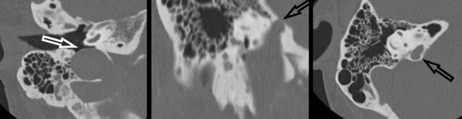
Vascular channel dehiscence or variantDò hoặc biến thể kênh mạch máu
Venous tinnitus is heard as a continuous murmur that exaggerates in systole.
There seems to be an association with congenital variants such as a high riding, enlarged, or diverticulum of the jugular bulb, which can be best depicted on thin-sliced high-resolution CT.
Prevalence of sigmoid sinus diverticulum and dehiscence has been reported to be significantly higher in pulsatile tinnitus than in the general population. Ù tai tĩnh mạch được nghe như một tiếng thổi liên tục, phóng đại trong thì tâm thu.
Dường như có sự liên quan với các biến thể bẩm sinh như hình ảnh bóng cảnh có độ cao, to hoặc túi thừa, có thể được mô tả tốt nhất trên CT độ phân giải cao cắt lát mỏng.
Tỷ lệ túi thừa xoang sigma và ù tai đã được báo cáo là cao hơn đáng kể trong ù tai do xung so với dân số nói chung.
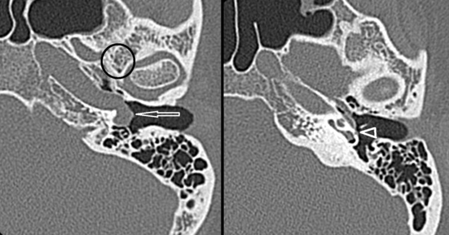
Aberrant course of ICA or stapedial arteryĐường đi bất thường của động mạch cảnh trong và động mạch gian sườn
An aberrant course of the internal carotid artery and persistence of the stapedial artery are congenital variants that need to be recognized on imaging studies.
An aberrant course of the internal carotid artery in the middle ear may mimic a soft tissue mass or paraganglioma at otoscopy.Một đường đi bất thường của động mạch cảnh trong và sự tồn tại của động mạch chũm là những biến thể bẩm sinh cần được nhận ra trên các nghiên cứu hình ảnh.
Một đường đi bất thường của động mạch cảnh trong ở tai giữa có thể giống với khối mô mềm hoặc u cận hạch khi soi tai.
On the left an Aberrant course of the internal carotid artery (arrow) and persistence of the stapedial artery (arrowhead) on thin-sliced CT.
Note the absence of the foramen spinosum (encircled).Bên trái là đường đi bất thường của động mạch cảnh trong (mũi tên) và sự tồn tại của động mạch chạy trên (đầu mũi tên) trên CT lát mỏng.
Lưu ý sự vắng mặt của lỗ gai (bao quanh).
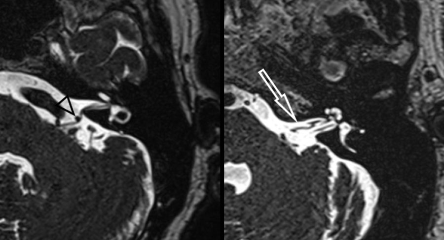
Vascular loops, neurovascular conflictVòng mạch, xung đột mạch thần kinh
Vascular loops and elongated arteries are occasionally described as a possible cause of pulsatile tinnitus.
Considering the presence of these vascular loops and elongations also in asymptomatic patients, other possible causes of pulsatile tinnitus need to be ruled out in those subjects. Các vòng mạch và động mạch kéo dài đôi khi được mô tả là nguyên nhân có thể gây ra ù tai.
Xem xét sự hiện diện của các vòng mạch và sự kéo dài cũng ở những bệnh nhân không có triệu chứng, các nguyên nhân khác có thể gây ù tai do rung cần phải được loại trừ ở những đối tượng đó.
Heavy T2 weighted CISS images on the left demonstrates a neurovascular conflict of the posterior inferior cerebellar artery with compression and displacement of the facial nerve and vestibulocochlear nerve (left image, black arrowhead). Hình ảnh CISS trọng số T2 nặng ở bên trái cho thấy xung đột mạch máu thần kinh của động mạch tiểu não dưới sau với sự chèn ép và di lệch của dây thần kinh mặt và dây thần kinh tiền đình (hình ảnh bên trái, đầu mũi tên màu đen).
The right image shows a grade 3 (>50%) vascular loop of the anterior inferior cerebellar artery within the left internal acoustic canal (white arrow).Hình ảnh bên phải cho thấy vòng mạch máu độ 3 (> 50%) của động mạch tiểu não trước dưới trong ống nội âm bên trái (mũi tên màu trắng).
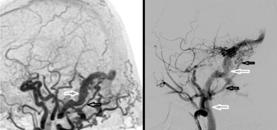 AVF: abnormal early contrast filling of the sigmoid sinus (left), venous drainage of the sigmoid sinus into the jugular vein (right)AVF: bất thường lấp đầy sớm của xoang sigma (trái), tĩnh mạch dẫn lưu xoang sigmoid vào tĩnh mạch cảnh (phải)
Arteriovenous fistula (AVF)Rò động tĩnh mạch (AVF)
An AVF is an, usually acquired, abnormal connection between an artery and a vein without an intervening nidus.
Located along the dura or within a dural sinus, these are called dural AVF. AVF là một kết nối bất thường, thường mắc phải giữa động mạch và tĩnh mạch mà không có ổ cắm can thiệp.
Nằm dọc theo màng cứng hoặc trong xoang màng cứng, chúng được gọi là AVF màng cứng.
Dural arteriovenous fistula (dAVF) located in the right sigmoid sinus as identified by 4D-CTA and DSA. Dò động tĩnh mạch màng cứng (dAVF) nằm ở xoang sigma phải được xác định bằng chụp 4D-CTA và DSA.
Left 4D-CTA lateral subtracted MIP demonstrating abnormal early contrast filling of the sigmoid sinus (white arrow) consistent with dAVF.
Hypertrophic occipital artery identified as arterial feeder (black arrow). Chụp 4D-CTA bên trái cho thấy bất thường lấp đầy chất cản quang sớm của xoang sigma (mũi tên trắng) phù hợp với dAVF.
Động mạch chẩm phì đại được xác định là động mạch nuôi dưỡng (mũi tên màu đen).
Right DSA, selective contrast injection of the external carotid artery showing a hypertrophic tortuous occipital artery (black arrows).
Venous drainage of the sigmoid sinus into the jugular vein (white arrows).DSA phải, tiêm thuốc cản quang chọn lọc động mạch cảnh ngoài cho thấy động mạch chẩm xoắn quá phát (các mũi tên đen).
Dẫn lưu tĩnh mạch của xoang sigma vào tĩnh mạch cảnh (các mũi tên trắng).
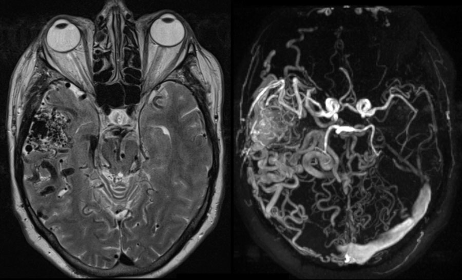 AVM located in the right temporal fossaAVM nằm ở hố thái dương phải
Arteriovenous malformation (AVM)Dị dạng động tĩnh mạch (AVM)
An AVM located in the head and neck region can be the cause of pulsatile tinnitus. AVM nằm ở vùng đầu và cổ có thể là nguyên nhân gây ù tai.
Typically, an AVM develops in adolescence or young adulthood but can remain occult for a long period. Thông thường, AVM phát triển ở tuổi vị thành niên hoặc thanh niên nhưng có thể vẫn còn ẩn giấu trong một thời gian dài.
T2-W (left) and phase-contrast MRA (right) demonstrating intracranial arteriovenous malformation (AVM) located in the right temporal fossa.T 2 - W (trái) và MRA pha tương phản (phải) cho thấy dị dạng động tĩnh mạch nội sọ (AVM) nằm ở hố thái dương phải.
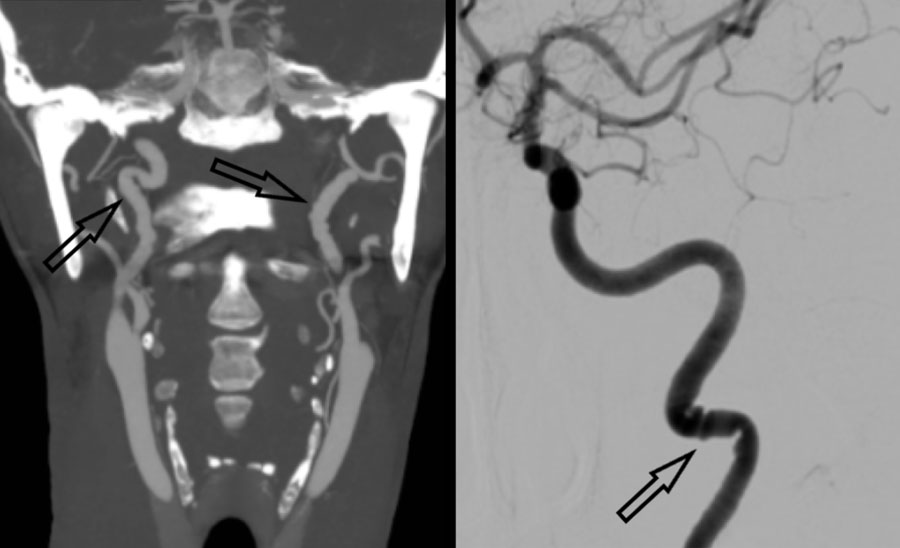 FMD with “string of beads” pattern on MIP CTA and DSA (black arrows)FMD với mẫu “chuỗi hạt” trên MIP CTA và DSA (các mũi tên đen)
Vessel wall pathologyBệnh lý thành mạch
Vessel wall pathology like atherosclerosis, FMD or dissection can be a cause of pulsatile tinnitus.Bệnh lý thành mạch như xơ vữa động mạch, FMD hoặc bóc tách có thể là nguyên nhân gây ù tai.
In the elderly population, atherosclerotic disease of the carotid or vertebral arteries is thought to be the most common cause of pulsatile tinnitus.
In a significant stenosed or occluded artery, increased vascular flow on the contralateral side could lead to pulsatile tinnitus as a symptom. Ở người cao tuổi, bệnh xơ vữa động mạch cảnh hoặc động mạch đốt sống được cho là nguyên nhân phổ biến nhất gây ù tai.
Trong một động mạch bị hẹp hoặc tắc nghẽn đáng kể, tăng lưu lượng mạch máu ở phía đối diện có thể dẫn đến ù tai như một triệu chứng.
Fibromuscular dysplasia (FMD) is a segmental nonatheromatous, non-inflammatory vascular disease of unknown etiology.
Often it is a disease of the young leading to vascular stenosis and cerebral ischemia. Loạn sản xơ cơ (FMD) là một bệnh mạch máu không viêm, không phải do u xơ, không rõ nguyên nhân.
Thường là bệnh lý của người trẻ tuổi dẫn đến hẹp mạch máu và thiếu máu cục bộ não.
On the left a classical imaging appearance of FMD the so-called “string of beads” pattern on MIP CTA and DSA (black arrows).Bên trái là hình ảnh cổ điển của FMD được gọi là “mẫu chuỗi hạt” trên MIP CTA và DSA (các mũi tên màu đen).
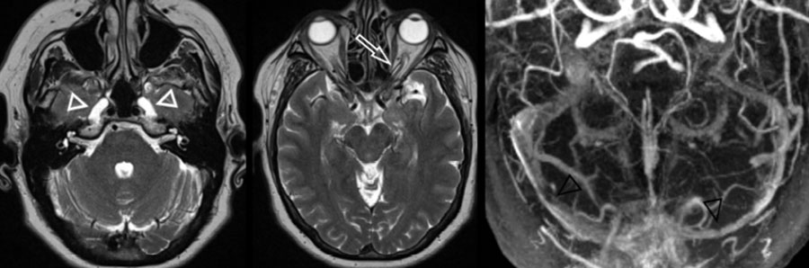 Intracranial hypertension with enlarged Meckel cave (left image, white arrowheads), prominent subarachnoid space around the optic nerve (middle image, white arrow) and bilateral venous sinus stenosis (right image, black arrowheads)Tăng áp lực nội sọ với hang Meckel mở rộng (hình bên trái, đầu mũi tên màu trắng), vùng dưới nhện nổi xung quanh dây thần kinh thị giác (hình giữa, mũi tên màu bạch) và hẹp xoang tĩnh mạch hai bên (hình phải, đầu mũi mũi tên màu đen)
Idiopathic intracranial hypertensionTăng áp lực nội sọ tự phát
Idiopathic intracranial hypertension (IIH), which predominantly affects young obese women, may cause pulsatile tinnitus, although IIH is primarily characterized by symptoms of headache and blurred vision due to increased cerebrospinal fluid pressure. Tăng áp lực nội sọ vô căn (IIH), chủ yếu ảnh hưởng đến phụ nữ trẻ béo phì, có thể gây ù tai, mặc dù IIH chủ yếu được đặc trưng bởi các triệu chứng đau đầu và mờ mắt do tăng áp lực dịch não tủy.
The exact pathophysiology of IIH is unknown but can develop in patients with a history of dural sinus thrombosis.Sinh lý bệnh chính xác của IIH không rõ nhưng có thể phát triển ở những bệnh nhân có tiền sử huyết khối xoang màng cứng.
Dural sinus stenosis or compression can also be observed in IHH.
It is therefore advised to perform MRV or CTV in a patient with pulsatile tinnitus and suspicion of IIH. Hẹp xoang màng cứng hoặc chèn ép xoang cũng có thể được quan sát thấy trong IHH.
Do đó, nên thực hiện MRV hoặc CTV ở bệnh nhân bị ù tai có nhịp đập và nghi ngờ IIH.
On the left a typical case of intracranial hypertension with enlarged Meckel cave (left image, white arrowheads), prominent subarachnoid space around the optic nerve (middle image, white arrow) and bilateral venous sinus stenosis (right image, black arrowheads).Bên trái là một trường hợp điển hình của tăng áp lực nội sọ với hang Meckel mở rộng (hình ảnh bên trái, đầu mũi tên màu trắng), khoảng dưới nhện nổi bật xung quanh dây thần kinh thị giác (hình giữa, mũi tên màu xanh) và hẹp xoang tĩnh mạch hai bên (hình bên phải, đầu mũi Tên màu đen).
ReferencesTài liệu tham khảo
-
Pegge SA, et al. Pulsatile Tinnitus: Differential Diagnosis and Radiological Work-Up. Curr Radiol Rep. 2017;5(1):5. Pegge SA, et al. Pulsatile Tinnitus: chẩn đoán phân biệt and Radiological Work-Up. Curr Radiol Rep. 2017; 5 (1): 5.
-
Hoang JK, Loevner LA. Evaluation of Tinnitus and Hearing Loss in the Adult. 2020 Feb 15. In: Hodler J, Kubik-Huch RA, von Schulthess GK, editors. Diseases of the Brain, Head and Neck, Spine 2020–2023: Diagnostic Imaging [Internet]. Cham (CH): Springer; 2020. Chapter 15. Hoang JK, Loevner LA. Đánh giá ù tai và mất thính lực ở người lớn. 2020 Feb 15. Trong: Hodler J, Kubik-Huch RA, von Schulthess GK, biên tập viên. Bệnh não, đầu cổ, cột sống 2020–2023: Chẩn đoán hình ảnh [Internet]. Cham (CH): Springer; 2020. Chương 15.
-
S. Vattoth, R. Shah and J.K. Curé. A Compartment-Based Approach for the Imaging Evaluation of Tinnitus. American Journal of Neuroradiology February 2010, 31 (2) 211-218. S. Vattoth, R. Shah và J.K. Curé. Tiếp cận theo khoang trong đánh giá ù tai bằng chẩn đoán hình ảnh. American Journal of Neuroradiology, tháng 2/2010, tập 31, số 2, tr. 211–218.
-
Tunkel DE, Bauer CA, Sun GH, et al. Clinical Practice Guideline: Tinnitus. Otolaryngol Head Neck Surg 2014, 151(2S) S1–S40. Tunkel DE, Bauer CA, Sun GH, et al. Clinical Practice Guideline: Ù tai. Otolaryngol Head Neck Surg 2014, 151(2S) S1–S40.
-
Kircher ML, Standring RT, Leonetti JP. Neuroradiologic assessment of pulsatile tinnitus. J Otolaryngol Head Neck Surg. 2008;139(Suppl):144. Kircher ML, Standring RT, Leonetti JP. Đánh giá thần kinh học về ù tai do nhịp đập. J Otolaryngol Head Neck Surg. 2008; 139 (Suppl): 144.
-
Mattox DE, Hudgins P. Algorithm for evaluation of pulsatile tinnitus. Acta Otolaryngol. 2008;128:427–31. Mattox DE, Hudgins P. Thuật toán đánh giá ù tai. Acta Otolaryngol. 2008; 128:427 – 31
-
Joustra J, et al. Richtlijn tinnitus. Nederlandse Vereniging voor Keel–Neus–Oorheelkunde en Heelkunde van het Hoofd Halsgebied. 2016. Joustra J, et al. Richtlijn ù tai. Nederlandse Vereniging voor Keel–Neus–Oorheelkunde en Heelkunde van het Hoofd Halsgebied. 2016.