
Algoritm for Ankle Fractures 2.0Thuật toán đánh giá gãy xương cổ chân 2.0
Radiology department of the Amsterdam University Medical Centre in Amsterdam and Alrijne hospital in Leiderdorp in the NetherlandsKhoa X quang của Trung tâm Y tế Đại học Amsterdam tại Amsterdam và Bệnh viện Alrijne ở Leiderdorp, Hà Lan
Ngày đăng / cập nhật: 01-08-2021
Bản gốc trên radiologyassistant.nl ↗ · tải về lúc 2026-11-19 20:19 · bản dịch máy có chuẩn hóa thuật ngữ chẩn đoán hình ảnh
In this lecture we present a simple algoritm that helps you to find:
Trong bài giảng này, chúng tôi trình bày một thuật toán đơn giản giúp bạn tìm:
- All ankle fractures, even the ones that are not visible on the X-rays
Tất cả các loại gãy cổ chân, kể cả những loại không nhìn thấy trên phim X-quang - Predict rupture of ligaments even if you can’t see them on the images
Dự đoán đứt dây chằng ngay cả khi bạn không thể nhìn thấy chúng trên hình ảnh - Determine whether the ankle is stable or unstable, just by looking at the radiographs Xác định xem cổ chân ổn định hay không ổn định, chỉ bằng cách nhìn vào phim X-quang
The algoritm is based on the Weber-classification, because it is simple and everybody knows it. Since the Weber-classification is a simplification of the Lauge-Hansen classification, it will help you to understand the different stages of Lauge-Hansen.Thuật toán dựa trên phân loại Weber, vì nó đơn giản và mọi người đều biết nó. Vì phân loại Webers là một sự đơn giản hóa của phân loại Lauge-Hansen, nó sẽ giúp bạn hiểu các giai đoạn khác nhau của Lauge- Hansen.
IntroductionMở đầu
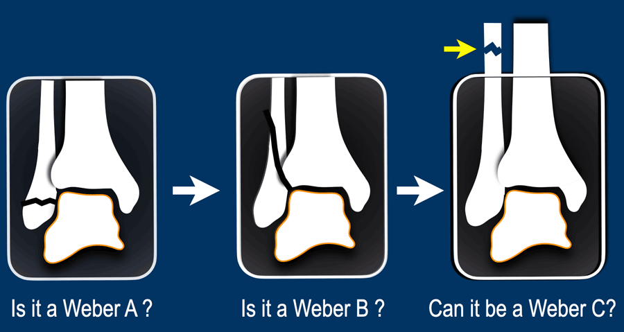
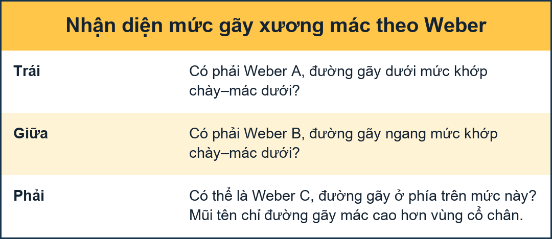
Algoritm for ankle fracturesThuật toán điều trị gãy cổ chân
Step 1
The first question you should ask yourself is:
Is it a Weber type A fracture?
Is there an avulsion of the lateral malleolus. That’s easy. Bước 1
Câu hỏi đầu tiên bạn nên tự hỏi mình là:
Có phải gãy Weber loại A không?
Có sự nhả của mắt cá ngoài. Điều đó dễ.
Step 2
If it is not a type A. Then the next question is:
Is it a Weber type B fracture?
This oblique fracture at the level of the syndesmosis is always easy to find on the x-rays.Bước 2
Nếu không phải là type A thì câu hỏi tiếp theo là:
Có phải gãy Weber loại B không?
Gãy chéo này ở mức khớp sợi chày–mác luôn dễ tìm thấy trên phim X-quang.
Step 3
If it is not type A or type B, then the last question is:
Can this be a Weber type C fracture?
These fractures are usually not visible on x-rays of the ankle, because the fibula fracture is too high, but the algoritm provides clues for the detection of these fractures.Bước 3
Nếu không phải là type A hay type B thì câu hỏi cuối cùng là:
Đây có thể là gãy Weber loại C không?
Những loại gãy này thường không nhìn thấy được trên phim X-quang cổ chân, vì xương mác quá cao, nhưng thuật toán cung cấp các đầu mối để phát hiện những loại gãy xương này.
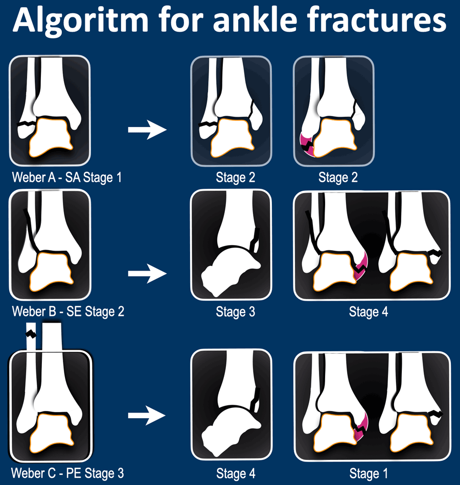
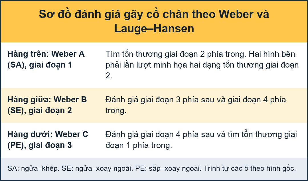
When we've answered the above questions, then we end up in one of these three categories.
In each of these categories we need to determine the stage of the fracture, which tells us if the ankle is stable or unstable.Khi chúng ta đã trả lời các câu hỏi trên, thì chúng ta sẽ kết thúc ở một trong ba loại này.
Trong mỗi loại này, chúng ta cần xác định giai đoạn gãy xương, cho chúng ta biết nếu cổ chân ổn định hay không ổn định.
In Weber A stage 1 is stable.
However when there is also a vertical or push-off fracture of the medial malleolus, then it is stage 2 and the ankle is unstable, as the ring of stability is broken in two places.Trong Weber A giai đoạn 1 ổn định.
Tuy nhiên khi cũng có gãy dọc hoặc gãy đẩy mắt cá trong thì đó là giai đoạn 2 và cổ chân không ổn định, vì vòng ổn định bị gãy ở hai nơi.
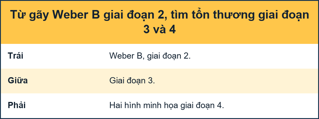
In Weber B stage 2 is stable, but stage 3 and 4 are unstable.Trong Weber B giai đoạn 2 ổn định, nhưng giai đoạn 3 và 4 không ổn định.
In Weber C finding a high fibula fracture means unstable stage 3. Since this fracture is usually not visible on x-rays of the ankle, you wanna study the ankle x-rays to look for stage 1 and 4, which can be a clue to image the whole lower leg to look for a high fibula fracture.Trong Weber C, phát hiện gãy xương mác cao có nghĩa là giai đoạn 3 không ổn định. Vì loại gãy này thường không nhìn thấy được trên phim X-quang cổ chân, bạn nên quan sát phim X-Quang cổ chân để tìm giai đoạn 1 và 4, đây có thể là một đầu mối để chụp ảnh toàn bộ cẳng chân để tìm gãy xương hàm cao.
These stages in Weber B and C always follow a strict order.
This means that when you find a stage 3 fracture, there already must be a stage 1 and 2 even if you can't see them.
We will discuss these stages in the next chapter.Các giai đoạn này trong Weber B và C luôn tuân theo một trật tự nghiêm ngặt.
Điều này có nghĩa là khi bạn tìm thấy gãy xương giai đoạn 3, thì phải có gãy xương ở giai đoạn 1 và 2 ngay cả khi bạn không thể nhìn thấy chúng.
Chúng ta sẽ thảo luận về các giai đoạn này trong chương tiếp theo.
Fracture stagesGiai đoạn gãy xương
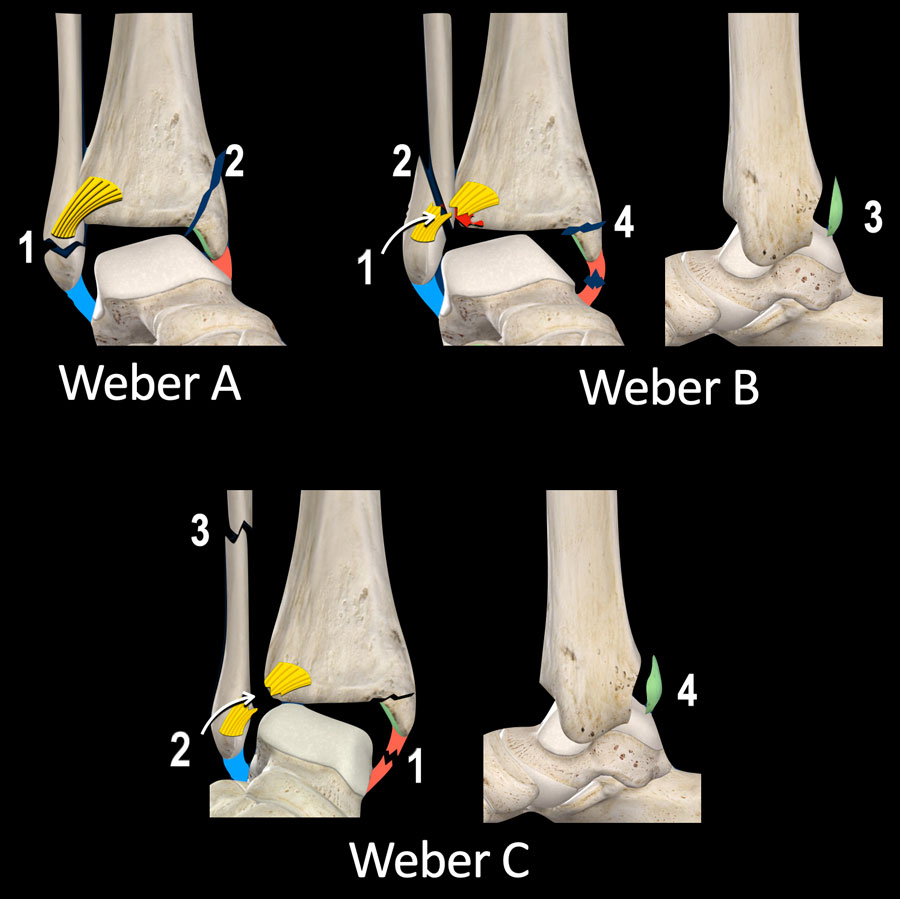
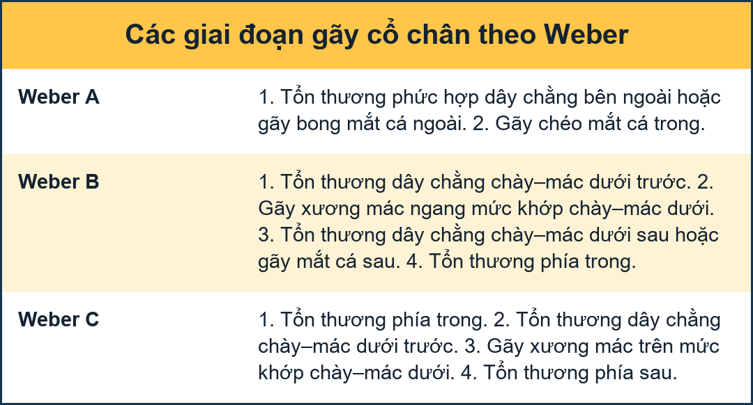
This is an overview of the stages of ankle fractures. It was originally described by Christian Lauge-Hansen, a Danish pathologist in 1950 and later copied by Bernhard Georg Weber in 1972, a member of the AO-group.Đây là tổng quan về các giai đoạn của gãy xương cổ chân. Nó được mô tả lần đầu bởi Christian Lauge-Hansen, một nhà bệnh học người Đan Mạch vào năm 1950 và sau đó được Bernhard Georg Weber sao chép vào năm 1972, một thành viên của nhóm AO.
Stages of Weber ACác giai đoạn của Weber A
- Lateral avulsionNhổ bên
- Medial vertical push-off fracture
Gãy đẩy dọc xương đùi
Stages of Weber BCác giai đoạn của Weber B
- Rupture of anterior syndesmosis or less common Tillaux fracture (avulsion of tibial attachment). Vỡ xương phần trước khớp chày–mác dưới hoặc ít gặp hơn gãy Tillaux (xẹp bám chày).
- Oblique fibula fracture at level of syndesmosis.Gãy xương mác xiên ở mức khớp sợi chày–mác.
- Tertius avulsion fracture or rupture of posterior syndesmosisTertius gãy bong điểm bám hoặc vỡ phần sau khớp sợi chày–mác
- Avulsion fracture of medial malleolus or rupture of medial collateral bands
Gãy bong điểm bám của mắt cá trong hoặc vỡ các dải đối xứng trong
Stages of Weber CCác giai đoạn của Weber C
- Avulsion fracture of medial malleolus or rupture of medial collateral bandsGãy bong điểm bám của mắt cá trong hoặc vỡ các dải đối xứng trong
- Rupture of anterior syndesmosis or less common Tillaux fracture (avulsion of tibial attachment). Vỡ xương phần trước khớp chày–mác dưới hoặc ít gặp hơn gãy Tillaux (xẹp bám chày).
- High twist-like fibula fracture above the level of the syndesmosis.Gãy xương mác xoắn cao trên mức khớp sợi chày–mác.
- Tertius avulsion fracture or rupture of posterior syndesmosis
Tertius gãy bong điểm bám hoặc vỡ phần sau khớp sợi chày–mác
Weber B and Weber C fractures are very different in the type of fibula fracture.
In Weber B there is a oblique oriented push-off fracture at the level of the syndesmosis, while in Weber C there is a twist-like fracture above the level of the syndesmosis.Weber B và Weber C rất khác nhau về loại gãy xương mác.
Ở Weber B có một vết gãy hướng chéo ở mức khớp dính, trong khi ở Weber C có một vết nứt xoắn trên mức khớp díh.
Notice however, that there are many similarities between Weber B and C with only differences in the order of events. For instance a tertius fracture is either stage 3 in a Weber B or a stage 4 in Weber C fracture.Tuy nhiên, cần lưu ý rằng có nhiều điểm tương đồng giữa Weber B và C chỉ khác nhau về thứ tự các sự kiện Ví dụ, gãy xương tertius là giai đoạn 3 trong Weber B hoặc giai đoạn 4 trong gãy Weber C.
Another important thing to realize is that traction on a ligament results in either a rupture of that ligament or an avulsion fracture at the insertion of that ligament.Một điều quan trọng khác cần nhận ra là lực kéo trên dây chằng dẫn đến đứt dây chằng đó hoặc gãy bong điểm bám khi chèn dây chằng vào.
Step 1 - is it a Weber A / SABước 1 - có phải là Weber A / SA
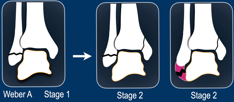
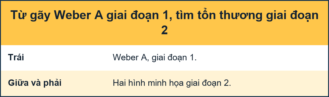
Weber A is the result of a pull-off or avulsion on the lateral side due to extreme supination of the foot with adduction. This is stage 1 and is stable.
Lauge-Hansen calls this supination-adduction (SA).Weber A là kết quả của một cú kéo hoặc nhổ ở bên ngoài do ngửa bàn chân quá mức khi khép bàn chân. Đây là giai đoạn 1 và ổn định.
Lauge-Hansen gọi đây là suy diễn ngửa (SA).
Although not that common, the injury may proceed and cause a push off fracture on the medial side resulting in a vertical fracture of the medial malleolus . This is always stage 2 and is unstable, whether you see a fracture of the lateral malleolus or not. In rare case you may find a vertical fracture of the medial malleolus in combination with a collateral band rupture on the lateral side.Mặc dù không phổ biến, chấn thương có thể tiến triển và gây ra gãy đẩy ra ở phía trong dẫn đến gãy dọc mắt cá trong. Đây luôn là giai đoạn 2 và không ổn định, cho dù bạn có thấy gãy mắt cá ngoài hay không. Trong một số ít trường hợp, bạn có thể tìm thấy gãy dốc mắt cá trong kết hợp với đứt dải băng bên ở phía bên.
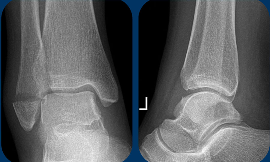 Stage 1: stable ankle fractureGiai đoạn 1: gãy ổn định cổ chân
Stage 1Giai đoạn 1
Here a typical avulsion or pull-off fracture of the lateral malleolus.
The avulsion fragment is quite large. More commonly there is a small avulsion fragment.Đây là một kiểu gãy nhô ra hoặc gãy kéo ra điển hình của mắt cá ngoài.
Đoạn nhổ khá lớn, thường gặp hơn là đoạn nhổ nhỏ.
This is a stage 1 stable Weber A fracture.Đây là gãy Weber A ổn định giai đoạn 1.
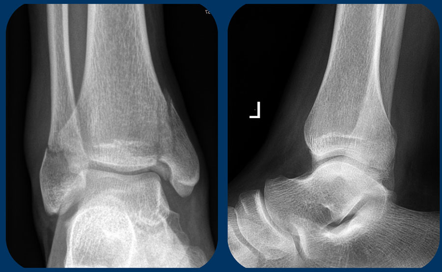
Stage 2Giai đoạn 2
This case is more challenging.
This fibula fracture is at the level of the syndesmosis and maybe some people would call this a Weber B fracture, but it doesn't look like a Weber B. A weber B fracture is oblique and more vertical because it is a push-off fracture, which we will discuss later.Trường hợp này khó khăn hơn.
Gãy xương mác này nằm ở mức khớp sợi chày–mác và có thể một số người gọi đây là gãy Weber B, nhưng nó không giống như Weber B. Gãy WeberB là gãy chéo và thẳng đứng hơn vì nó là gãy đẩy ra, điều mà chúng ta sẽ thảo luận sau.
Just by looking at the images, you can understand the trauma mechanism (scroll).Chỉ cần nhìn vào các hình ảnh, bạn có thể hiểu được cơ chế chấn thương (scroll).
This is a stage 2 - unstable bimalleolar Weber A fracture.Đây là gãy Weber A không vững ở giai đoạn 2, thường gặp ở trẻ em.
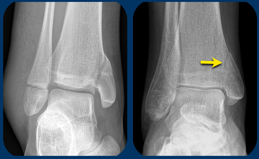
The left image is another example of an unstable bimalleolar fracture stage 2 Weber A.Hình ảnh bên trái là một ví dụ khác về gãy xương ống nhỏ không ổn định giai đoạn 2 Weber A.
The right image shows a vertical fracture of the medial malleolus (arrow). This is always stage 2 and unstable. It means that there already is stage 1, because the trauma mechanism always follows this strict order, first stage 1 and then stage 2.Hình bên phải cho thấy gãy dọc mắt cá trong (mũi tên) luôn là giai đoạn 2 và không ổn định, có nghĩa là đã có giai đoạn 1, vì cơ chế chấn thương luôn tuân theo trật tự chặt chẽ này, giai đoạn 1 trước rồi mới đến giai đoạn 2.
The ring of stability is broken in two places (scroll).Vòng ổn định bị đứt ở hai nơi (vòng cuộn).
Step 2 - is it a Weber B / SERBước 2 - có phải là Weber B / SER
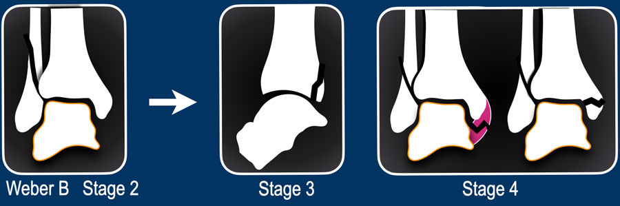
If it is not a Weber A fracture, then the next question is: is it a Weber B?Nếu không phải là Weber A thì câu hỏi tiếp theo là: có phải là Webers B?
When you see a Weber B fracture, which is always good visible on either the AP- or the lateral view, the only thing you need to check is whether there is an unstable stage 3 with posterior injury or even stage 4 with medial injury.Khi bạn thấy gãy Weber B, luôn nhìn thấy rõ trên góc nhìn AP hoặc bên, điều duy nhất bạn cần kiểm tra là liệu có giai đoạn 3 không ổn định với chấn thương sau hay thậm chí giai đoạn 4 với chấn thương trong hay không.
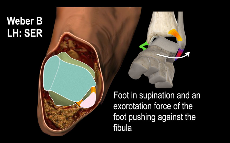
Weber B is the most common ankle fracture accounting for 60-70% of all ankle fractures.Weber B là loại gãy cổ chân hay gặp nhất chiếm 60-70% các loại gãy.
Just like a Weber C fracture it is the result of an exorotation force applied by the foot.
The only difference is that in Weber B the foot is in supination and the injury starts on the lateral side where the tension is, while in Weber C the injury starts on the medial side due to the position of the foot in pronation.Giống như gãy Weber C, nó là kết quả của lực đẩy ra của bàn chân.
Sự khác biệt duy nhất là ở Weber B, bàn chân nằm ngửa và chấn thương bắt đầu ở phía bên nơi có lực căng, trong khi ở Weber C, chấn thương bắt nguồn từ phía trong do vị trí của bàn chân nằm sấp.
Lauge-Hansen called this fracture mechanism supination exorotation or SER.
The fracture mechanism that leads to a Weber C fracture is called pronation exorotation or PER and we will discuss it in the next section.Lauge-Hansen gọi cơ chế gãy xương này là cơ chế ngửa ra ngoài hoặc SER.
Cơ chế gãy xương dẫn đến gãy Weber C được gọi là sấp ngửa hoặc PER và chúng ta sẽ thảo luận về nó trong phần tiếp theo.
Scroll through the images to see how the injury follows a clockwise fixed order.Cuộn qua các hình ảnh để xem thương tổn theo thứ tự cố định theo chiều kim đồng hồ như thế nào.
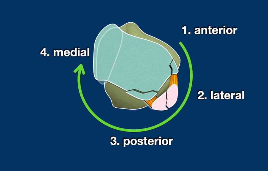
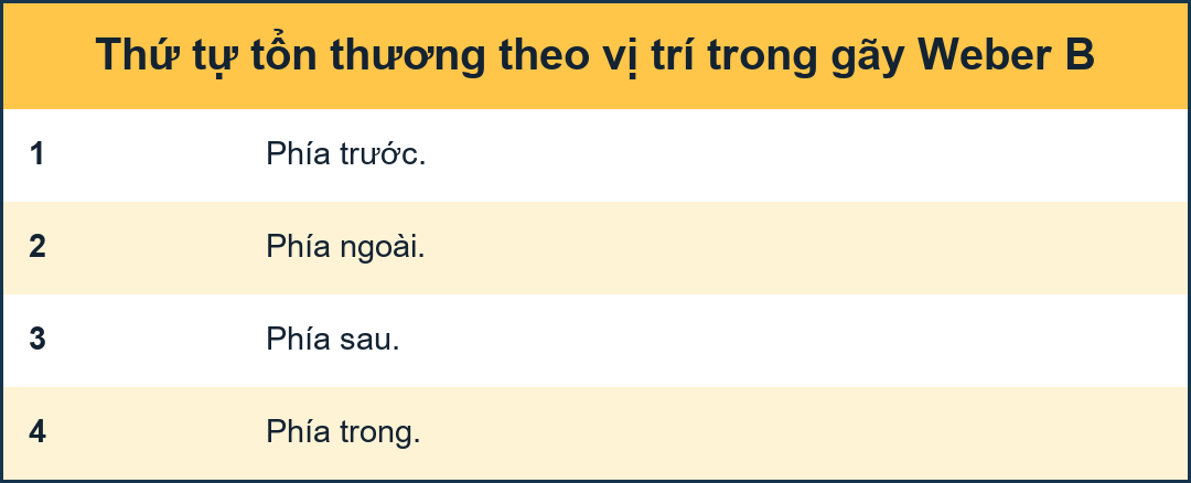
Things become very easy once you remember the fixed order of the injuries:
Mọi thứ trở nên rất dễ dàng khi bạn nhớ thứ tự cố định của các chấn thương:
- Anterior
Tension in anterior syndesmosis results in rupture of the syndesmosis or avulsion of the tibial attachment - Tillaux fracture.Trước
Căng ở phần trước khớp chày–mác dưới dẫn đến vỡ khớp sợi chày–mác hoặc bong bám chày - Gãy Tillaux. - Lateral
Exorotational force on the fibula results in an oblique - push off fracture.Bên
Lực tác động lên xương mác dẫn đến gãy xương chéo. - Posterior
As the talus pushes the fibula fragment in a posterior direction, the tension in the posterior syndesmosis will result in band rupture or avulsion of the malleolus tertius.Phía sau
Khi xương sên đẩy mảnh xương mác theo hướng sau, lực căng ở khớp sợi chày–mác sau sẽ dẫn đến đứt hoặc nhổ xương mắt cá. - Medial
As the talus moves further away in a posterolateral direction, this will create enormous tension in the medial collateral band resulting in a band rupture or avulsion of the medial malleolus.Trung gian
Khi xương sên di chuyển ra xa hơn theo hướng sau ngoài, điều này sẽ tạo ra sự căng thẳng rất lớn trong dải bên trong dẫn đến đứt dải hoặc nhổ mắt cá trong.
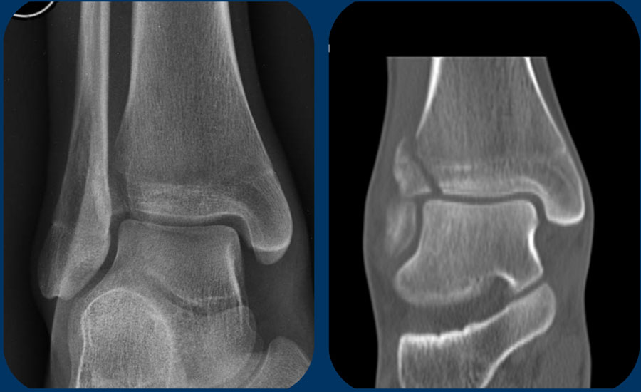
Tillaux fractureGãy Tillaux
The injury mechanism that causes a Weber B fracture can stop at any stage. Most commonly we see stage 2 which is the oblique fibula fracture, but sometimes the injury stops at stage 1.Cơ chế chấn thương gây gãy Weber B có thể dừng ở bất kỳ giai đoạn nào, thường gặp nhất là giai đoạn 2 là gãy xương mác chéo, nhưng đôi khi chấn thương dừng ở giai đoạn 1.
Usually this is a rupture of the anterior syndesmosis and we don't see anything on the x-rays, but the patient will have a lot of pain on this specific anterolateral spot. The tension in the anterior syndesmosis can sometimes lead to an avulsion of the tibial attachment of the anterior syndesmosis, which is called a Tillaux fracture. An avulsion of the fibular attachment is even more rare.Thông thường đây là một vết rách dây chằng chày trước và chúng ta không thấy gì trên phim X-quang, nhưng bệnh nhân sẽ đau nhiều ở vị trí này. Sự căng thẳng ở dây chằng trước đôi khi có thể dẫn đến sự bong ra của dây chằng bám chày của dây chèn chày trước, được gọi là gãy Tillaux. Sự bong ra dây chằng dính chày thậm chí còn hiếm hơn.
Images
On the AP-view and the coronal CT-reconstruction we see a Tillaux fracture as stage 1. No other fracture was seen.Hình ảnh
Trên phim cắt lớp vi tính và chụp cắt lớp vi tinh, chúng tôi thấy gãy Tillaux là giai đoạn 1. Không thấy gãy nào khác.
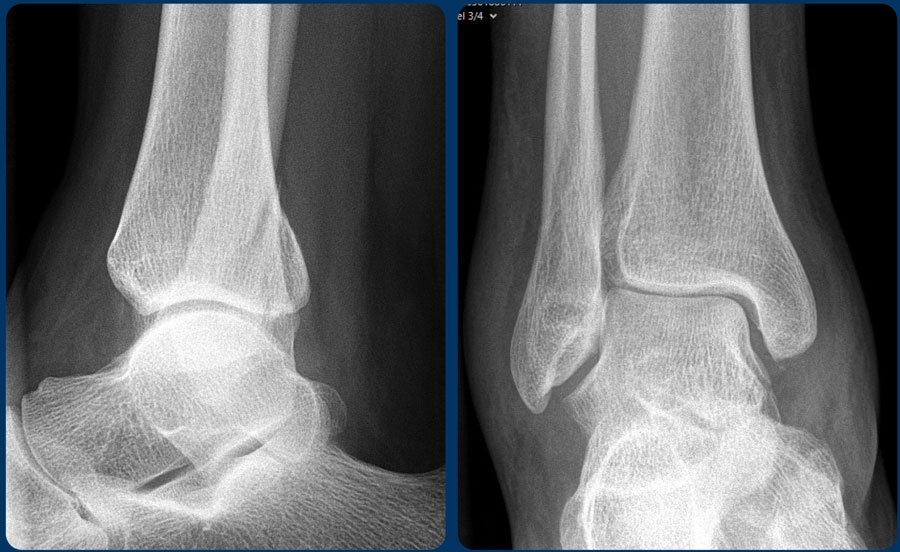
Study these images. You may have to click on them to get a larger view.Nghiên cứu những hình ảnh này. Bạn có thể phải nhấp vào chúng để có được một cái nhìn lớn hơn.
First Question: Is it a Weber A, B or could it be a Weber C? Then try to figure out the stage and determine whether the ankle is stable or unstable.Câu hỏi đầu tiên: Có phải là Weber A, B hay Weber C? Sau đó cố gắng tìm ra giai đoạn và xác định xem cổ chân có ổn định hay không ổn định.
You can look at the next images for a discussion of the images.Bạn có thể xem các hình ảnh tiếp theo để thảo luận về các hình ảnh.
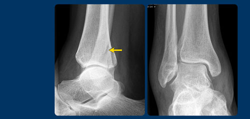
Scroll through the images.Cuộn qua các hình ảnh.
It is a stage 4 Weber B fracture, which is unstable.Đây là gãy Weber B giai đoạn 4, không vững.
Step 3 - Could it be a Weber C / PERBước 3 - Có thể là một Weber C / PER
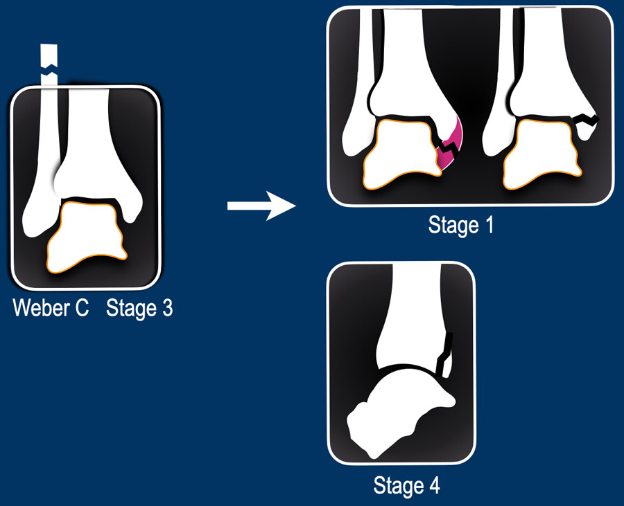
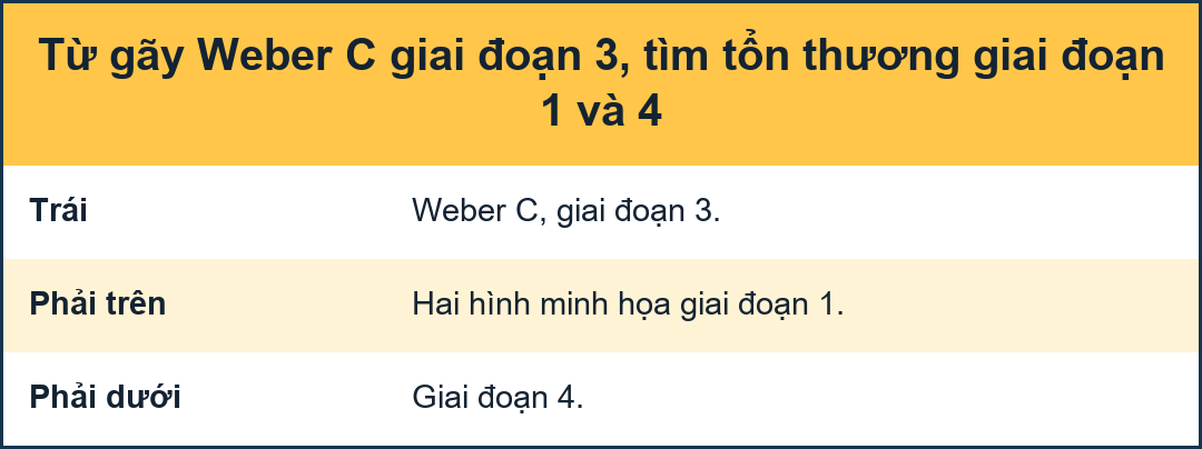
When the x-rays of the ankle show no obvious fracture like a Weber A or B, then the question is: could this be a Weber C fracture?
Since the fibula fracture in a Weber C is most commonly not visible on the x-rays of the ankle, this can be a tough question to answer.
We will have to look for additional findings that lead us to the right answer and that will help us to make the decision to do additional imagesKhi phim X-quang cổ chân không cho thấy gãy rõ ràng như Weber A hay B, thì câu hỏi đặt ra là: đây có thể là gãy Weber C không?
Vì gãy xương mác trong Weber C thường không nhìn thấy được trên phim X-quang cổ chân, đây có thể là một câu hỏi khó trả lời.
Chúng ta sẽ phải tìm kiếm những phát hiện bổ sung dẫn chúng ta đến câu trả lời đúng và điều đó sẽ giúp chúng ta đưa ra quyết định thực hiện các hình ảnh bổ sung
Since the fibula fracture is already stage 3, we wanna look for:
Vì gãy xương mác đã ở giai đoạn 3, chúng tôi muốn tìm:
- stage 1 which is the medial injury, where it all starts. Any pain or soft tissue swelling on the medial side can be a first indication that we are dealing with a Weber C fracture.
Giai đoạn 1 là tổn thương ở giữa, bắt đầu từ đó, đau hoặc sưng mô mềm ở bên trong có thể là dấu hiệu đầu tiên cho thấy gãy Weber C. - stage 2 is injury to the anterior syndesmosis, which is usually not visible, unless there is a Tillaux fracture.
Giai đoạn 2 là tổn thương phần trước khớp chày–mác dưới, thường không nhìn thấy được, trừ khi có gãy Tillaux. - stage 4 is injury to the posterior syndesmosis, which sometimes can not be seen, but will be suspected if there is a widening of the ankle fork or when there is an avulsion of the malleolus tertius like we see in the illustration.Giai đoạn 4 là tổn thương khớp sợi chày–mác sau, đôi khi không thể nhìn thấy, nhưng sẽ bị nghi ngờ nếu có sự mở rộng của mắt cá chân hoặc khi có sự nhổ của mắt cá lớn như chúng ta thấy trong hình minh hoạ.
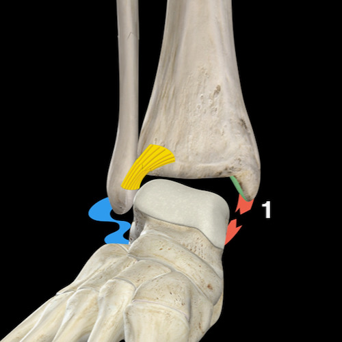
Mechanism of Weber CCơ chế của Weber C
The mechanism of a Weber C is a result of a position of the foot in pronation with an exorotation force applied by the foot and that is why Lauge-Hansen calls it pronation exorotation (PER).Cơ chế của Weber C là kết quả của vị trí của bàn chân sấp với lực đẩy của bàn chân và đó là lý do tại sao Lauge-Hansen gọi nó là sấp, đẩy (PER).
The stages are:
Các giai đoạn là:
- Due to the pronation there is enormous stress on the medial collateral bands and that’s where the injury will start with either a band rupture or an avulsion of the medial malleolus (stage 1).
Do sấp có áp lực rất lớn lên các dải bên trong và đó là nơi chấn thương sẽ bắt đầu với một vết rách dải hoặc một vết rát của mắt cá trong (giai đoạn 1). - As the exorotation force continues the anterior syndesmosis will rupture (stage 2).
Khi lực đẩy ra tiếp tục, phần trước khớp chày–mác dưới sẽ vỡ (giai đoạn 2). - As a result of the pronation of the foot, the bands on the lateral side are all without any tension. As the exorotation of the foot continues the distal fibula follows this exorotation movement, while the proximal fibula is held in position at the proximal tibiofibular joint and you get a twist-like fracture somewhere above the level of the syndesmosis (stage 3).
Do sấp bàn chân, các dải ở bên ngoài đều không có bất kỳ lực căng nào Khi bàn chân tiếp tục đi ra, xương mác đầu xa theo chuyển động đi ra này, trong khi xương mác gần được giữ ở vị trí ở khớp chày mác đầu gần và bạn sẽ bị gãy xương xoắn ở đâu đó trên mức khớp sợi chày–mác (giai đoạn 3). - And finally in stage 4 there will be a rupture of the posterior syndesmosis or tertius avulsion (stage 4). Và cuối cùng ở giai đoạn 4 sẽ có một sự vỡ của khớp sợi chày–mác sau hoặc nhổ tertius (giai đoạn 4).
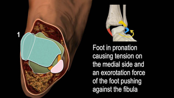
In the axial plane you can see that the injury starts on the medial side due to the pronation of the foot and follows a clockwise rotation through anterior and lateral to posterior.
And just like in a Weber B, you can get injury to the anterior syndesmosis, fibula and finally the posterior syndesmosis.
Trong mặt phẳng ngang, bạn có thể thấy rằng chấn thương bắt đầu ở phía trong do sấp bàn chân và theo chiều kim đồng hồ xoay qua phía trước và bên ra sau.
Và cũng giống như trong Weber B, bạn có thể bị thương ở phần trước khớp chày–mác dưới, xương mác và cuối cùng là khớp sợi chày–mác sau.
The two differences between Weber B and C are:
Hai điểm khác biệt giữa Weber B và C là:
- Weber B starts anterolateral due to the supination of the foot, while Weber C starts on the medial side due to the pronation of the foot.
Weber B bắt đầu ở phía trước bên do ngửa bàn chân, trong khi Weber C bắt đầu ở bên trong do sấp bàn chân. - Exorotation of the foot in Weber B results in a oblique push-off fracture because the fibula is held against the talus due to the supination, while in Weber C the fibula is quite loose resulting in a twist-like high fibula fracture.Sự mở rộng của bàn chân ở Weber B dẫn đến gãy xương chéo vì xương mác được giữ chống lại xương sên do ngửa, trong khi ở Weber C xương mác khá lỏng lẻo dẫn đến gãi xương mác cao giống như xoắn.
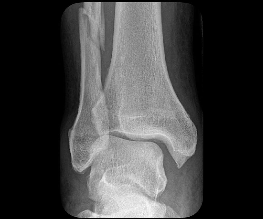
Sometimes we are lucky, because the fibula fracture is visible on the x-rays of the ankle.
Then we know we are looking at an unstable stage 3 weber C fracture.Đôi khi chúng ta may mắn, vì có thể nhìn thấy vết nứt xương mác trên phim X-quang cổ chân.
Vậy chúng ta biết mình đang tìm gãy Weber C giai đoạn 3 không vững.
In this case there is widening of the medial clear space between the medial malleolus and the talus indicating a rupture of the medial collateral bands (stage 1).Trong trường hợp này, có sự mở rộng khoảng trống trong giữa mắt cá trong và xương sên cho thấy sự đứt gãy của các dải bên trong (giai đoạn 1).
We have to assume that there also is a rupture of the anterior syndesmosis (stage 2).Chúng tôi phải giả định rằng cũng có một sự vỡ của phần trước khớp chày–mác dưới (giai đoạn 2).
There also could be a stage 4 injury to the posterior syndesmosis.Cũng có thể có tổn thương giai đoạn 4 ở khớp sợi chày–mác sau.
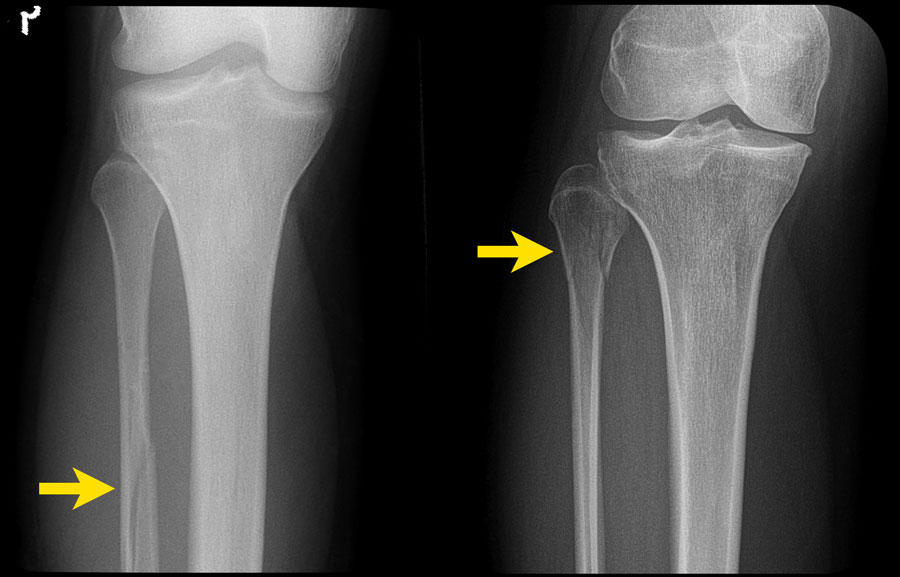
In many cases however, the fibula fracture is higher up and we need additional x-rays to find the fracture.Tuy nhiên, trong nhiều trường hợp, gãy xương mác ở vị trí cao hơn và cần chụp thêm X-quang để tìm ra chỗ gãy.
It is our job as radiologists to find clues on the x-rays of the ankle that will lead us to these high fibula fractures and the algoritm will help us.Công việc của chúng tôi là bác sĩ X-quang để tìm ra manh mối trên phim X-quaNG cổ chân sẽ dẫn chúng ta đến những vết gãy xương mác cao này và thuật toán sẽ giúp chúng ta.
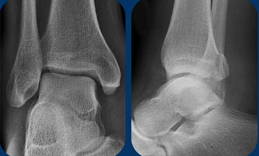 Click on the images to enlargeNhấp vào hình ảnh để phóng to
Study the images.
Use the algoritm and ask yourself if it is a Weber A or B or that it could be a Weber C.
Then determine the stage.Nghiên cứu hình ảnh.
Sử dụng thuật toán và tự hỏi liệu đó là Weber A hay B hay đó có thể là Weber C.
Sau đó xác định giai đoạn.
Then continue for a discussion of this case.Sau đó tiếp tục thảo luận về trường hợp này.
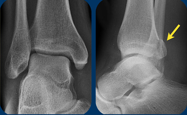
- At first sight it just looks like only a tertius fracture.
Thoạt nhìn nó chỉ giống như một vết gãy xương bánh chè. - When we look at the algoritm, you can see, that a tertius can be found in a Weber B fracture in stage 3 and in a Weber C fracture in stage 4.
Since this patient doesn't have a Weber B, it must be a Weber C fracture. An isolated tertius fracture is very uncommon and probably does not exist.
Khi chúng ta nhìn vào bản số liệu, bạn có thể thấy, có thể tìm thấy một tertius trong gãy Weber B ở giai đoạn 3 và trong gãy weber C ở giai đoạn 4.
Vì bệnh nhân này không có Weber B nên chắc chắn phải là gãy Weber C. Gãy xương đơn độc là rất hiếm gặp và có thể không tồn tại. - Now we recognize the soft tissue swelling on the medial side, which is stage 1.
Bây giờ chúng ta nhận ra sưng mô mềm ở phía trong, đó là giai đoạn 1. - On the enlarge view we also recognize a small avulsion fracture.
Trên hình ảnh phóng to, chúng tôi cũng nhận ra một gãy bong điểm bám nhỏ.
Having detected stage 1 and 4 of a Weber C type of trauma mechanism, we now are sure that there must also be a stage 3, which is a high fibula fracture.Sau khi phát hiện được cơ chế chấn thương Weber C loại 1 và 4, chúng tôi khẳng định chắc chắn có thể có gãy xương mác loại 3, gãy xương hàm lớn.
Charity
Hoạt động từ thiện
All the profits of the Radiology Assistant go to Medical Action Myanmar which is run by Dr. Nini Tun and Dr. Frank Smithuis sr, who is a professor at Oxford university and happens to be the brother of Robin Smithuis.Toàn bộ lợi nhuận của Radiology Assistant được chuyển cho Medical Action Myanmar, do bác sĩ Nini Tun và bác sĩ Frank Smithuis Sr. điều hành. Frank Smithuis Sr. là giáo sư tại Đại học Oxford và là anh em ruột của Robin Smithuis.
Click here to watch the video of Medical Action Myanmar and if you like the Radiology Assistant, please support Medical Action Myanmar with a small gift.Bấm để xem video về Medical Action Myanmar. Nếu thấy Radiology Assistant hữu ích, bạn có thể đóng góp cho Medical Action Myanmar.