
Non-Meniscal pathologyBệnh lý ngoài sụn chêm
Radiology department of the Washington University School of Medicine, St. Louis, USA and the Rijnland hospital in Leiderdorp, the NetherlandsKhoa X quang của Trường Đại học Y Washington, St. Louis, Hoa Kỳ và Bệnh viện Rijnland ở Leiderdorp, Hà Lan
Ngày đăng / cập nhật: 2005-8-2 0
Bản gốc trên radiologyassistant.nl ↗ · tải về lúc 2026-11-19 20:19 · bản dịch máy có chuẩn hóa thuật ngữ chẩn đoán hình ảnh
This article is based on a presentation given by David Rubin and adapted for the Radiology Assistant by Robin Smithuis.
This review focusses on all the non-meniscal pathology of the knee.
See the article entitled Knee MRI - meniscal pathology for the pathology of the meniscus.
Interactive cases are presented in the menubar.
Bài viết này dựa trên một bài thuyết trình của David Rubin và được Robin Smithuis chuyển thể cho Trợ lý X quang.
Bài tổng quan này tập trung vào tất cả các bệnh lý không phải đau của khớp gối.
Xem bài viết có tựa đề MRI khớp gối - bệnh lý sụn chêm để biết bệnh lý của sụn chêm.
Các trường hợp tương tác được trình bày trong thanh thực đơn.
by David Rubin and Robin SmithuisDavid Rubin và Robin Smithuis
LigamentsDây chằng
Anterior Cruciate LigamentDây chằng chéo trước
The ACL has interesting anatomy. It is an intra-articular structure, but it is extra-synovial.
The synovium folds over the ligament. So at arthroscopy they look through the synovium.
Sometimes when there is a tear ,the synovium layer is intact and only a hemorrhagic ACL is seen.
The ACL is composed of 3-5 layers of fibers. Between the fibers there can be fat or synovium or sometimes a little bit of fluid. This explains why the ACL is not black on PD-images.
Do not look at the ACL on PD-images because this may give a false impression of pathology.
Only look at the ACL on T2W-images and even on these images the ACL does not have to be entirely black.
ACL có giải phẫu khá thú vị, là một cấu trúc nội khớp, nhưng nó là ngoài màng hoạt dịch.
Màng hoạt dịch gấp lại trên dây chằng, vì vậy khi nội soi khớp, họ nhìn qua màng hoạt dịch.
Đôi khi có rách, lớp màng hoạt dịch còn nguyên vẹn và chỉ thấy ACL xuất huyết.
ACL bao gồm 3-5 lớp sợi, giữa các sợi có thể có mỡ hoặc màng hoạt dịch hoặc đôi khi có một ít chất lỏng, điều này giải thích tại sao ACL không có màu đen trên ảnh chụp PD.
Không nhìn vào dây chằng chéo trước trên hình ảnh PD vì điều này có thể gây ấn tượng sai về bệnh lý.
Chỉ nhìn vào ACL trên hình ảnh T2W và ngay cả trên những hình ảnh này, ACL không phải là màu đen hoàn toàn.
Criteria for the normal ACL are: Tiêu chuẩn cho ACL bình thường là:
- Fiber-orientation as steep or steeper than the intercondylar roof. Định hướng sợi là dốc hoặc dốc hơn so với mái liên lồi cầu.
- Fibers all the way from the tibia to the femur. Sợi xơ đi từ xương chày đến xương đùi.
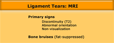
So on MR the primary signs of a tear are: discontinuity on T2, abnormal orientation or non-visualisation.
Many secondary signs of tears have been described, but these are not helpfull, since we have to rely on direct visualisation of the ligament.
Only bone bruises can be a helpfull secondary sign.Vì vậy, trên cộng hưởng từ, các dấu hiệu chính của rách là: mất liên tục trên T2, hướng bất thường hoặc không nhìn thấy được.
Nhiều dấu hiệu thứ phát của rách đã được mô tả, nhưng những dấu hiệu này không hữu ích, vì chúng ta phải dựa vào việc nhìn trực tiếp dây chằng.
Chỉ có các vết bầm ở xương có thể là một dấu hiệu thứ phát hữu ích.
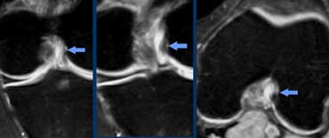
Notice that on coronal and axial images fibers of the ACL are right next to the bone of the intercondylar notch (arrows).
There should never be any fluid between these ACL-fibers and the bone of the lateral condyle ('empty notch sign').
Also notice that the PCL is also composed of many fibers.Trên ảnh mặt phẳng đứng ngang và ngang, các bó sợi dây chằng chéo trước (ACL) nằm sát xương ở khuyết gian lồi cầu (mũi tên). Bình thường không có dịch giữa các sợi ACL ở vị trí này và xương lồi cầu ngoài. Khoảng dịch tại đây tạo dấu hiệu khuyết gian lồi cầu trống. Lưu ý dây chằng chéo sau cũng gồm nhiều bó sợi.
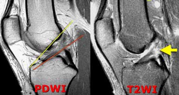 LEFT: Acute ACL-tear. ACL fibers too flat compared to condylar roof.RIGHT: Discontinuity of fibers.Bên trái: ACL-Tear cấp tính. Sợi ACL quá phẳng so với mái Condylar. Phải: Sự gián đoạn của sợi.
Anterior Cruciate Ligament (2).
The case on the left shows a ligament that's too flat and we see disrupted fibers so there is abnormal orientation and discontinuity.
Based on these images we cannot differentiate between complete tear, high grade partial tear or partial tear.
MRI does not accurately differentiate between partial or complete ACL tear.
But yes we can differentiate between high grade or low grade injury.
A high grade injury is 'not able to see 50% of the fibers'.
So if the othopaedic surgeons operate on a high grade injury, they will either find a totally torn ACL or a high grade partial tear, that needs to be repaired.
On the other hand if most of the fibers appear to be intact on MR indicating a low grade ACL tear, they will find an intact or partially torn ACL, that is stable and doesn't need any treatment.Dây chằng chéo trước (2).
Trường hợp bên trái cho thấy dây chằng quá phẳng và chúng ta thấy các sợi bị đứt đoạn nên có sự định hướng bất thường và gián đoạn.
Trên các hình ảnh này chúng tôi không thể phân biệt được rách hoàn toàn, rách một phần độ cao hay rách một Phần.
MRI không phân biệt chính xác giữa đứt dây chằng chéo trước một phần hoặc hoàn toàn.
Nhưng có thể phân biệt được giữa mức độ nặng và nhẹ.
Một chấn thương mức độ cao là 'không thể nhìn thấy 50% số sợi'.
Vì vậy, nếu phẫu thuật viên chỉnh hình phẫu thuật trên một chấn thương mức độ cao, họ sẽ tìm thấy một dây chằng chéo trước bị rách hoàn toàn hoặc rách một phần mức độ cao cần phải được sửa chữa.
Mặt khác, nếu hầu hết các sợi có vẻ còn nguyên vẹn trên MR cho thấy rách ACL mức độ thấp, họ sẽ tìm thấy một ACL còn nguyên vẹn hoặc bị rách một phần, ổn định và không cần điều trị.
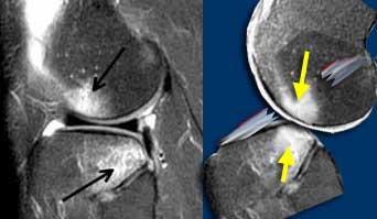 LEFT: ACL-tear with bone bruises on lateral side. RIGHT: Anterior condyle hits the posterior part of the tibia. ACL torn due to stretching.TRÁI: Đoạn chỏm xương đùi bên ngoài có bầm tím xương bên phải: lồi cầu trước chạm vào phần sau xương chày, đứt dây chằng do kéo giãn.
Anterior Cruciate Ligament (3).
Bone bruises appear in a very typical location indicating the dislocation, that was the cause of the ACL-tear.
Dây chằng chéo trước (3).
Các vết bầm tím xuất hiện ở một vị trí rất điển hình cho thấy trật khớp, đó là nguyên nhân gây ra rách dây chằng chéo trước.
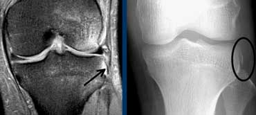 Segond fracture indicating ACL-tearGãy Segond cho thấy rách dây chằng chéo trước
Anterior Cruciate Ligament (4)
On X-rays an important indirect sign of an ACL-tear is a Segond fracture.
Difficult to see on MR, but much more easy to see on radiographs.
A Segond fracture is an avulsion fracture at the attachment of the lateral collateral band due to internal rotation and varus stress.
In 75-100% there will also be a tear of the ACL.
Dây chằng chéo trước (4)
Trên X-quang, dấu hiệu gián tiếp quan trọng của rách dây chằng chéo trước là gãy Segond.
Khó nhìn thấy trên cộng hưởng từ, nhưng dễ nhìn thấy hơn nhiều trên phim X quang.
Gãy Segond là gãy gãy bong điểm bám ở vị trí bám của đai bên do xoay trong và ứng suất giãn tĩnh mạch chủ.
Trong 75-100% trường hợp cũng sẽ có rách dây chằng chéo trước.
The unhappy triad or O'Donoghues syndrome is a different combination of injuries.
The unhappy triad injury commonly occurs in contact sports such as football when the knee is hit from the outside.
This causes an injury to three knee structures:Hội chứng bộ ba không hạnh phúc hoặc hội chứng O 'Donoghues là một sự kết hợp khác nhau của các thương tích.
Tổn thương bộ ba không hài lòng thường xảy ra trong các môn thể thao va chạm như bóng đá khi đầu gối bị đánh từ bên ngoài.
Điều này gây ra tổn thương cho ba cấu trúc khớp gối:
- ACL tearRách dây chằng chéo trước
- MCL tear (medial collateral ligament)Rách MCL (dây chằng bên trong)
- Medial meniscal tearRách sụn chêm trong
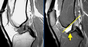 Torn ACL. ACL fibers are too flat (yellow arrow) compared to intercondylar roof (Blumensaat's line).Mỏng ACL. Sợi ACL quá phẳng (mũi tên màu vàng) so với mái liên lồi cầu (đường Blumensaat).
Anterior Cruciate Ligament (5)
Case on the left shows a torn ACL.
Fibers have an abnormal orientation (too flat).
Yet it is difficult to see if these are attached to the femur.
The acute angulation in the ligament is due to fact that the ACL and PCL have scarred together (see below).Dây chằng chéo trước (5)
Trường hợp bên trái cho thấy một dây chằng chéo trước bị rách.
Sợi có hướng bất thường (quá phẳng).
Tuy nhiên, rất khó để thấy nếu chúng được gắn vào xương đùi.
Góc cấp tính trong dây chằng là do thực tế là ACL và PCL có sẹo với nhau (xem bên dưới).
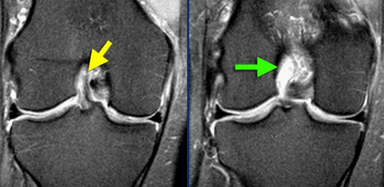 LEFT: ACL-fibers have a normal orientation but do not connect to femurcondyle. RIGHT: Empty notch sign: fluid against the interior part of the lateral condyleTrái: sợi ACL có hướng bình thường nhưng không kết nối với femurcondyle. Bên phải: Dấu hiệu Notch trống: Chất lỏng chống lại phần bên trong của ống dẫn bên
Sometimes it is easier to see whether these fibers are attached in the coronal plane.
Against the interior part of the lateral condyle there never should be fluid.
If this is the case it is called the 'empty notch sign' indicating that the ACL is torn from it's attachment to the femur.Đôi khi dễ dàng hơn để xem liệu các sợi này có được gắn vào trong mặt phẳng đứng ngang hay không.
Chống lại phần bên trong của lồi cầu ngoài không bao giờ nên có dịch.
Nếu đây là trường hợp, nó được gọi là 'dấu hiệu notch rỗng' cho thấy dây chằng chéo trước bị rách khỏi phần bám vào xương đùi.
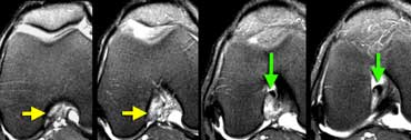 In the axial plane there is an empty notch sign (yellow arrows) where there should be ACL attached to the condyle. At a lower level (going right) the torn ACL is scarred to the PCL (green arrows)Trong mặt phẳng trục có một dấu hiệu notch trống (mũi tên màu vàng) nơi cần có ACL gắn liền với lồi cầu.Ở một mức độ thấp hơn (đi bên phải) ACL bị rách được sẹo để PCL (mũI tên màu xanh lá cây)
Also in the axial plane there should be ligament next to the condyle.
At a lower level we see the torn ACL attached to the posterior cruciate ligament.
They have scarred together.
This is a very common appearance of a chronic ACL tear.
This scarring leads to the acute angulation of the ligament.
Even though the ACL is connected to the PCL it is not strong enough and still needs reconstruction.Cũng trong mặt phẳng ngang nên có dây chằng bên cạnh lồi cầu.
Ở mức thấp hơn, chúng ta thấy ACL bị rách gắn với dây chằng chéo sau.
Chúng có sẹo cùng nhau.
Đây là một biểu hiện rất phổ biến của rách dây chằng chéo trước mạn tính.
Sẹo này dẫn đến sự gấp góc cấp tính của dây chằng.
Mặc dù dây chằng chéo trước được nối với dây chằng dọc chậu trước nhưng nó không đủ mạnh và vẫn cần được tái tạo.
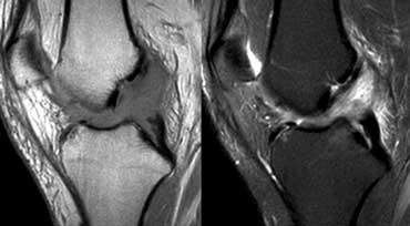 ACL Mucoid degeneration. This patient had an operation for another reason and the surgeons called it normal because it looks normal.Thóai hóa nhầy dây chằng chéo trước Bệnh nhân này đã phẫu thuật vì một lý do khác và các bác sĩ phẫu thuật gọi nó là bình thường vì nó trông bình thường.
Anterior Cruciate Ligament (6)
Case on the left shows a non-visualisation of the ACL on a PD-image. But the lesson is 'do not look at ligaments on a PD-image'.
If you want to judge the ACL-ligament look at the T2W-images.
The T2W-images show fibers going all the way from the tibia to the femur with a normal orientation. So the ACL is intact.
This is a case of mucoid degeneration. Normally between the ACL-fibers there can be synovium or fat.
In normal aging that can change into gelatinous material.
This has no effect on the strenght of the ACL.
Dây chằng chéo trước (6)
Trường hợp bên trái cho thấy ACL không nhìn thấy trên ảnh chụp cắt lớp điện toán, nhưng bài học là không nhìn vào dây chằng trên ảnh chụp PD.
Nếu bạn muốn đánh giá dây chằng ACL, hãy nhìn vào hình ảnh T2W.
Hình ảnh T2W cho thấy các sợi đi từ xương chày đến xương đùi với hướng bình thường, do đó, ACL vẫn còn nguyên vẹn.
Đây là một trường hợp thóai hóa niêm mạc, thường giữa các sợi ACL có thể có màng hoạt dịch hoặc mỡ.
Trong quá trình lão hóa bình thường có thể biến đổi thành dạng sệt.
Điều này không ảnh hưởng đến sức mạnh của dây chằng chéo trước.
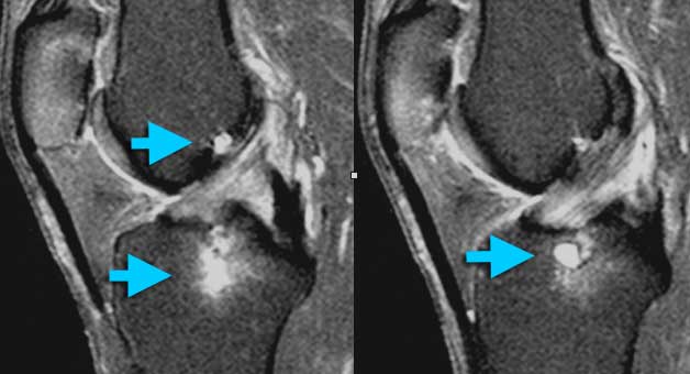 ACL Mucoid degeneration with cyst-formation (intra-osseus ganglion). Mucoid material is squeezed from between the ACL-fibers into the bone. At arthroscopy ACL and bone looked normal.ACL Thóai hóa niêm mạc với hình thành nang (hạt trong xương) Vật liệu niêm mạc được ép từ giữa các sợi ACL vào xương. Khi nội soi khớp ACL và xương có vẻ bình thường.
Anterior Cruciate Ligament (7)
Another case of ACL Mucoid degeneration.
Often this is associated with cyst-formation in the bone.
You could call it ganglion cyst, but you could also call it normal because it has no clinical meaning.
This is part of normal aging.Dây chằng chéo trước (7)
Một trường hợp thóai hóa dạng nhầy của dây chằng chéo trước.
Thông thường điều này có liên quan đến sự hình thành nang trong xương.
Bạn có thể gọi nó là u nang hạch, nhưng bạn cũng có thể gọi là bình thường vì nó không có ý nghĩa lâm sàng.
Đây là một phần của quá trình lão hóa bình thường.
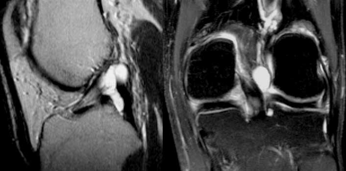 ACL ganglion cystU nang hạch ACL
Anterior Cruciate Ligament (8)
Case on the left shows cyst seperate from ACL unlike mucoid degenaration.
This is a ganglion cyst. Probably also a form of degeneration.
The difference with Mucoid degeneration is that these cysts can be symptomatic.
Sometimes these cysts will drained under ultrasound guidance.
Be sure to use a very large needle, because it is very thick material.Dây chằng chéo trước (8)
Trường hợp bên trái cho thấy nang tách ra từ ACL không giống như sự thóai hóa của niêm mạc.
Đây là một nang hạch, có thể là một dạng thóai hóa.
Sự khác biệt với thóai hóa Mucoid là những nang này có thể có triệu chứng.
Đôi khi những nang này sẽ được dẫn lưu dưới hướng dẫn siêu âm.
Đảm bảo sử dụng kim rất lớn, vì nó là vật liệu rất dày.
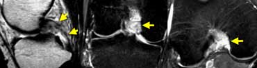 Acute PCL-tear: In the coronal and axial plane most of the fibers are gone so it's a high grade tear.Xơ tuỷ cấp tính: Ở đoạn vành và mặt phẳng ngang hầu hết các sợi đều biến mất nên là một vết rách cấp tính.
Posterior Cruciate LigamentDây chằng chéo sau
We use the same criteria for all the other ligaments in the body.
The case on the left shows a high grade PCL tear.Chúng tôi sử dụng cùng tiêu chuẩn cho tất cả các dây chằng khác trong cơ thể.
Trường hợp bên trái cho thấy rách PCL độ cao.
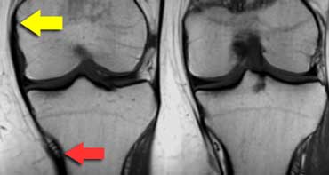 MCL from medial epicondyle (yellow arrow) to below the inferomedial geniculate vessels (red arrow) about 7 centimetres below the joint space.MCL từ lồi cầu trong (mũi tên màu vàng) đến dưới các mạch máu trong (mũi tên màu đỏ) khoảng 7 cm dưới khe khớp.
Medial collateral ligamentDây chằng bên trong
The superficial medial collateral ligament (MCL) extends from the medial epicondyle to insert not just near the joint but 7 cm below the joint space.
At that point there are three landmarks: the inferomedial geniculate artery and paired veins (figure).
The deep part of the MCL, even when it is normal, you may not be able to see.
It is closely applied to the medial meniscus and the superficial MCL.
Dây chằng bên trong nông (MCL) kéo dài từ lồi cầu trong để chèn vào không chỉ gần khớp mà còn cách khoảng khớp 7 cm.
Tại thời điểm đó có ba mốc: động mạch sinh dục trong và các tĩnh mạch ghép đôi (hình).
Phần sâu của MCL, ngay cả khi nó bình thường, bạn có thể không nhìn thấy.
Nó được áp dụng chặt chẽ cho sụn chêm trong và MCL bề mặt.
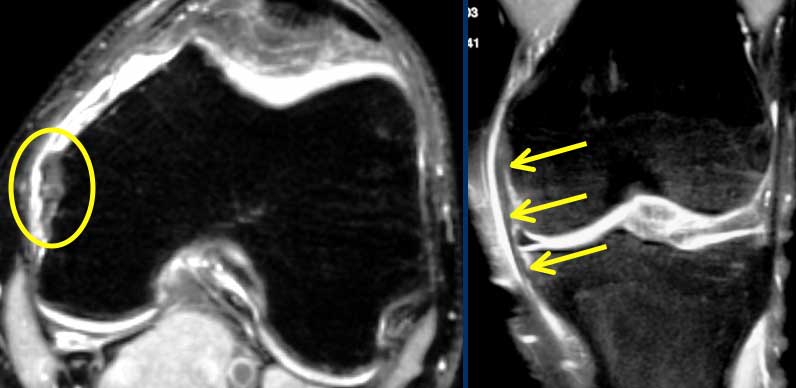 Grade 1 MCL SprainMCL Sprain độ 1
Medial collateral ligament (2)
The case on the left shows a Grade I sprain of the medial collateral ligament.Dây chằng bên trong (2)
Trường hợp bên trái cho thấy đứt dây chằng bên trong mức độ I.
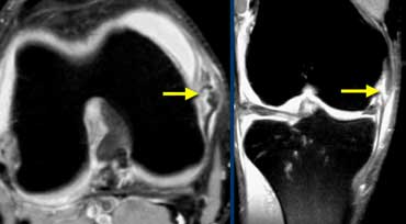 Grade II MCL SprainMCL độ II
Medial collateral ligament (3)
The case on the left shows a Grade II sprain of the medial collateral ligament.Dây chằng bên trong (3)
Trường hợp bên trái cho thấy đứt dây chằng bên trong cấp độ II.
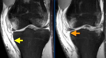 Superficial MCL-tear (yellow arrow) and deep MCL tear (orange arrow).MCL-tear nông (mũi tên màu vàng) và MCL-tear sâu (mũa tên màu cam).
Medial collateral ligament (4)
The case on the left shows a superficial MCL that is torn from it's attachment on the tibia.
Remember it should be attached 7 cm below the joint line.
Deep MCL is also torn the ligament is absent.Dây chằng bên trong (4)
Trường hợp bên trái cho thấy một MCL nông bị rách ra khỏi phần đính kèm trên xương chày.
Nhớ rằng nó phải được gắn bên dưới đường khớp 7 cm.
MCL sâu cũng bị rách dây chằng không có.
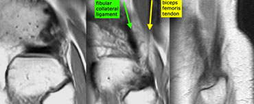
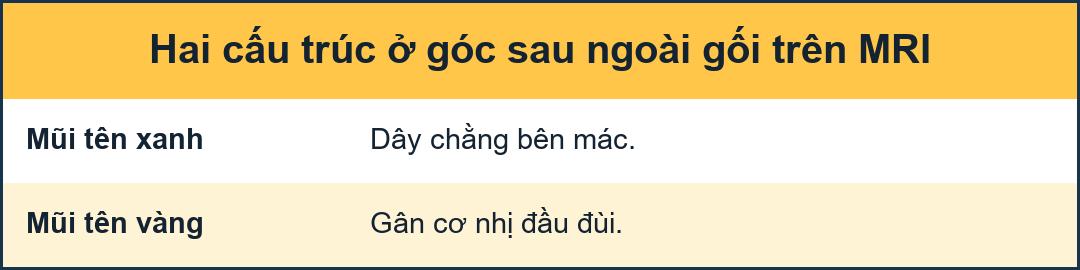 Posterolateral corner anatomy from medial to lateral on sagittal images. Fibular collateral ligament (green) and Biceps femoris tendon (yellow) form a letter V and insert as conjoined tendon on fibular head.Giải phẫu góc sau ngoài từ trong ra ngoài trên ảnh mặt phẳng đứng dọc Dây chằng bên mác (màu xanh lá) và gân cơ nhị đầu đùi (mào vàng) tạo thành chữ V và chèn vào như gân dính trên đầu mác.
Posterolateral Corner injuryChấn thương góc sau bên
Normal anatomy
Posterolateral corner contains seven or eight structures.
Only three of them are important to us because they are visible on MR and because the surgeon might want to fix them.
These structures are:
1. Fibular collateral ligament
2. Biceps femoris muscle and tendon.
3. Popliteal tendon
The fibular collateral ligament together with the tendon of the biceps femoris form the letter V on sagittal images.
They inserts on the fibulahead as the conjoined tendon.Giải phẫu bình thường
Góc sau bên chứa bảy hoặc tám cấu trúc.
Chỉ có ba trong số chúng quan trọng đối với chúng tôi vì chúng hiển thị trên MR và vì bác sĩ phẫu thuật có thể muốn sửa chúng.
Các cấu trúc này là:
1. Dây chằng phụ
2. Cơ bắp và gân cơ bắp tay.
3. Gân popliteal
Dây chằng phụ cùng với gân của xương đùi xương đùi tạo thành chữ V trên ảnh mặt phẳng đứng dọc.
Chúng chèn vào fibulahead dưới dạng gân dính liền.
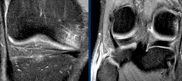 LEFT: bone bruise anteromedial. RIGHT: biceps femoris tendon is not attached to the fibula.Bên trái: Xương trước vết bầm tím. Phải: Gân xương đùi không gắn vào xương mác.
Posterolateral corner injury (2)
On the left a football player, who was hit in the front part of the knee.
The image on the far left shows a bone bruise anteromedially.
So you suspect ligamentous injury on the contralateral side, which is the posterolateral corner.
The next image shows a normal popliteus tendon but biceps femoris tendon is not attached to the fibula.Chấn thương góc sau bên (2)
Bên trái là một cầu thủ bóng đá, bị đánh vào phần trước của đầu gối.
Hình ảnh ở xa bên trái cho thấy một vết bầm xương ở phía trước trong.
Vì vậy, bạn nghi ngờ tổn thương dây chằng ở phía đối diện, đó là góc sau bên.
Hình ảnh tiếp theo cho thấy gân khoeo bình thường nhưng gân cơ nhị đầu đùi không gắn với xương mác.
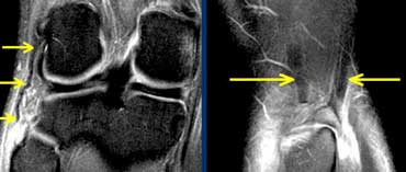 LEFT: distal rupture of fibular collateral ligament.RIGHT: biceps femoris tendon and collateral ligament do not attach.TRÁI: đứt dây chằng bên mác ngoài, bên phải: gân cơ nhị đầu đùi và dây chằng không dính.
On the left more images of the same patient located more anteriorly.
The fibular collateral ligament has a normal proximal attachment but is not attached to the fibula.
On a sagittal plane there is a gap between biceps femoris tendon and collateral ligament on one side and the fibular head on the other.
These findings indicate a conjoined tendon rupture.Bên trái có thêm hình ảnh của cùng một bệnh nhân nằm ở phía trước.
Dây chằng bên mác có đầu gần bình thường nhưng không gắn vào mác.
Trên mặt phẳng đứng dọc có khoảng cách giữa gân cơ nhị đầu đùi và dây chằng bên ngoài ở một bên và chỏm xương mác ở bên kia.
Những dấu hiệu này cho thấy đứt gân dính.
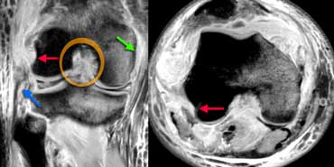 LEFT: cruciate lig.rupture (orange), med coll lig rupture (green), proximal rupture of the fibular collateral ligament (blue).RIGHT: no attachment of popliteus tendon (red arrows)Trái: cruciate lig.rupture (cam), med coll lig vỡ (xanh lá cây), gần như vỡ của sợi thủy tinh màu xanh lá cây (xanh dương). PHẢI: không có đính kèm của dây chằng poplitus (đỏ các mũi tên)
Posterolateral corner injury (3)
On the left PD-fatsat images after severe injury.
There are bone bruises and many ligaments are ruptured.
There is a posterolateral corner injury with proximal rupture of the fibular collateral ligament.
There is also a rupture of the popliteus tendon because it is not attached proximally.Chấn thương góc sau bên (3)
Trên ảnh PD-fatsat trái sau chấn thương nặng.
Có những vết bầm tím ở xương và nhiều dây chằng bị đứt.
Có một chấn thương góc sau bên với đứt đoạn gần dây chằng bên mác.
Cũng có một vết rách của gân khoeo vì nó không được gắn vào đầu gần.
Cysts, Bursae and RecessesU nang, bao hoạt dịch và lõm
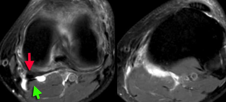 LEFT: popliteal cyst originating between semimembranosus tendon (red arrow) and gastrocnemius muscle and tendon (green arrow).RIGHT: ruptured popliteal cyst with fluid tracking down the muscles.Trái: Nang khoeo xuất phát giữa gân cơ bán màng (mũi tên đỏ) và cơ–gân cơ bụng chân (mũi tên xanh). Phải: Nang khoeo vỡ, dịch lan xuống dọc theo các lớp cơ.
There are about 12 named bursae and recesses in the knee.
Some very common and others uncommon.
These are synovial lined structures.
The most common recess is the popliteal or Baker's cyst.
The origin is between the semimembranosus and gastrocnemius tendon. Có khoảng 12 túi và hốc được đặt tên trong khớp gối.
Một số rất phổ biến và một số khác không phổ biến.
Đây là những cấu trúc có lớp hoạt dịch.
Ngõ vào phổ biến nhất là khoeo hoặc nang Baker.
Nguồn gốc là giữa gân cơ bán gân và gân cơ cẳng chân.
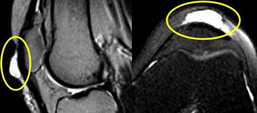 Prepatellar bursitisViêm bao hoạt dịch trước xương bánh chè
On the left the typical imaging findings of prepatellar bursitis.Bên trái là các dấu hiệu hình ảnh điển hình của viêm bao hoạt dịch trước xương bánh chè.
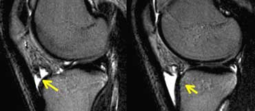 Deep infrapatellar bursitisViêm bao hoạt dịch dưới xương bánh chè
An uncommon form of bursitis is the deep infrapatellar bursitis.
Sometimes associated with Osgood-Schlatter.Một dạng viêm bao hoạt dịch không phổ biến là viêm bao hoạt động dưới xương bánh chè.
Đôi khi liên quan đến Osgood-Schlatter.
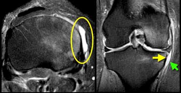 Medial collateral ligament bursitis located between the deep MCL (yellow arrow) and the superficial MCL (green arrow).Viêm bao thanh dịch dây chằng bên trong nằm giữa MCL sâu (mũi tên màu vàng) và MCL nông (mũI tên màu xanh lá cây).
These bursae are all named by the structures next to them.
So a bursitis of the bursa between the deep MCL and the superficial MCL is called a medial collateral ligament bursitis.Các túi này đều được đặt tên theo các cấu trúc bên cạnh chúng.
Vì vậy, viêm bao hoạt dịch của bao hoạt dịch giữa MCL sâu và MCL nông được gọi là viêm bao hoạt động dây chằng bên trong.
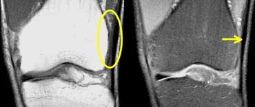 Iliotibial Band Friction syndrome: no fat between iliotibial band (yellow arrow) and the lateral condyle.Hội chứng ma sát dải chậu: không có mỡ giữa dải chùi (mũi tên màu vàng) và lồi cầu ngoài.
Cysts, Bursae and Recesses (2)
Adventitial bursae are bursae, that are formed in places where normally there is no bursa>
The bursa is formed due to abnormal friction.
U nang, bao hoạt dịch và lõm (2)
Bursae sinh dục là các túi thanh dịch, được hình thành ở những nơi mà bình thường không có túi thanh dịch>
Túi thanh dịch được hình thành do ma sát bất thường.
Iliotibial Band Friction syndrome
A common place for abnormal friction is between the iliotibial band and the lateral condyle in speedwalkers, bicyclists and sometimes runners.
When a bursa is formed in this location it is called the 'Iliotibial Band Friction syndrome'.
On the left a speedwalker with lateral knee pain.
Between iliotibial band and the lateral condyle there should be fat, but in this case it is missing.Hội chứng ma sát dải Iliotibial
Một vị trí phổ biến cho ma sát bất thường là giữa dải chậu và lồi cầu ngoài ở người chạy bộ nhanh, người đi xe đạp và đôi khi là người chạy bộ.
Khi một túi thanh dịch được hình thành ở vị trí này, nó được gọi là 'hội chứng ma sát dải i-li-ib'.
Bên trái, một người chạy bộ với đau đầu gối bên.
Giữa dải chậu-miệng và lồi cầu ngoài nên có mỡ, nhưng trong trường hợp này nó bị thiếu.
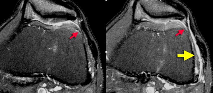 Iliotibial Band Friction syndrome : Fluid within a bursa is seen between the iliotibial tract and the femur (yellow arrows). Joint fluid stops at the red arrows.Hội chứng ma sát dải chậu chậu: Dịch trong bao hoạt dịch được nhìn thấy giữa đường chậu chỏm và xương đùi (mũi tên màu vàng). Dịch khớp dừng lại ở các mũi tên màu đỏ.
Same patient. On axial images fluid within a bursa is seen between the iliotibial tract and the underlying femur.
Sometimes fluid in this location has to be differentiated from joint fluid.
You have to look at all the images.
In this case the joint fluid stops at the red arrows. Cùng một bệnh nhân Trên ảnh mặt phẳng ngang dịch trong bao hoạt dịch được nhìn thấy giữa đường chậu và xương đùi bên dưới.
Đôi khi dịch ở vị trí này phải được phân biệt với dịch khớp.
Bạn phải nhìn vào tất cả các hình ảnh.
Trong trường hợp này, dịch khớp dừng lại ở các mũi tên màu đỏ.
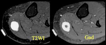 Patient with a palpable mass. Marker is placed. Enhancing lesion due to a neurofibroma.Bệnh nhân có khối u sờ thấy được, đánh dấu được đặt tổn thương ngấm thuốc do u sợi thần kinh.
Remember that not everything that's bright on a T2W-image is fluid.
You have to be suspicious, if there is something, that looks like a fluid collection, but it is not in a location, where there normally is a bursa, cyst or recess.
Give Gadolineum to differentiate cystic from solid.Hãy nhớ rằng không phải mọi thứ sáng trên hình ảnh T2W đều là chất lỏng.
Bạn phải nghi ngờ, nếu có gì đó, trông giống như một khối dịch, nhưng nó không ở một vị trí, nơi thường có một bao hoạt dịch, nang hoặc hốc.
Cho Gadolineum để phân biệt nang với đặc.
Quadriceps and Patellar tendonGân cơ tứ đầu đùi và gân bánh chè
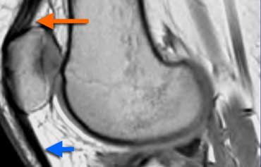 Normal Extensor mechanism: The quadriceps tendon comes in three layers (orange arrow). Patellar tendon (blue arrow) and quadriceps tendon have a sharp posterior demarcation.Cơ chế kéo dài bình thường: Gân cơ tứ đầu có ba lớp (mũi tên màu cam). Gân xương bánh chè (mũi tên màu xanh) và gân cơ tứ đầu có đường phân cách sắc nét phía sau.
Normal Extensor mechanism
The extensor mechanism of the knee is composed of the quadriceps muscle and tendon, the patella and the patellar tendon.
The quadriceps tendon is made of four tendons but comes in three layers on sagittal images.
It has a broad attachment all the way from the front of the patella almost to the back.
Cơ chế mở rộng bình thường
Cơ chế duỗi của khớp gối bao gồm cơ tứ đầu đùi và gân, xương bánh chè và gân bánh chè.
Gân cơ tứ đầu đùi được cấu tạo từ bốn gân nhưng có ba lớp trên hình ảnh dọc.
Nó có một phần bám rộng từ mặt trước xương bánh chè gần như đến mặt sau.
The tendons of the quadriceps aswell as the patellar tendon are homogeneous in signal but don't have to be black on PD-images.
They have a sharp posterior demarcation.
There should be no focal thickness.Các gân cơ tứ đầu đùi cũng như gân bánh chè đều đồng nhất về tín hiệu nhưng không nhất thiết phải có màu đen trên ảnh chụp cắt lớp điện toán.
Chúng có ranh giới sau rõ ràng.
Không nên có độ dày khu trú.
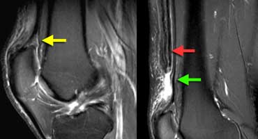 Partial quadriceps tendon tear: T2W-images.LEFT: Abnormal attachment of tendon.RIGHT: Most of tendon is retracted (red arrow) deep part (vastus intermedius) is still intact.Rách một phần gân cơ tứ đầu đùi trên T2W. Trái: Điểm bám gân bất thường. Phải: Phần lớn gân co rút (mũi tên đỏ), nhưng lớp sâu của gân cơ rộng trung gian còn nguyên.
Quadriceps tendon tear
The case on the left shows an abnormal quadriceps attachment.
There is only one layer and the attachment does not go from the front of the patella to nearly the back.
In such a case extra images higher up have to be made after repositioning of the coil to see what's going on up there.
The missing part of the torn quadriceps tendon is retracted.
The deep part is still intact Rách gân cơ tứ đầu đùi
Trường hợp bên trái cho thấy một sự gắn kết bất thường của cơ tứ đầu.
Chỉ có một lớp và phần bám dính không đi từ mặt trước xương bánh chè đến gần mặt sau.
Trong trường hợp như vậy, phải chụp thêm hình ảnh ở phía trên sau khi định vị lại cuộn dây để xem điều gì đang xảy ra ở đó.
Phần bị mất của gân cơ tứ đầu đùi bị rút lại.
Phần sâu vẫn còn nguyên vẹn
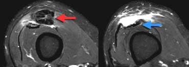 LEFT: Torn tendon with pre-existing tendinopathy (red arrow). RIGHT: Intact vastus intermedius tendon.Trái: Gân rách trên nền bệnh lý gân từ trước (mũi tên đỏ). Phải: Gân cơ rộng trung gian còn nguyên.
Same patient, axial images.
The torn quadriceps tendon is very thick indicating tendinopathy.
Normal tendons do not tear, so always look for signs of pre-existing tendinopathy.
Anywhere in the body, if you see a tendon that looks torn, but there is no pre-existing tendinopathy, think hard, if you really have the right diagnosis.
An image below this level shows normal vastus intermedius muscle and tendon.Ảnh mặt phẳng ngang của cùng bệnh nhân. Gân cơ tứ đầu đùi bị rách và rất dày, gợi bệnh lý gân. Theo bài gốc, luôn tìm dấu hiệu bệnh lý gân có từ trước khi thấy gân rách. Nếu hình ảnh gợi rách nhưng không có dấu hiệu bệnh lý gân trước đó, cần xem xét lại chẩn đoán. Ảnh dưới mức này cho thấy cơ và gân cơ rộng trung gian bình thường.
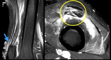 LEFT: Partial quadriceps tear. Only rectus femoris tendon is torn (blue arrow). RIGHT: Pre-existing tendinopathy (yellow cirkle) on axial image.TRÁI: Rách một phần cơ tứ đầu đùi, chỉ rách gân cơ thẳng đùi (mũi tên xanh), bên phải: Có tiền sử bệnh lý gân (xơ gan vàng) trên hình ảnh trục.
Another example of a partially torn quadriceps tendon.Một ví dụ khác về rách gân cơ tứ đầu đùi.
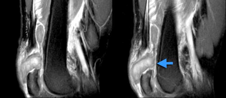 Complete quadriceps tear. Sag T2W-images. No continuity. Hematoma in between.Rách tứ đầu đùi hoàn toàn, Sag T2W, không liên tục, có máu tụ ở giữa.
If there is no continuity between the patella and the quadriceps tendons it is a complete tear.Nếu không có sự liên tục giữa xương bánh chè và gân cơ tứ đầu đùi thì đó là một vết rách hoàn toàn.
Jumper's Knee
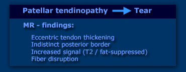
Jumper's knee is a spectrum from tendinopathy to tear.
Just the same as with the quadriceps tendon or any other tendon the MR shows a spectrum from eccentric tendon thickening, indistinct posterior border, increased signal on T2W-images and finally fiber disruption.Đầu gối của Jumper
Đầu gối là một phổ từ bệnh lý gân đến rách.
Cũng giống như với gân cơ tứ đầu đùi hoặc bất kỳ gân nào khác, cộng hưởng từ cho thấy một phổ từ dày gân lệch tâm, bờ sau không rõ, tín hiệu tăng lên trên hình ảnh T2W và cuối cùng là đứt sợi.
 Patellar tendinopathy and Partial patellar tendon tearBệnh lý gân bánh chè và rách gân bánh răng một phần
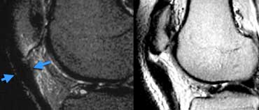
Patellar tendinopathy
Patient on the left is a professional ballet dancer with pain underneath the knee cap.
Patellar tendon proximally is too thick. Posterior border is indistinct. In patella a little bit of edema ( or bone bruise). If left untreated could end up like...
Partial patellar tendon tear. Image on the right of a different patient.Bệnh lý gân bánh chè
Bệnh nhân bên trái là một vũ công ballet chuyên nghiệp bị đau dưới mũ đầu gối.
Gân bánh chè ở đầu gần quá dày, bờ sau không rõ, ở xương bánh chè có phù nề (hoặc bầm xương) nhẹ, nếu không điều trị có thể kết cục như sau:
Rách gân bánh chè một phần Hình ảnh bên phải của một bệnh nhân khác.
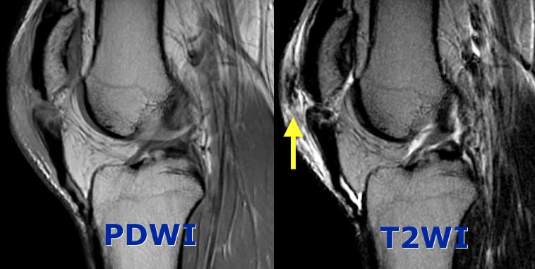 Complete Patellar tendon tear. Image on the right shows hemorrhagic bursitis ( low signal in bursa).Rách gân bánh chè hoàn toàn Hình ảnh bên phải cho thấy viêm bao hoạt dịch xuất huyết (tín hiệu thấp trong bao hoạt dịch).
Complete Patellar tendon tear. Images on the left show no continuity between fibers and patella. The tendon is thickened.Rách gân bánh chè hoàn toàn Hình ảnh bên trái cho thấy không có sự liên tục giữa các sợi và xương bánh chè Gân dày lên.
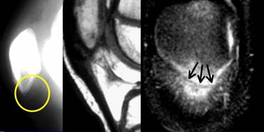 Patellar sleeve avulsion. Fractureline only visible on the coronal images.Tay áo bánh chè bị bong, chỉ thấy đường gãy trên ảnh mặt phẳng đứng ngang.
Patellar sleeve avulsion.
In children we have a different situation. They don't develop tendinopathy.
The case on the left shows images of a girl who had pain beneath the patella after doing gymnastics.
Although the X-ray is normal there accually is a fracture through the cartilage part of the lower pole.
On MR it looks just like the jumper's knee above. Only on coronal images the dark fractureline within the bright cartilage is visible.
Usually these fractures are sutured.
When these lesions are unrecognized they heal with ossification just below the patella.Nhổ tay áo bánh chè.
Ở trẻ em chúng ta có một tình huống khác. Chúng không phát triển bệnh lý gân.
Trường hợp bên trái cho thấy hình ảnh một cô gái bị đau bên dưới xương bánh chè sau khi tập thể dục.
Mặc dù X-quang bình thường nhưng có một vết nứt xuyên qua phần sụn của cực dưới.
Trên cộng hưởng từ, nó trông giống như đầu gối của người nhảy ở trên, chỉ trên ảnh mặt phẳng đứng ngang mới thấy đường gãy tối màu trong sụn sáng.
Thông thường những vết gãy này được khâu lại.
Khi những tổn thương này không được công nhận, chúng sẽ lành lại với sự cốt hóa ngay dưới xương bánh chè.
Patellar dislocationTrật bánh chè
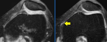
Normal anatomy
The patellar cartilage is the thickest in the body. It should have smooth contours.
The most important part of the medial retinaculum is the medial patellofemoral ligament which inserts all the way posteriorly just in front of the MCL.
Giải phẫu bình thường
Sụn bánh chè là phần dày nhất trong cơ thể, có đường viền nhẵn.
Phần quan trọng nhất của dây chằng trong là dây chằng chè đùi trong, chèn vào phía sau ngay trước MCL.
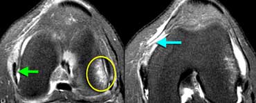 LEFT: Bone bruise lateral condyle (yellow circle). Normal MCL (green arrow) but missing patellar femoral ligament anterior to it.RIGHT: Medial patellar femoral ligament thorn from femoral attachment.TRÁI: Bầm xương lồi cầu ngoài (vòng tròn màu vàng), MCL bình thường (mũi tên xanh lá cây), nhưng mất dây chằng đùi trước xương bánh chè, bên phải: gai dây chằng trong xương bánh rà từ chèn ép đùi.
Case on the left is a female soccerplayer who twisted her knee.
Four MR-images from caudal to cranial demonstrate all the imaging features of a patellar dislocation with rupture of the medial patellar femoral ligament .
The patella was dislocated and the medial facet has bumped onto the lateral condyle.
The patella has spontaneously reduced.
Trường hợp bên trái là một nữ cầu thủ bóng đá bị vặn khớp gối.
Bốn hình ảnh cộng hưởng từ từ xương đuôi đến sọ cho thấy tất cả các đặc điểm hình ảnh của trật khớp bánh chè có đứt dây chằng đùi trong xương bánh chè.
Xương bánh chè bị trật và mặt trong bị va vào lồi cầu ngoài.
Xương bánh chè đã tự giảm.
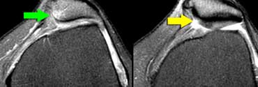 LEFT: Bone bruise medial patella (green arrow).RIGHT: Cartilage fracture.Bên trái: xương bánh chè trung gian bầm tím (mũi tên màu xanh lá cây). Bên phải: Gãy sụn.
Bone bruise may be complicated by cartilage fracture.Bầm tím xương có thể phức tạp do gãy sụn.
Patellar dislocation (2)
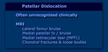
Patellar dislocation is a common condition, but clinically often unrecognized because the patella after the dislocation comes back in it's normal position.
The patient comes with a swollen painfull knee which could be anything from ACL-, MCL- or meniscal tear to a fracture.
So the MRI-findings are important in recognizing this condition.
Patients who have loose bodies or continuing dislocation may undergo operation with retinaculum repair.Trật bánh chè (2)
Trật bánh chè là một tình trạng phổ biến, nhưng lâm sàng thường không được nhận ra vì xương bánh chè sau khi trật trở lại vị trí bình thường.
Bệnh nhân có sưng đau đầu gối, có thể là bất cứ thứ gì từ rách ACL, MCL hoặc rách sụn chêm đến gãy xương.
Vì vậy, các phát hiện trên MRI rất quan trọng trong việc nhận biết tình trạng này.
Bệnh nhân có thể bị lỏng cơ thể hoặc tiếp tục trật khớp có thể được phẫu thuật sửa chữa võng mạc.
Bone and CartilageXương và sụn
Normal and abnormal bone marrowBình thường và bất thường tủy xương
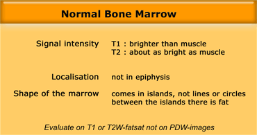
In adults the bone marrow is largely composed of fat.
Normal islands of red marrow may produce confusing images.
Red bone marrow can be pronounced in young women, cigarette smoking, high altitude, hemoglobinopathy or for no reason at all.
As long as the criteria on the left are fullfilled it is normal. Ở người lớn, tủy xương phần lớn bao gồm chất béo.
Các đảo tuỷ đỏ bình thường có thể tạo ra những hình ảnh khó hiểu.
tủy xương đỏ có thể được phát hiện ở phụ nữ trẻ, hút thuốc lá, độ cao, bệnh huyết sắc tố hoặc không có lý do gì cả.
Miễn là các tiêu chí bên trái được điền đầy đủ thì đó là bình thường.
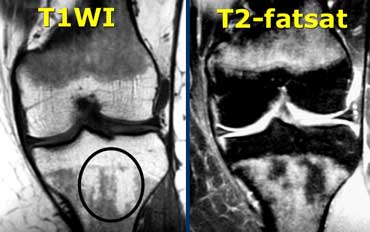
Normal red marrow on the left.
Restricted to the metaphysis and not into the epiphysis.
Comes in islands.
On T1 brighter than muscle. Tuỷ đỏ bình thường bên trái.
Hạn chế ở phần đầu và không vào phần đầu.
Đi vào đảo.
Trên T1 sáng hơn cơ.
 Patient with leukemia and abnormal bone marrowBệnh nhân bạch cầu mạn tính có bất thường tủy xương
Abnormal Bone Marrow
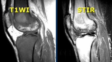
Case on the left shows abnormal bone marrow.
On T1W the signal intensity is lower than muscle.
On T2W-images the signal is very bright.
The abnormal signal comes into the epiphysis. Bất thường tủy xương
Trường hợp bên trái cho thấy bất thường tủy xương.
Trên T1W cường độ tín hiệu thấp hơn cơ.
Trên hình ảnh T2W, tín hiệu rất sáng.
Tín hiệu bất thường đi vào đầu xương.
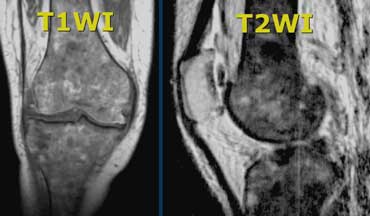 Abnormal bone marrow. There is abnormal signal intensity in the form of circles in metaphysis and epiphysis.Bất thường tuỷ xương.Có bất thường cường độ tín hiệu dưới dạng các vòng tròn trong siêu hình và siêu hình.
Another case with abnormal marrow.
In this case the marrow is too dark on T1 and T2 due to iron deposition in the marrow after many blood transfusions in a patient with hemosiderosis. Một trường hợp khác có tuỷ bất thường.
Trong trường hợp này tuỷ quá sẫm màu ở T1 và T2 do sự lắng đọng sắt trong tuỷ sau nhiều lần truyền máu ở bệnh nhân bị hemosiderosis.
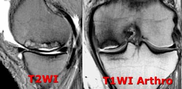 Avascular Necrosis: fluid underneath the cartilage just like a crescent sign on a X-ray. Cartilage is normal.Hoại tử mạch máu: dịch bên dưới sụn giống như dấu hiệu hình lưỡi liềm trên phim X-quang. Sụn bình thường.
Avascular NecrosisHoại tử mạch máu
The most common marrow abnormality is Avascular Necrosis (AVN).
Some people will say 'AVN, Osteochondrosis Dissecans and Stress fracture all look the same'.
There is however a distinct difference.
AVN has the following features:
1. Focal abnormality is subchondral and originates in the bone.
2. Normal cartilage (until it collapses).
3. Wedge shaped marrow edema due to bone infarction.Bất thường tuỷ thường gặp nhất là hoại tử mạch máu (AVN).
Một số người sẽ nói rằng 'AVN, Osteochondrosis Dissecans và gãy xương do stress đều trông giống nhau'.
Tuy nhiên, có một sự khác biệt rõ rệt.
AVN có các đặc điểm sau:
1. Bất thường khu trú là dưới sụn và bắt nguồn từ xương.
2. Sụn bình thường (cho đến khi nó sụp đổ).
3. Phù tuỷ hình nêm do xương nhồi máu.
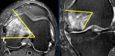 Wedge-shaped bone infarction in AVNXương hình nêm nhồi máu trong AVN
The wedge-shaped pattern of bone marrow edema is just the same as any other infarction in the body i.e. liver infarction or kidney-infarction.Hình dạng phù tuỷ xương hình nêm giống như bất kỳ nhồi máu nào khác trong cơ thể, tức là nhồi máu gan hoặc nhồi máu thận.
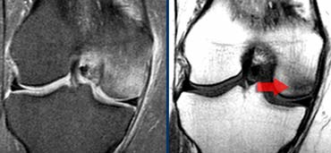
Insufficiency fractureGãy xương không đủ
On the left a different entity, but the patient had the same symptoms.
Acute onset of medial pain.
There is diffuse marrow edema on T2W-image.
On T1W-image the focal abnormality is not directly subchondral.
The abnormality on the T1 is more inside the edema.
On the T1W-image a dark line is visible indicating a insufficiency fracture.
This patient will get better with no weight bearing. Bên trái là một thực thể khác, nhưng bệnh nhân có các triệu chứng tương tự.
Đau trong khởi phát cấp tính.
Có phù tuỷ lan toả trên hình ảnh T2W.
Trên hình ảnh T1W, bất thường khu trú không trực tiếp dưới sụn.
Sự bất thường trên T1 là bên trong phù nề.
Trên hình ảnh T1W có thể nhìn thấy một đường tối cho thấy một vết gãy không có xương.
Bệnh nhân này sẽ tốt hơn mà không cần dùng tạ.
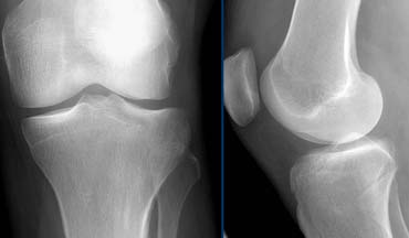
On the left another patient with knee pain after trauma.
There is some effusion but otherwise the x-rays are normal. Bên trái một bệnh nhân khác bị đau gối sau chấn thương.
Có một số tràn dịch nhưng nếu không thì phim X-quang bình thường.
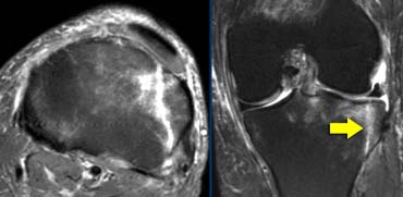 Tibiaplateau fracture. This also is a osteochondral fracture as the fracture line extends all the way through the cartilage to the joint surface.Gãy mâm chày, gãy xương sụn khớp, gãy sụn chêm, gãy mâm xôi, gãy thân xương, gãy đầu xương, gãY xương sụt, gãy chêm,...
In the same patient the MRI shows an obvious tibiaplateau fracture.
The point is that any patient who is unable to bear weight in the hip, knee or ankle with normal X-rays needs another study. Trong cùng một bệnh nhân, MRI cho thấy gãy xương chày.
Vấn đề là bất kỳ bệnh nhân nào không thể chịu được trọng lượng ở hông, đầu gối hoặc cổ chân với X-quang bình thường đều cần một nghiên cứu khác.
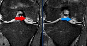 Unstable Osteochondritis DissecansViêm xương sụn không ổn định Dissecans
Osteochondritis DissecansViêm xương sụn Dissecans
The diagnosis Osteochondritis Dissecans is usually made on X-rays.
The question for MRI is whether it is stable or unstable.
The case on the left is unstable for two reasons:
- small cysts at the base of the lesion (red arrow)
- even more important is fluid at the base of the lesion (blue arrow)
Notice that this layer of fluid is different from AVN where the fluid is between the cartilage and the bone.Chẩn đoán Viêm xương sụn Dissecans thường được thực hiện trên X-quang.
Câu hỏi đặt ra cho MRI là liệu nó ổn định hay không ổn định.
Ca bên trái không ổn định vì hai lý do:
- nang nhỏ ở đáy tổn thương (mũi tên màu đỏ)
- quan trọng hơn là dịch ở đáy tổn thương (mũi tên xanh)
Lưu ý rằng lớp dịch này khác với AVN, nơi dịch nằm giữa sụn và xương.
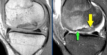 Unstable Osteochondritis Dissecans. The only helpfull sign for the diagnosis unstable OD is the fluid (yellow arrow) not the edema nor the break in the osteochondral surface (green arrow).Viêm xương sụn không ổn định Dấu hiệu hữu ích duy nhất để chẩn đoán OD không ổn định là dịch (mũi tên màu vàng) không phải là phù nề cũng như vỡ trên bề mặt xương sụN (mũI tên màu xanh lá cây).
Not helpfull for the discussion stable versus unstable OD are
- bone marrow edema (could be stable or unstable)
- break in the osteochondral surface.
So the case on the left is unstable because there is fluid at the base of the lesion.Không hữu ích cho các cuộc thảo luận ổn định so với không ổn định OD là
- phù tủy xương (có thể ổn định hoặc không ổn định)
- Bẻ gãy bề mặt xương sụn.
Vậy trường hợp bên trái không ổn định vì có dịch ở đáy tổn thương.
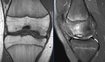
The case on the left shows a OD with bone marrow edema and a break in the osteochondral surface.
But since there is no fluid we cannot tell if this is stable or unstable.
At operation the OD was found to be stableTrường hợp bên trái cho thấy một OD với phù tủy xương và một vết rách trên bề mặt xương sụn.
Nhưng vì không có dịch nên chúng ta không thể biết được dịch này ổn định hay không ổn định.
Tại thời điểm mổ, OD được đánh giá là ổn định.
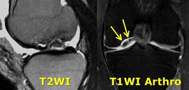
In those cases where you cannot tell whether the lesion is stable or unstable MR-arthrogram is helpfull.
We look for Gadolineum tracking around the osteochondral lesion.Trong những trường hợp mà bạn không thể biết được tổn thương là ổn định hay không ổn định, chụp cộng hưởng từ khớp là hữu ích.
Chúng tôi tìm kiếm Gadolineum theo dõi xung quanh tổn thương xương sụn.
ReferencesTài liệu tham khảo
- Rubin DA: Magnetic resonance imaging of chondral and osteochondral injuries. Top Magn Reson Imaging 1998; 9:348-359 Rubin DA: Hình ảnh cộng hưởng từ của chấn thương chondral và u xương. Hình ảnh Reson Magn hàng đầu 1998; 9: 348-359
- Rubin DA, Kettering JM, Towers JD, Britton CA: MR imaging of knees having isolated and combined ligament injuries. AJR, 1998; 170:1207-1213 Rubin DA, Kettering JM, Towers JD, Britton CA: Hình ảnh MR của đầu gối bị chấn thương dây chằng bị cô lập và kết hợp. AJR, 1998; 170: 1207-1213