
US-guided injection of jointsTiêm khớp dưới hướng dẫn siêu âm
Department of Radiology of the Medical Center, Leeuwarden, the Rijnland Hospital, Leiderdorp and the Jeroen Bosch Hospital, 's-Hertogenbosch, The NetherlandsKhoa Chẩn đoán hình ảnh của Trung tâm Y tế, Leeuwarden, Bệnh viện Rijnland, Leiderdorp và Bệnh viện Jeroen Bosch, 's-Hertogenbosch, Hà Lan
Ngày đăng / cập nhật: 2012-12-22
Bản gốc trên radiologyassistant.nl ↗ · tải về lúc 2026-11-19 20:19 · bản dịch máy có chuẩn hóa thuật ngữ chẩn đoán hình ảnh
This article describes the application of Ultrasound guidance for diagnostic and therapeutic joint injections.Bài viết này mô tả ứng dụng hướng dẫn siêu âm trong chẩn đoán và điều trị tiêm khớp.
Ultrasound is a valuable alternative to procedures performed either blind or under fluoroscopic or CT guidance.Siêu âm là một phương pháp thay thế có giá trị cho các thủ thuật được thực hiện dưới hướng dẫn mù hoặc dưới hướng dẫn của đèn huỳnh quang hoặc CT.
ShoulderVai
Glenohumeral jointKhớp Glenohumeral
Anterior approach Tiếp cận trước
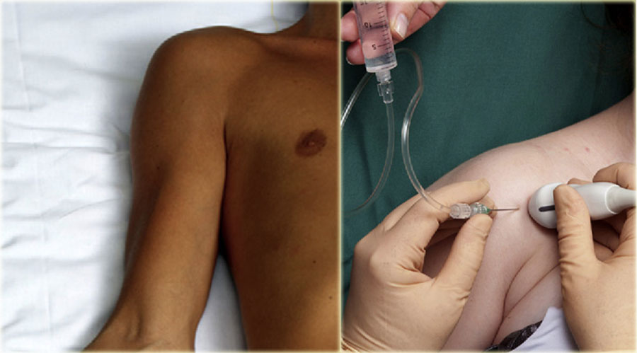
In the anterior approach the patient is lying supine with the extended arm externally rotated (figure).Trong cách tiếp cận trước, bệnh nhân nằm ngửa với cánh tay duỗi ra ngoài (hình).
The transducer is placed ventrally parallel to the long axis of the subscapular tendon.Đầu dò được đặt song song với trục dài của gân dưới vai.
The grey line on the side of the transducer indicates the long axis.Đường màu xám ở bên cạnh đầu dò cho biết trục dài.
Local anaesthetics are not needed if needles are used with a diameter of 21-gauge or thinner.Không cần gây tê cục bộ nếu kim được sử dụng với đường kính 21 gauge hoặc mỏng hơn.
For joint aspirations one may need to use a larger bore needle due to high viscosity of the aspirate. In such cases local anaesthetics are indicated.Đối với chọc hút khớp, có thể cần sử dụng kim khoan lớn hơn do độ nhớt cao của dịch hút. Trong những trường hợp như vậy, gây tê cục bộ được chỉ định.
To facilitate injection of medication or contrast, one may use a connection tube in between the needle and the syringe, the latter being held and managed by an assistant.Để tạo điều kiện thuận lợi cho việc tiêm thuốc hoặc thuốc cản quang, người ta có thể sử dụng một ống nối giữa kim và ống tiêm, ống tiêM sau đó được giữ và quản lý bởi một trợ lý.
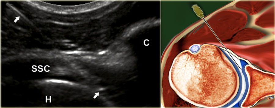
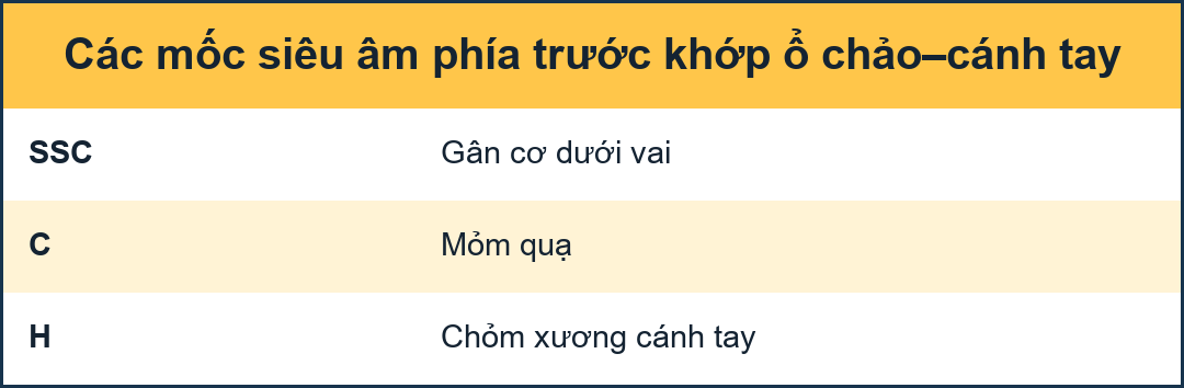 The landmarks one should look for are the medial contour of the humeral head and medial to this the coracoid process (C)Các mốc cần được quan sát là đường viền trong của đầu xương cánh tay và trong đó là mỏm quạ (C).
A 22-gauge, 50mm needle is used connected to a syringe containing the contrast media, held by an assistant who upon proper needle position injects 15-20 mL of the contrast medium.Một kim 22 gauge, 50 mm được sử dụng kết nối với một ống tiêm chứa chất cản quang, được giữ bởi một trợ lý, người mà khi ở vị trí kim thích hợp sẽ tiêm 15-20 mL chất cản Quang.
The needle is advanced perpendicular to the medial edge of the humeral head, penetrating the subscapular tendon.Kim được đẩy vuông góc với mép trong của đầu xương cánh tay, xuyên qua gân dưới vai.
If one hits the cartilage of the humeral head, the needle should be pulled back 1 or 2 mm, slightly angled by about 15◦ and then advanced tangentially to the head into the joint with the bevel of the needle facing into the joint (figure).Nếu một kim đâm vào sụn đầu xương cánh tay, kim nên được kéo lại 1 hoặc 2 mm, hơi nghiêng khoảng 15◦ và sau đó tiến tiếp tuyến với đầu vào khớp với mặt vát của kim hướng vào khớp (hình).
No resistance to injection should be felt and one should see the contrast flow freely into the joint and if present into the subscapular recess.Không có kháng cự để tiêm nên được cảm nhận và người ta sẽ thấy chất cản quang chảy tự do vào khớp và nếu có trong hốc dưới vai.
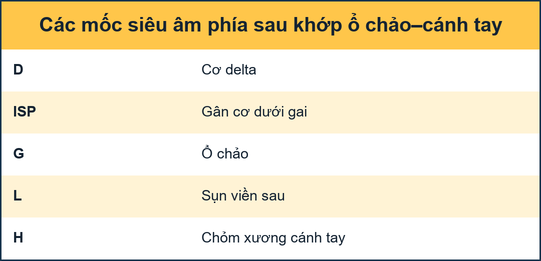
Posterior approachTiếp cận từ sau
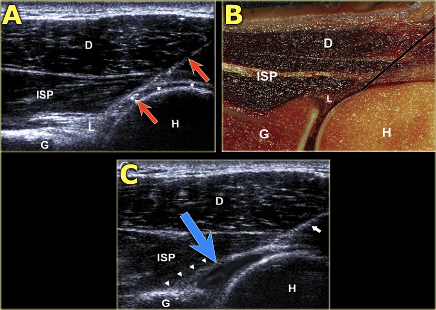
A. The needle is in the intra-articular position with the tip underneath the infraspinatus tendon (ISP) and posterior labrum (L) and bordering the hyaline cartilage (asterisks) of the humeral head. A. Kim ở vị trí trong khớp với đầu kim nằm bên dưới gân cơ dưới gai (ISP) và xương ức sau (L) và giáp với sụn chêm (dấu hoa thị) của đầu xương cánh tay.
B. Corresponding cadaver section showing the optimal needle track (white line).B. Phần xác tương ứng cho thấy đường kim tối ưu (đường trắng).
C. Sonogram after injection of 15 mL contrast. The correct intra-articular position of the needle can be visualized real-time during injection, but is also confirmed by the 'comma'-like configuration of the posterior labrum (arrowheads), which is lifted by the intraarticularly injected fluid. C. Siêu âm sau khi tiêm thuốc cản quang 15 mL Vị trí trong khớp chính xác của kim có thể được nhìn thấy theo thời gian thực trong quá trình tiêm, nhưng cũng được xác nhận bởi cấu hình giống như dấu phẩy của xương ức sau (các đầu mũi tên), được nâng lên bởi dịch tiêm trong khớp.
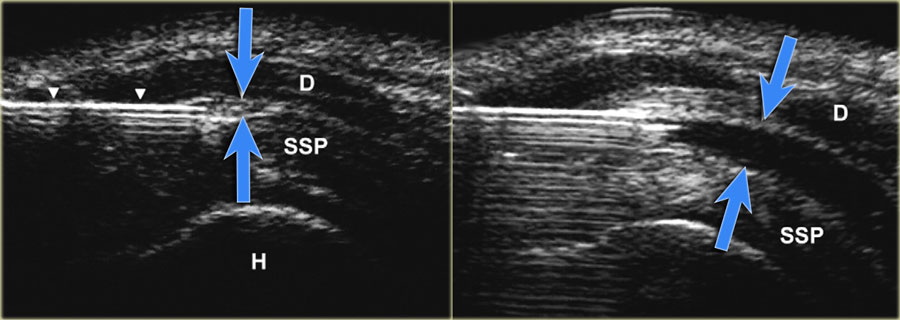
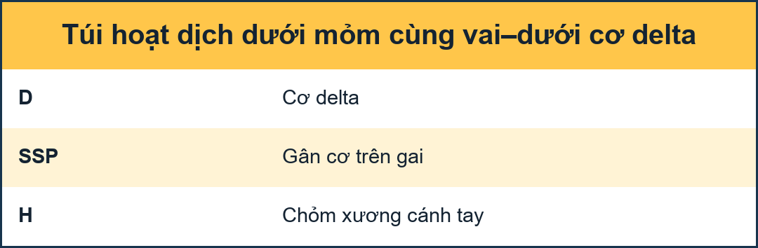 US-image showing a long axis view of the supraspinatus tendon (SSP). The advancing needle under real-time US-guidance has entered the subacromial bursa between the deltoid and SSP-muscle. Dilatation of the subacromial–subdeltoid bursa after injection of 5 mL fluid (blue arrows).Hình ảnh siêu âm cho thấy hình ảnh trục dài của gân cơ trên gai (SSP). Kim chọc dưới hướng dẫn siêu âm thời gian thực đã đi vào bao hoạt dịch dưới mỏm cùng vai giữa cơ delta và cơ SSP. Sự giãn của bao hoạt dịch hạ mỏm đồng vai-dưới mỏm đùi sau khi tiêm 5 mL dịch (các mũi tên màu xanh).
Subacromial bursaTúi hoạt dịch dưới mỏm cùng vai
The subacromial-subdeltoid bursa is a synovial lined space, which contains no observable or only a minimal amount of fluid.Túi thanh dịch dưới mỏm cùng vai-dưới cơ delta là một không gian được lót bằng màng hoạt dịch, không chứa hoặc chỉ chứa một lượng chất lỏng tối thiểu.
The bursa consists of two bursal leaves. The outer and inner leaves are fused with the deltoid muscle fascia and rotator cuff, respectively. The bursal leaves can easily glide over each other, thus facilitating the range of movement of the shoulder.Túi thanh dịch bao gồm hai lá bao hoạt dịch, lá ngoài và lá trong được hợp nhất với cân cơ delta và chóp xoay, tương ứng, các lá bao hoạt động có thể dễ dàng lướt qua nhau, do đó tạo điều kiện cho phạm vi vận động của vai.
Blind subacromial injection of drugs into the subacromial bursa is a frequently performed therapy by general practitioners and orthopedic surgeons. The incidence of injections that miss the subacromial bursa range from 12% to 70%.Tiêm thuốc mù vào bao hoạt dịch dưới mỏm cùng vai là một liệu pháp thường được thực hiện bởi các bác sĩ đa khoa và bác sĩ phẫu thuật chỉnh hình, tỷ lệ tiêm thuốc bỏ sót bao hoạt dịch nằm dưới mỏM cùng vai dao động từ 12% đến 70%.
ElbowKhuỷu
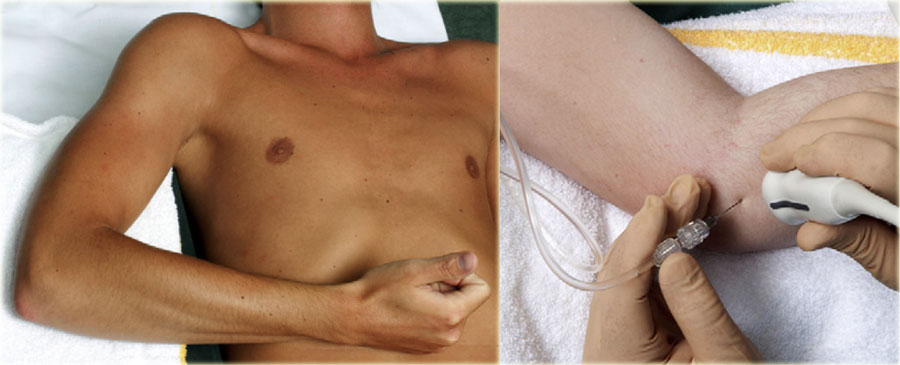
For injection of the elbow the patient is supine with the arm in 90◦ flexion, raised and resting on a cushion. The joint space between the radial head and the capitulum is easily palpated. The hand is pronated or may be turned into the thumb up position, which is necessary to open the joint maximally.Đối với tiêm khuỷu tay, bệnh nhân nằm ngửa với cánh tay gấp 90◦, nâng lên và nghỉ ngơi trên đệm. Khoảng trống giữa đầu quay và đầu gối dễ dàng sờ thấy. Bàn tay được sấp hoặc có thể được xoay vào vị trí ngón tay cái, cần thiết để mở khớp tối đa.
The transducer is placed over the joint visualizing the joint space. The needle (22 gauge, 30 mm) is directed at a slight craniocaudal angle on the dorsolateral side of the joint toward the joint space aiming toward the articular surface of the radial head.Đầu dò được đặt trên khớp để quan sát không gian khớp. Kim (22 gauge, 30 mm) được hướng vào một góc sọ nhẹ ở phía lưng bên của khớp về phía không gian khớp hướng về phía bề mặt khớp của đầu quay.
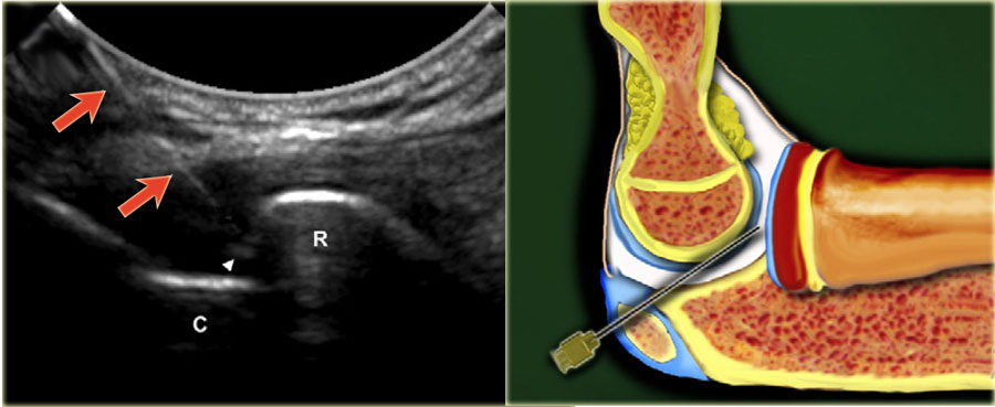
When seen to have entered the joint and upon feeling the cartilage of the radial head, the needle is slightly pulled back to ensure that the bevel is free from the cartilage and facing into the joint.Khi nhìn thấy đã vào khớp và khi sờ thấy sụn đầu quay, kim được kéo nhẹ trở lại để đảm bảo rằng vát không có sụn và hướng vào khớp.
5-8 mL of contrast media is injected. No resistance to injection should be felt.Tiêm 5-8 mL thuốc cản quang Không cảm thấy kháng thuốc khi tiêm.
Wrist and handCổ tay và bàn tay
Radiocarpal jointXạ trị khớp cổ tay
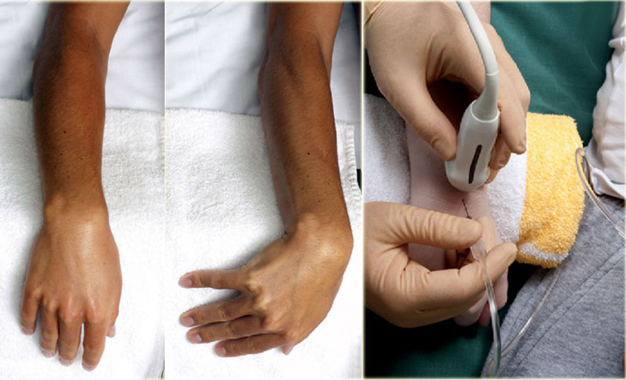
The patient is supine with the wrist resting flexed over a 45◦ sponge or a rolled-up towel.Bệnh nhân nằm ngửa, cổ tay gập trên miếng bọt biển 45 ◦ hoặc khăn cuộn lại.
In some cases it may be helpful to hold the wrist in ulnar deviation in order to open the joint space even more.Trong một số trường hợp, giữ cổ tay ở tư thế xoay trụ có thể hữu ích để mở rộng không gian khớp nhiều hơn.
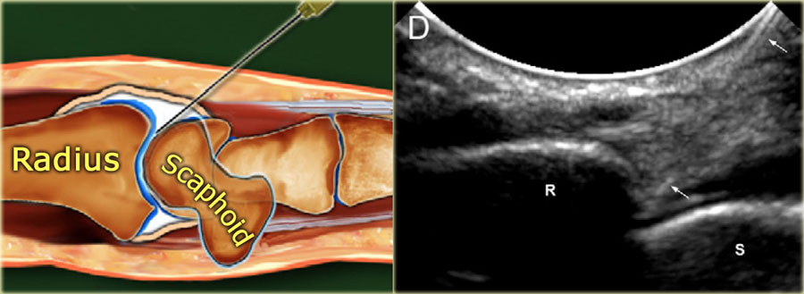
The space between the radius and the scaphoid is identified on ultrasound.Khoảng cách giữa xương quay và xương thuyền được xác định trên siêu âm.
A 23-25-gauge, 30 mm needle is advanced under ultrasound guidance into the joint directed toward the articular surface of the radius until one feels contact with the radius.Một kim 23-25 gauge, 30 mm được đưa dưới hướng dẫn siêu âm vào khớp hướng về phía bề mặt khớp của xương quay cho đến khi cảm thấy tiếp xúc với xương quay.
After ensuring that the tip of the needle is free from the radial cartilage 2-4 mL contrast is injected.Sau khi đảm bảo rằng đầu kim không bị dính vào sụn quay, tiêm 2-4 mL thuốc cản quang.
The bevel of the needle is facing toward the joint space and the contrast is seen to flow into the joint.Vòng vát của kim hướng về phía khe khớp và thấy được độ tương phản chảy vào khớp.
Distal radioulnar joint (DRUJ)Khớp quay xa (DRUJ)
A linear array transducer is axially positioned dorsally over the distal radius and ulna. Along the short axis of the transducer a 23-25- gauge, 30 mm needle is inserted being directed from proximally to distally in a caudal direction. A total amount of 0.5-1 mL is injected according to rising pressure during injection.Đầu dò mảng tuyến tính được đặt theo trục dọc theo bán kính đầu xa và xương trụ. Dọc theo trục ngắn của đầu dò, một kim 23-25 gauge, 30 mm được chèn vào được hướng từ đầu gần đến đầu xa theo hướng đuôi. Tổng lượng 0,5-1 mL được tiêm theo áp suất tăng trong quá trình tiêm.
Carpal, carpometacarpal and interphalangeal jointsKhớp cổ tay, cổ tay-mông cổ và khớp gian đốt ngón
Physicians and specialists routinely perform intra-articular punctures and injections on small wrist and finger joints to relieve joint effusion or to inject drugs.Các bác sĩ và chuyên gia thường xuyên thực hiện chọc và tiêm nội khớp vào các khớp cổ tay và ngón tay nhỏ để làm giảm tràn dịch khớp hoặc để tiêm thuốc.
The failure rate and frequency of occurrence of peri-articular injections are high: 15% - 32%, especially with the joints of the little finger and the DIP joints.Tỷ lệ thất bại và tần suất xuất hiện tiêm quanh khớp cao: 15% - 32%, đặc biệt ở khớp ngón út và khớp DIP.
Unintended peri-articular drug injection moreover may affect the surrounding ligaments or tendons, leading to serious complications.Tiêm thuốc quanh khớp ngoài ý muốn có thể ảnh hưởng đến các dây chằng hoặc gân xung quanh, dẫn đến các biến chứng nghiêm trọng.
A dorsal approach using a 23-25-gauge needle is preferable. Although high-frequency linear array transducers with frequencies from 18 to 12 MHz are often used for scanning the superficial soft tissue structures of the wrist and hand, a small footprint transducer may allow better access to the small peripheral joints.Cách tiếp cận mặt lưng bằng kim 23-25 gauge là thích hợp hơn Mặc dù các đầu dò mảng tuyến tính tần số cao với tần suất từ 18 đến 12 MHz thường được sử dụng để quét các cấu trúc mô mềm bề mặt của cổ tay và bàn tay, một đầu dò dấu chân nhỏ có thể cho phép tiếp cận tốt hơn với các khớp nhỏ ngoại vi.
Generally, 0.5-1 mL of contrast material is instilled after confir- mation of correct needle placement.Nói chung, 0,5-1 mL chất cản quang được thấm nhuần sau khi đặt kim đúng cách.
Sacroiliac JointKhớp cùng chậu
The sacroiliac joint has been implicated as a source of low back and lower extremity pain, which is thought to be caused by sacroiliitis.Khớp cùng chậu được cho là nguyên nhân gây đau thắt lưng và chi dưới, được cho là do viêm khớp cùng chậu.
Treatment consists of intra-articular injection of corticosteroids. Diagnostic injections or blocks are frequently performed, to distinguish between the probable causes of low back pain, because in 15-25% this is generated by the SI joint.Điều trị bao gồm tiêm corticosteroid nội khớp. Tiêm chẩn đoán hoặc khối thường được thực hiện, để phân biệt giữa các nguyên nhân có thể gây đau lưng thấp, bởi vì trong 15-25% trường hợp này là do khớp SI gây ra.
Upper level SI joint injection Tiêm khớp trên
The axially orientated transducer is moved from the level of the fifth lumbar vertebra caudally, depicting the dorsal surface of the sacrum with the median and lateral sacral crest, the gluteal surface of the ilium, and the first posterior sacral foramen.Đầu dò định hướng theo trục được di chuyển từ mức của đốt sống thắt lưng thứ năm, mô tả bề mặt lưng của xương cùng với mào xương cùng giữa và bên, bề mặt mông của chậu và lỗ xương cùng sau đầu tiên.
The needle is inserted along the short axis of the transducer into the hypoechoic cleft located between the surface of the sacrum and the contour of the ilium. Angulations of needle insertion are adjusted to the orientation of the hypoechoic cleft of the SI joint, which presents cranially a more medial to lateral orientation, and caudally a more vertical orientation.Kim được đưa dọc theo trục ngắn của đầu dò vào khe giảm âm nằm giữa bề mặt của xương cùng và đường viền của xương chậu Các góc của kim được điều chỉnh theo hướng của khe giảm âm của khớp SI, hướng về phía sọ theo hướng từ trong ra ngoài và hướng thẳng đứng hơn.
Lower level SI joint injection Tiêm khớp cùng bên dưới
From the upper level the transducer is moved downward by delineation of the median and lateral sacral crest, at the dorsal surface of the sacrum and the gluteal surface of the ilium until the second posterior sacral foramen is visualized.Từ tầng trên, đầu dò được di chuyển xuống dưới bằng cách phác thảo đỉnh xương cùng giữa và bên, ở mặt lưng của xương cùng và mặt mông của chậu cho đến khi hình dung được lỗ xương cùng sau thứ hai.
As with the upper level, the needle is inserted into the hypoechoic cleft between the sacrum and ilium.Cũng như với mức trên, kim được đưa vào khe giảm âm giữa xương cùng và xương chậu.
HipKhớp háng
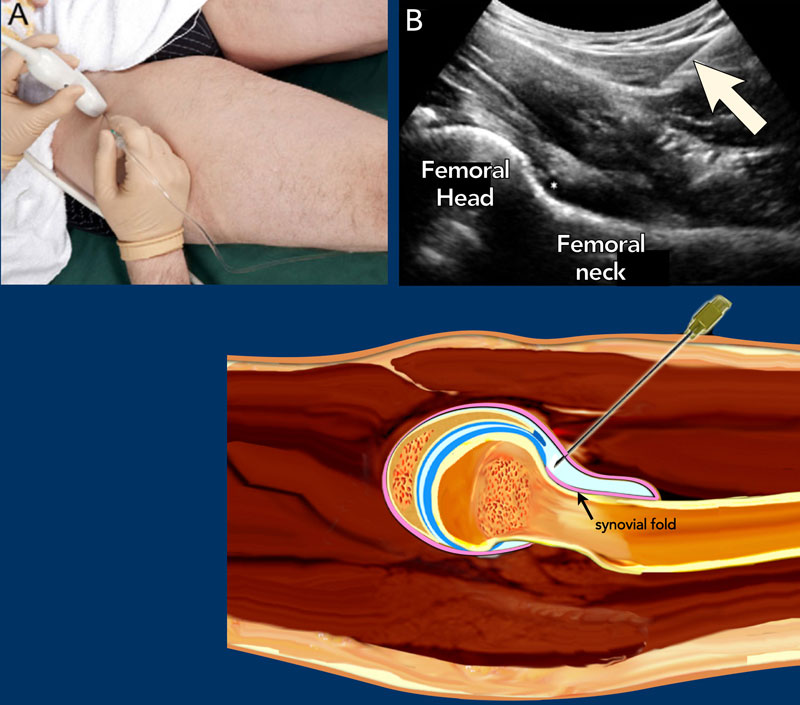
The patient is placed supine. The leg is held in slight endorotation and abduction thereby reducing tension on the capsular structures and moving the iliopsoas tendon and bursa medially out of the intended needle path.
Preferably a 5-3.5 MHz curved array transducer is used, which provides the necessary penetration depth.
Usually a 21 gauge needle with a length of 9 cm is used for the average adult. In smaller adults or children a 23-gauge, 5 cm hypodermic needle might be used.Bệnh nhân được đặt nằm ngửa, chân được giữ trong tư thế nhẹ và giạng, do đó làm giảm sức căng trên các cấu trúc bao và di chuyển gân cơ thắt lưng chậu và bao hoạt dịch ra ngoài đường đi kim dự định.
Tốt nhất là sử dụng đầu dò mảng cong 5 - 3,5 MHz, cung cấp độ sâu thâm nhập cần thiết.
Thông thường, một kim 21 gauge với chiều dài 9 cm được sử dụng cho người lớn trung bình Ở người lớn nhỏ hơn hoặc trẻ em, có thể sử dụng kim tiêm dưới da 23 gauge, 5 cm.
Local anaesthetic may be injected prior to the main injection but this entails extra manipulation as well as non-contrast fluid (anaesthetic) in the joint leaving less room for the contrast in the limited joint space as well as possibly 2 punctures.Có thể tiêm thuốc tê tại chỗ trước khi tiêm chính nhưng điều này đòi hỏi phải thao tác thêm cũng như dịch không cản quang (gây tê) trong khớp để ít chỗ cho thuốc cản quang trong không gian khớp hạn chế cũng như có thể là 2 lỗ thủng.
The needle may inadvertently be withdrawn from the joint after anaesthetic injection during the switch to connect the syringe with contrast. This could be avoided by using a three-way connector between the two syringes containing the anaesthetic and the injection fluid (contrast or medication).
The needle is advanced at a caudo-cranial angle along the long axis of the transducer aiming for the anterior recess near the junction of the femoral neck with the femoral head (see US and illustration).Có thể vô tình rút kim ra khỏi khớp sau khi tiêm thuốc tê trong quá trình chuyển đổi để kết nối ống tiêm với thuốc cản quang. Điều này có thể tránh được bằng cách sử dụng một đầu nối ba chiều giữa hai ống tiêM có chứa thuốc gây mê và dịch tiêm (thuốc cản quang hoặc thuốc).
Kim được đẩy theo góc sọ dọc theo trục dài của đầu dò hướng về phía lõm trước gần điểm nối của cổ xương đùi với đầu xương đùi (xem siêu âm và hình minh hoạ).
The bevel of the needle should be facing toward the joint.
When the needle makes contact with the femoral head-neck junction it is slightly retracted to prevent inadvertently injection underneath the synovial fold of the hip joint capsule (see drawing).
If correctly positioned within the joint capsule, 10-15 mL contrast media or medication is injected and the anterior recess will distend by the injected fluid confirming the intra-articular positioning.Mép vát của kim phải hướng về phía khớp.
Khi kim tiếp xúc với khớp đầu cổ xương đùi, nó sẽ hơi rút lại để tránh vô tình tiêm bên dưới nếp hoạt dịch của bao khớp háng (xem bản vẽ).
Nếu vị trí chính xác trong bao khớp, tiêm 10-15 mL thuốc cản quang hoặc thuốc và phần lõm trước sẽ bị giãn ra bởi dịch tiêm xác nhận vị trí trong khớp.
KneeGối
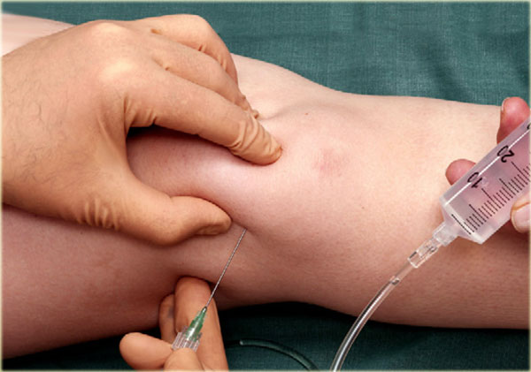
Indications for CT or MR arthrography of the knee are evaluation of the post-operative meniscus, query intra-articular bodies, evaluation of the stability of osteochondral lesions and evaluation of articular cartilage. One may also be requested to inject medication such as corticosteroids and/or a local anaesthetic.Chỉ định chụp CT hoặc MR khớp gối là đánh giá sụn chêm sau phẫu thuật, truy vấn các cơ quan nội khớp, đánh giá độ ổn định của tổn thương xương sụn và đánh giá sụn khớp. Người ta cũng có thể được yêu cầu tiêm thuốc như corticosteroid và / hoặc gây tê tại chỗ.
For injection we do not use ultrasound guidance but use the standard "blind" procedure introducing the needle (21-gauge, 50 mm) behind the patella using a lateral midpatellar approach.Đối với tiêm chúng tôi không sử dụng dẫn hướng siêu âm mà sử dụng quy trình "mù" tiêu chuẩn đưa kim (21 gauge, 50 mm) vào phía sau xương bánh chè bằng cách tiếp cận giữa xương bánh rào bên.
The patella is lateralized and the needle introduced from the mid lateral side aiming toward the centre of the patella indicated by the left forefinger.Xương bánh chè được nghiêng và kim được đưa vào từ phía giữa bên nhằm vào trung tâm của xương bánh chè có chỉ định bằng ngón trỏ trái.
The needle is introduced horizontally aiming posterior to the centre of the patella until one makes contact with the lateral patellar facet or the lateral femoral condyle and when felt to be in the joint 40 mL contrast media is injected.Kim được đưa vào theo chiều ngang hướng về phía sau trung tâm xương bánh chè cho đến khi kim tiếp xúc với mặt bên xương bánh rào hoặc lồi cầu xương đùi bên và khi cảm thấy ở trong khớp, tiêm 40 mL thuốc cản quang.
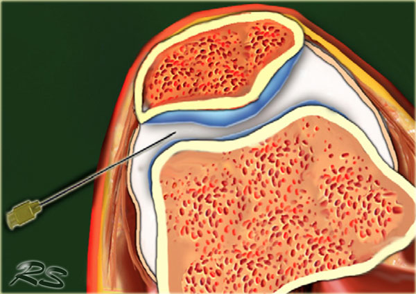
Prior to CT or MR one can choose to apply a tight bandage above the patella thereby forcing contrast from the suprapatellar recess into the joint space proper.Trước khi chụp CT hoặc MR, người ta có thể chọn băng chặt phía trên xương bánh chè, do đó buộc thuốc cản quang từ hốc trên xương bánh răng vào khoang khớp thích hợp.
Ankle and footMắt cá chân và bàn chân
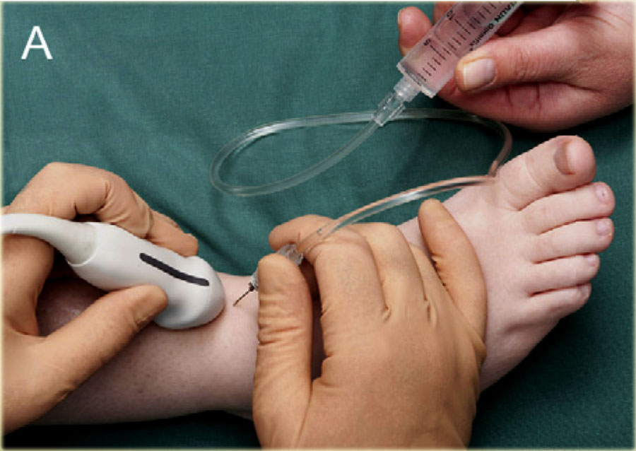 The foot is slightly plantar flexed. The long axis of the transducer is indicated by the grey line on the side, being in a sagittal plane.Bàn chân hơi gập lại. Trục dài của đầu dò được chỉ ra bởi đường màu xám ở bên cạnh, nằm trong mặt phẳng đứng dọc.
Tibiotalar jointKhớp răng hàm lớn
CT or MR arthrography may be used to query ligamentous, osteochondral or chondral injury, eval- uation for free bodies or query stability of ostechondral lesions.
For injection of the ankle (tibiotalar joint) the patient is supine with the foot in slight plantar flexion.Chụp CT hoặc MR có thể được sử dụng để tìm kiếm tổn thương dây chằng, xương sụn hoặc sụn, đánh giá sự tự do hoặc tìm sự ổn định của tổn thương xương sụt.
Đối với tiêm khớp cổ chân (khớp xương đùi), bệnh nhân nằm ngửa, bàn chân gập nhẹ.
The medial side of the tibiotalar joint is investigated anteriorly with ultrasound to deter- mine a suitable place for injection, at the same time checking for any excessive joint fluid.Siêu âm đánh giá phía trong khớp chày để xác định vị trí tiêm, đồng thời kiểm tra dịch khớp thừa.
We use a small curved array 8 MHz transducer but if preferred one can use an 18-12 MHz linear array transducer. The long axis of the probe is held in a sagittal plane.Chúng tôi sử dụng một đầu dò mảng cong 8 MHz nhỏ nhưng nếu thích có thể sử dụng đầu dò tuyến tính mảng 18-12 MHz. Trục dài của đầu dò được giữ trong mặt phẳng đứng dọc.
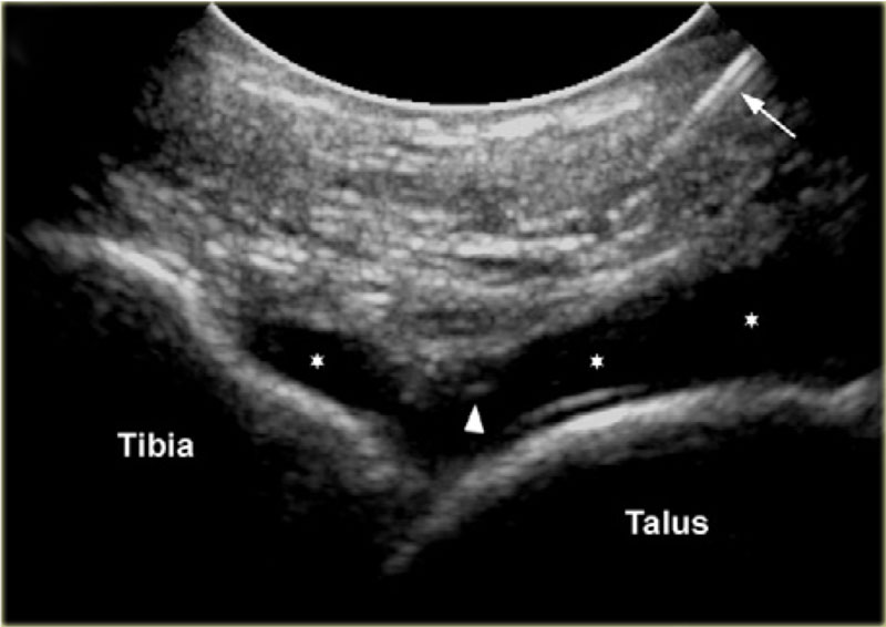
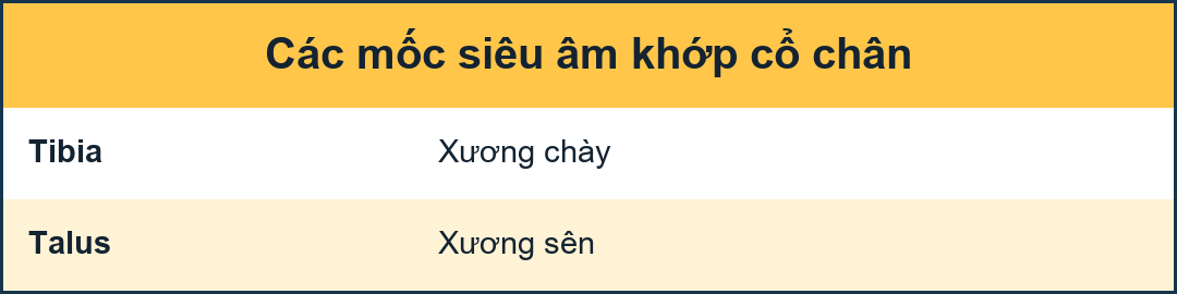 Sonogram showing the needle (arrow) and the needle tip (arrowhead) and the injected contrast media in the tibiotalar joint.Siêu âm cho thấy kim (mũi tên) và đầu kim (đầu mũi tên) và môi trường thuốc cản quang được tiêm vào khớp chày.
The needle, usually 22-gauge (length: 30 mm), is introduced in line with the long imaging axis of the transducer on the medial side of the anterior joint space, medial to the anterior tibial ligament, avoiding ligaments and vessels.Kim, thường là 22 gauge (chiều dài: 30 mm), được đưa vào phù hợp với trục hình ảnh dài của đầu dò ở phía trong của khoang khớp trước, ở giữa dây chằng chày trước, tránh dây chằng và mạch máu.
One should identify the talar dome and the overhanging anterior tibial lip. The needle is angled caudo-cranially into the joint under the ventral lip of the distal tibia aiming for the articular surface of the distal tibia.Cần xác định xương sên và môi chày trước nhô ra, kim được đặt góc cạnh theo đường sọ vào khớp dưới môi bụng của đầu xa xương chày, hướng về phía bề mặt khớp của đầu xa.
Contact is felt and once again one ensures that the needle tip is free from the tibial cartilage and that the bevel is facing into the joint.Tiếp xúc được cảm nhận và một lần nữa đảm bảo rằng đầu kim không có sụn chày và góc vát hướng vào khớp.
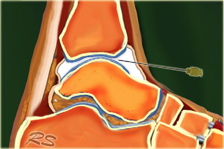
8-10 ml of contrast is injected into the tibiotalar joint and one sees the anterior capsule swells up with the fluid.8-10 ml thuốc cản quang được tiêm vào khớp chày và người ta thấy bao trước phồng lên cùng với dịch.
There should be no resistance to injection or pain experienced by the patient.Không nên có sự kháng cự với tiêm hoặc đau của bệnh nhân.
Posterior subtalar jointKhớp dưới cẳng chân sau
The subtalar or talocalcaneal joint is composed of 3 facets: a broad posterior facet representing the primary articulating surface, a medially located middle facet in which the sustentaculum tali articulates with the medial process of the talus, and an anterior facet. Subtalar arthrography may be performed via an anterolat- eral, posterolateral or posteromedial approach. 2-4 ml of contrast material is injected into the posterior subtalar joint.Khớp dưới xương ức hoặc khớp bàn ngón gồm 3 mặt: mặt sau rộng đại diện cho mặt khớp chính, mặt giữa nằm ở giữa, trong đó mỏm móc nối với mặt trong của xương sên và mặt trước Chụp động mạch dưới xương ổ có thể được thực hiện thông qua đường tiếp cận trước-sau, sau ngoài hoặc sau trong. 2-4 ml chất cản quang được tiêm vào khớp dưới xương đuôi sau.
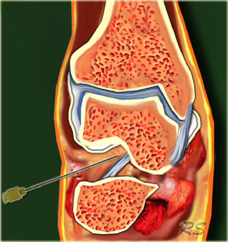
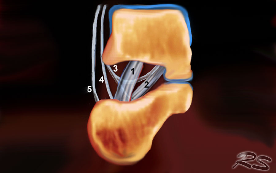 Schematic drawing in a coronal view of the right sinus tarsi. Displayed are the course and attachment sites of the cervical ligament (1); the interosseous talo- calcaneal ligament (2); and the medial (3), intermediate (4), and lateral (5) roots of the inferior extensor retinaculum.Sơ đồ hình ảnh chụp vành xoang phải, thể hiện đường đi và vị trí bám của dây chằng cổ (1); dây chằng gót-dây chằng sên xương gót (2); và rễ trong (3), rễ giữa (4), rễ ngoài (5) của võng mạc duỗi dưới.
Sinus tarsiXoang cổ chân
The sinus tarsi is a cone-shaped cavity that courses in a postero- medial to anterolateral direction. It is located in the lateral aspect of the foot between the neck of the talus and the anterosuperior surface of the calcaneus.Xoang tarsi là một khoang hình nón, hướng từ sau trong đến trước ngoài, nằm ở mặt ngoài của bàn chân, giữa cổ xương sên và mặt trước trên xương gót.
The tarsal sinus continues medially as the tarsal canal, which is a funnel-shaped space between the talus and the calcaneus.Xoang sụn tiếp tục ở giữa như ống sụn, là một khoảng trống hình phễu giữa xương sụn và xương gót.
It contains fat, an arterial anastomosis, joint cap- sules, nerve endings, and five ligamentous structures-the medial, intermediate, and lateral roots of the inferior extensor retinaculum; the cervical ligament; and the interosseous talocalcaneal ligament (figure).Nó chứa mỡ, một động mạch nối, nắp khớp, đầu dây thần kinh và năm cấu trúc dây chằng - rễ trong, trung gian và bên của võng mạc duỗi dưới; dây chằng cổ tử cung; và dây chằng ụ đùi (hình).
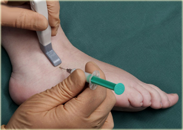
This space can be the cause of foot pain in the sinus tarsi syn- drome. The first step in treatment is infiltration of the sinus tarsi with a mixture of Depomedrol and local anaesthetic (Lidocaine). This can be challenging for the surgeon in a non-guided approach but is reasonably easily and accurately achieved with ultrasound guidance.Không gian này có thể là nguyên nhân gây đau chân trong hội chứng sụn viền xoang. Bước đầu tiên trong điều trị là xâm nhập vào xoang sụn bằng hỗn hợp Depomedrol và gây tê tại chỗ (Lidocaine).Điều này có thể gây khó khăn cho phẫu thuật viên trong cách tiếp cận không có hướng dẫn nhưng đạt được một cách dễ dàng và chính xác với hướng dẫn siêu âm.
US-guided injection of the sinus tarsi at the right-hand side with a lateral approach. The transducer is held in a coronal oblique plane. The needle is introduced along the long axis of the transducer.Tiêm dưới hướng dẫn siêu âm của tarsi xoang ở bên phải với cách tiếp cận bên. Đầu dò được giữ trong một mặt phẳng xiên vành. Kim được đưa dọc theo trục dài của đầu dò.
The sinus tarsi can easily be visualized using ultrasound.Siêu âm có thể dễ dàng quan sát được xoang tarsi.
The patient turns onto the contralateral side laying the foot to be treated with its medial surface against the table top, the lateral side of the foot being uppermost.Bệnh nhân xoay sang phía đối diện đặt bàn chân để được điều trị với bề mặt trong của nó chống lại mặt bàn, mặt bên của bàn chân là trên cùng.
The transducer is held in a coronaloblique plane with regards to the foot.Đầu dò được giữ trong mặt phẳng nghiêng về phía bàn chân.
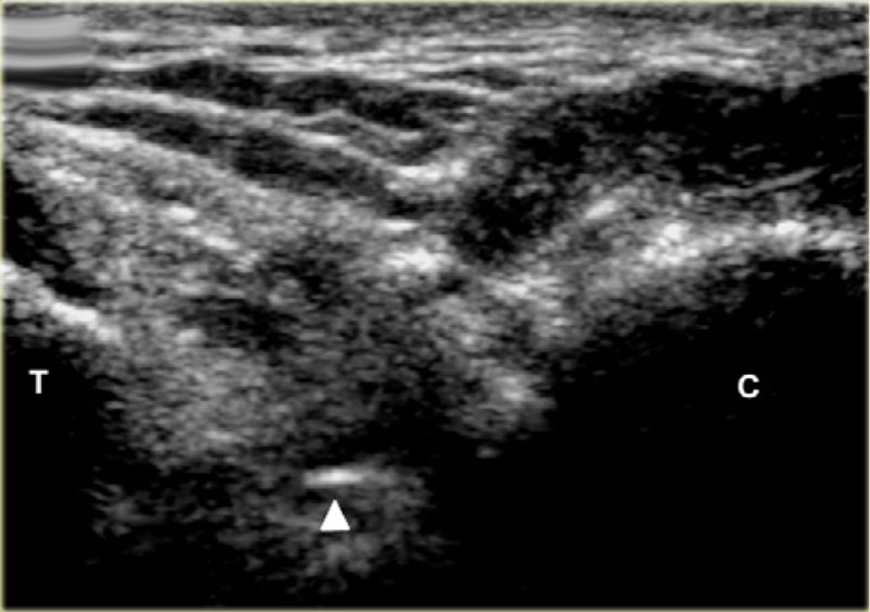
The sinus tarsi is identified as a triangular space between the anterior process of the calcaneus and the talar neck.Xương sên được xác định là khoảng trống hình tam giác giữa mỏm trước xương gót và cổ xương sên.
The tip of the needle (arrow head) is seen within the cone shaped sinus tarsi, which is bordered by the talus (T) and calcaneus (C). Đầu kim (đầu mũi tên) được nhìn thấy trong xoang hình nón, được bao quanh bởi xương sên (T) và xương gót (C).
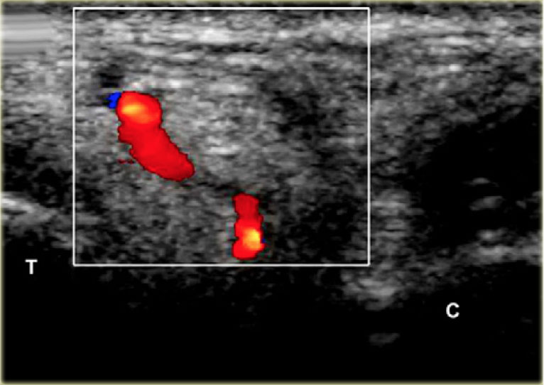
Depending on the degree of inflammation there may be hyperemia of the space and there may be intervening vessels visible, which one wishes to avoid. This is relatively easy, especially with colour doppler Tuỳ thuộc vào mức độ viêm, có thể có tăng huyết áp của không gian và có thể có các mạch máu can thiệp có thể nhìn thấy, điều mà người ta muốn tránh Điều này tương đối dễ dàng, đặc biệt là với doppler màu
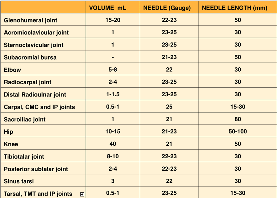
Volumes of injectionKhối lượng tiêm
Click on the image below to look at the video of Medical Action Myanmar (MAM), an NGO run by Frank Smithuis and Nini Tun.
MAM has 12 clinics and 2100 community health workers all over Myanmar.
You can also visit the website of MAM
All donations are extremely well spend.Nhấp vào hình ảnh bên dưới để xem video Hành động Y tế Myanmar (MAM), một tổ chức phi chính phủ do Frank Smithuis và Nini Tun điều hành.
MAM có 12 phòng khám và 2100 nhân viên y tế cộng đồng trên khắp Myanmar.
Bạn cũng có thể truy cập vào trang web của MAM
Tất cả các khoản quyên góp đều được chi tiêu rất tốt.
ReferencesTài liệu tham khảo
-
US-guided injection of the upper and lower extremity joints.
Tiêm dưới hướng dẫn siêu âm khớp chi trên và dưới.
by Collins JM, Smithuis R, Rutten MJ. Eur J Radiol. 2012 Oct;81(10):2759-70 bởi Collins JM, Smithuis R, Rutten MJ. Euro J Radiol. 2012 Tháng Mười;81(10): 2759-70