
CraniosynostosisDính khớp sọ sớm
Department of Radiology of Akershus University Hospital in Lørenskog, Norway, Departments of Radiology and Plastic Surgery of Erasmus Medical Center in Rotterdam and Department of Radiology of Alrijne Hospital in Leiderdorp, the NetherlandsKhoa Chẩn đoán hình ảnh của Bệnh viện Đại học Akershus ở Lørenskog, Na Uy, Khoa ChẩN đoán hình ảnh và Phẫu thuật tạo hình của Trung tâm Y tế Erasmus ở Rotterdam và Khoa Chẩns đoán hình ảnh Bệnh viện Alrijne ở Leiderdorp, Hà Lan
Bản gốc trên radiologyassistant.nl ↗ · tải về lúc 2026-11-19 20:19 · bản dịch máy có chuẩn hóa thuật ngữ chẩn đoán hình ảnh
In this review we will discuss imaging in craniosynostosis.
Craniosynostosis refers to the premature closure of sutures of the skull and results in an abnormal head shape.
It is a rare disorder with a prevalence of around 1 in 1500.
Most cases occur already prenatally and will be diagnosed in the first few months of life.
It may also be diagnosed intrauterine, but antenatal imaging is beyond the scope of this article.Trong bài tổng quan này, chúng tôi sẽ thảo luận về hình ảnh trong dính khớp sọ sớm.
dính khớp sọ sớm đề cập đến việc đóng sớm các mũi khâu của hộp sọ và dẫn đến hình dạng đầu bất thường.
Đây là một rối loạn hiếm gặp với tỷ lệ hiện hành khoảng 1 trên 1500.
Hầu hết các trường hợp xảy ra trước khi sinh và sẽ được chẩn đoán trong vài tháng đầu đời.
Nó cũng có thể được chẩn đoán trong tử cung, nhưng hình ảnh trước sinh nằm ngoài phạm vi của bài viết này.
IntroductionMở đầu
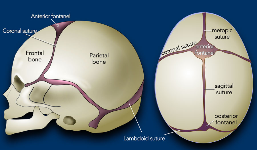

Normal skullHộp sọ bình thường
In the first few years of life the brain grows fast and the skull grows along with it.
This requires open sutures for enchondral growth.
At 1 year of age around 65% of skull growth is accomplished, with the growth rate thereafter slowing down, and reaching around 90% at 5 years of age.Trong những năm đầu đời, não phát triển nhanh và hộp sọ phát triển cùng với nó.
Điều này đòi hỏi phải có các mũi khâu mở để tăng trưởng nội sọ.
Ở tuổi 1, khoảng 65% sự phát triển của hộp sọ được thực hiện, với tốc độ tăng trưởng sau đó chậm lại, và đạt khoảng 90% ở tuổi 5.
Most craniosynostoses are the result of an intrinsic defect in the suture.
Craniosynostosis may be part of a syndrome and in those cases the craniosynostosis is often multi-sutural and there may be other anomalies (cardiac, kidney and skeletal).Hầu hết các trường hợp craniosynostoses là kết quả của một khiếm khuyết nội tại trong mũi khâu.
dính khớp sọ sớm có thể là một phần của hội chứng và trong những trường hợp đó, dính khớp sọ sớm thường đa ổ và có thể có các dị tật khác (tim, thận và xương).
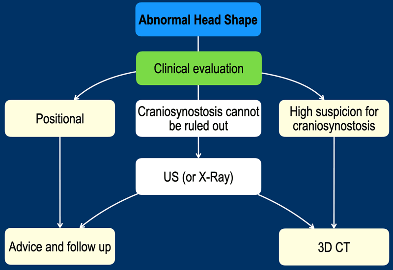
Clinical evaluationĐánh giá lâm sàng
In the past decades there has been an increase in the number of children seeking medical attention because of an abnormal head shape, which is now reported as high as 1 in 5. Most cases are a positional plagiocephaly rather than a real craniosynostosis. This is often related to the advice of sleeping on the back to prevent sudden infant death syndrome. Trong những thập kỷ qua, số trẻ em tìm đến chăm sóc y tế ngày càng tăng do hình dạng đầu bất thường, hiện nay được báo cáo là 1/5. Hầu hết các trường hợp là bệnh sọ não vị trí chứ không phải là dính khớp sọ sớm thật, điều này thường liên quan đến lời khuyên ngủ ngửa để ngăn ngừa hội chứng đột tử ở trẻ sơ sinh.
A well-trained clinician will often be able to recognize the typical presentation of a positional plagiocephaly and distinguish from its main differential diagnosis of a unilateral lambdoid stenosis (see below).
The parents may then be reassured because the condition is self-limiting and with adequate advice usually reversible.Một bác sĩ lâm sàng được đào tạo tốt thường có thể nhận ra biểu hiện điển hình của bệnh sụn viền vị trí và phân biệt với chẩn đoán phân biệt chính của hẹp một bên xương cừu (xem bên dưới).
Sau đó, cha mẹ có thể yên tâm vì tình trạng này có thể tự giới hạn và với lời khuyên đầy đủ thường có thể hồi phục.
In case of a clinical high suspicion of a craniosynostosis timely reference to a specialized center may be advisable before diagnostic imaging is started (figure).Trong trường hợp nghi ngờ lâm sàng cao về dính khớp sọ sớm, có thể nên tham khảo kịp thời đến một trung tâm chuyên khoa trước khi bắt đầu chẩn đoán hình ảnh (hình).
Overview craniosynostosesTổng quan về craniosynostoses
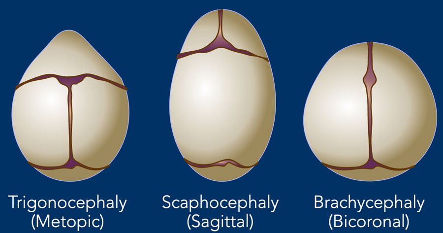

Unisutural and BicoronalUnisutural và Bicoronal
In craniosynostosis the skull deformity is present from birth and will be progressive.
Timely diagnosis is important, as earlier treatment leads to a better outcome. Trong dính khớp sọ sớm, biến dạng hộp sọ xuất hiện từ khi sinh ra và sẽ tiến triển.
Chẩn đoán kịp thời là rất quan trọng, vì điều trị sớm hơn dẫn đến kết quả tốt hơn.
In unisutural and bicoronal craniosynostosis a specific head shape may be expected, depending on the involved suture.
This is depicted in the illustration.Trong dính khớp sọ sớm không khớp và hai giác, có thể mong đợi một hình dạng đầu cụ thể, tuỳ thuộc vào mũi khâu liên quan.
Điều này được mô tả trong hình minh hoạ.
In multisutural involvement the changes are more complex and cannot easily be predicted.Trong tổn thương đa rễ, những thay đổi phức tạp hơn và không thể dễ dàng dự đoán.
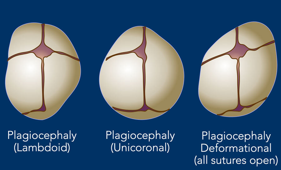

PlagiocephalyBệnh não mủ
In plagiocephaly, also known as flat head syndrome, there is an asymmetrical flattening of one side of the skull.
A mild and widespread form of plagiocephaly is caused by remaining in a supine position for prolonged periods, as discussed above.
This is called deformational plagiocephaly. There is no synostosis and all the sutures are open.Trong bệnh sọ não, còn được gọi là hội chứng đầu phẳng, có một sự phẳng không đối xứng của một bên của hộp sọ.
Một dạng bệnh sán lá phổ biến và nhẹ là do nằm ngửa trong thời gian dài, như đã thảo luận ở trên.
Đây được gọi là tật đầu bẩm sinh biến dạng, không có khớp nối và tất cả các mũi khâu đều mở.
The synostotic form of plagiocephaly can be on the anterior side due to unicoronal closure or on the posterior side due to closure of a lambdoid suture on one side.Thể dính của plagiocephaly có thể ở phía trước do khâu một bên hoặc ở phía sau do khâu một mũi khâu ở một bên.
ImagingHình ảnh
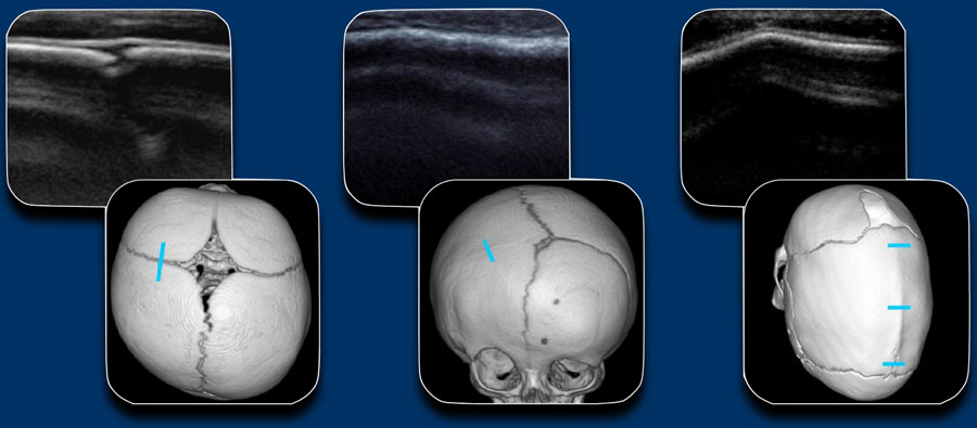
When a craniosynostosis is suspected, the first line imaging will be ultrasound or x-rays, depending on local expertise and preferences. Khi nghi ngờ dính khớp sọ sớm, hình ảnh đầu tiên sẽ là siêu âm hoặc chụp X-quang, tuỳ thuộc vào chuyên môn và sở thích của địa phương.
Because of lack of radiation, ultrasound is the preferred method.
In case of high clinical suspicion low dose 3D CT is preferred.Do thiếu bức xạ, siêu âm là phương pháp được ưu tiên.
Trong trường hợp nghi ngờ lâm sàng cao, CT 3D liều thấp được ưu tiên hơn.
The figure shows the US of a normal suture, an unilateral coronal stenosis and a sagittal stenosis with ridge.Hình cho thấy siêu âm của một mũi khâu bình thường, hẹp vành một bên và hẹp dọc có chóp.
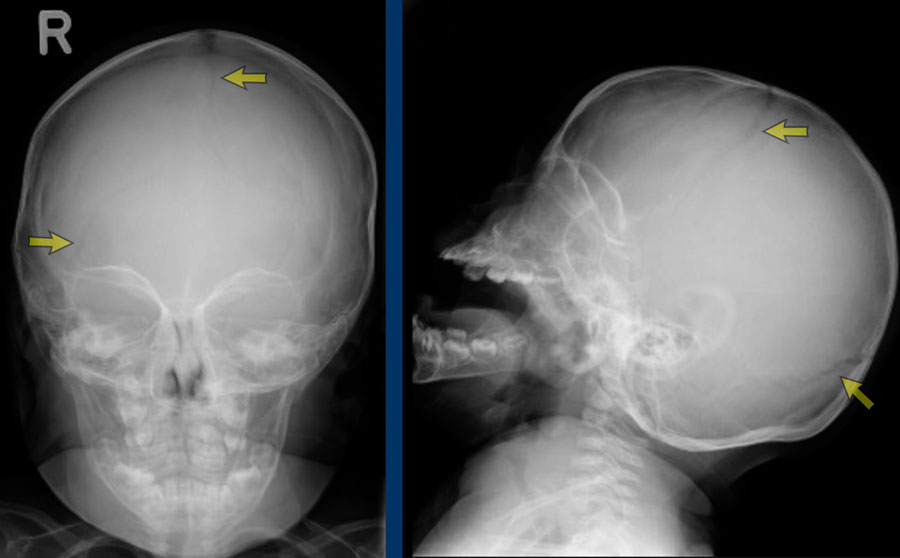
Skull radiographs
demonstrating open sutures.
X quang sọ cho thấy các mũi khâu mở.
Be aware that radiographs have a limited value for assessment of the sagittal suture and a partial closure of the sagittal suture may be overlooked. Lưu ý rằng phim X quang có giá trị hạn chế để đánh giá đường khâu dọc và có thể bỏ qua khâu kín một phần đường khâu dốc.
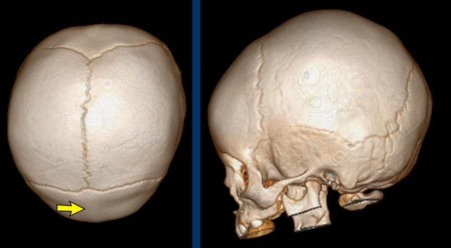
3D CTChụp cắt lớp 3D
The images show open sutures in a child with a physiologically closed metopic suture (arrow).Hình ảnh cho thấy các mũi khâu mở ở một đứa trẻ có mũi khâu cơ địa đóng sinh lý (mũi tên).
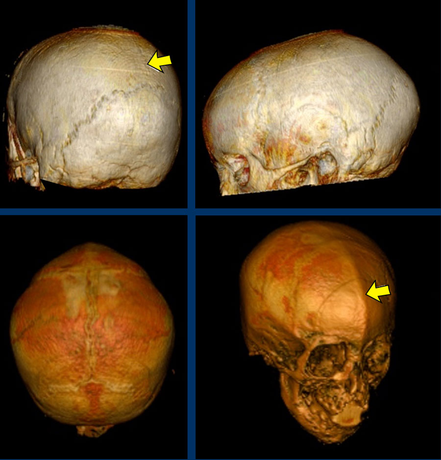 Images courtesy of Dr K.A. Eley and Dr C. DelsoHình ảnh do bác sĩ K.A. Eley và bác sĩ C. Delso cung cấp
MRI
It is also possible to evaluate the sutures with black bone MRI, but as MRI often requires anaesthesia this will mainly be done in conjunction with an indication for evaluation of intracranial abnormalities and complications.Cũng có thể đánh giá các mũi khâu bằng MRI xương đen, nhưng vì MRI thường đòi hỏi phải gây mê nên chủ yếu sẽ được thực hiện cùng với chỉ định đánh giá các bất thường và biến chứng nội sọ.
Images
Black bone MRI demonstrating
closed sagittal suture (top) and closed metopic suture (down) in two different
patients.Hình ảnh
CHT xương đen cho thấy có sự khác biệt giữa hai nhóm bệnh nhân có chỉ dọc đóng (trên) và chỉ metopic đóng (dưới).
Positional plagiocephalyBệnh sỏi thận vị trí
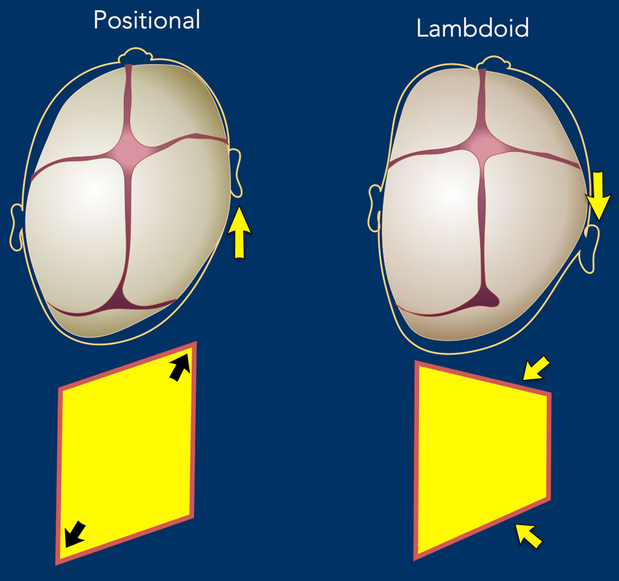

Positional or deformational plagiocephaly refers to the flattening of the head due to the baby’s preferred position.
It is not a synostosis, but the most common form of temporary skull deformity.Tật sụp mi lệch vị trí hoặc biến dạng đề cập đến sự phẳng của đầu do vị trí ưa thích của em bé.
Đây không phải là một khớp thần kinh, nhưng là dạng phổ biến nhất của biến dạng sọ tạm thời.
Typically, the entire side is pushed forward, hence the ear will be seen more anterior than on the contralateral side (arrow).
Thông thường, toàn bộ bên bị đẩy về phía trước, do đó tai sẽ được nhìn thấy phía trước nhiều hơn so với bên đối diện (mũi tên).
This is in contradiction to a posterior plagiocephaly resulting from a unilateral lambdoid suture stenosis, where the mastoid and the ear are pulled down and drawn back.Điều này trái ngược với một bệnh lý sụn viền sau do hẹp chỉ khâu lambdoid một bên, trong đó xương chũm và tai bị kéo xuống và kéo ra sau.
The drawing illustrates the differences. Bản vẽ minh hoạ sự khác biệt.
In positional plagiocephaly the form of the head resembles a parallelogram.Trong bệnh sỏi bể thận vị trí, hình dạng của đầu giống như một hình bình hành.
In unilateral lambdoid stenosis the form of the head resembles a trapezium, because one side of the head is restricted in its growth.Trong hẹp một bên xương cừu, hình dạng của đầu giống như một hình thang, bởi vì một bên của đầu bị hạn chế trong sự phát triển của nó.
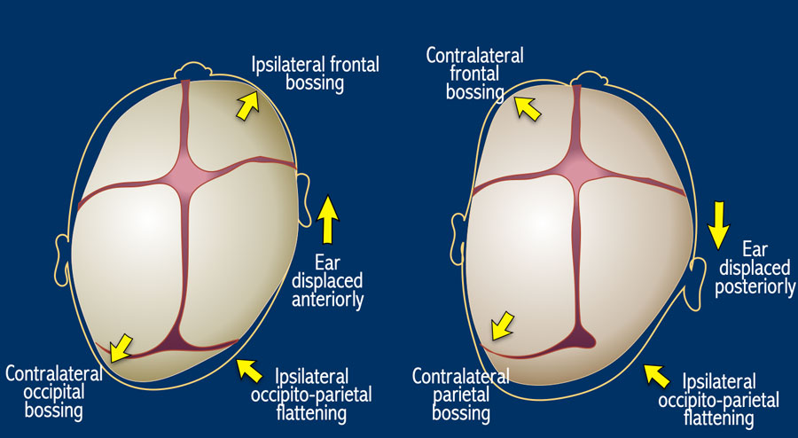

Here the same illustration with now all the differences between deformational and lambdoid plagiocephaly.Đây là minh hoạ tương tự với tất cả sự khác biệt giữa biến dạng và bệnh sụn đầu nhỏ.
NOTE: unilateral coronal stenosis will also result in the ear being positioned more forward – however, flattening will be seen frontal (anterior plagiocephaly) instead of occipital and typically the eye is drawn up on the affected side. Chú ý: Hẹp vành một bên cũng sẽ dẫn đến tai được đặt về phía trước nhiều hơn - tuy nhiên, sẽ thấy phẳng ở trán (dạng đầu trước) thay vì chẩm và thường mắt được vẽ lên ở bên bị ảnh hưởng.
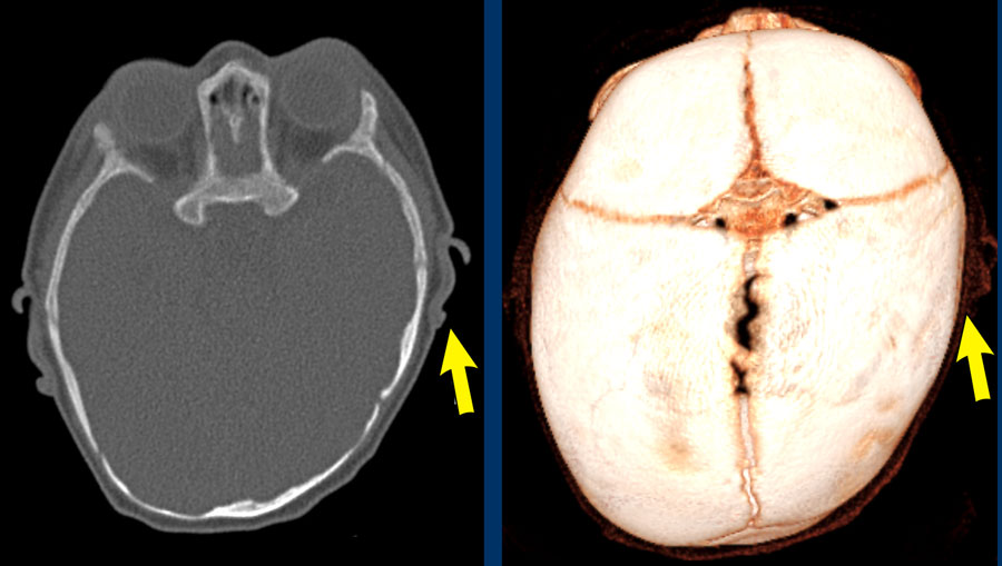
Images
Hình minh họa
Positional
plagiocephaly. The left ear is pushed forward.
Note: Adequate clinical
evaluation and/or ultrasound should have obviated the need for CT. Tật sụp mi tư thế Tai trái bị đẩy về phía trước.
Lưu ý: Đánh giá lâm sàng và / hoặc siêu âm đầy đủ sẽ loại bỏ được nhu cầu chụp CT.
Monosutural craniosynostosisDính khớp sọ sớm đơn sắc
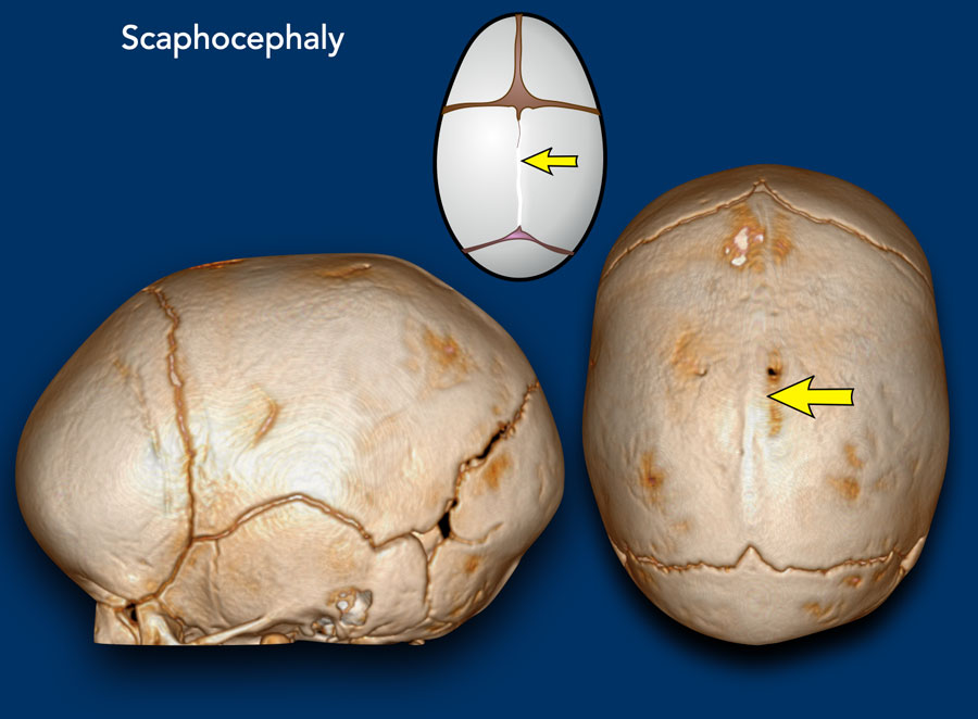

ScaphocephalyBệnh sọ não
Scaphocephaly means ‘boat shaped’ skull and results
from a premature closure of the sagittal suture (arrow).
The skull is narrow and long.Đầu thuyền có nghĩa là hộp sọ hình thuyền và là kết quả của việc khâu khâu dọc (mũi tên) sớm.
Hộp sọ hẹp và dài.
It is the most common monosutural craniosynostosis.
Sometimes it is called
dolichocephaly, as ‘dolicho’ means long.Đây là loại dính khớp sọ sớm đơn sừng phổ biến nhất.
Đôi khi nó được gọi là dolichocephaly, vì 'dolicho' có nghĩa là dài.
Images
Scaphocephaly: The head has a short laterolateral and a long anteroposterior diameter.
Hình ảnh
Đầu ngắn: Đầu có đường kính ngắn và dài.

Trigonocephaly
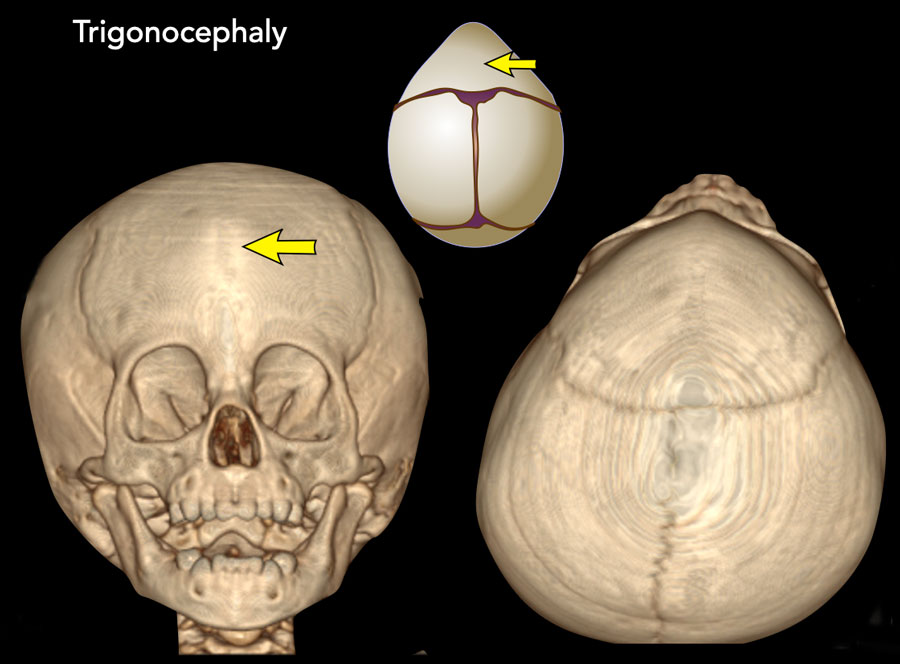
Trigonocephaly refers to the triangular head form resulting from premature closure of the metopic suture (arrows). Hình tam giác đầu là hình dạng đầu tam giác do khâu lỗ cơ địa sớm (các mũi tên).
Physiological closure can already start as early
as three months of age, so premature closure typically presents early.
The
frontal head volume is small with a pointy forehead and hypotelorism. Sự đóng cửa sinh lý có thể bắt đầu sớm nhất là ba tháng tuổi, vì vậy đóng cửa sớm thường xuất hiện sớm.
Thể tích trán nhỏ với trán nhọn và hai mắt dưới.
Only pronounced cases require surgery and if there is clinically no indication for surgery, a CT is not needed.Chỉ những trường hợp rõ rệt mới cần phẫu thuật và nếu lâm sàng không có chỉ định phẫu thuật thì không cần chụp CT.
Images
Trigonocephaly: Pointy forehead and
hypotelorism. Seen from above the lateral orbital rim will be
visible.Hình ảnh
Hình tam giác đầu: Vầng trán nhọn và hai mắt dưới, nhìn từ trên vành ổ mắt bên sẽ thấy được.

BrachycephalyĐầu ngắn
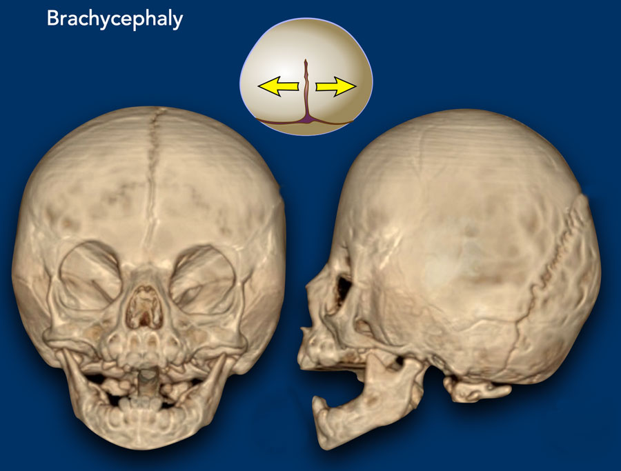
Brachycephaly results from the premature closure
of both coronal sutures.
The frontal skull base is small with
shallow eye sockets with the supraorbital rim drawn back and the head is shortened (‘brachy’) in the anteroposterior direction. Sụp đầu nhanh là kết quả của việc đóng sớm cả hai mũi khâu vành.
Đế sọ trán nhỏ với các hốc mắt nông với vành trên ổ mắt kéo ra sau và đầu được rút ngắn ('brachy') theo hướng trước sau.
Images
Bilateral coronal synostosis with brachycephaly.Hình ảnh
Bệnh nhân có hẹp đầu ngắn hai bên.

Synostotic Anterior plagiocephalyBệnh sụn đầu nhỏ trước xương ức
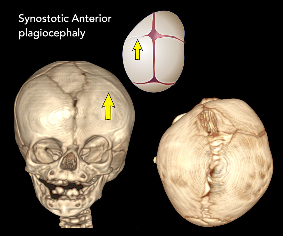
One of the coronal sutures has closed prematurely.
The
forehead is flattened on the affected side with the eye socket drawn up, also
called a harlequin’s eye, and the nose may be deviated.Một trong những mũi khâu ở vành bị đóng sớm.
Vầng trán bị dẹt ở phía bị ảnh hưởng với hốc mắt được vẽ lên, còn được gọi là mắt của harlequin, và mũi có thể bị lệch.
Images
Left sided coronal synostosis. The left forehead is
drawn back with the eye socket lifted up.Hình ảnh
Mối liên kết động mạch vành bên trái Trán trái được kéo ra sau với ổ mắt được nâng lên.

Synostotic posterior plagiocephalyBệnh sỏi màng phổi sau nối liền
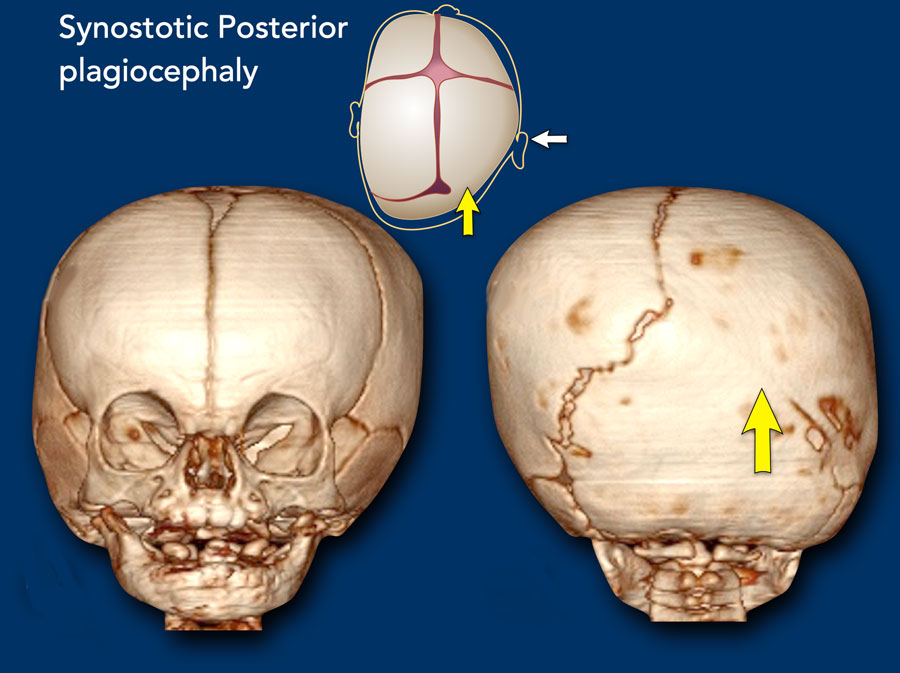
In this case the lambdoid suture has closed
prematurely.
It is the main differential diagnosis for positional plagiocephaly
(see above). Trong trường hợp này, mũi khâu lambdoid đã đóng sớm.
Đây là chẩn đoán phân biệt chính cho bệnh sán lá vị trí (xem ở trên).
There is a risk for Chiari 1 malformation and MRI of the brain and craniocervical junction is advised.Có nguy cơ dị dạng Chiari 1 và nên chụp MRI não và khớp sọ-cổ.
Images
Right sided lambdoid synostose.
Contralateral occipitoparietal bossing is evident.Hình ảnh
Mối liên kết lambdoid bên phải.
Rõ ràng là có hiện tượng chẩm ngoài tâm thu đối bên.
Multisutural craniosynostosisDính khớp sọ sớm đa rễ
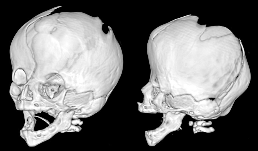
Multisutural craniosynostosis are complex
deformations.
They are usually linked to syndromes and there are several
genetic defects identified.Dính khớp sọ sớm đa rễ là biến dạng phức tạp.
Chúng thường liên quan đến các hội chứng và có một số khiếm khuyết di truyền được xác định.
The best known
craniosynostoses syndromes are Pfeiffer/Crouzon, Apert, Saethre-Chotzen and
Muenke.
In the most severe cases all sutures will eventually close.
Such a
‘pansynostosis’ is a severe condition. Các hội chứng craniosynostoses được biết đến nhiều nhất là Pfeiffer / Crouzon, Apert, Saethre-Chotzen và Muenke.
Trong những trường hợp nặng nhất, tất cả các mũi khâu cuối cùng sẽ đóng lại.
'Hẹp dính khớp' là một tình trạng nặng.
Images
Saethre-Chotzen syndrome. At two months of age there is a bilateral coronal
synostosis with a severely deformed head shape. The fontanel is grossly
enlarged.Hình ảnh
Hội chứng Saethre-Chotzen Ở hai tháng tuổi có một khớp thần kinh vành hai bên với hình dạng đầu bị biến dạng nghiêm trọng, thóp to ra.
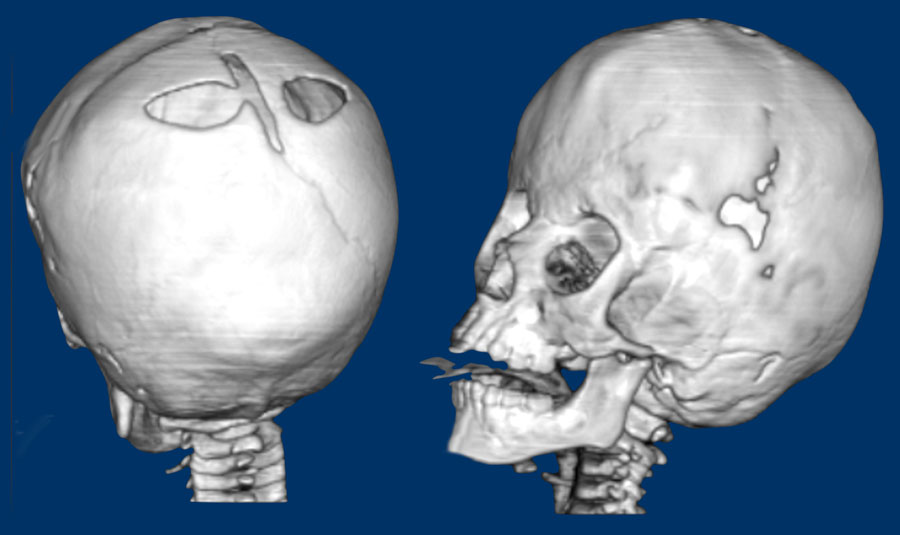
Same patient as above. Cùng một bệnh nhân như trên.
At three years
old there is microcephaly from a near pan-synostosis.
Note enlarged bilateral
parietal foramina. Lúc 3 tuổi, trẻ có đầu nhỏ do dính gần như toàn bộ các khớp sọ. Lưu ý các lỗ xương đỉnh hai bên giãn rộng.
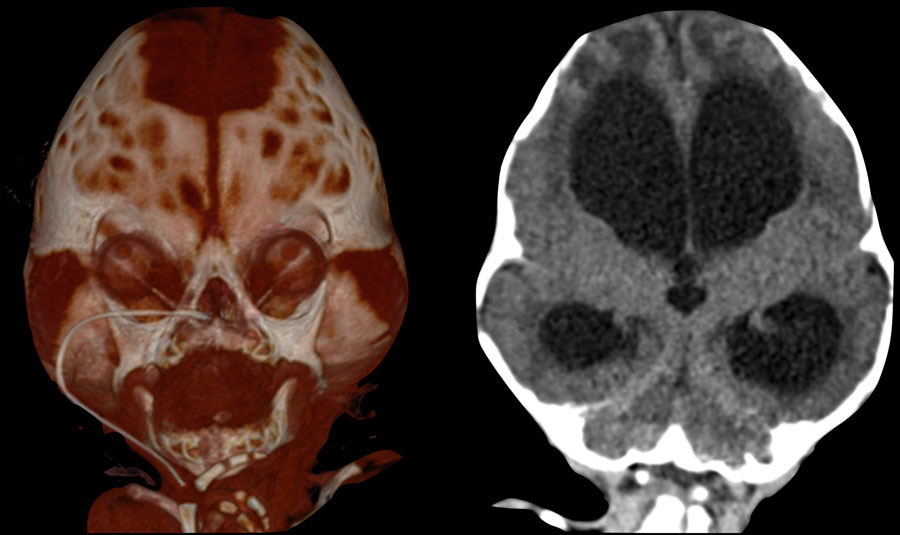
Three-month-old with Pfeiffer syndrome.Trẻ 3 tháng tuổi mắc hội chứng Pfeiffer.
There is a bicoronal synostosis with enlarged anterior and mastoid
fontanels.
Temporal bulging give rise to a trilobate appearance, also called
‘clover leaf deformity’. Có một khớp thần kinh hai vỏ với thóp trước và thóp chũm to ra.
Sự phình ra của thái dương làm tăng hình dạng ba thuỳ, còn được gọi là ‘biến dạng lá cỏ ba lá ’.
Note the ventriculomegaly with periventricular edema.Lưu ý đến cơn to não thất kèm theo phù quanh não thất.
Intracranial complicationsBiến chứng nội sọ
 Raised intracranial pressureTăng áp lực nội sọ
MRI has a main role in detecting associated intracranial malformations and complications from multisutural craniosynostosis. Cộng hưởng từ có vai trò chính trong phát hiện các dị dạng và biến chứng liên quan đến nội sọ từ dính khớp sọ sớm đa rễ.
Images
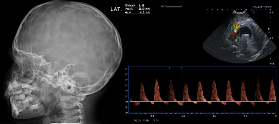
Raised intracranial
pressure.
LEFT: Impressions on a skull radiograph have a low sensitivity,
especially in young children, but when seen, are highly suspicious for raised
intracranial pressure.
RIGHT: Hydrocephalus
with inverted diastolic flow indicating high intracranial pressure. Hình ảnh
Tăng áp lực nội sọ. Trái: Các dấu ấn trên phim sọ có độ nhạy thấp, nhất là ở trẻ nhỏ. Nếu thấy, cần nghĩ nhiều đến tăng áp lực nội sọ. Phải: Não úng thủy kèm đảo chiều dòng chảy thì tâm trương, gợi tăng áp lực nội sọ.

Signs that may indicate raised intracranial pressure are listed in the table on the left.Các dấu hiệu có thể cho thấy áp lực nội sọ tăng lên được liệt kê trong bảng bên trái.
One of these findings is non-specific,
but a combination of findings should raise suspicion.
Increased intracranial
pressure may result in venous collaterals which may be important in
preoperative planning.
Chiari malformation may result in spinal syrinx.Một trong những dấu hiệu này không đặc hiệu, nhưng sự kết hợp của các dấu hiệu nên gây nghi ngờ.
Tăng áp lực nội sọ có thể dẫn đến tổn thương tĩnh mạch bàng hệ, đây là yếu tố quan trọng trong lập kế hoạch trước mổ.
Dị dạng Chiari có thể dẫn đến ống tuỷ.
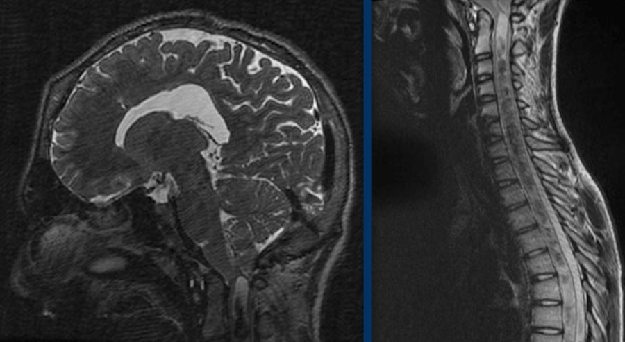
The images are of a patient with increased intracranial pressure.
Hình ảnh là một bệnh nhân có tăng áp lực nội sọ.
There is tonsillar herniation with severe
crowding at the craniocervical junction.
This has resulted in a syrinx over the
entire length of the spinal cord. Có thóat vị amidan với sự chèn ép nặng nề ở ngã ba sọ-cổ.
Điều này đã dẫn đến một syrinx trên toàn bộ chiều dài của tủy sống.
Postoperative complicationsBiến chứng sau mổ
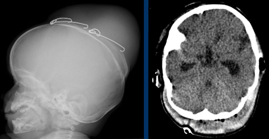
Surgery should be conducted timely in order to reach an optimal outcome. Phẫu thuật nên được tiến hành kịp thời để đạt được kết quả tối ưu.
Timing is dependent on the deformity, but usually preferably under the age of 6 months. Post-operative complications include blood loss, infection, brain swelling and CSF leak. Thời gian phẫu thuật phụ thuộc vào độ biến dạng, nhưng thường là dưới 6 tháng tuổi. Biến chứng sau phẫu thuật bao gồm mất máu, nhiễm trùng, sưng não và rò rỉ dịch não tuỷ.
Images
Direct complications:
LEFT Large effusion/hematoma
from migrating spring.
RIGHT
subgaleal hematoma with severe blood loss.Hình ảnh
Biến chứng trực tiếp:
TRÁI Tràn dịch / khối máu tụ lớn từ mùa xuân di cư.
Máu tụ dưới màng cứng bên phải, mất máu nhiều.
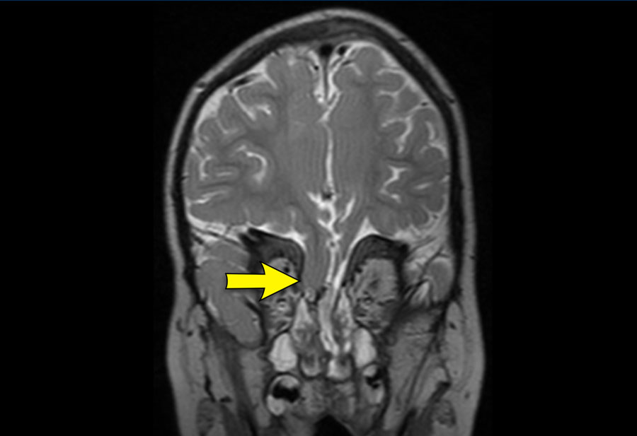
Image
Liquorroe due to CSF leak from a frontobasal
herniation in the upper nasal passage after midface surgery in a patient with Apert syndrome. Hình ảnh
Rỉ dịch não tuỷ do thóat vị trán ở đường mũi trên sau phẫu thuật vùng giữa mặt ở bệnh nhân có hội chứng Apert.
PitfallsCạm bẫy
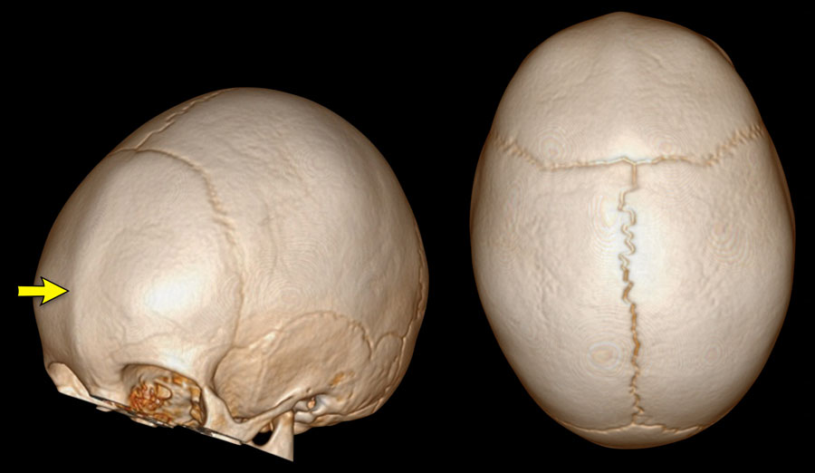 Metopic ridgeSườn núi Metopic
A metopic ridge alone is not equivalent to
trigonocephaly.
Children present at a later age, the frontal skull has a normal
volume and there is no hypotelorism. Chỉ riêng một sườn núi metopic không tương đương với tật đầu tam giác.
Trẻ em xuất hiện muộn hơn, hộp sọ trán có thể tích bình thường và không có giảm thị lực.
It is a common deformity that can be
clinically diagnosed and does not need imaging.
As
with mild trigonocephaly, there is no need for surgery.Đây là một dị tật thường gặp có thể chẩn đoán lâm sàng và không cần chẩn đoán hình ảnh.
Cũng như tật đầu ba nhẹ, không cần phẫu thuật.
Images
Slight metopic ridge in a nine-month-old (arrow). Note that
the scan was taken for a different purpose.
Metopic ridge can be clinically
diagnosed and should not be imaged.
The anterior fontanelle has already closed
but apart from the metopic suture, the other sutures are still open. Hình ảnh
Sườn núi metopic nhẹ ở trẻ chín tháng tuổi (mũi tên) Lưu ý rằng việc chụp ảnh được thực hiện cho một mục đích khác.
Sườn núi Metopic có thể được chẩn đoán lâm sàng và không nên chụp ảnh.
Phần thóp trước đã đóng nhưng ngoài mũi khâu cơ thắt, các mũi khâu khác vẫn còn mở.
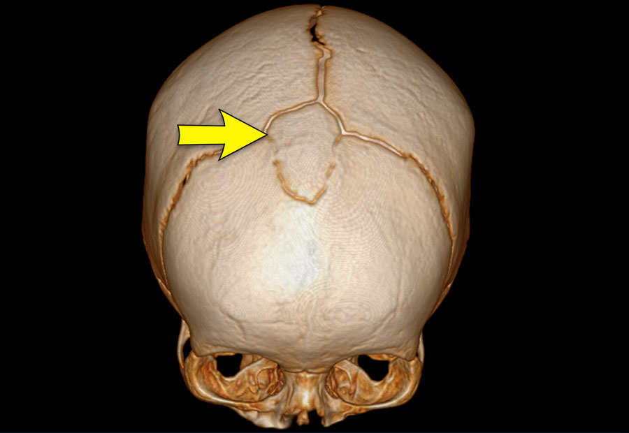
Absent anterior fontanelleKhông có thóp trước
An absent anterior fontanelle does not have to
be a reason for concern.
As long as the skull has a normal shape and
circumference, and the sutures are patent – it does not have any clinical
implications. Không có thóp trước không phải là một lý do để lo lắng.
Miễn là hộp sọ có hình dạng và chu vi bình thường, và các mũi khâu là bằng sáng chế - nó không có bất kỳ ý nghĩa lâm sàng nào.
Image
Large wormian bone occupying the anterior
fontanelle (bregmatic bone) in a six-month-old.
The skull has a normal shape
and the sutures are open. The metopic suture has fused physiologically. Hình ảnh
Một xương Worm lớn chiếm thóp trước (xương bregma) ở trẻ 6 tháng tuổi. Hình dạng sọ bình thường và các khớp sọ còn mở. Khớp trán đã đóng sinh lý.