
Normal Values in Pediatric UltrasoundGiá trị bình thường trong siêu âm nhi khoa
Radiology Departement of the Maastricht University Hospital, Academical Medical Centre in Amsterdam and the Alrijne hospital in Leiden, the NetherlandsPhòng X quang của Bệnh viện Đại học Maastricht, Trung tâm Y khoa Học thuật ở Amsterdam và Bệnh viện Alrijne ở Leiden, Hà Lan
Ngày đăng / cập nhật: 2018-02-09 update 2025-10-27Cập nhật 2018-02-09 2025-10-27
Bản gốc trên radiologyassistant.nl ↗ · tải về lúc 2026-11-19 20:19 · bản dịch máy có chuẩn hóa thuật ngữ chẩn đoán hình ảnh
This document provides an overview of normal reference values for ultrasound examinations in neonates and children.
The items are in an alphabethic order.
Select one of the items listed on the left for details.Tài liệu này cung cấp một cái nhìn tổng quan về các giá trị tham chiếu bình thường cho việc siêu âm ở trẻ sơ sinh và trẻ em.
Các mục được sắp xếp theo thứ tự bảng chữ cái.
Chọn một trong các mục được liệt kê ở bên trái để biết chi tiết.
AdrenalBướu
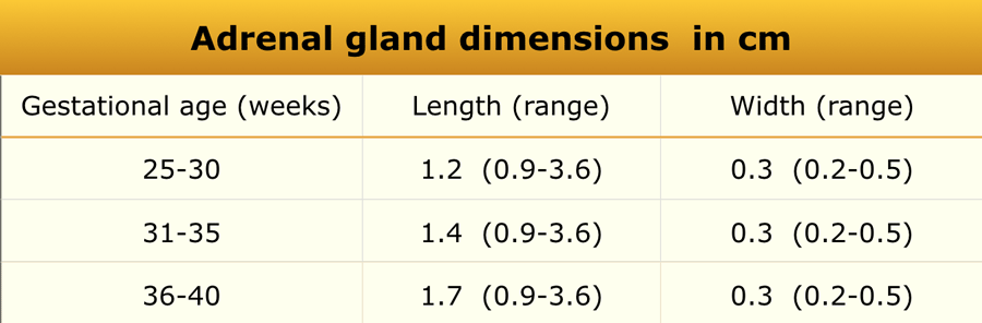
Materials and Methods
An ultrasonographic study was performed in 92 infants.Phương pháp nghiên cứu
Nghiên cứu siêu âm được thực hiện trên 92 trẻ nhũ nhi.


MeasurementsĐo lường
- Length (L): Defined as the maximum cephalocaudal dimension of the adrenal gland, measured in either the coronal or sagittal plane.Chiều dài (L): Được xác định là kích thước cực đại của tuyến thượng thận, được đo bằng vành hoặc mặt phẳng đứng dọc.
- Width (w): Defined as the maximum thickness of one of the limbs.Chiều rộng (w): Được xác định là độ dày tối đa của một trong các chi.
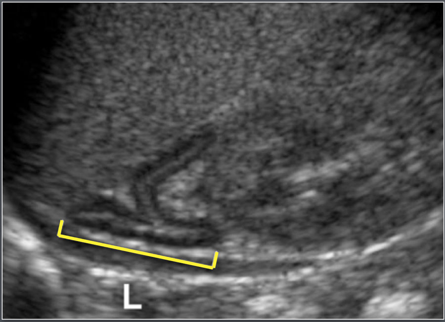
Causes of Adrenal EnlargementNguyên nhân của sự mở rộng tuyến thượng thận
- Congenital adrenal hyperplasiaTăng sản thượng thận bẩm sinh
- Adrenal hemorrhageXuất huyết thượng thận
- Adrenal neuroblastomaU nguyên bào thần kinh thượng thận
AppendixRuột thừa
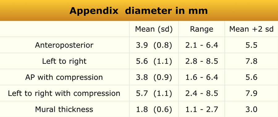
Materials and Methods
An ultrasonographic study was conducted in 146 consecutive patients (62 boys and 84 girls; mean age, 7 years; age range, 2–15 years).
Exclusion criteriaPhương pháp nghiên cứu
Nghiên cứu siêu âm tim được thực hiện trên 146 bệnh nhân (62 nam và 84 nữ, tuổi trung bình 7 tuổi, độ tuổi từ 2-15 tuổi).
Tiêu chuẩn loại trừ
- Cystic fibrosisXơ nang
- Acute abdominal painĐau bụng cấp tính
- Previous appendectomyCắt ruột thừa trước đây
- Age <2 years (due to difficulty in performing the examination)Tuổi < 2 tuổi (do khó thực hiện khám)
The appendix was successfully visualized in 120 children.Ruột thừa đã được hiển thị thành công ở 120 trẻ.
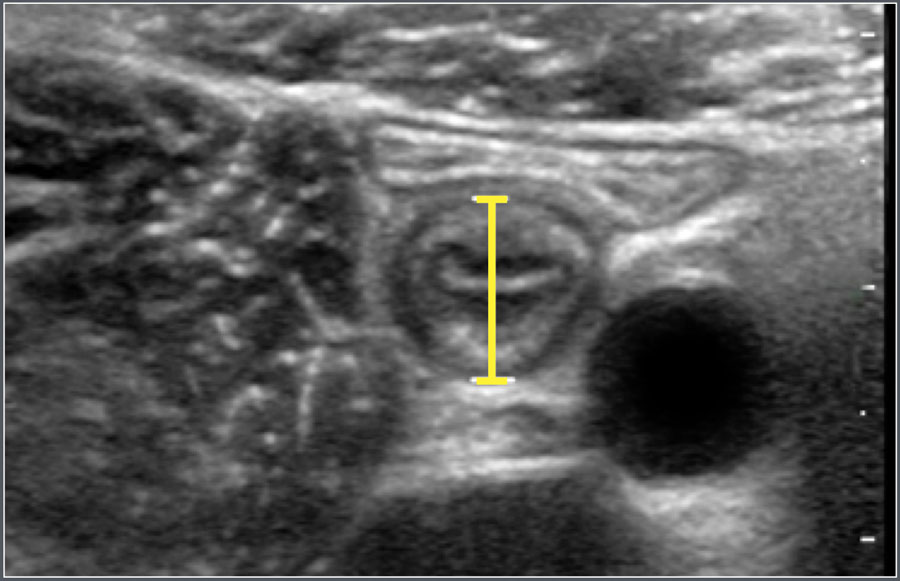
Causes of Appendiceal EnlargementNguyên nhân của phì đại ruột thừa
- Acute appendicitisCấp tính viêm ruột thừa
- Cystic fibrosisXơ nang
- Lymphoid hypertrophy (due to immune deficiency or viral enteritis)Phì đại lympho (do suy giảm miễn dịch hoặc viêm ruột do virus)
- Intraluminal gas, mucus, or fecal materialKhí trong lòng, chất nhầy, hoặc chất thải
- Mucocele
BladderBàng quang
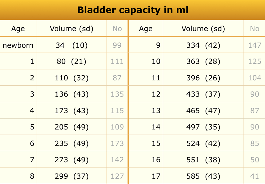
Bladder volumeThể tích bàng quang
Materials and Methods
A total of 3,376 children were included in this ultrasonographic study.
Note: The total number of patients listed in the results tables may differ, as not all age subgroups were represented.Phương pháp nghiên cứu
Tổng cộng có 3.376 trẻ được đưa vào nghiên cứu.
Lưu ý: Tổng số bệnh nhân được liệt kê trong bảng kết quả có thể khác nhau, vì không phải tất cả các nhóm tuổi đều được đại diện.
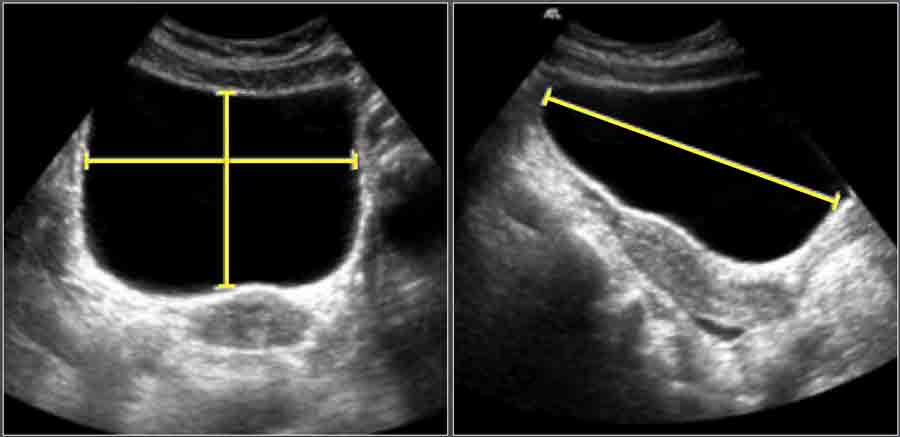
Measurement TechniqueKỹ thuật đo lường
- Length (L): Measured on a longitudinal scan from the bladder neck to the fundus.Chiều dài (L): Đo trên phim chụp dọc từ cổ bàng quang đến đáy.
- Depth (D): Measured perpendicular to the longitudinal plane at the level of maximum cross-sectional area, from the anterior to posterior mucosal surface in the midline.Độ sâu (D): Đo vuông góc với mặt phẳng dọc ở mức diện tích mặt cắt ngang tối đa, từ bề mặt niêm mạc trước ra sau ở đường giữa.
- Width (W): Measured perpendicular to D at its midpoint.Chiều rộng (W): Đo vuông góc với D tại điểm giữa của nó.
Bladder Volume Calculation
Bladder volume was calculated using the ellipsoid formula:Tính toán khối lượng bàng quang
Thể tích bàng quang được tính theo công thức ellipsoid:
Volume (cm³)=L×D×W×0.523Volume (cm³)=L×D×W×0.523Thể tích (cm3)=L×D×W×0.523 Thể tích ( cm3)=L × D × W × 0.523
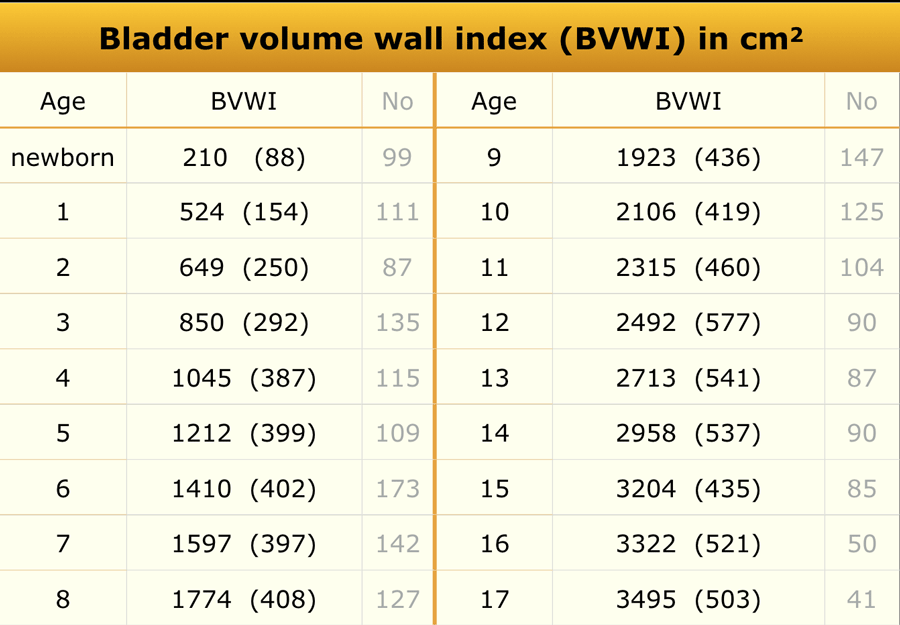
Bladder wall thicknessĐộ dày thành bàng quang
Materials and Methods
A total of 3,376 children were included.
Bladder wall thickness was measured only when the residual bladder volume was <10% of the pre-voided volume.Phương pháp nghiên cứu
Tổng số là 3.376 trẻ được đưa vào nghiên cứu.
Độ dày thành bàng quang chỉ được đo khi thể tích bàng quang tồn dư < 10% thể tích trước khi đo.
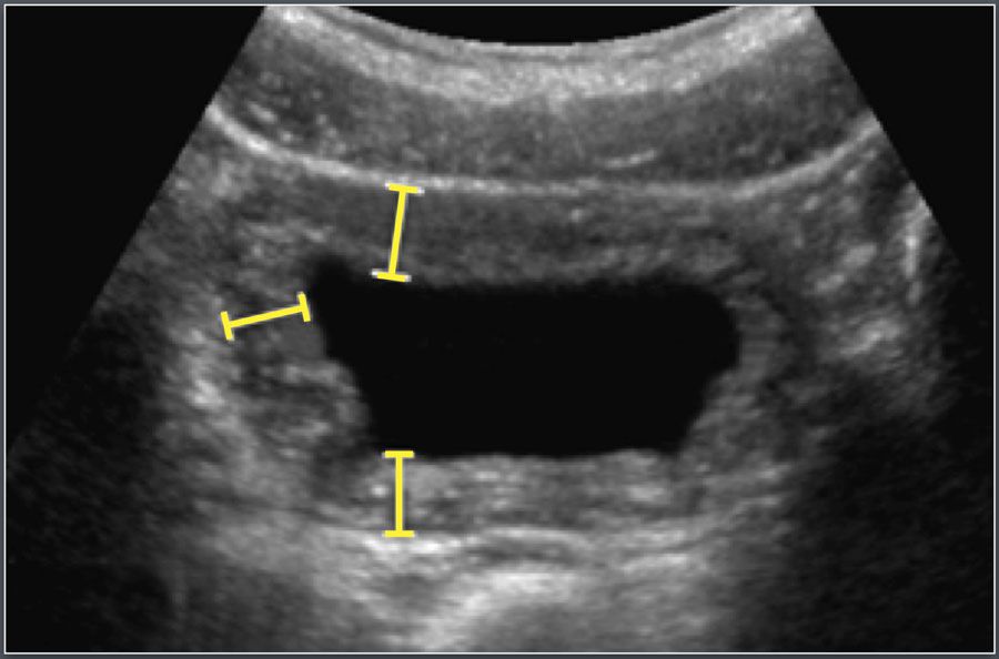
Measurement Technique
The bladder wall was evaluated on a zoomed transverse image of the emptied bladder.
Measurements were taken at three sites:Kỹ thuật đo lường
Thành bàng quang được đánh giá trên hình ảnh cắt ngang thu nhỏ của bàng quang trống.
Các phép đo được thực hiện tại ba địa điểm:
- AnterolateralTrước ngoài
- LateralNgoài
- PosterolateralSau bên
The mean of these three measurements was used for analysis.Giá trị trung bình của ba phép đo này được sử dụng để phân tích.
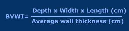
Bladder Volume Wall Thickness Index (BVWTI)
Bladder wall thickness depends on the degree of bladder filling and bladder capacity.
Therefore, it is expressed as the Bladder Volume Wall Thickness Index (BVWI).Chỉ số độ dày thành khối lượng bàng quang (BVWTI)
Độ dày thành bàng quang phụ thuộc vào mức độ đầy bàng quang và dung tích bàng quang.
Do đó, nó được thể hiện dưới dạng Chỉ số độ dày thành thể tích bàng quang (BVWI).
It helps normalize bladder wall measurements across different degrees of bladder distension and improves diagnostic accuracy for lower urinary tract abnormalities, especially in children.Nó giúp bình thường hóa các phép đo thành bàng quang ở các mức độ chướng bàng quang khác nhau và cải thiện độ chính xác trong chẩn đoán các bất thường đường tiết niệu dưới, đặc biệt là ở trẻ em.
Causes of Bladder Wall ThickeningNguyên nhân của dày thành bàng quang
- CystitisViêm bàng quang
- Dysfunctional voidingRối loạn chức năng đi tiểu
- Posterior urethral valvesVan niệu đạo sau
BowelRuột
Materials and Methods
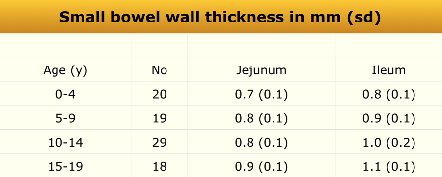
The study population consisted of 128 patients (57 males and 71 females). Of these, 86 individuals were aged between 1 and 19 years (only data pertaining to this subgroup are presented).Phương pháp nghiên cứu
Số lượng bệnh nhân nghiên cứu là 128 người (57 nam và 71 nữ), trong đó có 86 người từ 1 đến 19 tuổi (chỉ có dữ liệu liên quan đến nhóm này được trình bày).
Bowel wall thickness was measured on transverse ultrasound sections and included the following histologic layers: mucosa, lamina propria, muscularis mucosae, submucosa, and muscularis propria.Độ dày thành ruột được đo trên siêu âm cắt ngang và bao gồm các lớp mô học sau: niêm mạc, lớp đệm, lớp cơ niêm, lớp dưới niêm mạc và lớp cơ niên.
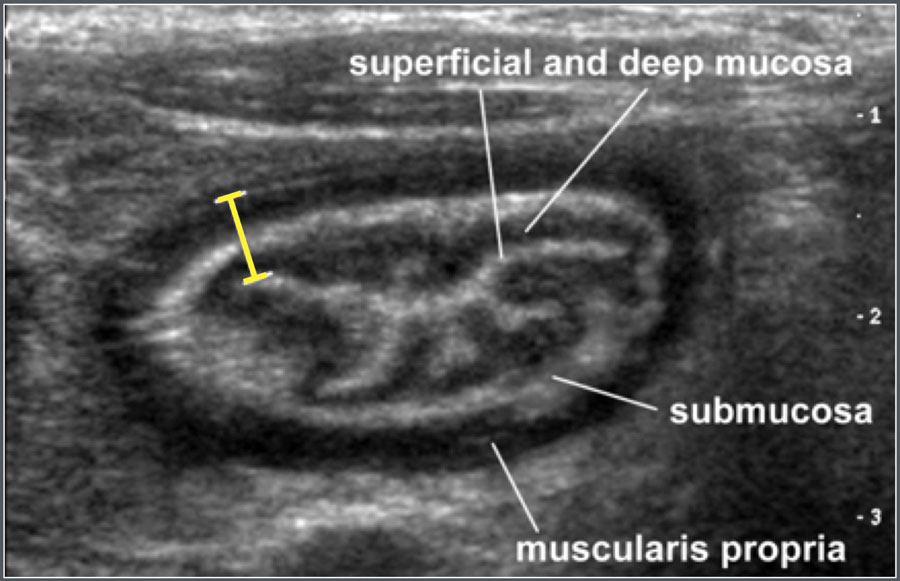

Ultrasonographic example
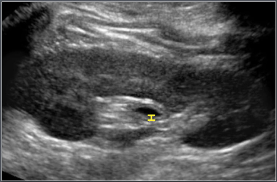
Measurement of the terminal ileum wall thickness in a 12-year-old male patient with cystic fibrosis.Ví dụ siêu âm
Đo độ dày thành đoạn cuối hồi tràng ở một bệnh nhân nam 12 tuổi bị xơ nang.
Common causes of small bowel wall thickening include:Nguyên nhân thường gặp của dày thành ruột nhỏ bao gồm:
- Henoch–Schönlein purpuraBan xuất huyết Henoch-Schönlein
- Crohn’s diseaseBệnh Crohn
- Lymphoma
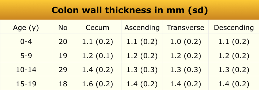
In the same study, colonic wall thickness was also measured.Trong nghiên cứu này, độ dày thành đại tràng cũng được đo.
Common causes of colonic wall thickening include:Các nguyên nhân thường gặp của dày thành đại tràng bao gồm:
- Inflammatory bowel disease (IBD)Bệnh viêm ruột (IBD)
- Hemolytic–uremic syndrome (HUS)Hội chứng tan máu-urê huyết (HUS)
- Pseudomembranous colitisViêm đại tràng giả mạc
Common Bile DuctỐng mật chủ
Materials and MethodsPhương pháp nghiên cứu
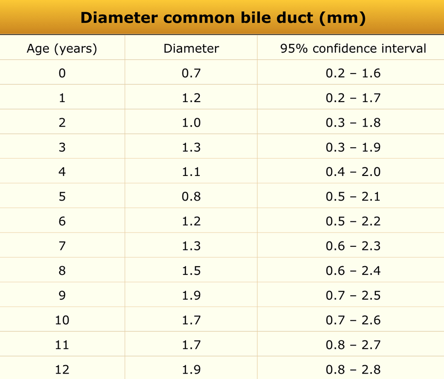
A total of 173 consecutive pediatric patients (100 boys and 73 girls) referred for abdominal ultrasonography unrelated to hepatobiliary pathology were included. The age range was 1 day to 13 years (median 5.0 years).Tổng cộng có 173 bệnh nhi (100 nam và 73 nữ) được đưa vào nghiên cứu không liên quan đến bệnh lý gan mật, tuổi từ 1 ngày đến 13 tuổi (trung vị 5,0 tuổi).
In all subjects, the diameter of the common bile duct measured ≤ 3.3 mm.Đường kính ống mật chủ đo được ở tất cả các đối tượng ≤ 3,3 mm.
Image
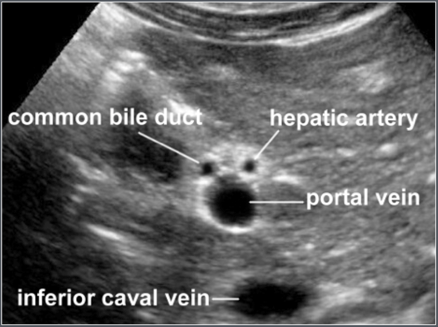
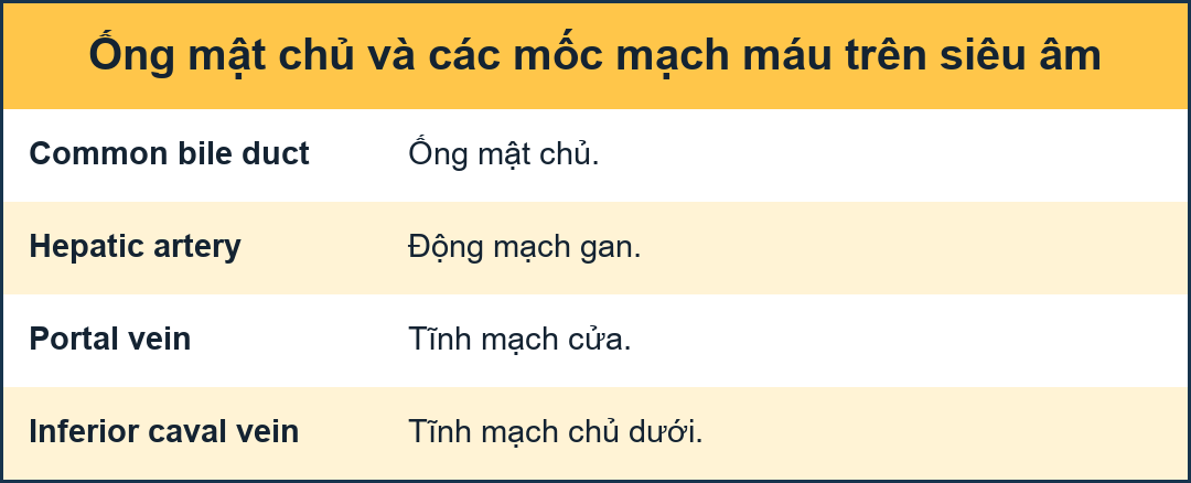
Transverse ultrasonographic image of the common bile duct and surrounding anatomy.Hình ảnh
Hình ảnh siêu âm cắt ngang ống mật chủ và giải phẫu xung quanh.
GalbladderTrang chủ
Materials and Methods
Ultrasonographic gallbladder volume was calculated using the ellipsoid formula:
Volume = length × width × height × 0.52Phương pháp nghiên cứu
Thể tích túi mật trên siêu âm được tính theo công thức ellipsoid:
Thể tích = chiều dài × chiều rộng × chiều cao × 0,52
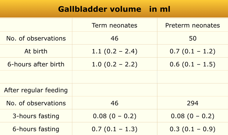
The study included 50 preterm infants (mean gestational age 31.7 ± 2.5 weeks, mean birth weight 1556 ± 441 g) and 46 term infants (mean gestational age 38.3 ± 1.2 weeks, mean birth weight 3253 ± 440 g).Nghiên cứu gồm 50 trẻ sinh non (tuổi thai trung bình 31,7 ± 2,5 tuần, cân nặng trung bình 1556 ± 441 g) và 46 trẻ sinh đủ tháng (tuổi sinh trung bình 38,3 ± 1,2 tuần, cân trọng trung bình 3253 ± 440 g).
Measurements were obtained shortly after birth, following 6 hours of fasting, and again at 5–7 days of age after 3-hour and 6-hour fasting intervals following regular milk feeding.Các phép đo được thực hiện ngay sau khi sinh, sau 6 giờ nhịn ăn, và một lần nữa ở ngày thứ 5-7 sau khi nhịn ăn 3 giờ và 6 giờ sau khi cho bú sữa thường xuyên.
Common causes of a small gallbladder include:Các nguyên nhân thường gặp của túi mật nhỏ bao gồm:
- Biliary atresiaTeo đường mật
- Postprandial (after fatty meal) contractionCo thắt sau ăn (sau bữa ăn nhiều chất béo)
HipKhớp háng
Anterior recessLõm trước
Materials and Methods
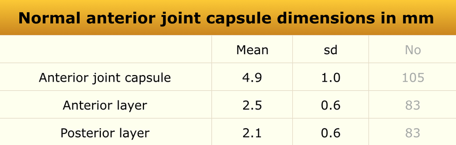
An ultrasonographic study was performed in 58 healthy children and 105 children with unilateral transient synovitis, aged 1.7–12.8 years. Phương pháp nghiên cứu
Nghiên cứu siêu âm được thực hiện trên trẻ 58 khoẻ mạnh và 105 có viêm màng hoạt dịch thóang qua một bên, tuổi 1.7–12.8.
All examinations were performed with the patient in the supine position and the hips in a neutral position.Tất cả các lần khám đều được thực hiện với bệnh nhân ở tư thế nằm ngửa và hông ở tư thế trung tính.
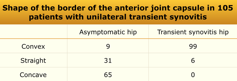
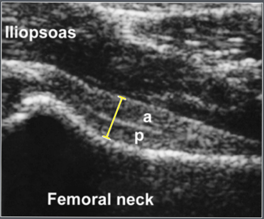
The anterior joint capsule was measured, including both its anterior and posterior layers. The anterior contour of the joint capsule was also evaluated.
No statistically significant correlation was found between age and anterior joint capsule thickness.Đo bao khớp trước, bao gồm cả lớp trước và lớp sau, đánh giá đường viền bao khớp trước.
Không có mối tương quan có ý nghĩa thống kê giữa tuổi và độ dày bao khớp trước.
A difference greater than 2 mm between sides or an effusion >2 mm was considered abnormal.Chênh lệch lớn hơn 2 mm giữa các bên hoặc tràn dịch > 2 mm được coi là bất thường.

Shape of the border of the anterior joint capsuleHình dạng bờ bao khớp trước
The anterior contour of the joint capsule can be evaluated.
Ultrasonographic measurement of the anterior joint capsule.
Both anterior (a) and posterior (p) layer can be identified.Có thể đánh giá đường viền trước bao khớp.
Đo siêu âm bao khớp trước.
Cả hai lớp trước (a) và sau (p) có thể được xác định.
Common causes of hip joint effusion include:Nguyên nhân thường gặp của tràn dịch khớp khớp háng bao gồm:
- Transient synovitisViêm màng hoạt dịch thóang qua
- Septic arthritisViêm khớp nhiễm khuẩn
- Juvenile idiopathic arthritis (JIA)Viêm khớp tự phát vị thành niên (JIA)
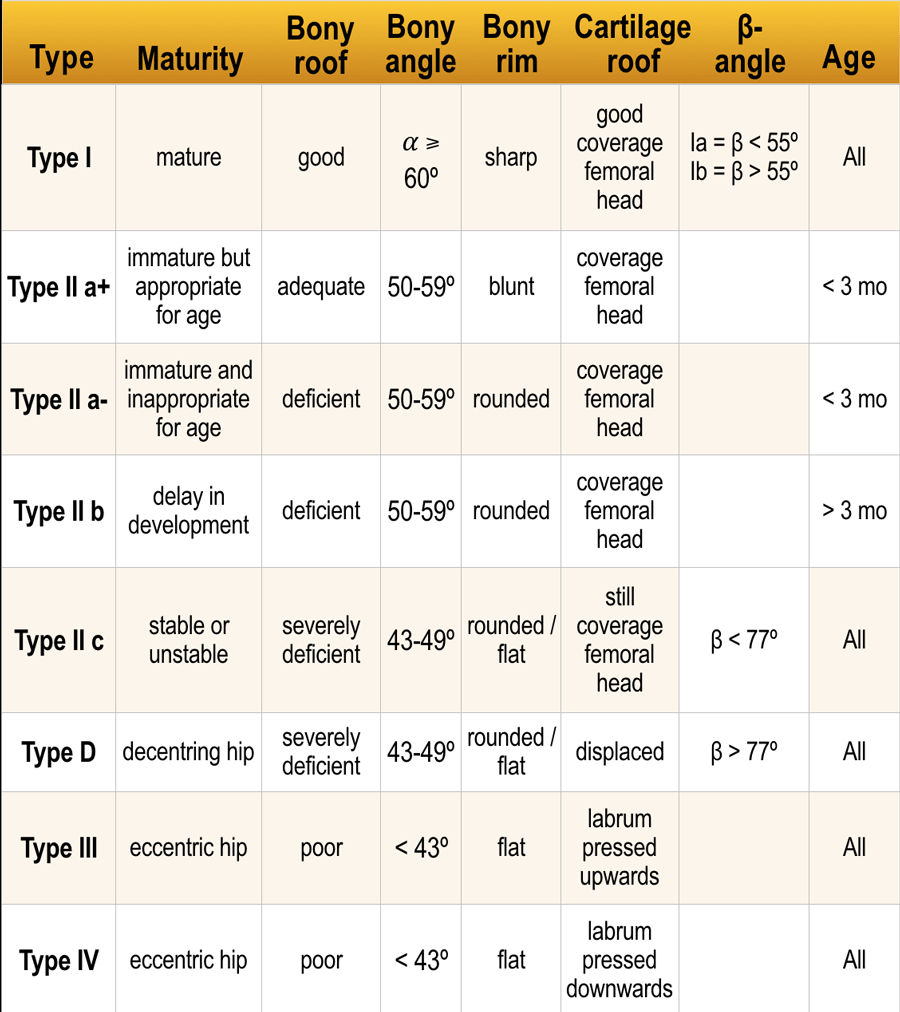
Developmental Dysplasia of the Hip (Graf’s Classification)
Loạn sản phát triển xương hông (Phân loại của Graf)
- Type I: Mature, centered hip joint with a well-developed acetabular roof and an angular or slightly blunt bony rim.Loại I: Khớp háng trưởng thành, có tâm với phần mái ổ cối phát triển tốt và vành xương hơi dẹt hoặc góc cạnh.
- Type II: Centered hip joint with a deficiently developed acetabular roof and a rounded bony rim.Loại II: Khớp háng giữa với phần ổ cối phát triển kém và vành xương tròn.
- Type III: Decentered hip joint with a poorly developed acetabular roof and a flattened bony rim.
Loại III: Khớp háng mất trung tâm với phần mái ổ cối kém phát triển và vành xương dẹt.
Click here for article on Developmental Dysplasia of the Hip.Nhấp vào đây để xem bài viết về loạn sản phát triển của khớp háng.
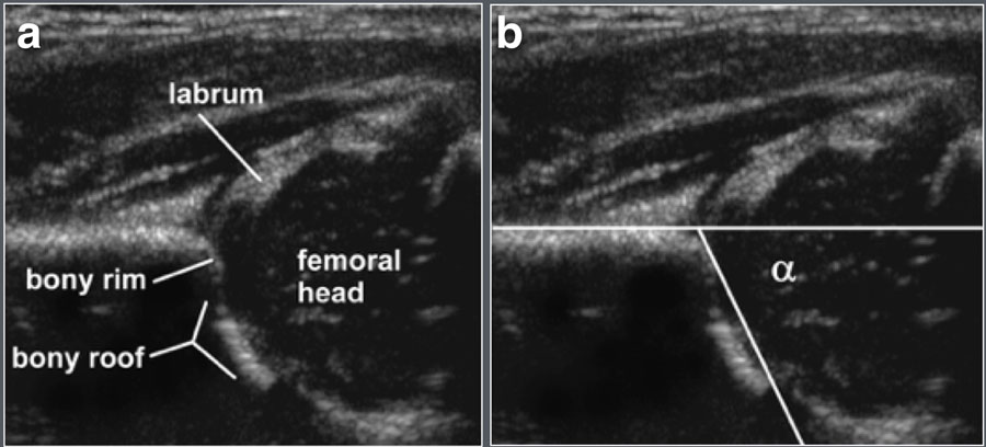

Ultrasonographic parameters:Các thông số siêu âm:
- Coronal plane view of the hip jointHình mặt phẳng đứng ngang của khớp háng
- Measurement of the α (alpha) angleĐo góc α (alpha)
KidneyBệnh thận
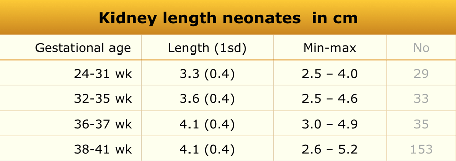
Preterm and Term BabiesTrẻ sơ sinh non tháng và đủ tháng
Materials and Methods
Ultrasonographic evaluation was performed in 261 healthy newborn infants.
The craniocaudal dimension of each kidney was measured using standard ultrasonography.Phương pháp nghiên cứu
Đánh giá siêu âm được thực hiện trên 261 trẻ sơ sinh khoẻ mạnh.
Kích thước sọ não của mỗi quả thận được đo bằng siêu âm tiêu chuẩn.
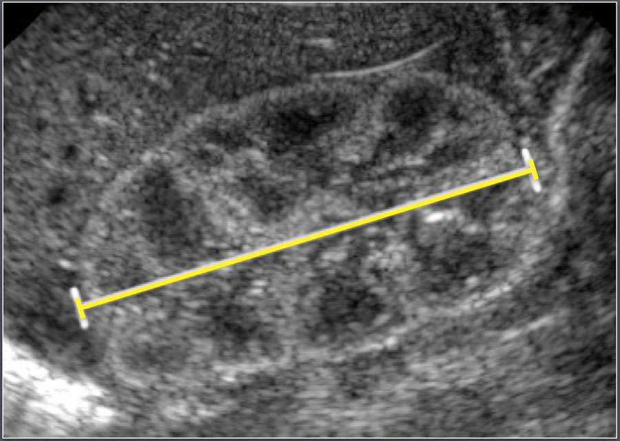
Ultrasonographic measurement of the length of a neonatal kidney.Đo siêu âm chiều dài thận của trẻ sơ sinh.
In neonates, the renal parenchyma is typically more echogenic than the liver parenchyma, which is a normal finding at this age.Ở trẻ sơ sinh, nhu mô thận thường có nhiều tiếng vang hơn nhu mô gan, đây là một dấu hiệu bình thường ở độ tuổi này.
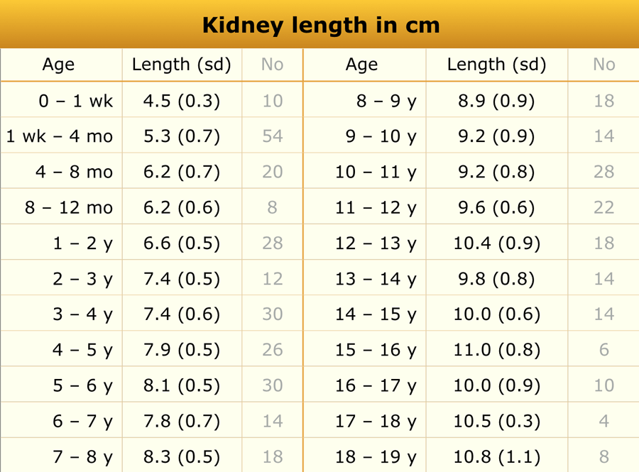
ChildrenTrẻ em
Materials and Methods
The study included 203 pediatric patients. Exclusion criteria included a history of malignancy, steroid use, upper urinary tract abnormalities, vesicoureteral reflux (VUR) greater than grade I, urologic surgery, or abnormal renal sonographic findings.Phương pháp nghiên cứu
Nghiên cứu gồm 203 bệnh nhi, tiêu chuẩn loại trừ bao gồm tiền sử ác tính, sử dụng steroid, bất thường đường tiết niệu trên, trào ngược bàng quang niệu quản (VUR) lớn hơn độ I, phẫu thuật tiết niệu hoặc bất thường trên siêu âm thận.
On average, the left kidney measured 1.9 mm longer than the right.Trung bình, thận trái dài hơn thận phải 1,9 mm.
Common causes of renal enlargement include:Các nguyên nhân phổ biến của thận to bao gồm:
- Duplicated collecting systemHệ thống thu thập dữ liệu
- Nephritis (infectious and non-infectious)Viêm thận (nhiễm trùng và không nhiễm trùng)
- Leukemia or lymphomaBệnh bạch cầu hoặc u lympho
- Autosomal recessive polycystic kidney disease (ARPKD)Bệnh thận đa nang lặn trên nhiễm sắc thể thường (ARPKD)
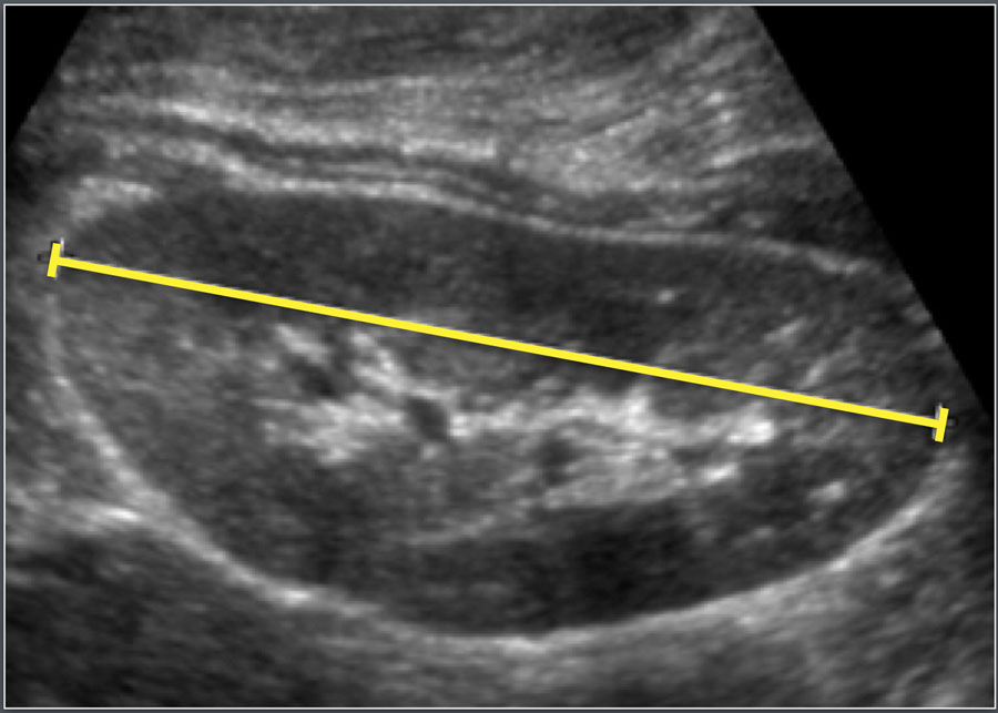
Ultrasonographic measurement of the length of a kidney.Đo siêu âm chiều dài của thận.
Renal volumeThể tích thận
Materials and Methods
A total of 3,376 children were included in this ultrasonographic study.
Phương pháp nghiên cứu
Tổng cộng có 3.376 trẻ được đưa vào nghiên cứu.
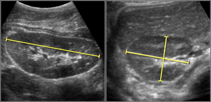
Renal volume was calculated using the ellipsoid formula:
Volume = Length × Width × Depth × 0.523Thể tích thận được tính theo công thức ellipsoid:
Thể tích = Dài × Rộng × Dài x 0,523
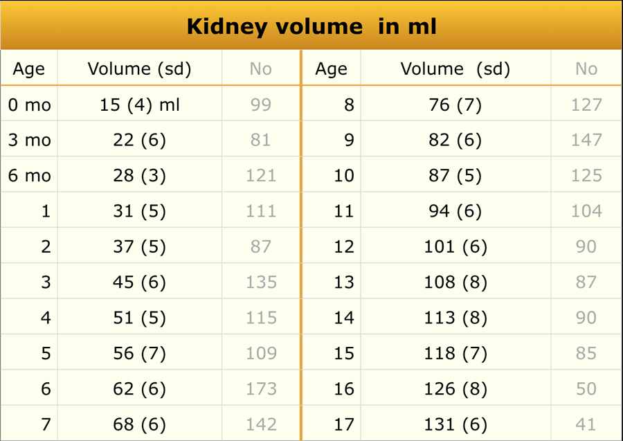
The total renal volume was determined by summing the right and left kidney volumes.
The reported values represent the average of both kidneys.Thể tích thận toàn phần được xác định bằng cách cộng thể tích thận phải và trái.
Các giá trị được báo cáo đại diện cho mức trung bình của cả hai thận.
 Adapted from reference 18Chuyển thể từ tài liệu tham khảo 18
Thickness of the wall of the collecting systemĐộ dày thành của hệ thống thu gom
Materials and Methods
Ultrasonographic examination of 48 renal collecting systems in 24 healthy children (aged 3 days–12.6 years) was performed.
The wall of the collecting system was identifiable in all kidneys, ranging from 0 (not visible) to 0.8 mm in thickness.
A wall thickness ≥ 1 mm was considered abnormal.Phương pháp nghiên cứu
Siêu âm 48 hệ thống thu thập nước tiểu ở 24 trẻ em (từ 3 ngày tuổi đến 12,6 tuổi) được thực hiện.
Thành của hệ thống thu thập có thể nhận dạng được ở tất cả các thận, có độ dày từ 0 (không nhìn thấy) đến 0,8 mm.
Bề dày thành ≥ 1 mm được coi là bất thường.
Common causes of collecting system wall thickening include:Các nguyên nhân phổ biến của dày thành hệ thống thu thập bao gồm:
- Urinary tract infection (UTI)Nhiễm trùng đường tiết niệu (UTI)
- Intermittent dilatation (e.g., vesicoureteral reflux)Giãn không liên tục (ví dụ, trào ngược bàng quang niệu quản)
- Recent or resolved dilatationNở miệng gần đây hoặc đã được giải quyết
LiverGan
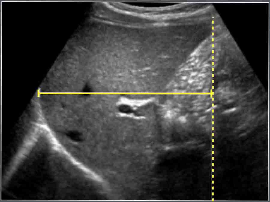
The craniocaudal dimension of the liver is measured along the midclavicular line using ultrasonography.Kích thước sọ não của gan được đo dọc theo đường giữa xương đòn bằng siêu âm.
Causes of hepatomegaly include:Nguyên nhân gây gan to bao gồm:
- LeukemiaBệnh bạch cầu
- Storage diseases (e.g., glycogen storage disorders)Bệnh dự trữ (ví dụ, rối loạn dự trữ glycogen)
- Neonatal hepatitisViêm gan sơ sinh
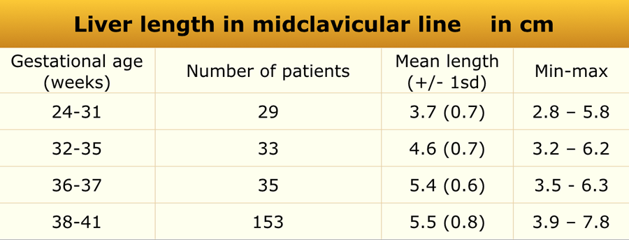
NewbornsTrẻ sơ sinh
Materials and Methods
In newborns (n = 261), the craniocaudal dimension of the liver was measured along the midclavicular line using ultrasonography.Phương pháp nghiên cứu
Ở trẻ sơ sinh (n = 261), kích thước sọ não của gan được đo dọc theo đường giữa xương đòn bằng siêu âm.
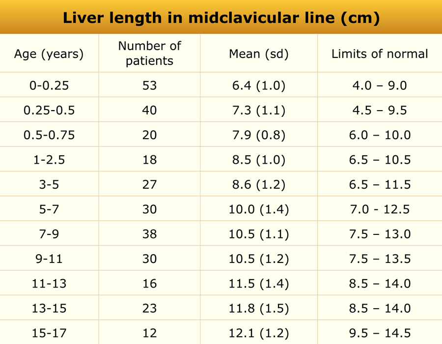
ChildrenTrẻ em
Material and methods
US study in 307 healthy children. Đối tượng và phương pháp nghiên cứu
Nghiên cứu siêu âm trên 307 trẻ khoẻ mạnh.
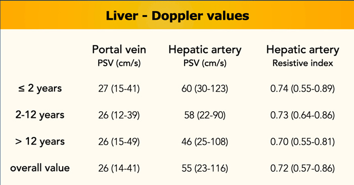
Doppler parametersCác thông số Doppler
Materials and Method
An additional study of 100 healthy children (age range 0–17.9 years, median 7.8 years) established reference values for:Đối tượng và phương pháp nghiên cứu
Một nghiên cứu bổ sung trên 100 trẻ khoẻ mạnh (tuổi từ 0 đến 17,9 tuổi, trung vị 7,8 tuổi) đã thiết lập các giá trị tham chiếu cho:
- Portal vein peak systolic velocityVận tốc tâm thu đỉnh tĩnh mạch cửa
- Hepatic artery peak systolic velocityVận tốc đỉnh tâm thu động mạch gan
- Hepatic artery resistive index (RI)Chỉ số điện trở động mạch gan (RI)
(reference).(tham khảo).
Findings:Kết quả:
- Portal vein velocity was not age-independentVận tốc tĩnh mạch cửa không phụ thuộc vào tuổi.
- Hepatic artery velocity and RI decreased with increasing ageVận tốc động mạch gan và RI giảm theo tuổi
Mesenteric lymph nodesHạch mạc treo
Materials and Methods
This retrospective study included 61 children (36 boys, 25 girls; mean age 10.7 years, range 1.1–17.3 years) who underwent non-contrast abdominal CT for evaluation of suspected or confirmed renal stones. Lymph node size was evaluated on these images, and findings were extrapolated to ultrasonographic reference values.Phương pháp nghiên cứu
Nghiên cứu hồi cứu 61 trẻ (36 nam, 25 nữ; tuổi trung bình 10,7 tuổi, khoảng 1,1-17,3) được chụp CT bụng không cản quang để đánh giá sỏi thận nghi ngờ hoặc xác định. Kích thước hạch bạch huyết được đánh giá trên các hình ảnh này và các kết quả được ngoại suy ra các giá trị tham chiếu siêu âm.
Findings:
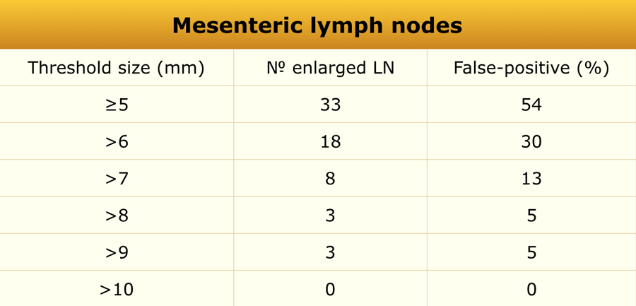
Enlarged mesenteric lymph nodes (short-axis diameter > 5 mm) were observed in 33 of 61 children (54%), predominantly in the right lower quadrant (88%).
Based on these results, a short-axis diameter > 8 mm may represent a more accurate threshold for pediatric mesenteric lymphadenopathy.Kết quả:
Mức độ giãn hạch mạc treo (đường kính trục ngắn > 5 mm) được quan sát thấy ở 33 trong số 61 trẻ (54%), chủ yếu ở vùng bụng dưới phải (88%).
Dựa trên những kết quả này, đường kính trục ngắn > 8 mm có thể là ngưỡng chính xác hơn cho bệnh hạch ở mạc treo ở trẻ em.
False-positive rate for enlarged mesenteric lymph nodes with varying lymph node threshold size is seen in the table.Tỷ lệ dương tính giả đối với hạch mạc treo mở rộng với kích thước ngưỡng hạch bạch huyết khác nhau được thấy trong bảng.
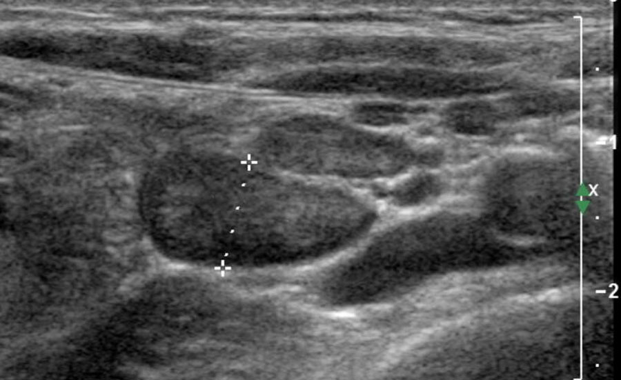
Common pathological causes of enlarged mesenteric lymph nodes includeCác nguyên nhân bệnh lý thường gặp của hạch mạc treo phì đại bao gồm:
- Intestinal lymphomaU lympho ruột
- Lymphogenic metastasisDi căn hạch
- Specific infectious enteritis (e.g., tuberculosis)Viêm ruột nhiễm trùng đặc hiệu (ví dụ, lao)
OvaryBuồng trứng
Materials and Methods
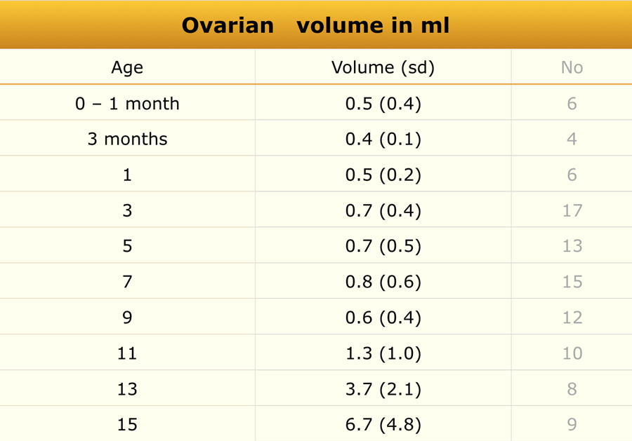
Ultrasonographic measurements of uterine and ovarian volumes were performed in 178 healthy girls.
Phương pháp nghiên cứu
Đo thể tích buồng trứng và tử cung bằng siêu âm được thực hiện ở 178 bé gái khoẻ mạnh.
Common causes of ovarian enlargement include:Các nguyên nhân phổ biến của phì đại buồng trứng bao gồm:
- Precocious pubertyDậy thì sớm
- Ovarian torsionXoắn buồng trứng
- Polycystic ovarian diseaseBệnh buồng trứng đa nang
- Teratoma or dermoid cystU quái hoặc nang bì
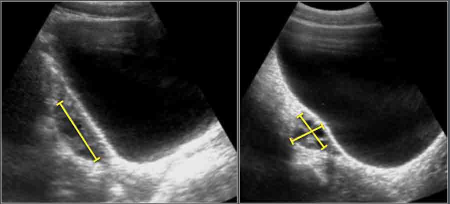
Ovarian volume is calculated using the formula:Thể tích buồng trứng được tính theo công thức:
- Length x Width x Height x 0.523.Chiều dài x Chiều rộng x Chiều cao x 0,523.
PancreasTuyến tụy
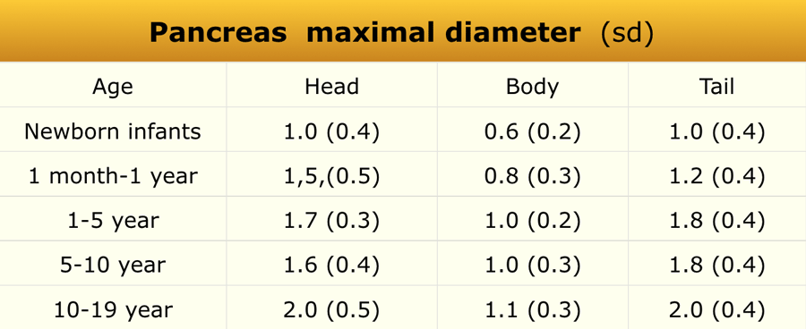
Materials and Methods
This retrospective ultrasonographic study included 273 pediatric patients (sex distribution not specified).Phương pháp nghiên cứu
Nghiên cứu này được thực hiện trên 273 bệnh nhi (không phân bố giới tính).
Echogenicity findings:Các dấu hiệu siêu âm:
- Low echogenicity in 27 cases (10%)Có 27 trường hợp có độ vang thấp (10%).
- Isoechoic in 145 cases (53%)Đồng âm trong 145 trường hợp (53%)
- Increased echogenicity in 101 cases (37%)Tăng cường siêu âm trong 101 trường hợp (37%)
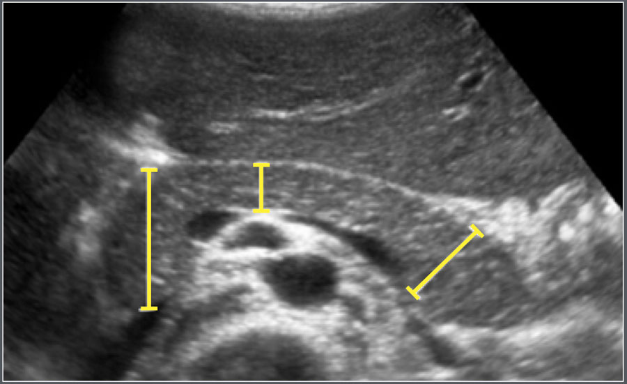
The maximum anteroposterior (AP) diameters of the head, body, and tail of the pancreas were measured on transverse or oblique ultrasound images.Đường kính tối đa trước sau (AP) của đầu, thân và đuôi tụy được đo trên hình ảnh siêu âm ngang hoặc chéo.
Common causes of pancreatic enlargement include:Các nguyên nhân phổ biến của phì đại tụy bao gồm:
- Traumatic pancreatitisViêm tụy chấn thương
- Viral pancreatitisVirus viêm tụy
- Drug-induced pancreatitisViêm tụy do thuốc
Portal veinTĩnh mạch cửa
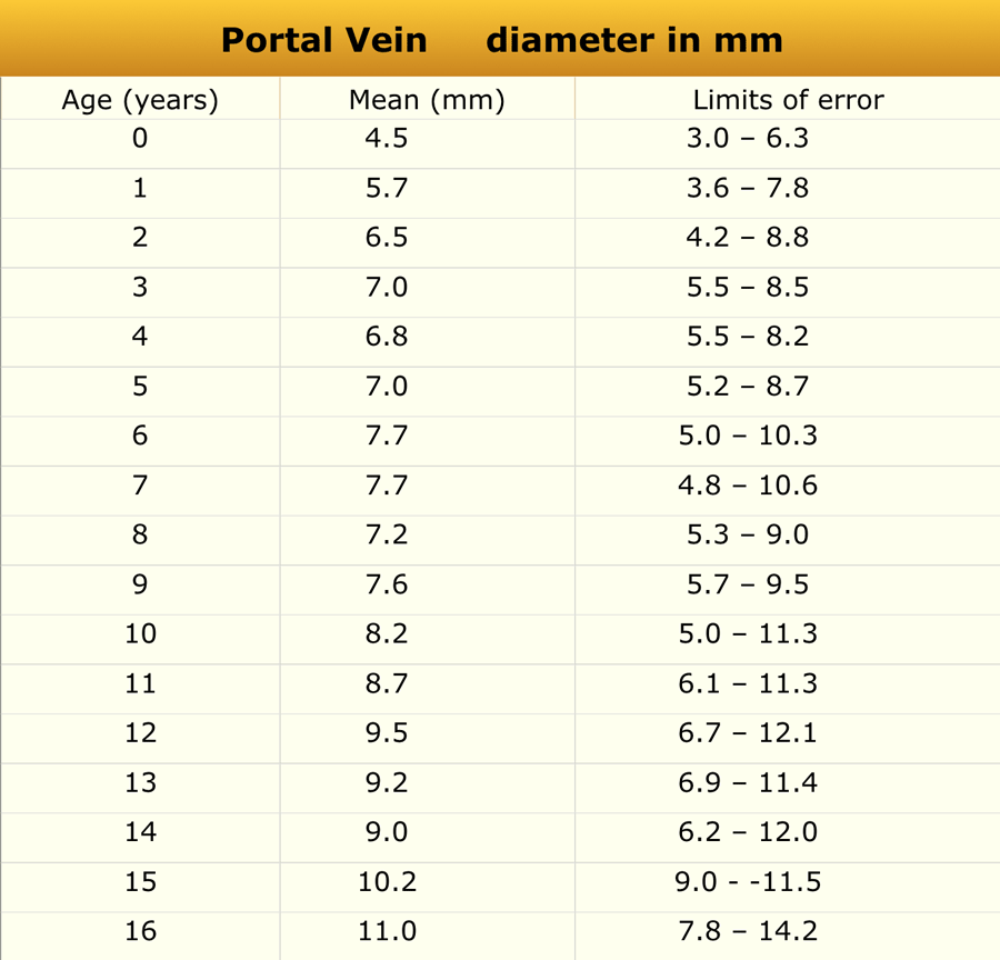
Materials and Methods
A study was conducted in 150 children aged 0–16 years without clinical evidence of hepatic or intestinal disease who underwent abdominal ultrasonography.Phương pháp nghiên cứu
Một nghiên cứu được thực hiện ở 150 trẻ em từ 0-16 tuổi không có bằng chứng lâm sàng về bệnh gan hoặc ruột được siêu âm bụng.
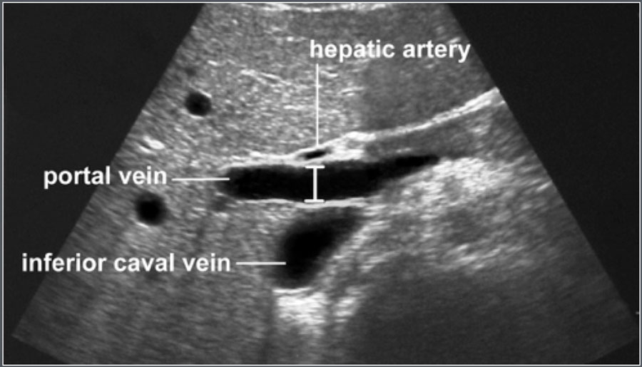
 Measurement of portal vein diameterĐo đường kính tĩnh mạch cửa
The portal vein was visualized in the longitudinal plane from the splenomesenteric junction to the hepatic hilum.
The maximum anteroposterior diameter was measured at the point where the hepatic artery crosses the portal vein.Tĩnh mạch cửa được quan sát trên mặt phẳng dọc từ chỗ nối lách - mạc treo gan đến rốn gan.
Đường kính tối đa trước sau được đo tại điểm động mạch gan đi qua tĩnh mạch cửa.
SpleenLách
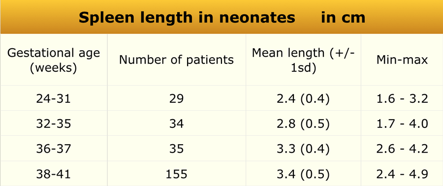
Preterm and term babiesTrẻ sơ sinh non tháng và đủ tháng
Material and methods
US study in 261 healthy newborn infants.
Craniocaudal dimension of the spleen was determined with ultrasonography.Đối tượng và phương pháp nghiên cứu
Nghiên cứu siêu âm trên 261 trẻ sơ sinh khoẻ mạnh.
Kích thước sọ-đầu của lách được xác định bằng siêu âm.
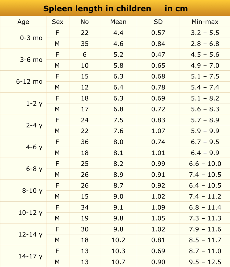
ChildrenTrẻ em
Materials and Methods
Ultrasonographic examination included 512 healthy children (238 boys, 274 girls; age range 1 day to 17 years) and 96 preterm infants (gestational age 25–35 weeks).
None of the subjects had medical conditions affecting spleen size.Phương pháp nghiên cứu
Khám siêu âm bao gồm 512 trẻ khoẻ mạnh (238 bé trai, 274 bé gái; tuổi từ 1 ngày đến 17 tuổi) và 96 trẻ sinh non (tuổi thai 25-35 tuần).
Không có đối tượng nào có bệnh lý ảnh hưởng đến kích thước lách.
Common causes of splenomegaly include:Các nguyên nhân phổ biến của lách to bao gồm:
- Portal hypertensionTăng áp lực tĩnh mạch cửa
- LeukemiaBệnh bạch cầu
- Systemic infections (e.g., Epstein–Barr virus, cytomegalovirus)Nhiễm trùng toàn thân (ví dụ, Epstein-Barr virus, cytomegalovirus)
- Hematologic disorders (e.g., spherocytosis, thalassemia)Rối loạn huyết học (ví dụ, tăng hồng cầu hình cầu, thalassemia)
- Storage diseasesBệnh lí kho
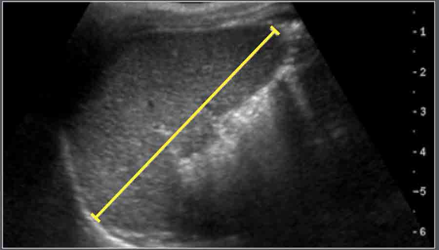
Measurement
Spleen length was defined as the maximal distance between the most superomedial and inferolateral points on a longitudinal coronal view, ideally through the splenic hilum.Đo lường
Chiều dài lách được xác định là khoảng cách tối đa giữa các điểm trên trong và dưới ngoài nhất trên mặt cắt vành dọc, lý tưởng là qua rốn lách.
Subarachnoid spaceKhông gian dưới màng nhện
Materials and Methods
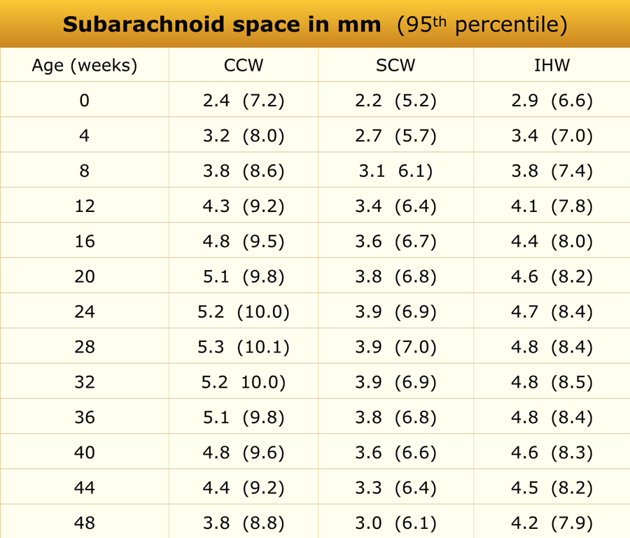
Ultrasonographic assessment of the subarachnoid space was performed in 278 full-term healthy Chinese infants.
Measurements were taken in the coronal plane at the level of the foramen of Monro.
Phương pháp nghiên cứu
Đánh giá siêu âm khoang dưới nhện được thực hiện trên 278 trẻ sơ sinh Trung Quốc đủ tháng khoẻ mạnh.
Các phép đo được thực hiện trong mặt phẳng đứng ngang ở mức của lỗ Monro.
Mean values were derived from regression equations, and 95% confidence intervals were calculated from the published data.Giá trị trung bình được tính từ phương trình hồi quy và khoảng tin cậy 95% được tính từ dữ liệu đã công bố.
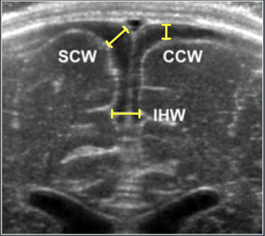

Ultrasonographic parametersCác thông số siêu âm
- SCW: Sinu-cortical widthSCW: Chiều rộng vỏ xoang
- CCW: Cranio-cortical widthCCW: Chiều rộng vỏ sọ
- IHW: Interhemispheric widthIHW: Chiều rộng liên bán cầu
Common causes of subarachnoid space enlargement includeCác nguyên nhân phổ biến của phì đại khoang dưới nhện bao gồm:
- Benign enlargement of the subarachnoid spaces of infancy (BESSI)Sự mở rộng lành tính của khoang dưới nhện ở trẻ sơ sinh (BESSI)
- Cerebral atrophyTeo não
- Dural sinus thrombosisHuyết khối xoang màng cứng
TesticleTinh hoàn
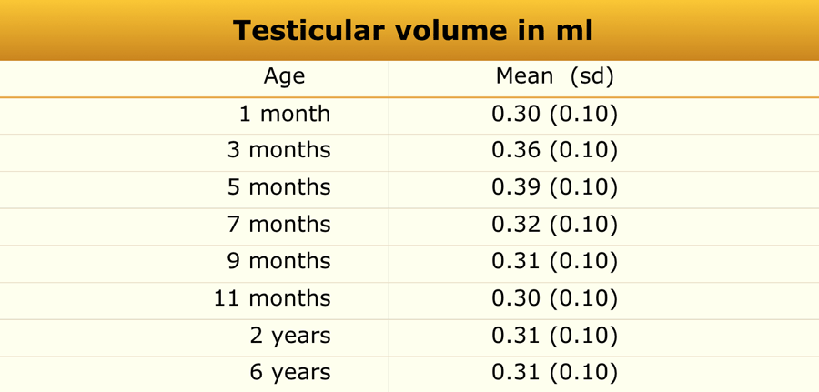
Materials and Methods
Ultrasonographic measurements were obtained from 344 boys of various ethnic backgrounds.
No significant differences were noted between ethnicities or between the right and left testes.Phương pháp nghiên cứu
Các số đo siêu âm được thực hiện trên 344 bé trai thuộc các dân tộc khác nhau.
Không có sự khác biệt có ý nghĩa thống kê giữa các dân tộc hay giữa tinh hoàn phải và tinh hoàn trái.
Common causes of testicular enlargement includeCác nguyên nhân phổ biến của tinh hoàn to bao gồm
- LeukemiaBệnh bạch cầu
- Precocious pubertyDậy thì sớm
- Testicular torsionXoắn tinh hoàn
- Epididymo-orchitisViêm màng ngoài tim-tinh hoàn
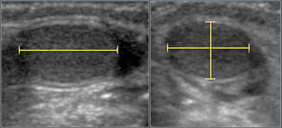
Testicular volume was calculated using the formula:
Length × Width × Height × 0.523Thể tích tinh hoàn được tính theo công thức:
Chiều dài × Chiều rộng × Chiều cao × 0,523
ThymusTuyến ức
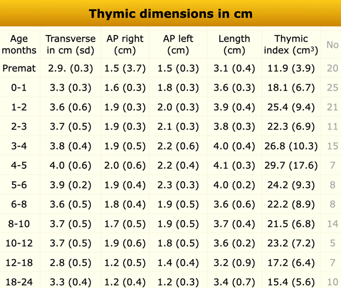
Materials and Methods
Mediastinal ultrasonography was performed in 151 healthy infants (79 boys and 72 girls) with no stress-related factorsaffecting thymic size.Phương pháp nghiên cứu
Siêu âm trung thất được thực hiện ở 151 trẻ khoẻ mạnh (79 nam và 72 nữ) không có yếu tố liên quan đến stress ảnh hưởng đến kích thước tuyến ức.
Common causes of thymic enlargement include:Các nguyên nhân phổ biến của phì đại tuyến ức bao gồm:
- Rebound thymic hyperplasiaTăng sản tuyến ức hồi phục
- T-cell lymphoma or leukemiaU lympho tế bào T hoặc bệnh bạch cầu
- ThymomaU tuyến giáp
- Langerhans cell histiocytosisBệnh mô bào Langerhans
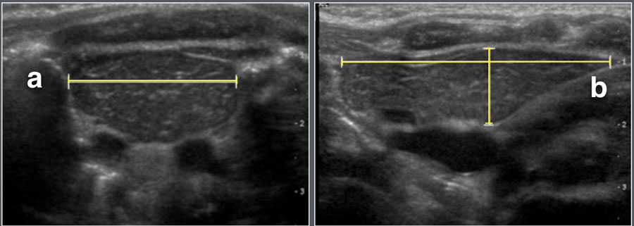
Measurements includedCác phép đo bao gồm
- Maximum transverse diameterĐường kính ngang tối đa
- Right and left lobe anteroposterior diametersĐường kính trước sau thuỳ phải và trái
- Craniocaudal (longitudinal) lengthChiều dài sọ (dọc)
The thymic index was calculated as:
Transverse diameter × largest sagittal areaChỉ số thymic được tính như sau:
Đường kính ngang × diện tích dọc lớn nhất
Thyroid glandTuyến giáp
Materials and Methods
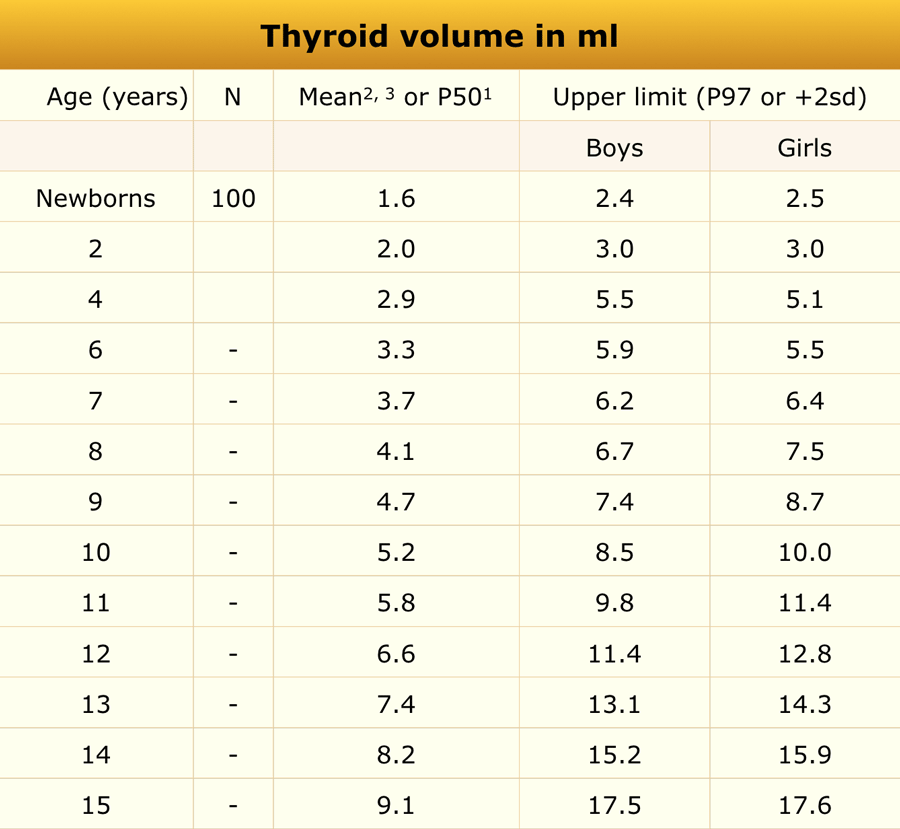
Ultrasonographic evaluation of thyroid volume was conducted in:Phương pháp nghiên cứu
Đánh giá thể tích tuyến giáp bằng siêu âm được thực hiện trong:
- 100 English neonates (within the first week of life)100 trẻ sơ sinh Anh (trong tuần đầu tiên của cuộc đời)
- A subset of iodine-sufficient European children (n = 5,709, aged 6–15 years)Một nhóm nhỏ trẻ em châu Âu đủ iốt (n = 5.709, tuổi từ 6-15 tuổi)
- A group of German children (n = 252, aged 2–4 years)
Một nhóm trẻ em Đức (n = 252, từ 2-4 tuổi)
Common causes of thyroid enlargement includeCác nguyên nhân phổ biến của phì đại tuyến giáp bao gồm
- Hashimoto’s thyroiditisViêm tuyến giáp Hashimoto
- Graves’ diseaseBệnh Graves
- Multinodular goiterBướu giáp đa nhân
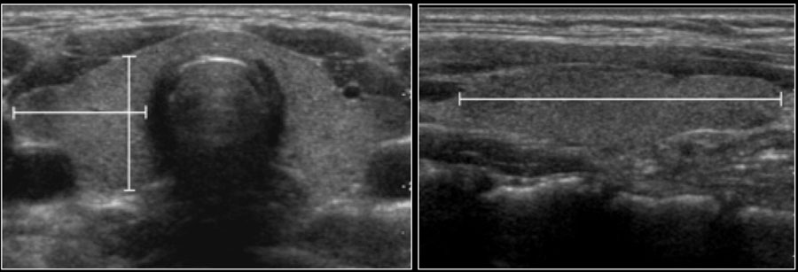
Thyroid volume was calculated as the sum of both lobes, excluding the isthmus.
The ellipsoid formula was used:
Volume = Length × Width × Height × 0.52Thể tích tuyến giáp được tính bằng tổng của cả hai thuỳ, không tính đến eo tuyến giáp.
Công thức tính ellipsoid được sử dụng:
Thể tích = Dài × Rộng × Cao × 0,52
UterusTử cung
Materials and Methods
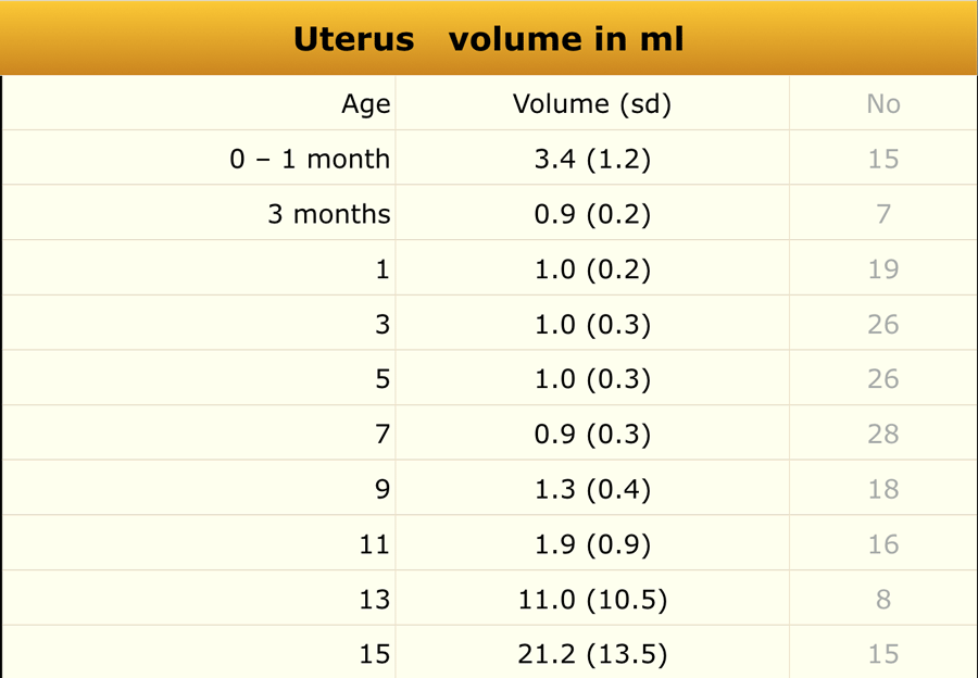
Ultrasonographic measurements of uterine and ovarian volumes were performed in 178 healthy girls.
Phương pháp nghiên cứu
Đo thể tích buồng trứng và tử cung bằng siêu âm được thực hiện ở 178 bé gái khoẻ mạnh.
Common causes of uterine enlargement includeCác nguyên nhân phổ biến của tử cung to bao gồm
- Precocious pubertyDậy thì sớm
- Hydro- or hematometrocolposHydro- hoặc hematometrocolpos
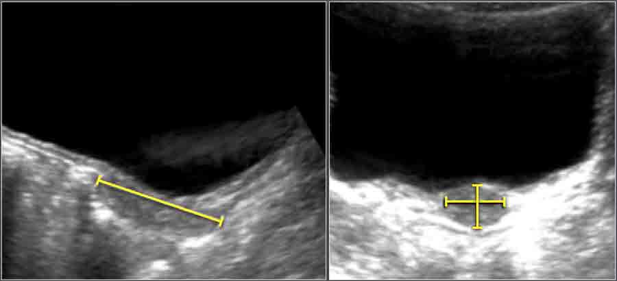
Uterine volume was calculated using the ellipsoid formula:
Length × Width × Height × 0.523Thể tích tử cung được tính theo công thức ellipsoid:
Chiều dài × Chiều rộng × Chiều cao × 0,523
VentriclesTrang chủ
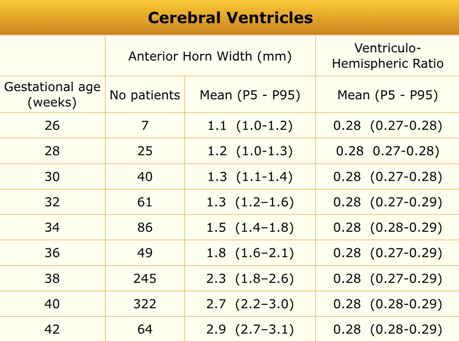
Materials and Methods
An ultrasonographic study was performed in 1,483 neonates (gestational age 25–42 weeks) examined on day 3 of life.
Infants with perinatal asphyxia, CNS infection, intracranial hemorrhage, or craniospinal malformations were excluded.Phương pháp nghiên cứu
Một nghiên cứu siêu âm được thực hiện ở trẻ sơ sinh 1,483 (tuổi thai 25-42 tuần) được khám vào ngày 3 của cuộc đời.
Trẻ bị ngạt chu sinh, nhiễm trùng thần kinh trung ương, xuất huyết nội sọ, dị dạng sọ não được loại trừ.
Common causes of ventriculomegaly includeCác nguyên nhân thường gặp của bệnh não thất to bao gồm
- Congenital malformations (e.g., holoprosencephaly)Dị tật bẩm sinh (ví dụ, tật não ba đầu)
- Obstructive hydrocephalusNão úng thủy do tắc nghẽn
- Communicating hydrocephalusNão úng thủy thông thương
- Cerebral atrophyTeo não
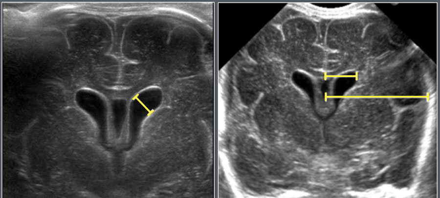
MeasurementsĐo lường
- Anterior horn width (AHW)Chiều rộng sừng trước (AHW)
- Ventriculohemispheric ratio (VHR)
Measured on coronal images at the level of the foramen of Monro.Tỷ lệ thể tích tĩnh mạch (VHR)
Đo trên ảnh mặt phẳng đứng ngang ở mức của lỗ Monro.
ReferencesTài liệu tham khảo
-
Delange F, Benker G, Caron P, et al. Thyroid volume and urinary iodine in European schoolchildren: standardization of values for assessment of iodine deficiency. Eur J Endocrinol 1997;136:180-7. Delange F, Benker G, Caron P, et al. Thể tích tuyến giáp và iốt nước tiểu ở học sinh châu Âu: tiêu chuẩn hóa các giá trị để đánh giá tình trạng thiếu iốt. Eur J Endocrinol 1997; 136: 180-7.
-
Menken KU, Engelhardt S, Olbricht T. [Thyroid gland volume and urinary iodine excretion in children 2-16 years of age]. Dtsch Med Wochenschr 1992;117:1047-51. Menken KU, Engelhardt S, Olbricht T. [Thể tích tuyến giáp và bài tiết i-ốt trong nước tiểu ở trẻ 2-16 tuổi]. DTSCH Med Wochenschr 1992; 117: 1047-51.
-
Perry RJ, Hollman AS, Wood AM, et al. Ultrasound of the thyroid gland in the newborn: normative data. Arch Dis Child Fetal Neonatal Ed 2002;87:F209-11. Perry RJ, Hollman AS, Wood AM, et al. Siêu âm tuyến giáp ở trẻ sơ sinh: dữ liệu chuẩn. Arch Dis Child Fetal Neonatal Ed 2002; 87: F 209-11.
-
Sondhi V, Gupta G, Gupta PK, et al. Establishment of nomograms and reference ranges for intra-cranial ventricular dimensions and ventriculo-hemispheric ratio in newborns by ultrasonography. Acta Paediatr 2008;97:738-44. Sondhi V, Gupta G, Gupta PK, et al. Thiết lập các hình ảnh đồ và các khoảng tham chiếu cho kích thước thất trong sọ và tỷ lệ thất-bán cầu não ở trẻ sơ sinh bằng siêu âm. Acta Paediatr 2008; 97: 738-44.
-
Lam WW, Ai VH, Wong V, et al. Ultrasonographic measurement of subarachnoid space in normal infants and children. Pediatr Neurol 2001;25:380-4. Lam WW, Ai VH, Wong V, et al. Đo siêu âm khoang dưới nhện ở trẻ sơ sinh và trẻ em bình thường. Pediatr Neurol 2001; 25: 380-4.
-
Soyupak SK, Narli N, Yapicioglu H, et al. Sonographic measurements of the liver, spleen and kidney dimensions in the healthy term and preterm newborns. Eur J Radiol 2002;43:73-8. Soyupak SK, Narli N, Yapicioglu H, et al. Các phép đo siêu âm kích thước gan, lách và thận ở trẻ sơ sinh khoẻ mạnh và non tháng. Eur J Radiol 2002; 43: 73-8.
-
Konus OL, Ozdemir A, Akkaya A, et al. Normal liver, spleen, and kidney dimensions in neonates, infants, and children: evaluation with sonography. AJR Am J Roentgenol 1998;171:1693-8. Konus OL, Ozdemir A, Akkaya A, et al. Kích thước gan, lách và thận bình thường ở trẻ sơ sinh, trẻ sơ sinh và trẻ em: đánh giá bằng siêu âm. AJR Am J Roentgenol 1998; 171: 1693-8.
-
Hernanz-Schulman M, Ambrosino MM, Freeman PC, et al. Common bile duct in children: sonographic dimensions. Radiology 1995;195:193-5. Hernanz-Schulman M, Ambrosino MM, Freeman PC, et al. ống mật chủ ở trẻ em: kích thước siêu âm. X quang 1995; 195: 193-5.
-
Patriquin HB, Perreault G, Grignon A, et al. Normal portal venous diameter in children. Pediatr Radiol 1990;20:451-3. Patriquin HB, Perreault G, Grignon A, et al. Đường kính tĩnh mạch cửa bình thường ở trẻ em Pediatr Radiol 1990; 20: 451-3.
-
Ho ML, Chen JY, Ling UP, et al. Gallbladder volume and contractility in term and preterm neonates: normal values and clinical applications in ultrasonography. Acta Paediatr 1998;87:799-804. Ho ML, Chen JY, Ling UP, et al. Thể tích và độ co bóp túi mật ở trẻ sơ sinh đủ tháng và non tháng: giá trị bình thường và ứng dụng lâm sàng trong siêu âm. Acta Paediatr 1998; 87: 799-804.
-
Megremis SD, Vlachonikolis IG, Tsilimigaki AM. Spleen length in childhood with US: normal values based on age, sex, and somatometric parameters. Radiology 2004;231:129-34. Megremis SD, Vlachonikolis IG, Tsilimigaki AM. Chiều dài lách ở trẻ em với siêu âm: giá trị bình thường dựa trên tuổi, giới tính và các thông số đo thân thể. X quang 2004; 231: 129-34.
-
Siegel MJ, Martin KW, Worthington JL. Normal and abnormal pancreas in children: US studies. Radiology 1987;165:15-8. Siegel MJ, Martin KW, Worthington JL. Tuyến tụy bình thường và bất thường ở trẻ em: Nghiên cứu siêu âm. X quang 1987; 165: 15-8.
-
Haber HP, Stern M. Intestinal ultrasonography in children and young adults: bowel wall thickness is age dependent. J Ultrasound Med 2000;19:315-21. Haber HP, Stern M. Siêu âm ruột ở trẻ em và thanh niên: độ dày thành ruột phụ thuộc vào tuổi. J Ultrasound Med 2000; 19:315-21.
-
Wiersma F, Sramek A, Holscher HC. US features of the normal appendix and surrounding area in children. Radiology 2005;235:1018-22. Wiersma F, Sramek A, Holscher HC. Đặc điểm siêu âm của ruột thừa bình thường và khu vực xung quanh ở trẻ em. X quang 2005; 235:1018-22.
-
Karmazyn B, Werner EA, Rejaie B, et al. Mesenteric lymph nodes in children: what is normal? Pediatr Radiol 2005;35:774-7. Karmazyn B, Werner EA, Rejaie B, et al. hạch mạc treo ở trẻ em: bình thường là gì? Pediatr Radiol 2005; 35: 774-7.
-
Rosenbaum DM, Korngold E, Teele RL. Sonographic assessment of renal length in normal children. AJR Am J Roentgenol 1984;142:467-9. Rosenbaum DM, Korngold E, Teele RL. Đánh giá chiều dài thận bằng siêu âm ở trẻ em bình thường. AJR Am J Roentgenol 1984; 142: 467-9.
-
Leung VY, Chu WC, Yeung CK, et al. Nomograms of total renal volume, urinary bladder volume and bladder wall thickness index in 3,376 children with a normal urinary tract. Pediatr Radiol 2007;37:181-8. Leung Vy, Chu WC, Yeung CK, et al. nomograms tổng thể tích thận, thể tích bàng quang và chỉ số độ dày thành bàng quang ở 3.376 trẻ có đường tiết niệu bình thường. Pediatr Radiol 2007; 37: 181-8.
-
Robben SG, Boesten M, Linmans J, et al. Significance of thickening of the wall of the renal collecting system in children: an ultrasound study. Pediatr Radiol 1999;29:736-40. Robben SG, Boesten M, Linmans J, et al. Tầm quan trọng của dày thành hệ thống đài–bể thận ở trẻ em: một nghiên cứu siêu âm. Pediatr Radiol 1999; 29: 736-40.
-
Kuijper EA, van Kooten J, Verbeke JI, et al. Ultrasonographically measured testicular volumes in 0- to 6-year-old boys. Hum Reprod 2008;23:792-6. Kuijper EA, van Kooten J, Verbeke JI, et al. Siêu âm đo thể tích tinh hoàn ở trẻ em từ 0 đến 6 tuổi. Hum Reprod 2008; 23: 792-6.
-
Haber HP, Mayer EI. Ultrasound evaluation of uterine and ovarian size from birth to puberty. Pediatr Radiol 1994;24:11-3. Haber HP, Mayer EI. Siêu âm đánh giá kích thước tử cung và buồng trứng từ sơ sinh đến dậy thì. Pediatr Radiol 1994; 24: 11-3.
-
Oppenheimer DA, Carroll BA, Yousem S. Sonography of the normal neonatal adrenal gland. Radiology 1983;146:157-60. Oppenheimer DA, Carroll BA, Yousem S. Siêu âm tuyến thượng thận bình thường ở trẻ sơ sinh. X quang 1983; 146: 157-60.
-
Robben SG, Lequin MH, Diepstraten AF, et al. Anterior joint capsule of the normal hip and in children with transient synovitis: US study with anatomic and histologic correlation. Radiology 1999;210:499-507. Robben SG, Lequin MH, Diepstraten AF, et al. Viên nang khớp trước của khớp háng bình thường và ở trẻ em bị viêm màng hoạt dịch thóang qua: Nghiên cứu siêu âm có mối tương quan về giải phẫu và mô học. X quang 1999; 210: 499-507.
-
Graf R. Hip Sonography. Berlin Heidelberg: Springer, 2006. Bản dịch gốc: Graf R. Hip Sonography. Berlin Heidelberg: Springer, 2006.
-
Yekeler E, Tambag A, Tunaci A, et al. Analysis of the thymus in 151 healthy infants from 0 to 2 years of age. J Ultrasound Med 2004;23:1321-6. Yekeler E, Tambag A, Tunaci A, et al. Phân tích tuyến ức ở 151 trẻ sơ sinh khoẻ mạnh từ 0 đến 2 tuổi. J Ultrasound Med 2004; 23: 1321-6.