
Gallbladder wall thickeningDày thành túi mật
Radiology Department of the Rijnland Hospital, Leiderdorp; the Groene Hart Hospital, Gouda and the Medical Centre Haaglanden, the Hague, the NetherlandsKhoa Chẩn đoán hình ảnh của Bệnh viện Rijnland, Leiderdorp; Bệnh viện Groene Hart, Gouda và Trung tâm Y tế Haaglanden, Hague, Hà Lan
Ngày đăng / cập nhật: 2006-02-01
Bản gốc trên radiologyassistant.nl ↗ · tải về lúc 2026-11-19 20:19 · bản dịch máy có chuẩn hóa thuật ngữ chẩn đoán hình ảnh
Thickening of the gallbladder wall is a relatively frequent finding at diagnostic imaging studies.
Historically, a thick-walled gallbladder has been regarded as proof of primary gallbladder disease, and it is a well-known hallmark feature of acute cholecystitis.
The finding itself, however, is non-specific and can be found in a wide range of gallbladder diseases and extracholecystic pathological conditions.
In this review we discuss and illustrate the various causes of a generalized thickened gallbladder wall.
If you encounter printing problems with the margins of the document, try to adjust the margins or the scale of the document in the print settings.Dày thành túi mật là một dấu hiệu tương đối thường gặp trong chẩn đoán hình ảnh.
Trong lịch sử, túi mật thành dày được coi là bằng chứng của bệnh túi mật nguyên phát, và nó là một đặc điểm nổi tiếng của viêm túi mật cấp tính.
Tuy nhiên, bản thân dấu hiệu này không đặc hiệu và có thể được tìm thấy trong một loạt các bệnh về túi mật và các tình trạng bệnh lý ngoài túi mật.
Trong bài tổng quan này, chúng tôi thảo luận và minh hoạ các nguyên nhân khác nhau của thành túi mật dày lên toàn thân.
Nếu bạn gặp vấn đề in ấn với lề của tài liệu, hãy thử điều chỉnh lề hoặc tỷ lệ của tài liệu trong cài đặt in.
IntroductionMở đầu
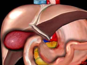 Distended hydropic and hyperemic gallbladder in acute cholecystitis due to stone obstruction in the gallbladder neck or cystic duct.Túi mật giãn rộng và tăng huyết áp trong viêm túi mật cấp tính do tắc nghẽn sỏi ở cổ túi mật hoặc ống túi mật.
Sonography, CT and MRI all allow direct visualization of the normal and thickened gallbladder wall.
Traditionally, sonography is used as the initial imaging technique for evaluating patients with suspected gallbladder disease, because of its high sensitivity in the detection of gallbladder stones, its real-time character, speed and portability [1].
However, CT has become popular for evaluating the acute abdomen and often is the first modality to detect gallbladder wall thickening [2], or it may be used as an adjunct to an inconclusive sonography or for staging of disease.
The potential value of MRI in the evaluation of gallbladder pathology has been shown [3], but it still plays little role.
Siêu âm, CT và MRI đều cho phép hình ảnh trực tiếp của thành túi mật bình thường và dày lên.
Theo truyền thống, siêu âm được sử dụng làm kỹ thuật hình ảnh ban đầu để đánh giá bệnh nhân nghi ngờ mắc bệnh túi mật, vì độ nhạy cao trong việc phát hiện sỏi túi mật, đặc tính thời gian thực, tốc độ và tính di động của nó [1].
Tuy nhiên, CT đã trở nên phổ biến để đánh giá bụng cấp tính và thường là phương thức đầu tiên để phát hiện dày thành túi mật [2], hoặc nó có thể được sử dụng như một phương pháp bổ trợ cho siêu âm không kết luận hoặc để phân giai đoạn bệnh.
Giá trị tiềm năng của MRI trong đánh giá bệnh lý túi mật đã được chứng minh [3], nhưng nó vẫn đóng vai trò rất ít.
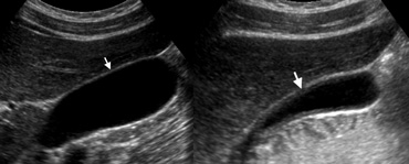 LEFT: US of a normal gallbladder after an overnight fast shows the wall as a pencil-thin echogenic line (arrow).RIGHT: US in the postprandial state shows pseudothickening of the gallbladderTrái: Siêu âm túi mật bình thường sau khi nhịn ăn qua đêm. Thành túi mật là một đường tăng âm rất mảnh (mũi tên). Phải: Siêu âm sau ăn cho thấy hình ảnh giả dày thành túi mật.
Normal gallbladderTúi mật bình thường
The normal gallbladder wall appears as a pencil-thin echogenic line at sonography.
The thickness of the gallbladder wall depends on the degree of gallbladder distention and pseudothickening can occur in the postprandial state. Thành túi mật bình thường xuất hiện dưới dạng một đường echogenic mỏng như bút chì khi siêu âm.
Độ dày của thành túi mật phụ thuộc vào mức độ căng giãn của túi mật và giả dày có thể xảy ra trong trạng thái sau ăn.
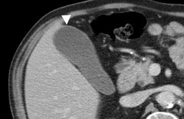 Contrast-enhanced CT shows the normal gallbladder wall as a thin rim of enhancing soft-tissueCT có tiêm thuốc cản quang cho thấy thành túi mật bình thường như một vành mỏng của mô mềm tăng cường
The normal gallbladder wall is usually perceptible at CT as a thin rim of soft-tissue density that enhances after contrast injection.Thành túi mật bình thường thường có thể cảm nhận được trên CT là một vành mỏng có mật độ mô mềm tăng cường sau khi tiêm thuốc cản quang.
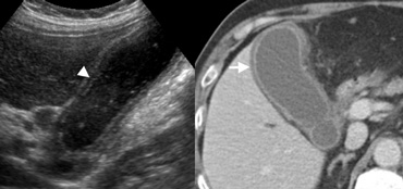 LEFT: US in a 59-year-old woman with acute cholecystitis shows the layered appearance of a thickened gallbladder wall, with a hypoechoic region between echogenic linesRIGHT: At contrast-enhanced CT the thick-walled gallbladder contains a hypodense outer layer (arrow) due to subserosal oedemaTRÁI: siêu âm ở một phụ nữ 59 tuổi có viêm túi mật cấp tính cho thấy hình dạng lớp của thành túi mật dày, với vùng giảm âm giữa các đường echogenicRIGHT: Tại CT có tiêm thuốc cản quang, túi mật có thành dày chứa lớp ngoài giảm tỷ trọng (mũi tên) do phù dưới màng cứng
Thickened gallbladder wallBức tường túi mật dày
Thickening of the gallbladder wall is a relatively frequent finding at diagnostic imaging studies.
A thickened gallbladder wall measures more than 3 mm, typically has a layered appearance at sonography [1], and at CT frequently contains a hypodense layer of subserosal oedema that mimics pericholecystic fluid [2] Dày thành túi mật là một dấu hiệu tương đối thường gặp trong chẩn đoán hình ảnh.
Thành túi mật dày hơn 3 mm, thường có hình dạng lớp trên siêu âm [1], và trên CT thường có lớp phù dưới màng cứng giảm tỷ trọng giống như dịch quanh túi mật [2].
Differential diagnosis of gallbladder wall thickeningChẩn đoán phân biệt của dày thành túi mật
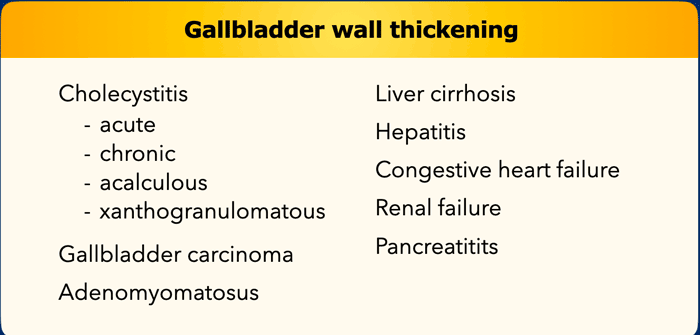
The differential diagnosis of gallbladder wall thickening is listed on the left.
Diffuse gallbladder wall thickening may produce a diagnostic problem, as it occurs in symptomatic and asymptomatic patients, and in patients with and without an indication for a cholecystectomy.
Chẩn đoán phân biệt của dày thành túi mật được liệt kê bên trái.
Khuếch tán dày thành túi mật có thể gây ra một vấn đề chẩn đoán, vì nó xảy ra ở những bệnh nhân có triệu chứng và không có triệu chứng, và ở những bệnh nhi có và không có chỉ định cắt bỏ túi mật.
Diffuse thickening of the gallbladder wall may occur in patients who do not have a primary gallbladder disease, but in whom the gallbladder is secondarily involved in an extrinsic pathological condition.
In these patients a cholecystectomy is unwarranted, and gallbladder abnormalities will usually return to normal after correction of its extrinsic cause.Sự dày lên lan toả của thành túi mật có thể xảy ra ở những bệnh nhân không mắc bệnh túi mật nguyên phát, nhưng trong đó túi mật thứ phát liên quan đến tình trạng bệnh lý bên ngoài.
Ở những bệnh nhân này, việc cắt bỏ túi mật là không chính xác, và các bất thường về túi mật thường sẽ trở lại bình thường sau khi điều chỉnh nguyên nhân bên ngoài.
Primary gallbladder diseaseBệnh túi mật nguyên phát
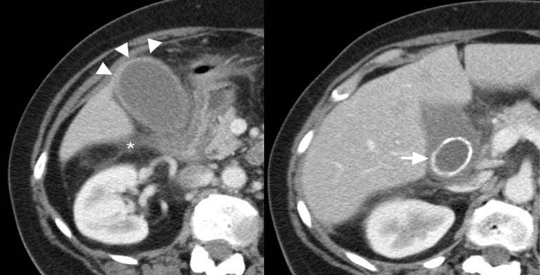 43-year-old woman with acute calculous cholecystitis. Contrast-enhanced CT shows a distended gallbladder (arrowheads) with a slightly thickened wall and subtle regional fat-stranding (asterisk). There is an impacted obstructing stone in the neck of the gallbladder (arrow).Nữ 43 tuổi bị viêm túi mật cấp do sỏi. CT có tiêm thuốc cản quang cho thấy túi mật căng (đầu mũi tên), thành hơi dày và thâm nhiễm mỡ quanh túi mật kín đáo (dấu sao). Có sỏi kẹt gây tắc ở cổ túi mật (mũi tên).
Acute cholecystitisViêm túi mật cấp tính
Acute cholecystitis is the fourth most common cause of hospital admissions for patients presenting with an acute abdomen [4], and it is the prime diagnostic concern when a thick-walled gallbladder is found at imaging.
This feature, however, is not pathognomonic for acute cholecystitis.Viêm túi mật cấp tính là nguyên nhân phổ biến thứ tư gây nhập viện cho bệnh nhân có bụng cấp tính [4], và đây là mối quan tâm chẩn đoán chính khi tìm thấy túi mật có thành dày trên hình ảnh.
Tuy nhiên, đặc điểm này không phải là đặc trưng cho viêm túi mật cấp tính.
Additional imaging signs that support the diagnosis of acute calculous cholecystitis are:
Các dấu hiệu hình ảnh bổ sung hỗ trợ chẩn đoán sỏi cấp viêm túi mật là:
- Obstructing gallstoneTắc nghẽn sỏi mật
- Hydropical dilatation of the gallbladderDãn túi mật qua nội soi
- Positive sonographic Murphy's sign ( i.e., pain elicited by pressure over the sonographically located gallbladder)Siêu âm dương tính Dấu hiệu Murphy (tức là đau do áp lực lên túi mật được siêu âm)
- Pericholecystic fat inflammation or fluidViêm hoặc dịch quanh túi mật
- Hyperemia of the gallbladder wall at power Doppler. Tăng huyết áp thành túi mật trên Doppler điện.
 Acute calculous cholecystitisSỏi cấp viêm túi mật
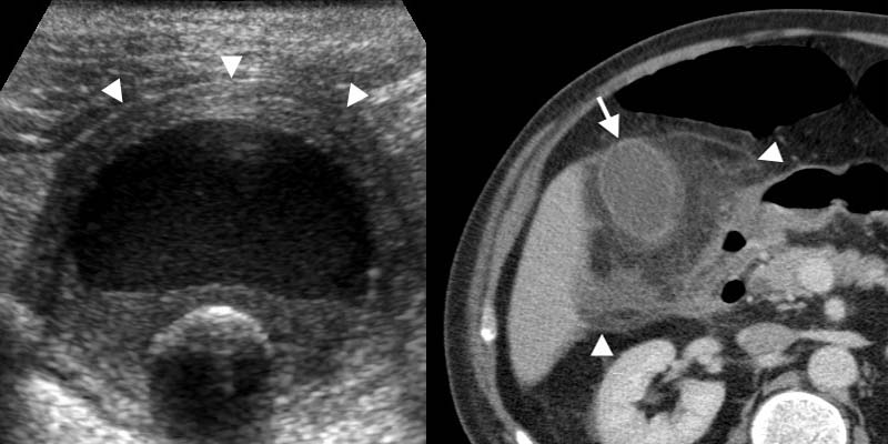
On the left images of a 62-year-old man with acute calculous cholecystitis.
Transverse sonogram at the spot of maximum tenderness shows a non-compressible hydropically distended thick-walled gallbladder (arrowheads), with an intraluminal stone and sludge or debris.
Contrast-enhanced CT depicts extensive fat inflammation (arrowheads) surrounding the
gallbladder (arrow).
Trên hình ảnh bên trái của một người đàn ông 62 tuổi bị sỏi viêm túi mật cấp tính.
Siêu âm ngang tại vị trí đau tối đa cho thấy túi mật dày có thành không nén được (các đầu mũi tên), có sỏi trong lòng túi mật và bùn hoặc mảnh vụn.
CT có tiêm thuốc cản quang cho thấy tình trạng viêm mỡ lan rộng (đầu mũi tên) xung quanh túi mật (mũi tên).
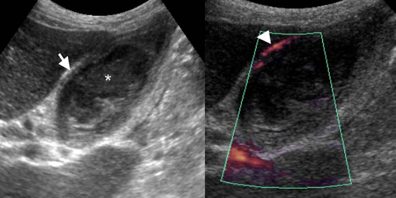 74-year-old man with acute acalculous cholecystitis. LEFT: US at the spot of maximum tenderness shows mural thickening of the gallbladder (arrow) that is completely filled with sludge (asterisk) without any stones. RIGHT: Power-Doppler sonography shows hypervascularity of the gallbladder wall (arrowhead), as a supporting sign of inflammation.Người đàn ông 74 tuổi bị sỏi viêm túi mật cấp tính. TRÁI: siêu âm tại điểm đau tối đa cho thấy thành túi mật dày lên (mũi tên) chứa đầy bùn (dấu hoa thị) mà không có sỏi. PHẢI: Siêu âm Doppler điện cho thấy tăng sinh mạch máu ở thành túi mật (đầu mũi tên), như một dấu hiệu hỗ trợ của tình trạng viêm.
Acalculous cholecystitisAcalculous viêm túi mật
Acute acalculous cholecystitis mainly occurs in critically ill patients, presumably due to increased bile viscosity from fasting and medication that causes cholestasis.
The imaging features are those of acute cholecystitis, except for the absence of stones whereas gallbladder sludge is usually present (Fig).
Because in critically ill patients gallbladder abnormalities are frequently found secondary to systemic disease (see below), acalculous cholecystitis can be difficult to diagnose [5].
In these patients a percutaneous cholecystostomy can be both diagnostic and therapeutic.Viêm túi mật cấp tính chủ yếu xảy ra ở những bệnh nhân nặng, có lẽ do tăng độ nhớt mật do nhịn ăn và thuốc gây ứ mật.
Các đặc điểm hình ảnh là những đặc điểm của viêm túi mật cấp tính, ngoại trừ không có sỏi trong khi bùn túi mật thường có mặt (Hình).
Bởi vì ở những bệnh nhân bị bệnh nặng, các bất thường về túi mật thường được tìm thấy thứ phát do bệnh hệ thống (xem bên dưới), viêm túi mật không vôi hóa có thể khó chẩn đoán [5].
Ở những bệnh nhân này, mở túi mật qua da có thể vừa chẩn đoán vừa điều trị.
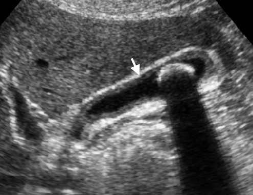 Chronic cholecystitis. Longitudinal sonogram of the gallbladder shows slight wall thickening (arrow) and an intraluminal non-obstructing stoneViêm túi mật mạn tính Siêu âm dọc túi mật cho thấy dày thành nhẹ (mũi tên) và sỏi không cản quang trong lòng túi mật
Chronic cholecystitisViêm túi mật mạn tính
Chronic cholecystitis is a term used clinically to refer to symptomatic gallbladder stones that cause transient obstruction, leading to a low-grade inflammation with fibrosis [1].
Correlation of the imaging finding of a stone-containing slightly thick-walled gallbladder with the clinical history is critical.
Viêm túi mật mạn tính là một thuật ngữ được sử dụng trên lâm sàng để chỉ sỏi túi mật có triệu chứng gây tắc nghẽn thóang qua, dẫn đến viêm mức độ thấp với xơ hóa [1].
Tương quan giữa hình ảnh túi mật có thành hơi dày chứa sỏi với tiền sử lâm sàng là rất quan trọng.
On the left images of a 49-year-old woman with chronic cholecystitis.
This patient had fasted overnight, so the wall-thickening does not represent physiologic contraction.
Correlation of these findings with her clinical history of recurrent colic-like right upper quadrant pain, due to transient gallbladder obstruction, is essential for the diagnosis.Trên hình ảnh bên trái của một phụ nữ 49 tuổi bị viêm túi mật mạn tính.
Bệnh nhân này đã nhịn ăn qua đêm, do đó, dày thành không biểu hiện sự co thắt sinh lý.
Mối liên quan giữa các dấu hiệu này với tiền sử lâm sàng của bệnh nhân đau bụng kiểu vùng bụng trên phải tái phát do tắc nghẽn túi mật thóang qua là rất cần thiết để chẩn đoán.
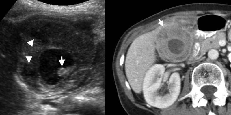 Xanthogranulomatous cholecystitis. LEFT: US shows marked wall thickening with intramural hypoechoic nodules (arrowheads), and an intraluminal stone (arrow).RIGHT: Contrast-enhanced CT shows a deformed and thickened gallbladder wall containing hypoattenuating nodulesViêm túi mật u hạt vàng. Trái: Siêu âm thấy thành túi mật dày rõ, có các nốt giảm âm trong thành (đầu mũi tên) và sỏi trong lòng (mũi tên). Phải: CT có tiêm thuốc cản quang cho thấy túi mật biến dạng, thành dày, có các nốt giảm tỷ trọng trong thành.
Xanthogranulomatous cholecystitisXanthogranulomatous viêm túi mật
Xanthogranulomatous cholecystitis is an unusual variant of chronic cholecystitis, characterized by a lipid-laden inflammatory process comparable to xanthogranulomatous pyelonephritis.Xanthogranulomatous viêm túi mật là một biến thể bất thường của viêm túi mật mạn tính, được đặc trưng bởi quá trình viêm chứa nhiều lipid tương đương với viêm thận bể thận xanthogranulomat.
Imaging studies show marked gallbladder wall thickening, often containing intramural nodules that are hypoechoic at sonography and hypoattenuating at CT, representing abscesses or foci of xanthogranulomatous inflammation.
These features overlap with those of gallbladder carcinoma, making preoperative distinction between these entities often impossible [6].
Chẩn đoán hình ảnh cho thấy dày thành túi mật rõ rệt, thường chứa các nốt trong thành là giảm âm trên siêu âm và giảm tỷ trọng trên CT, đại diện cho áp xe hoặc ổ viêm u hạt xantho.
Các đặc điểm này trùng lặp với các đặc điểm của ung thư biểu mô túi mật, làm cho việc phân biệt trước phẫu thuật giữa các thực thể này thường không thể [6].
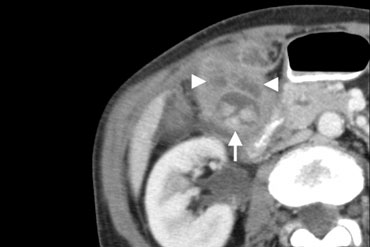 Xanthogranulomatous cholecystitis. Hypoattenuating nodules ( arrowheads) represent abscesses. The lumen contains several stones (arrow).Xanthogranulomatous viêm túi mật. Các nốt giảm tỷ trọng (các đầu mũi tên) đại diện cho các áp xe. Lumen chứa một số đá (mũi tên).
On the left a 71-year-old man with xanthogranulomatous cholecystitis.
Contrast-enhanced CT shows a deformed and thickened gallbladder wall containing hypoattenuating nodules .
These represent abscesses or foci of inflammation.
Bên trái là một người đàn ông 71 tuổi bị u hạt xantho viêm túi mật.
CT có tiêm thuốc cản quang cho thấy thành túi mật biến dạng và dày lên chứa các nốt giảm tỷ trọng.
Chúng đại diện cho áp xe hoặc ổ viêm.
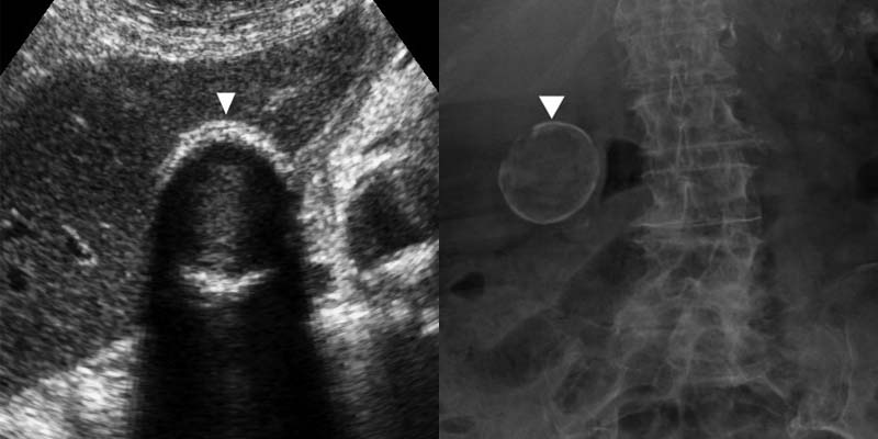 Porcelain gallbladder.Túi mật sứ.
Porcelain gallbladderTúi mật sứ
A porcelain gallbladder is a rare disorder in which chronic cholecystitis produces mural calcification.
In these patients a prophylactic cholecystectomy has been advocated because of its association with gallbladder carcinoma [4]. However, this association appears to be weak.
Túi mật sứ là một rối loạn hiếm gặp, trong đó viêm túi mật mạn tính tạo ra vôi hóa trên thành túi mật.
Ở những bệnh nhân này, cắt túi mật dự phòng đã được ủng hộ vì có liên quan đến ung thư biểu mô túi mật [4] Tuy nhiên, mối liên quan này dường như yếu.
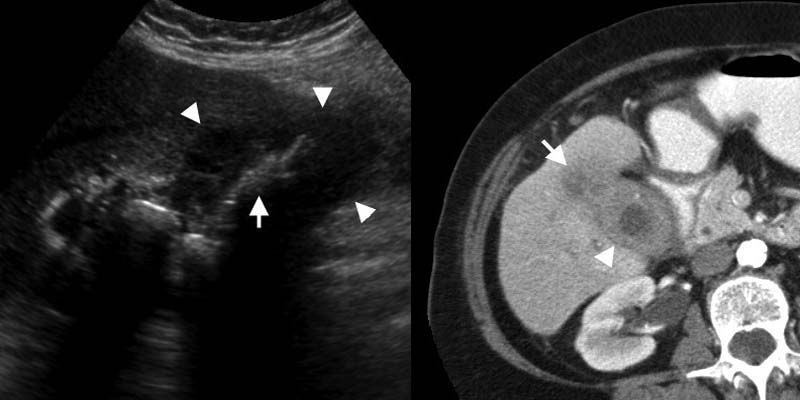 LEFT: Gallbladder carcinoma. US shows marked generalized wall thickening (arrowheads), replacing the gallbladder lumen. Multiple gallbladder stones (arrow) indicate the probable location of the filled lumen.RIGHT: Contrast-enhanced CT depicts a thick-walled gallbladder (arrowhead), with local infiltration of the mass in the adjacent liver (arrow).Trái: Ung thư biểu mô túi mật. Siêu âm cho thấy thành túi mật dày rõ, lan tỏa và chiếm chỗ lòng túi mật (đầu mũi tên). Nhiều viên sỏi (mũi tên) giúp xác định vị trí có thể là lòng túi mật đã bị lấp. Phải: CT có tiêm thuốc cản quang cho thấy túi mật thành dày (đầu mũi tên) và khối u xâm lấn gan kế cận (mũi tên).
Gallbladder carcinomaUng thư túi mật
Gallbladder carcinoma is the fifth most common malignancy of the gastrointestinal tract, and is found incidentally in 1% to 3% of cholecystectomy specimens [4].
It is often detected at a late stage of the disease, due to lack of early or specific symptoms.
Gallbladder carcinoma has various imaging appearances, ranging from a polypoid intra-luminal lesion to an infiltrating mass replacing the gallbladder, and it may also present as diffuse mural thickening.
Ung thư biểu mô túi mật là bệnh lý ác tính phổ biến thứ năm của đường tiêu hóa, và được tìm thấy tình cờ trong 1% đến 3% của mẫu cắt túi mật [4].
Nó thường được phát hiện ở giai đoạn muộn của bệnh, do thiếu các triệu chứng sớm hoặc cụ thể.
Ung thư biểu mô túi mật có nhiều hình ảnh khác nhau, từ tổn thương trong lòng túi mật đến khối thâm nhiễm thay thế túi mật, và nó cũng có thể biểu hiện như dày thành lan toả.
Associated findings such as invasion of adjacent structures, secondary bile duct dilatation, and liver or nodal metastases may help in differentiating a carcinoma from acute or xanthogranulomatous cholecystitis [2, 4].
In absence of these associated findings, it may not be possible to differentiate a carcinoma from xanthogranulomatous cholecystitis.Các dấu hiệu liên quan như xâm lấn các cấu trúc lân cận, giãn ống mật thứ phát, và di căn gan hoặc di căn hạch có thể giúp phân biệt ung thư biểu mô với viêm túi mật cấp tính hoặc xanthogranulomatous [2, 4].
Nếu không có những phát hiện liên quan này, có thể không phân biệt được ung thư biểu mô với xanthogranulomatous viêm túi mật.
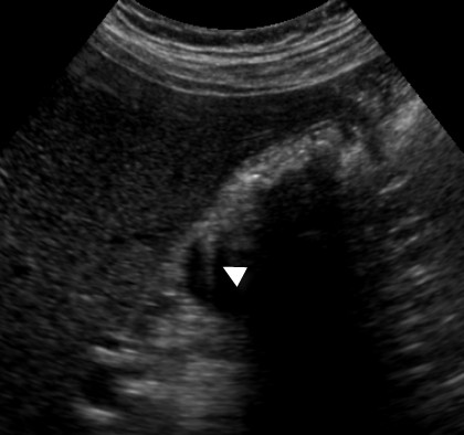 Adenomyomatosis in a 39-year-old woman. US shows mural thickening with calcifications with the characteristic 'comet-tail' reverberation artifact (arrow) due to small cholesterol crystals within Rokitansky-Aschoff sinuses.U tuyến ở phụ nữ 39 tuổi siêu âm cho thấy dày thành với vôi hóa với tạo âm vang 'đuôi cụt' đặc trưng (mũi tên) do các tinh thể cholesterol nhỏ trong xoang Rokitansky-Aschoff.
AdenomyomatosisU tuyến
Adenomyomatosis of the gallbladder is characterized by epithelial proliferation, muscular hypertrophia and intramural diverticula (Rokitansky-Aschoff sinuses), which may segmentally or diffusely involve of the gallbladder.
It is a benign condition that requires no specific treatment, occurring as an incidental finding in up to 9% of cholecystectomy specimens [6].
U cơ tuyến của túi mật được đặc trưng bởi sự tăng sinh biểu mô, phì đại cơ và túi thừa trong thành (xoang Rakitansky-Aschoff), có thể liên quan đến túi mật một cách phân đoạn hoặc lan toả.
Đây là một tình trạng lành tính không cần điều trị đặc hiệu, xảy ra dưới dạng phát hiện tình cờ trong tới 9% mẫu bệnh phẩm cắt túi mật [6].
The sonographic finding of cholesterol crystals, shown as 'comet-tail'
reverberation artifacts (Fig), within a thickened wall of the gallbladder strongly suggests this diagnosis.
Air may produce a similar artifact, however, patients with emphysematous cholecystitis are usually ill in contrast to those with adenomyomatosis.
MR imaging may be able to differentiate adenomyomatosis from gallbladder carcinoma by depicting Rokitansky-Aschoff sinuses [7].Phát hiện siêu âm các tinh thể cholesterol, được hiển thị dưới dạng các vết dội âm 'đuôi cụt' (Hình), trong một thành dày của túi mật gợi ý mạnh mẽ cho chẩn đoán này.
Không khí có thể tạo ra một hiện tượng tương tự, tuy nhiên, bệnh nhân viêm túi mật khí phế thũng thường bị bệnh trái ngược với những người bị u cơ tuyến.
Chụp cộng hưởng từ có thể phân biệt u cơ tuyến với ung thư túi mật bằng cách mô tả các xoang Rokitansky-Aschoff [7].
Secondary gallbladder involvementTổn thương túi mật thứ phát
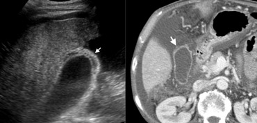 56-year-old man with liver cirrhosisLEFT: US depicts wall thickening (arrow), surrounded by ascites. Note the irregular cirrhotic liver parenchyma. RIGHT: At contrast-enhanced CT the wall of the gallbladder (arrow) appears nearly normal, because subserosal oedema can not be well differentiated from surrounding ascites at CT.Người đàn ông 56 tuổi bị xơ gan TRÁI: siêu âm mô tả thành dày lên (mũi tên), được bao quanh bởi cổ trướng. Lưu ý nhu mô gan xơ gan không đều. PHẢI: Trên CT cản quang, thành túi mật (mũi tên) gần như bình thường, do phù nề dưới thanh mạc không thể phân biệt rõ với báng bụng xung quanh trên CT.
Liver cirrhosisXơ gan
Systemic diseases such as hepatic dysfunction, heart failure, or renal failure may lead to diffuse gallbladder thickening [1, 2].
The exact pathophysiologic mechanism leading to oedema of the gallbladder wall in these diverse conditions is uncertain, but it is likely due to elevated portal venous pressure, elevated systemic venous pressure, decreased intravascular osmotic pressure, or a combination of these factors.
Hypoproteinemia has also been reported as a cause of extrinsic gallbladder disease, but this has been disputed [8].Các bệnh hệ thống như rối loạn chức năng gan, suy tim hoặc suy thận có thể dẫn đến dày túi mật lan toả [1, 2].
Cơ chế sinh lý bệnh chính xác dẫn đến phù nề thành túi mật trong những tình trạng đa dạng này là không chắc chắn, nhưng có thể là do áp lực tĩnh mạch cửa tăng cao, áp lực tĩnh mạc hệ thống tăng cao, giảm áp lực thẩm thấu nội mạch hoặc kết hợp các yếu tố này.
Giảm protein máu cũng đã được báo cáo là một nguyên nhân gây ra bệnh túi mật bên ngoài, nhưng điều này đã bị tranh cãi [8].
Liver cirrhosis, hepatitis and congestive right heart failure are relatively frequent causes.
The case on the left is a patient with liver cirrhosis.
The secondary gallbladder wall thickening is presumably due to elevated portal venous pressure and decreased intravascular osmotic pressure.Xơ gan, viêm gan và suy tim phải sung huyết là những nguyên nhân tương đối thường gặp. Trường hợp bên trái là một bệnh nhân bị xơ gan.
dày thành túi mật thứ phát có lẽ là do tăng áp lực tĩnh mạch cửa và giảm áp lực thẩm thấu nội mạch.
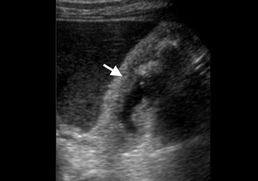 Drug-induced hepatitis with diffuse gallbladder wall thickeningViêm gan do thuốc với dày thành túi mật lan toả
HepatitisViêm gan
On the left a 75-year-old man with drug-induced hepatitis.
Longitudinal sonogram of a non-distended gallbladder shows diffuse wall thickening (arrow), and incidental cholelithiasis which may be confusing.
Bên trái là một người đàn ông 75 tuổi bị viêm gan do thuốc.
Siêu âm dọc của túi mật không giãn cho thấy dày thành lan toả (mũi tên) và sỏi mật ngẫu nhiên có thể gây nhầm lẫn.
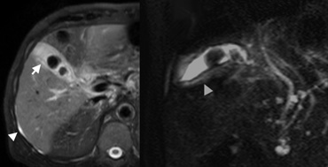 Drug-induced hepatitis.Viêm gan do thuốc.
In the same patient with the drug-induced hepatitis MR images were obtained to evaluate the bile ducts because of abnormal liver function tests.
On the far left Axial SPIR T2-weighted image (A) shows a small amount of ascites (arrowhead) which indicates that the thickened gallbladder wall (arrow) probably has an extrinsic systemic cause.
Next to it an oblique HASTE image for MR cholangiography that excludes choledocholithiasis.
Mural thickening of the gallbladder (arrowhead) is also shown.Trên cùng một bệnh nhân viêm gan do thuốc, hình ảnh cộng hưởng từ được thu thập để đánh giá ống mật do các xét nghiệm chức năng gan bất thường.
Trên hình ảnh T2 có trọng số trục SPIR (A) bên trái cho thấy một lượng nhỏ cổ trướng (đầu mũi tên) cho thấy thành túi mật dày lên (mũi tên) có thể do nguyên nhân hệ thống bên ngoài.
Bên cạnh đó là hình ảnh HASTE chéo cho chụp cộng hưởng từ đường mật loại trừ bệnh sỏi ống mật chủ.
Dày thành túi mật (đầu mũi tên) cũng được hiển thị.
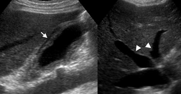 Diffuse gallbladder wall thickening in congestive right heart failureKhuếch tán dày thành túi mật trong suy tim phải sung huyết
Congestive right heart failureSuy tim phải sung huyết
On the left a 74-year-old man with congestive right heart failure.
Ultrasound depicts diffuse wall thickening of a stone-free painless gallbladder and large-caliber hepatic veins (arrowheads) and inferior vena cava, as supporting evidence of right heart failure.
Bên trái là một người đàn ông 74 tuổi bị suy tim phải sung huyết.
Siêu âm mô tả sự dày lên lan toả của túi mật không đau và không có sỏi và tĩnh mạch gan cỡ lớn (các đầu mũi tên) và tĩnh mạch chủ dưới, là bằng chứng hỗ trợ cho suy tim phải.
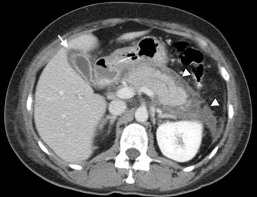 Pancreatitis in a 56-year-old man Contrast-enhanced CT shows peripancreatic inflammatory changes (arrowheads), and thickening of the wall of the gallbladder (arrow) which is secondarily involved in the pancreatic inflammation.Viêm tụy ở một người đàn ông 56 tuổi CT có tiêm thuốc cản quang cho thấy những thay đổi viêm quanh tụy (đầu mũi tên) và dày thành túi mật (mũi tên) thứ phát liên quan đến viêm tụy.
PancreatitisViêm tụy
Extracholecystic inflammation may secondarily involve the gallbladder causing wall thickening, due to direct spread of the primary inflammation, or less frequently due to an immunologic reaction [8]. Viêm ngoài túi mật thứ phát có thể liên quan đến túi mật gây dày thành, do sự lây lan trực tiếp của viêm nguyên phát, hoặc ít thường xuyên hơn do phản ứng miễn dịch [8].
Theoretically, it may be caused by any inflammation that extends to the region of the gallbladder, but only few are regularly encountered including hepatitis, pancreatitis (Figure), and pyelonephritis. Về mặt lý thuyết, nó có thể được gây ra bởi bất kỳ tình trạng viêm nào lan rộng đến vùng túi mật, nhưng chỉ có một số ít thường xuyên gặp phải bao gồm viêm gan, viêm tụy (Hình) và viêm thận bể thận.
Gallbladder wall thickening has also been reported in patients with infectious mononucleosis [9], and in patients with AIDS due to opportunistic infections or secondary neoplastic infiltration [2].Dày thành túi mật cũng đã được báo cáo ở những bệnh nhân bị tăng bạch cầu đơn nhân nhiễm trùng [9], và ở những bệnh nhi AIDS do nhiễm trùng cơ hội hoặc thâm nhiễm tân sinh thứ phát [2].
ConclusionKết luận
Diffuse gallbladder wall thickening can result from a broad spectrum of pathological conditions, including surgical and non-surgical diseases.
At times a definite imaging diagnosis may be impossible.
In most cases however, the cause can be determined by correlation of the associated imaging findings with the clinical presentation.Dày thành túi mật khuếch tán có thể là kết quả của một loạt các tình trạng bệnh lý, bao gồm các bệnh phẫu thuật và không phẫu thuật.
Đôi khi chẩn đoán hình ảnh xác định có thể không thể thực hiện được.
Tuy nhiên, trong hầu hết các trường hợp, nguyên nhân có thể được xác định bằng sự tương quan của các dấu hiệu hình ảnh liên quan với biểu hiện lâm sàng.
Charity
Hoạt động từ thiện
All the profits of the Radiology Assistant go to Medical Action Myanmar which is run by Dr. Nini Tun and Dr. Frank Smithuis sr, who is a professor at Oxford university and happens to be the brother of Robin Smithuis.Toàn bộ lợi nhuận của Radiology Assistant được chuyển cho Medical Action Myanmar, do bác sĩ Nini Tun và bác sĩ Frank Smithuis Sr. điều hành. Frank Smithuis Sr. là giáo sư tại Đại học Oxford và là anh em ruột của Robin Smithuis.
Click here or on the image below to watch the video of Medical Action Myanmar and if you like the Radiology Assistant, please support Medical Action Myanmar with a small gift.Nhấp vào đây hoặc vào hình ảnh bên dưới để xem video về Medical Action Myanmar và nếu bạn thích Trợ lý X quang, vui lòng hỗ trợ Medical Action Myanmar bằng một món quà nhỏ.
ReferencesTài liệu tham khảo
-
Rumack CM, Wilson SR, Charboneau JW. Diagnostic Ultrasound, 2nd ed. St.Louis:
Mosby, 1998:175-200
Rumack CM, Wilson SR, Charboneau JW. Siêu âm chẩn đoán, xuất bản lần thứ 2. St.Louis: -
Zissin R, Osadchy A, Shapiro M, Gayer G. CT of a thickened-wall gallbladder. Br J Radiol 2003; 76:137-143 Zissin R, Osadchy A, Shapiro M, Gayer G. CT của túi mật thành dày. BR J Radiol 2003; 76: 137-143
-
Jung SE, Lee JM, Lee K, et al. Gallbladder wall thickening: MR imaging and pathologic correlation with emphasis on layered pattern. Eur Radiol 2005; 15:694-701 Jung SE, Lee JM, Lee K, et al. dày thành túi mật: MR imaging and pathologic correlation with emphasis on layered pattern. Eur Radiol 2005; 15:694-701
-
Gore RM, Yaghmai V, Newmark GM, Berlin JW, Miller FH. Imaging of benign and malignant disease of the gallbladder. Radiol Clin N Am 2002; 40:1307-1323 Gore RM, Yaghmai V, Newmark GM, Berlin JW, Miller FH. Hình ảnh bệnh lý lành tính và ác tính của túi mật. Radiol Clin N. Am 2002; 40:1307-1323
-
Boland GWL, Slater G, Lu DSK, Eisenberg P, Lee MJ, Mueller PR. Prevalence and significance of gallbladder abnormalities seen on sonography in intensive care unit patients. AJR 2000; 174:973-977 Boland GWL, Slater G, Lu DSK, Eisenberg P, Lee MJ, Mueller PR. Tỷ lệ và ý nghĩa của bất thường túi mật trên siêu âm ở bệnh nhân hồi sức tích cực AJR 2000; 174:973-977
-
Levy AD, Murakat LA, Abbott RM, Rohrmann CA. Benign tumors and tumorlike lesions of the gallbladder and extrahepatic bile ducts: radiologic-pathologic correlation. RadioGraphics 2002; 22:387-413 Levy AD, Murakat LA, Abbott RM, Rohrmann CA. Các khối u lành tính và các tổn thương giống u của túi mật và ống mật ngoài gan: tương quan về X quang-Bệnh học. RadioGraphics 2002; 22:387-413
-
Yoshimitsu K, Honda H, Jimi M, et al. MR diagnosis of adenomyomatosis of the gallbladder and differentiation from gallbladder carcinoma: importance of showing Rokitansky-Aschoff sinuses. AJR 1999;172:1535-1540 Yoshimitsu K, Honda H, Jimi M, et al. Chẩn đoán MR của u tuyến túi mật và phân biệt với ung thư biểu mô túi mật: Tầm quan trọng của việc cho thấy các xoang rokitansky-aschoff. AJR 1999; 172: 1535-1540
-
Kaftori JK, Pery M, Green J, Gaitini D. Thickness of the gallbladder wall in patients with hypoalbuminemia: a sonographic study of patients on peritoneal dialysis. AJR 1987; 148:1117-1118 Kaftori JK, Pery M, Green J, Gaitini D. Độ dày thành túi mật ở bệnh nhân giảm albumin máu: nghiên cứu siêu âm ở bệnh nhân lọc màng bụng. AJR 1987; 148:1117-1118
-
Yamada K, Yamada H. Gallbladder wall thickening in mononucleosis syndromes. J Clin Ultrasound 2001; 29:322-325 Yamada K, Yamada H. dày thành túi mật trong hội chứng tăng bạch cầu đơn nhân. J Clin Ultrasound 2001; 29:322-325