
Biliary duct pathologyBệnh lý ống mật
Chief Gastrointestinal Radiology, University Department of Radiologic Pathology, Armed Forces Institute of Pathology, Washington DCTrưởng khoa X quang đường tiêu hóa, Khoa Bệnh học X quang, Viện Bệnh học Lực lượng Vũ trang, Washington DC
Ngày đăng / cập nhật: 2009-04-24
Bản gốc trên radiologyassistant.nl ↗ · tải về lúc 2026-11-19 20:19 · bản dịch máy có chuẩn hóa thuật ngữ chẩn đoán hình ảnh
This review is based on a presentation given by Angela Levy and adapted for the Radiology Assistant by Robin Smithuis.
We will discuss:Bài tổng quan này dựa trên một bài thuyết trình của Angela Levy và được Robin Smithuis điều chỉnh cho Trợ lý X quang.
Chúng ta sẽ thảo luận:
- Nonobstructive biliary disordersRối loạn đường mật không tắc nghẽn
- Obstructive biliary neoplasmsTắc nghẽn đường mật
by Angela D. Levybởi Angela D. Levy
IntroductionMở đầu
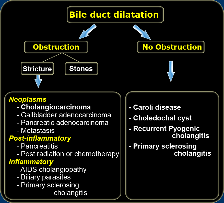
Differential Diagnosis of bile duct dilatationChẩn đoán phân biệt của nong đường mật
Whenever there is bile duct dilatation, the first priority is to look for obstruction.
Obstruction
If there is an obstruction, we first look for gallstones in the bile duct.
If there are no gallstones involved, we then look for strictures.
The differential diagnosis for a stricture is based on the location.Khi có giãn ống mật, ưu tiên hàng đầu là tìm kiếm tắc nghẽn.
Tắc nghẽn
Nếu có tắc nghẽn, đầu tiên chúng ta tìm sỏi mật trong ống mật, nếu không có sỏi mật, sau đó chúng ta tìm các chỗ hẹp.
chẩn đoán phân biệt cho hẹp được dựa trên vị trí.
- A distal stricture is most likely the result of a distal cholangiocarcinoma, pancreatic carcinoma or pancreatitis.Hẹp đầu xa thường là kết quả của ung thư đường mật đầu xa, ung thư biểu mô tuyến tụy hoặc viêm tụy.
- A stricture within the liver is likely due to gallbladder carcinoma or inflammatory strictures like PSC (Primary Sclerosing Cholangitis) or AIDS cholangiopathy.Hẹp trong gan có thể là do ung thư biểu mô túi mật hoặc hẹp do viêm như PSC (Viêm đường mật xơ cứng nguyên phát) hoặc bệnh đường mật AIDS.
- Metastatic disease can occur anywhere within the bilairy system.Bệnh di căn có thể xảy ra ở bất cứ đâu trong hệ thống động mạch đôi.
No Obstruction
Once we have excluded obstruction, we have to think about nonobstructive biliary diseases like:Không có tắc nghẽn
Một khi đã loại trừ được tắc mật, chúng ta phải nghĩ đến các bệnh lý đường mật không tắc như:
- Caroli diseaseBệnh Caroli
- Choledochal cystNang ống mật chủ
- Recurrent pyogenic cholangitisViêm đường mật tái phát
- Primary sclerosing cholangitisViêm xơ đường mật nguyên phát
Caroli DiseaseBệnh Caroli
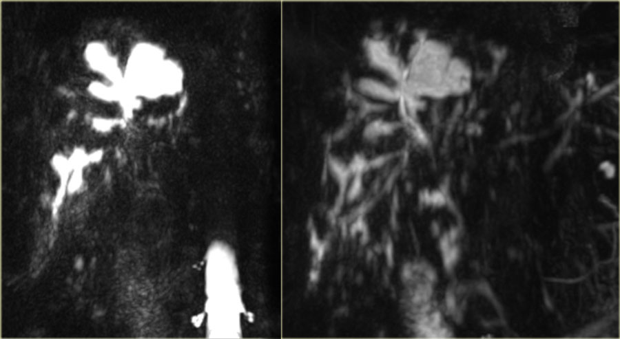 Saccular intrahepatic duct dilatation with normal sized choledochal duct in Caroli diseaseỐng gan trong gan nong với ống ống mật chủ có kích thước bình thường trong bệnh Caroli
Caroli disease is an autosomal recessive disease secondary to the ductal plate malformation.
It is associated with polycystic kidney disease, medullary sponge kidney and medullary cystic disease.
So looking at the kidneys can sometimes help you make this diagnosis.Bệnh Caroli là một bệnh di truyền lặn nhiễm sắc thể thường thứ phát do dị dạng đĩa ống dẫn.
Nó có liên quan đến bệnh thận đa nang, thận xốp tuỷ và bệnh nang tuỷ.
Vì vậy, nhìn vào thận đôi khi có thể giúp bạn đưa ra chẩn đoán này.
On the left we see images of a patient with Caroli disease.
Notice the intrahepatic duct dilatation and the normal caliber of the choledochal duct (extrahepatic bile duct).Bên trái chúng ta thấy hình ảnh của một bệnh nhân mắc bệnh Caroli.
Lưu ý sự giãn ống gan trong gan và tầm cỡ bình thường của ống dẫn mật (ống mật ngoài gan).
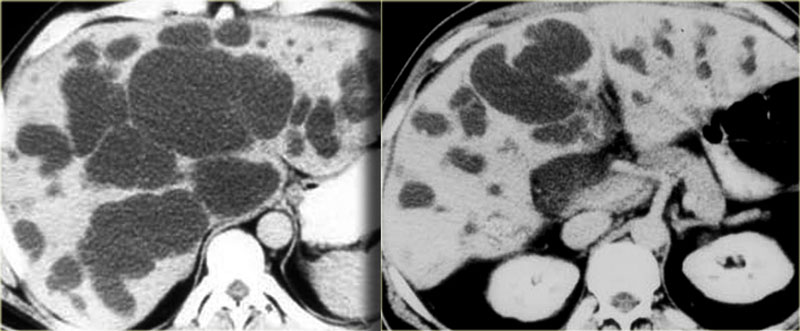 Saccular bile duct dilatation in Caroli diseaseDày ống mật chủ trong bệnh Caroli
The hallmark of Caroli disease is intrahepatic duct dilatation.
The dilatation can be very large and saccular as seen in the case on the left or it can be very linear.Dấu hiệu đặc trưng của bệnh Caroli là giãn ống gan.
Sự giãn nở có thể rất lớn và có túi như trong trường hợp bên trái hoặc nó có thể rất tuyến tính.
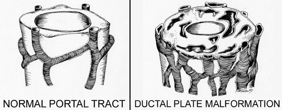
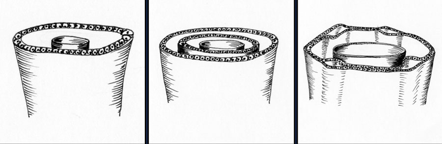 Normal development of the ductal plate (Illustration by Aletta Frazier)Sự phát triển bình thường của đĩa đệm ống động mạch (hình minh hoạ bởi Aletta Frazier)
Caroli Disease (2)
The duct dilatation in Caroli disease is due to a congenital malformation of the ductal plate, which is the precursor of the intrahepatic bile ducts.
On the left we see the normal development of the ductal plate.
Embryologically each bile duct begins as a single layer of cells that surrounds a portal vein.
This layer then duplicates.
Portions of this double layer fuse and resorb leaving unfused portions that become the bile ducts.
Bệnh Caroli (2)
Sự giãn nở ống dẫn trong bệnh Caroli là do dị tật bẩm sinh của tấm ống dẫn, là tiền thân của các ống mật trong gan.
Bên trái chúng ta thấy sự phát triển bình thường của tấm ống.
Về mặt phôi thai, mỗi ống mật bắt đầu như một lớp tế bào đơn bao quanh tĩnh mạch cửa.
Lớp này sau đó nhân đôi.
Các phần của lớp kép này kết hợp và tái hấp thu để lại các phần không được sử dụng trở thành các ống mật.
 Xem hình gốc (EN)
 Abnormal development of the ductal plate(Illustration by Aletta Frazier)Sự phát triển bất thường của tấm ống dẫn (hình minh hoạ bởi Aletta Frazier)
So in the normal situation each portal vein is surrounded by interconnecting bile ducts (left image).
However if the patient has ductal plate malformation, the bile ducts are too numerous and they are ectatic (right image).
Whether or not we see this on imaging depends on which portion of the bile ducts is affected.
If the large ducts are involved, we see this as Caroli disease.
However if only the very small ducts are involved, the result is congenital hepatic fibrosis.
If all ducts are involved, then there is a combination of fibrosis and Caroli disease, which is also known as the Caroli syndrome.Vì vậy, trong tình huống bình thường, mỗi tĩnh mạch cửa được bao quanh bởi các ống mật kết nối với nhau (hình bên trái).
Tuy nhiên, nếu bệnh nhân có dị dạng ống mật chủ, các ống mật quá nhiều và chúng bị giãn (hình ảnh bên phải).
Việc chúng ta có thấy điều này trên hình ảnh hay không phụ thuộc vào phần nào của ống mật bị ảnh hưởng.
Nếu các ống lớn có liên quan, chúng ta thấy đây là bệnh Caroli.
Tuy nhiên, nếu chỉ có các ống dẫn rất nhỏ, kết quả là xơ gan bẩm sinh.
Nếu tất cả các ống dẫn đều có liên quan, thì có sự kết hợp của xơ hóa và bệnh Caroli, còn được gọi là hội chứng Caroli.
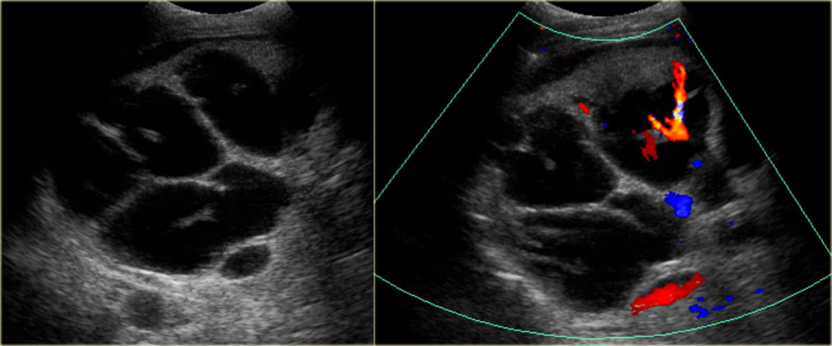 Central dot sign in Caroli diseaseDấu hiệu chấm trung tâm trong bệnh Caroli
Central dot signDấu chấm trung tâm
Most commonly the intrahepatic duct dilatation is segmental (83%) in distribution.
The diffuse form is less common (17%).
The shape of the dilatation is saccular in 76% or fusiform in 24% of the cases.
A very important sign is the central dot sign.
The central dot corresponds to the portal vein that is surrounded by dilated bile ducts.
Phổ biến nhất là giãn ống gan phân đoạn (83%).
Dạng lan toả ít gặp hơn (17%).
Hình dạng của nong là dạng túi trong 76% hoặc dạng hình thoi trong 24% trường hợp.
Một dấu hiệu rất quan trọng là dấu chấm trung tâm.
Chấm trung tâm tương ứng với tĩnh mạch cửa được bao quanh bởi các ống mật bị giãn.
On the left we see a dot within the dilated ducts.
When we put on the color doppler, we will notice that these structures contain blood flow and represent portal veins.Bên trái chúng ta thấy một chấm trong các ống dẫn nong.
Khi chúng tôi đặt doppler màu, chúng tôi sẽ nhận thấy rằng những cấu trúc này chứa dòng máu và đại diện cho các tĩnh mạch cửa.
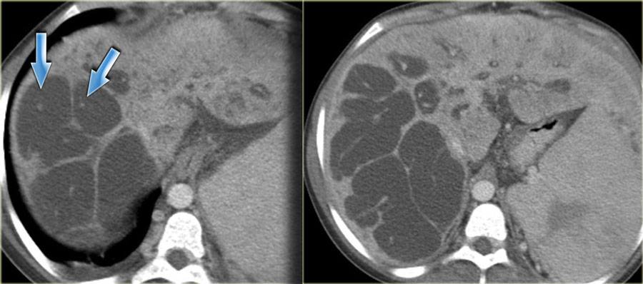 Central dot sign in Caroli diseaseDấu hiệu chấm trung tâm trong bệnh Caroli
Caroli Disease (3)
On the left CT-images of the same patient.
Notice the central dot sign and the segmental involvement.
This patient has cirrhosis with splenomegaly due to portal hypertension.Bệnh Caroli (3)
Trên hình ảnh CT bên trái của cùng một bệnh nhân.
Chú ý dấu hiệu chấm trung tâm và tổn thương theo phân đoạn.
Bệnh nhân này bị xơ gan với lách to do tăng áp lực tĩnh mạch cửa.
Extrahepatic duct dilatation is present in 53% of cases, secondary to cholangitis and stone or sludge passage.
These are secondary findings, that are not part of the primary disease.
When there is extensive fibrosis, these patients can develop cirrhosis over time.Dày ống gan ngoài gan là hiện tượng xảy ra trong 53% trường hợp, thứ phát do viêm đường mật và sỏi hoặc bùn đường mật.
Đây là những phát hiện thứ phát, không phải là một phần của bệnh nguyên phát.
Khi có xơ hóa lan toả, những bệnh nhân này có thể tiến triển thành xơ gan theo thời gian.
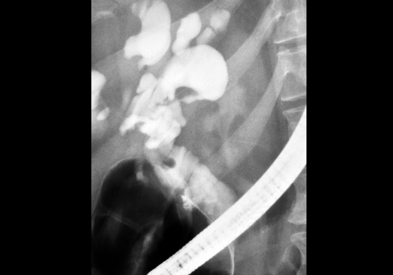 ERCP: Caroli disease with severe intrahepatic duct dilatation. No obstruction. Mild dilatation of the choledochal duct due to cholangitisERCP: Bệnh Caroli với tình trạng giãn ống gan nặng, không có tắc nghẽn, giãn ống mật chủ nhẹ do viêm đường mật.
Caroli Disease (4)
The cholangiogram is important in the work up of these patients, because obstruction has to be excluded.
This can be done with MRCP or ERCP, as is shown on the left.
There was no sign of obstruction.
The mild dilatation of the choledochal duct was the result of cholangitis.Bệnh Caroli (4)
Chụp đường mật rất quan trọng trong việc đánh giá các bệnh nhân này, bởi vì tắc nghẽn phải được loại trừ.
Điều này có thể được thực hiện với MRCP hoặc ERCP, như được hiển thị ở bên trái.
Không có dấu hiệu tắc nghẽn.
Ống mật chủ giãn nhẹ là hậu quả của viêm đường mật.
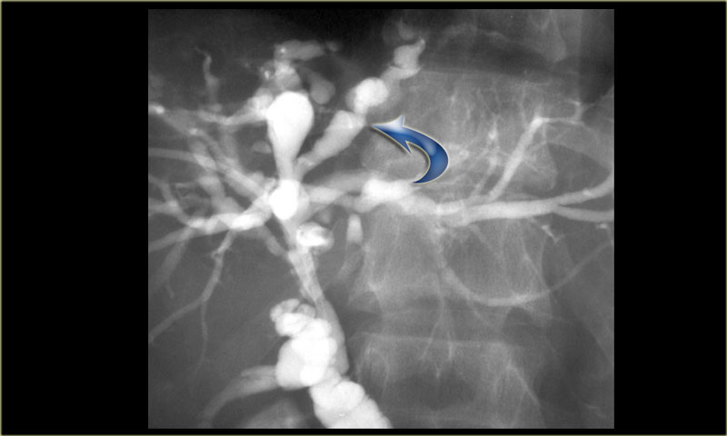
Study the image on the left.
Then continue reading.
There is focal dilatation with intermixing strictures of the bile ducts in segment IV (arrow).
The other bile ducts and the choledochal duct are normal.Nghiên cứu hình ảnh bên trái.
Sau đó tiếp tục đọc.
Có giãn ống mật chủ khu trú với hẹp ống mật ở đoạn IV (mũi tên).
Các ống mật khác và ống mật chủ bình thường.
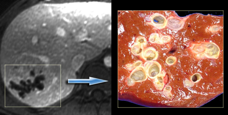
In some of the cases of Caroli disease the imaging findings may simulate a cystic neoplasm as is seen in the case on the left.
This case was originally diagnosed as a biliary cystadenoma.
However, the gross specimen demonstrates dilated bile ducts and ductal plate malformation was present microscopically.Trong một số trường hợp bệnh Caroli, các dấu hiệu hình ảnh có thể mô phỏng một khối u nang như trong trường hợp bên trái.
Ca bệnh này ban đầu được chẩn đoán là u nang đường mật.
Tuy nhiên, mẫu đại thể hiện ống mật dãn và dị dạng đĩa ống mật có mặt trên kính hiển vi.
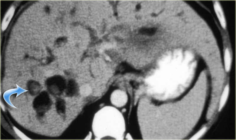 Caroli disease with intraductal stone formationBệnh Caroli với sỏi trong ống thận
Caroli Disease (5): Complications
Patient with Caroli disease are usually brought to our attention, when they develop complications.
These complications are commonly the result of biliary stasis, which leads to stone formation and infection.
Complications:Bệnh Caroli (5): Các biến chứng
Bệnh nhân mắc bệnh Caroli thường được chúng tôi chú ý, khi họ phát triển các biến chứng.
Những biến chứng này thường là kết quả của ứ mật, dẫn đến hình thành sỏi và nhiễm trùng.
Biến chứng:
- Intraductal stones Sỏi trong ống mật chủ
- Cholangitis and Abscess Viêm đường mật và áp xe
- Livercirrhosis Bệnh gan
- Cholangiocarcinoma Ung thư đường mật
On the left a patient with dilated bile ducts with intraductal stones (arrow) Bên trái là bệnh nhân có sỏi đường mật trong ống mật chủ (mũi tên)
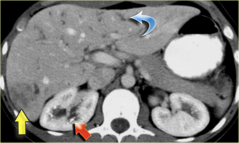
Study the image on the left.
Then continue reading.
The findings are:Nghiên cứu hình ảnh bên trái.
Sau đó tiếp tục đọc.
Kết quả nghiên cứu:
- Medullary sponge kidney with stone formation (red arrow)Miếng bọt biển tuỷ thận hình thành sỏi (mũi tên màu đỏ)
- Mild bile duct dilatation (blue arrow) Ống mật giãn nhẹ (mũi tên xanh)
- Round hypoattenuating mass in the right liver lobe Khối giảm tỷ trọng tròn ở thuỳ gan phải
The mass in the right lobe of the liver turned out to be an abscess.
Remember that liver abscesses in the early phase can look quite solid.
In the differential diagnosis we would also have to include a neoplasm, because patients with Caroli disease have an increased risk of developing a cholangiocarcinoma. Khối u ở thuỳ phải gan là một áp xe.
Hãy nhớ rằng áp xe gan trong giai đoạn đầu có thể trông khá cứng.
Trong chẩn đoán phân biệt, chúng ta cũng sẽ phải bao gồm một khối u, bởi vì bệnh nhân mắc bệnh Caroli có nguy cơ phát triển ung thư đường mật.
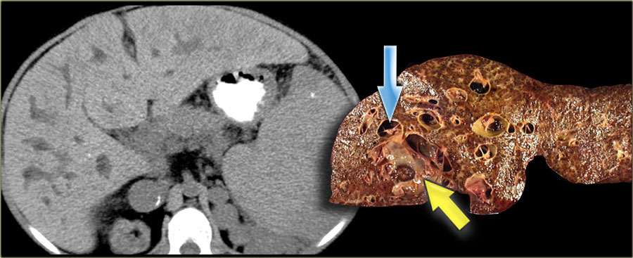 Cirrhosis in a patient with Caroli diseaseXơ gan ở bệnh nhân Caroli
Ultimately if there is substantial fibrosis and the entire liver is involved, these patients develop cirrhosis.
The patient on the left developed severe liver failure and a resection was performed.
Notice the intrahepatic bile duct dilatation, splenomegaly and dilated venous collaterals.
In the resected specimen there is a central dot sign (blue arrow) and a small pus collection (yellow arrow). Cuối cùng nếu có xơ hóa đáng kể và toàn bộ gan bị ảnh hưởng, những bệnh nhân này sẽ bị xơ gan.
Bệnh nhân bên trái bị suy gan nặng và được phẫu thuật cắt gan.
Lưu ý tình trạng giãn ống mật trong gan, lách to và giãn tĩnh mạch bàng hệ.
Trong mẫu bệnh phẩm đã cắt bỏ có dấu chấm trung tâm (mũi tên màu xanh) và một mủ nhỏ (mũI tên màu vàng).
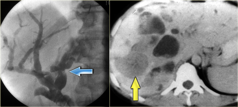 LEFT: Infiltrating cholangiocarcinoma with stricture in a patient with Caroli disease RIGHT: Intraductal cholangiocarcinomaTrái: thâm nhập vào ung thư đường mật với tình trạng nghiêm trọng ở bệnh nhân mắc bệnh Caroli Phải: ung thư đường mật nội dẫn
Cholangiocarcinoma can take on many forms in patients with Caroli disease.
The cholangiogram on the left demonstrates a focal stricture (blue arrow), which turned out to be a infiltrating cholangiocarcinoma.
The patient on the right has a cholangiocarcinoma in which the tumor was filling up the dilated ducts (yellow arrow).Ung thư đường mật có thể có nhiều dạng ở bệnh nhân mắc bệnh Caroli.
Chụp đường mật bên trái cho thấy một hẹp khu trú (mũi tên màu xanh), hóa ra là một ung thư đường mật thâm nhiễm.
Bệnh nhân bên phải có ung thư đường mật trong đó khối u đang lấp đầy các ống dẫn nong (mũi tên màu vàng).
Choledochal cystNang ống mật chủ
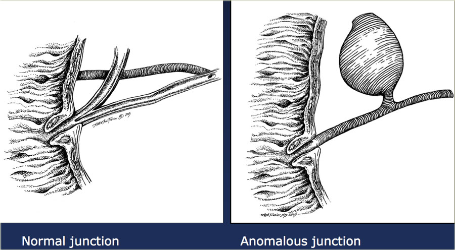

A choledochal cyst is a congenital dilatation of the extrahepatic bile duct.
These patients do not have a ductal plate abnormality.
The most common theory for the development of a choledochal cyst is that the dilatation is due to an underlying anomalous pancreatico-biliary junction.
In the anomalous junction the biliary and pancreatic duct join proximal to the sphincter of Oddi.
In these patients there is a long common channel.
The theory is that when the sphincter of Oddi contracts, pancreatic enzymes will flow into the bile duct and causing dilatation and in some cases, narrowing of the distal duct.Nang đường mật là tình trạng giãn bẩm sinh của ống mật ngoài gan. Những bệnh nhân này không có bất thường về tấm ống dẫn. Lý thuyết phổ biến nhất cho sự phát triển của nang đường mật là sự giãn nở là do một sự bất thường của đường mật tụy.
Trong chỗ nối bất thường, đường mật và ống tụy nối với đoạn gần cơ thắt của Oddi.
Ở những bệnh nhân này có một kênh phổ biến dài.
Lý thuyết cho rằng khi cơ thắt của Oddi co lại, các enzym tụy sẽ chảy vào ống mật và gây giãn và trong một số trường hợp, thu hẹp ống lượn xa.
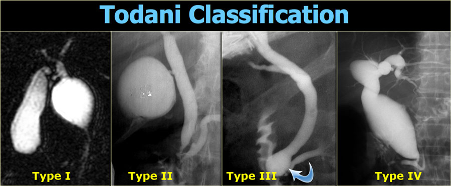
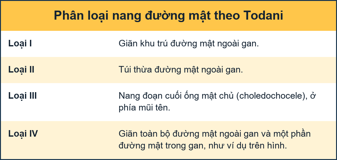
Todani ClassificationPhân loại Todani
This classification classifies the choledochal cysts into 5 cathegories.
Type V, which is not shown on the left is Caroli disease.
We now know, that Caroli is a different disease.
Type I is a true choledichal cyst with focal dilatation of the extrahepatic duct.
This is the most frequent type (90-95% of the cases).
Type IV is also a true choledichal cyst with dilatation of the entire extrahepatic duct with involvement of portions of the intrahepatic ducts.
The intrahepatic ducts taper normally to the periphery, indicating that there is no obstruction.
Type II and III are extremely rare and it is debatable whether or not these are true choledochal cysts.
Type II is a diverticulum of the extrahepatic duct and many believe that this entity is not related to an anomalous pancreatico-biliary junction.
Type III is a choledochocele, where there is dilatation of the distal part of the bile duct.
These patients also have a normal pancreaticobiliary junction.
Phân loại này phân loại các nang ống mật chủ thành 5 cathegories.
Loại V, không được hiển thị bên trái là bệnh Caroli.
Chúng ta biết rằng Caroli là một bệnh khác.
Loại I là một nang ống động mạch sọ thực sự với sự giãn nở khu trú của ống ngoài gan.
Đây là loại thường gặp nhất (90-95% số trường hợp).
Loại IV cũng là một nang ống động mạch cảnh thực sự với sự giãn nở của toàn bộ ống ngoài gan với sự tham gia của các phần của ống trong gan.
Các ống dẫn trong gan thường thon về phía ngoại vi, cho thấy không có tắc nghẽn.
Loại II và III là cực kỳ hiếm và gây tranh cãi liệu đây có phải là nang ống mật chủ thực sự hay không.
Loại II là một túi thừa của ống dẫn ngoài gan và nhiều người tin rằng thực thể này không liên quan đến sự bất thường của đường nối mật tụy.
Loại III là sa ống mật chủ, trong đó có giãn phần xa của ống mật.
Những bệnh nhân này cũng có một đường nối tụy-mật bình thường.
 Type IV choledochal cystU nang ống mật chủ type IV
Choledochal cyst (2)
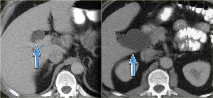
On the left a patient with dilatation of both the extrahepatic duct and part of the intrahepatic ducts.
So this is a type IV.
Notice that the peripheral ducts are normal, so this is not an obstructive pattern. Nang ống mật chủ (2)
Bên trái là một bệnh nhân có giãn cả ống ngoài gan và một phần ống trong gan.
Đây là loại IV.
Lưu ý rằng các ống ngoại vi là bình thường, vì vậy đây không phải là một kiểu tắc nghẽn.
 Type IV choledochal cystU nang ống mật chủ type IV
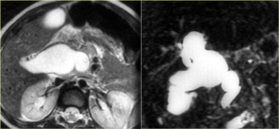
On the left another type IV choledochal cyst.
There is dilatation of the extrahepatic duct, cystic duct and a small portion of the left hepatic duct.
There is no intrahepatic dilatation.Bên trái là một nang ống mật chủ type IV khác.
Có giãn ống gan ngoài, ống nang và một phần nhỏ của ống gan trái.
Không có giãn trong gan.
 Cholangiocarcinoma within a choledocha cystUng thư đường mật trong nang ống mật chủ
Choledochal cyst (3)
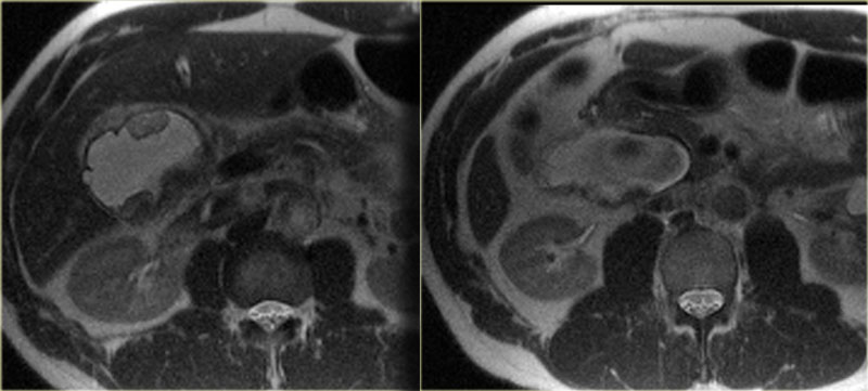
There is an association of bile duct adenocarcinoma and choledochal cysts.
These carcinomas can occur within the choledochal cyst, like the case on the left, or in the gallbladder or anywhere else in the biliary ducts.
In the bile ducts they can present as classic peripheral cholangiocarcinoma, Klatskin tumor or distal cholangiocarcinoma.Nang ống mật chủ (3)
Có mối liên quan giữa ống mật ung thư biểu mô tuyến và nang ống mật chủ.
Những ung thư biểu mô này có thể xảy ra trong nang ống mật chủ, như trường hợp bên trái, hoặc trong túi mật hoặc bất cứ nơi nào khác trong ống mật.
Trong ống mật chúng có thể có dạng ung thư đường mật ngoại vi cổ điển, u Klatskin hoặc ung thư đường mật đầu xa.
Recurrent Pyogenic Cholangitis (RPC)Viêm đường mật tái phát do vi khuẩn (RPC)
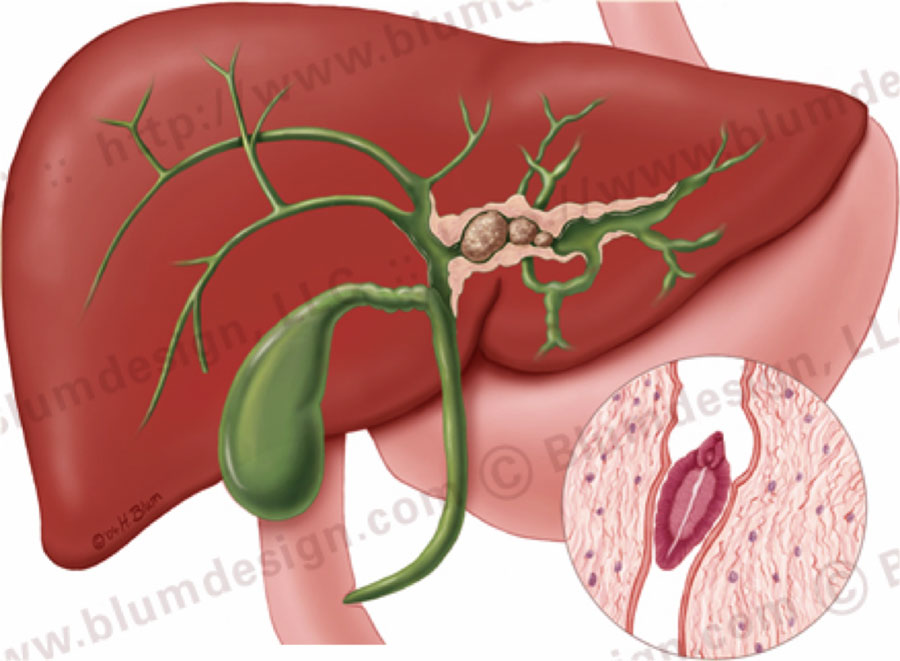 Recurrent pyogenic cholangitis. Illustration by Heike Blum.Viêm đường mật sinh mủ tái phát. Minh hoạ bởi Heike Blum.
Recurrent pyogenic cholangitis is an uncommon disease in the western world.
Most of these cases are seen in Asian countries.
The etiology is unknown, although some of these patients have biliary parasites.
The disease is characterized by the presence of intrahepatic pigmented stones and recurrent infection.
These patients are also at risk of developing biliary cirrhosis and cholangiocarcinoma.Viêm đường mật tái phát là một bệnh lý ít gặp ở các nước phương Tây.
Hầu hết các trường hợp này được thấy ở các nước châu Á.
Nguyên nhân chưa được biết, mặc dù một số bệnh nhân này có ký sinh trùng đường mật.
Bệnh được đặc trưng bởi sự hiện diện của sỏi sắc tố trong gan và nhiễm trùng tái phát.
Những bệnh nhân này cũng có nguy cơ bị xơ gan mật và ung thư đường mật.
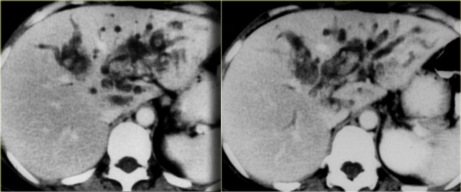 Recurrent pyogenic cholangitis of the left lobe with intrahepatic stonesViêm đường mật tái phát ở thuỳ trái có sỏi trong gan
The left lobe is the most common location of the disease due to the delayed drainage of the left system.
On the left a typical case.
There is focal dilatation of the bile ducts in the left lobe with stones.Thuỳ trái là vị trí phổ biến nhất của bệnh do hệ thống dẫn lưu trái bị trì hoãn.
Bên trái là một trường hợp điển hình.
Có sự giãn nở khu trú của các ống mật ở thuỳ trái với sỏi.
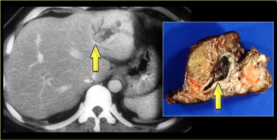 Recurrent pyogenic cholangitis with resected specimenViêm đường mật sinh mủ tái phát với bệnh phẩm được cắt bỏ
Recurrent pyogenic cholangitis (2)
On the left another example of recurrent pyogenic cholangitis.
There is intrahepatic lithiasis with focal diatation.
A case like this is indistinguishable from focal Caroli disease with secundary stone formation.Viêm đường mật tái phát (2)
Bên trái là một ví dụ khác về viêm đường mật tái phát.
Có sỏi trong gan với hình ảnh quay đầu dò khu trú.
Một trường hợp như vậy không thể phân biệt được với bệnh Caroli khu trú với sự hình thành sỏi cơ bản.
Primary Sclerosing CholangitisViêm đường mật xơ cứng nguyên phát
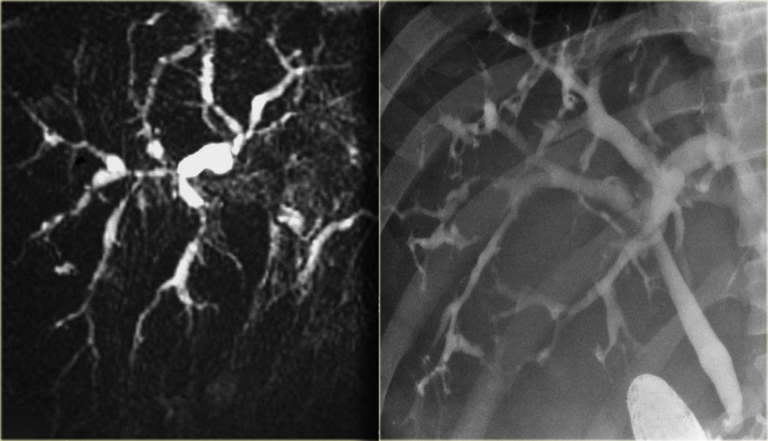 Primary sclerosing cholangitis: MRCP and ERCPViêm xơ đường mật nguyên phát: MRCP và ERCP
When we see intrahepatic bile duct dilatation with strictures and only mild dilatation, the first diagnosis we think of is primary sclerosing cholangitis (PSC).
We know however that there is a long differential diagnistic list which includes:Khi thấy giãn ống mật chủ kèm theo hẹp và chỉ giãn nhẹ, chẩn đoán đầu tiên chúng tôi nghĩ đến là viêm đường mật xơ hóa nguyên phát (PSC).
Tuy nhiên, chúng tôi biết rằng có một danh sách chẩn đoán phân biệt dài bao gồm:
- CholangiocarcinomaUng thư đường mật
- Metastatic disease (especially breast cancer)Bệnh di căn (đặc biệt là ung thư vú)
- Iatrogenic bile duct stricturesHẹp đường mật do điều trị
- Unusual biliary tumors (granular cell tumor) U đường mật bất thường (u tế bào hạt)
-
Secondary sclerosing cholangitis due to:Viêm xơ đường mật thứ phát do:
- Ischemia, for example post -ChemoembolisationThiếu máu cục bộ, ví dụ sau nút mạch hóa chất
- AIDS related cholangiopathyBệnh đường mật liên quan đến AIDS
The case on the left nicely demonstrates the bile duct strictures.
Notice that there is only mild dilatation, which is common in PSC.Trường hợp bên trái cho thấy rõ ràng sự hẹp ống mật.
Lưu ý rằng chỉ có giãn nhẹ, thường gặp trong PSC.
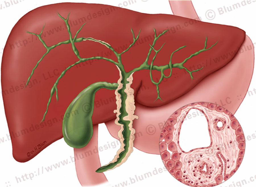 Primary sclerosing cholangitis with strictures both in the intra- and extrahepatic bile ducts. Illustration by Heike BlumViêm đường mật xơ hóa tiên phát với hẹp cả trong và ngoài ống mật gan. Minh hoạ bởi Heike Blum
The hallmark of PSC is strictures, but early on in the disease the strictures can be difficult to appreciate.
The underlying abnormality in PSC is fibrosis, which is of unknown etiology.
PSC is strongly associated with ulcerative colitis in up to 70% of patients, but it can also be associated with Crohn's disease of the large intestine.
The reason for the association with IBD is unknown, but it is thought to be the result of an immune response.Dấu hiệu của PSC là hẹp, nhưng ở giai đoạn sớm của bệnh, các hẹp có thể khó đánh giá.
Sự bất thường cơ bản trong PSC là xơ hóa, không rõ nguyên nhân.
PSC có liên quan chặt chẽ với viêm đại tràng thể loét ở 70% bệnh nhân, nhưng nó cũng có thể liên quan đến bệnh Crohn của ruột già.
Lý do cho sự liên quan với IBD là không rõ, nhưng nó được cho là kết quả của một phản ứng miễn dịch.
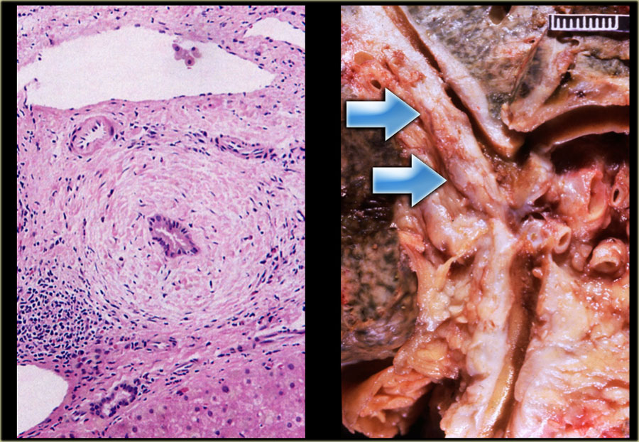 Chronic inflammation surrounding a bile duct in PSCViêm mạn tính quanh ống mật chủ trong PSC
Primary sclerosing cholangitis (2)
On the far left a histologic specimen demonstrating chronic inflammation surrounding the bile duct.
The gross specimen demonstrates the thickening of the wall of the bile duct (arrow) causing many strictures.
The strictures in PSC are short in the order of 3-5 mm in length, which is helpful to remember because if you are looking for cholangiocarcinoma, the malignant strictures usually exceed 10 mm in length.Viêm xơ đường mật nguyên phát (2)
Ở xa bên trái, một mẫu mô học cho thấy tình trạng viêm mạn tính xung quanh ống mật.
Mẫu đại thể cho thấy sự dày lên của thành ống mật (mũi tên) gây ra nhiều chỗ hẹp.
Các chỗ hẹp trong PSC ngắn theo thứ tự chiều dài 3-5 mm, điều này rất hữu ích vì nếu bạn đang tìm kiếm ung thư đường mật, các chỗ hẹp ác tính thường dài hơn 10 mm.
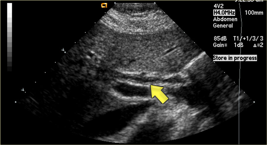 PSC with thickening of the wall of the bile duct (arrow)PSC với sự dày lên của thành ống mật (mũi tên)
Ultrasound findingsSiêu âm
One of the earliest features in PSC is on sonography where we see thickening of the wall of the bile duct as is seen in the image on the left.
This patient came for an ultrasound examination to rule out gallstones.
Notice that the intrahepatic ducts are normal.
The differential diagnosis would include PSC, AIDS-cholangitis and cholangiocarcinoma.
A cholangiocarcinoma would be rather unlikely, because there is no obstruction.
Continue with the CT.
Một trong những đặc điểm sớm nhất trong PSC là trên siêu âm, nơi chúng ta thấy sự dày lên của thành ống mật như được thấy trong hình ảnh bên trái.
Bệnh nhân này đến khám siêu âm để loại trừ sỏi mật.
Lưu ý rằng các ống dẫn trong gan bình thường.
chẩn đoán phân biệt bao gồm PSC, AIDS-viêm đường mật và ung thư đường mật.
ung thư đường mật sẽ khó xảy ra, vì không có tắc nghẽn.
Tiếp tục với CT.
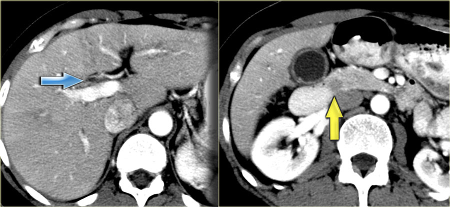 PSC with thickening of the wall of the bile duct (arrow)PSC với sự dày lên của thành ống mật (mũi tên)
CT findingsCác dấu hiệu trên CT
On the CT the liver looks quite normal.
However if you look at the common bile duct in the pancreatic head you will notice the soft tissue density.
The differential diagnosis would include an impacted stone or cholangiocarcinoma, but since this patient had no obstruction, it was thought to be the result of bile wall thickening.
The gallbladder wall is also thickened.
Trên CT, gan trông khá bình thường.
Tuy nhiên, nếu bạn nhìn vào ống mật chủ trong đầu tụy, bạn sẽ nhận thấy mật độ mô mềm.
chẩn đoán phân biệt có thể bao gồm sỏi nằm ngầm hoặc ung thư đường mật, nhưng vì bệnh nhân này không có tắc nghẽn nên được cho là kết quả của dày thành mật.
Thành túi mật cũng dày lên.
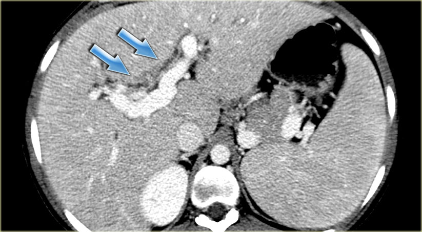 Primary sclerosing cholangitis. CT findingsViêm xơ đường mật nguyên phát. Các dấu hiệu trên CT
Primary sclerosing cholangitis (3)
On the left a patient with more severe disease.
We can see mild bile duct dilatation with a discontinuous pattern.Viêm xơ đường mật nguyên phát (3)
Bên trái là một bệnh nhân có bệnh nặng hơn.
Chúng ta có thể thấy giãn ống mật nhẹ với kiểu không liên tục.
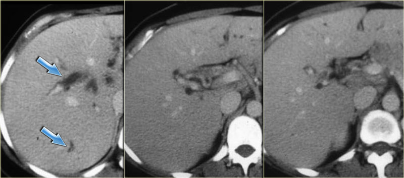 Primary sclerosing cholangitis. CT findingsViêm xơ đường mật nguyên phát. Các dấu hiệu trên CT
On the left a patient with more pronounced CT findings.
Study the image and then continue reading.
The findings are:Bên trái là một bệnh nhân có dấu hiệu CT rõ rệt hơn.
Nghiên cứu hình ảnh và sau đó tiếp tục đọc.
Kết quả nghiên cứu:
- Discontinuous dilatationGiãn không liên tục
- Bile wall thickening at the level of the porta hepatisDày thành mật ngang mức gan cửa
- Lymphadenopathy Bệnh hạch
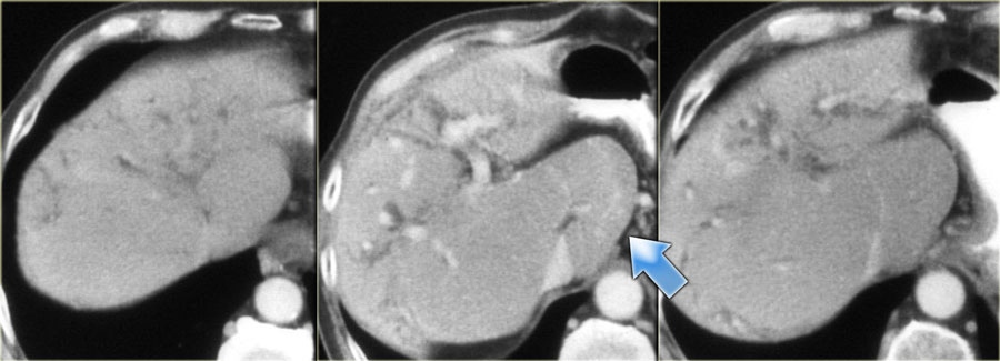 Primary sclerosing cholangitis. late CT findingsViêm xơ đường mật nguyên phát.các dấu hiệu muộn trên CT
Primary sclerosing cholangitis (4)
Late CT features are seen on the images on the left.
Study the images and then continue reading.
The findings are:Viêm xơ đường mật nguyên phát (4)
Các đặc điểm CT muộn được nhìn thấy trên các hình ảnh bên trái.
Nghiên cứu các hình ảnh và sau đó tiếp tục đọc.
Kết quả nghiên cứu:
- Morphologic changes of cirrhosis (volume loss in the right lobe and a large caudate lobe (arrow) Thay đổi hình thái xơ gan (mất thể tích ở thuỳ phải và thùy đuôi lớn (mũi tên)
- More pronounced dilatation of the bile ductsSự giãn nở rõ rệt hơn của các ống mật
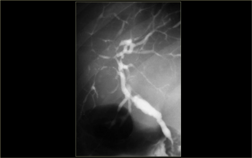 Primary sclerosing cholangitis. Cholangiographic findingsViêm xơ đường mật nguyên phát. Đặc điểm trên chụp đường mật
Cholangiography is used in the initial diagnosis of the disease, when there are only subtle strictures and in patients known with PSC to look for new strictures that are suspicious for carcinoma.
On cholangiography we can see:Chụp đường mật được sử dụng trong chẩn đoán ban đầu của bệnh, khi chỉ có những chỗ hẹp tinh tế và ở những bệnh nhân được biết đến với PSC để tìm những chỗ hẹp mới nghi ngờ là ung thư biểu mô.
Trên chụp đường mật chúng ta có thể thấy:
- Beading: alternating pattern of strictures and normal or slightly dilated ducts Beading: mô hình luân phiên của hẹp và ống thông thường hoặc hơi giãn
- Pruned-tree: distal bile ducts are narrowed and difficult to see Cây tỉa cành: ống mật đầu xa bị hẹp và khó nhìn thấy
- Mural irregularity: irregular luminal margin (best seen on the left in the extrahepatic duct) Thành ống mật không đều, thấy qua bờ lòng ống gồ ghề (rõ nhất ở ống mật ngoài gan trên ảnh bên trái).
- Diverticula: best seen in the next patientTúi thừa: nhìn thấy rõ nhất ở bệnh nhân kế tiếp
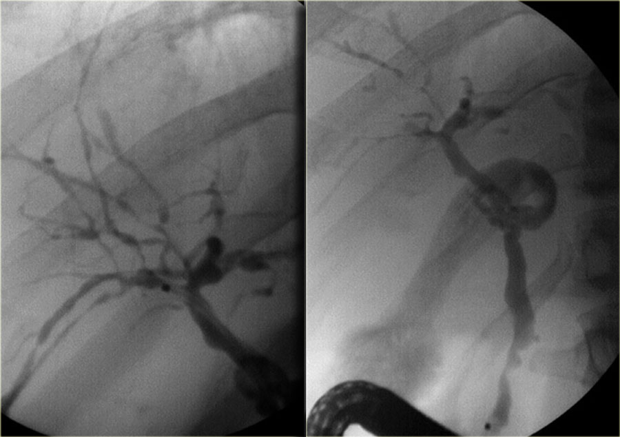 Primary sclerosing cholangitis. Cholangiographic findingsViêm xơ đường mật nguyên phát. Đặc điểm trên chụp đường mật
On the left the typical findings in PSC.
Notice the diverticula on the image on the right.
Diverticula are very specific for the diagnosis PSC.
So when you see these diverticula, you should immediately search for subtle strictures in the intrahepatic ducts.Bên trái là các dấu hiệu điển hình trong PSC.
Chú ý túi thừa trên hình ảnh bên phải.
Túi thừa rất đặc hiệu để chẩn đoán PSC.
Vì vậy, khi bạn nhìn thấy những túi thừa này, bạn nên tìm kiếm ngay lập tức các chỗ hẹp tinh vi trong các ống dẫn trong gan.
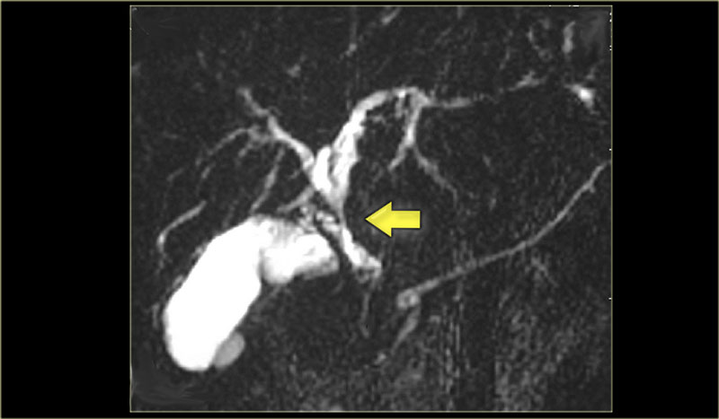 MRCP in PSC. There is a long stricture suspective of cholangiocarcinoma (arrow)MRCP trong PSC. Có một nghi ngờ hẹp dài của ung thư đường mật (mũi tên)
Primary sclerosing cholangitis (5)
On the left a MRCP in a pateint with PSC.
Notice the large stricture, which is quite worriesome for cholagiocarcinoma (arrow).
The strictures in PSC show an abrupt transission, while here we see 'shouldering', which indicates mass-efect.
In addition there is intrahepatic dilatation proximal to this stenotic area.Viêm xơ đường mật nguyên phát (5)
Bên trái là MRCP trong một bệnh nhân có PSC.
Lưu ý đến độ hẹp lớn, điều này khá đáng lo ngại đối với ung thư biểu mô sụn (mũi tên).
Các chỗ hẹp trong PSC cho thấy một sự chuyển đổi đột ngột, trong khi ở đây chúng ta thấy 'sự rung lắc', cho thấy khiếm khuyết khối lượng.
Ngoài ra còn có giãn trong gan ở đầu gần vùng hẹp này.
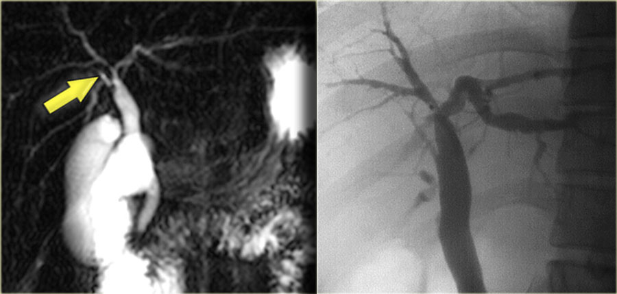 MRCP and ERCP in a patient with a stricture at the hilumNghiên cứu kết quả cộng hưởng từ và nội soi mật tụy ngược dòng trên bệnh nhân hẹp rốn phổi
On the left a MRCP in a patient demonstrating a stricture at the level of the hilum.
On MRCP this stricture looked long and worriesome for cholangiocarcinoma.
However, on the ERCP, the ducts have been distended with contrast and we can see that this is a short stricture compatible with the diagnosis of PSC.
During follow up this proved to be just PSC.Bên trái là MRCP ở một bệnh nhân có hẹp ở mức rốn phổi.
Trên MRCP, hẹp này có vẻ dài và đáng lo ngại đối với ung thư đường mật.
Tuy nhiên, trên ERCP, các ống dẫn đã bị giãn ra do thuốc cản quang và chúng ta có thể thấy đây là một hẹp ngắn tương thích với chẩn đoán PSC.
Trong quá trình theo dõi, kết quả này được chứng minh chỉ là PSC.
CholangiocarcinomaUng thư đường mật
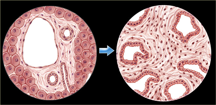 Normal columnar epithelium (left) transforms into adenocarcinoma (right)Biểu mô cột bình thường (trái) chuyển thành ung thư biểu mô tuyến (phải)
Cholangiocarcinoma (i.e., adenocarcinoma of the bile duct) arises from the columnar epithelium of the bile duct.
It is characterized by malignant glands within a desmoplastic stroma.
These tumors have an infiltrative growth pattern and do not have a capsule.Ung thư đường mật (tức là ung thư biểu mô tuyến của ống mật) phát sinh từ biểu mô cột của ống dẫn mật.
Nó được đặc trưng bởi các tuyến ác tính trong một mô đệm desmoplastic.
Những khối u này có mô hình tăng trưởng thâm nhiễm và không có bao.
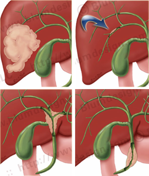 Typical locations of cholangiocarcinoma.Vị trí điển hình của ung thư đường mật.
There are four basic patterns of chlangiocarcinoma: (illustration on the left).Có bốn kiểu hình cơ bản của ung thư biểu mô đường mật: (hình minh hoạ bên trái).
Cholangiocarcinoma is an uncommon tumor, that is mostly seen in patients with underlying benign bilairy disease.
The incidence in the U.S. is 2000 to 2500 cases per year (coloncancer 150.000 per year).
In Asian countries the incidence is ten times greater due to more chronic biliary infection.
Ung thư đường mật là loại u ít gặp, chủ yếu xảy ra ở người có bệnh đường mật lành tính từ trước. Tại Hoa Kỳ, mỗi năm có 2.000–2.500 ca mới, so với 150.000 ca ung thư đại tràng. Ở các nước châu Á, tỷ lệ mắc cao gấp mười lần do nhiễm trùng đường mật mạn tính phổ biến hơn.
High risk groups are patients with:Nhóm nguy cơ cao là những bệnh nhân:
-
Autoimmune diseasesBệnh tự miễn
- PSC, ulcerative colitis, primary biliary cirrhosisPSC, viêm loét đại tràng, xơ gan mật nguyên phát
-
Congenital anatomic anomaliesDị tật bẩm sinh
- Caroli, choledochal cyst, anomalous pancreaticobiliary junctionCaroli, nang ống mật chủ, dị thường đường nối tụy-mật
- Abnormal tumor suppressor genes, FAP, NF1Gen ức chế u bất thường, FAP, NF1
-
InfectionNhiễm trùng
- Biliary parasites, recurrent pyogenic cholangitisKý sinh trùng đường mật, viêm đường mật mủ tái phát
Clinical presentation:Lâm sàng:
- JaundiceVàng da
- PainĐau
- Fever if secondary cholangitis Sốt nếu viêm đường mật thứ phát
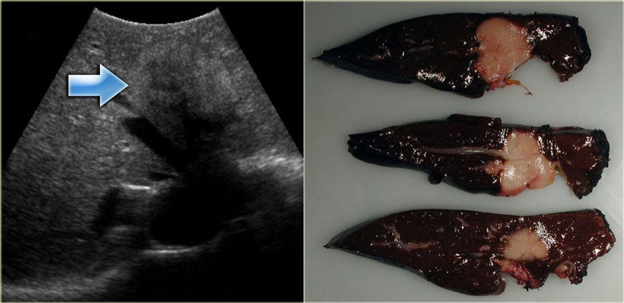 Intrahepatic cholangiocarcinoma. RadPath correlationTương quan ung thư đường mật. RadPath trong gan
Intrahepatic cholangiocarcinomaUng thư đường mật trong gan
These arise in the very small peripheral ducts.
These tumors have abundant fibrous stroma that can cause retraction of the liver capsule.
The tumor typically enhances in the equilibrium and delayed phases (5-10 minutes).Chúng phát sinh trong các ống dẫn rất nhỏ ở ngoại vi.
Những khối u này có nhiều chất xơ có thể gây co thắt bao gan.
Khối u thường tăng cường trong các giai đoạn cân bằng và chậm (5-10 phút).
Read more about intrahepatic cholangiocarcinoma in Liver : Masses Part II : common liver tumorsĐọc thêm về ung thư đường mật trong gan: Các khối Phần II: các khối u gan thường gặp
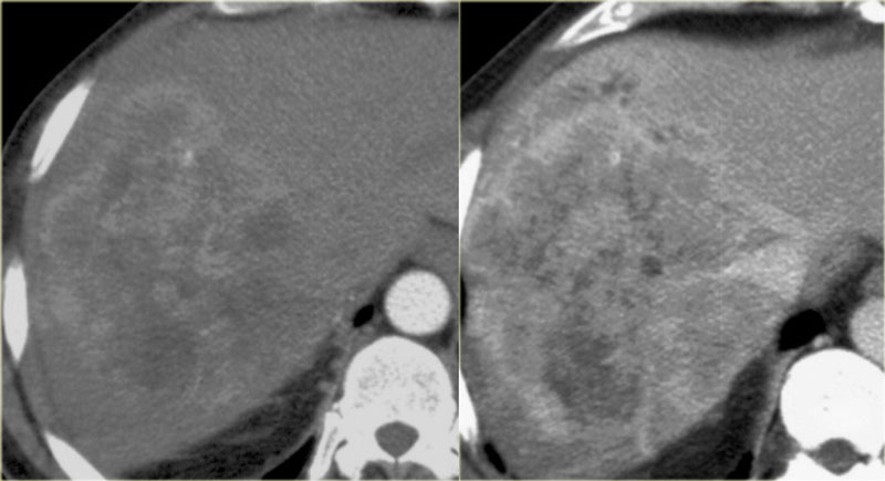 Early and late phase enhancement of a intrahepatic cholangiocarcinomaNghiên cứu sự tăng cường giai đoạn sớm và giai đoạn muộn của một ung thư đường mật trong gan
Although these tumors are usually quite heterogeneous because the contrast uptake is delayed and can be irregular.Mặc dù các khối u này thường khá không đồng nhất vì sự hấp thu chất cản quang bị trì hoãn và có thể không đều.
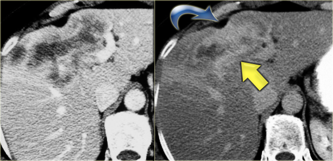 Intrahepatic cholangiocarcinoma. Capsular retraction and the late enhancementUng thư đường mật trong gan: Rút bao và ngấm thuốc muộn
The key findings to look for are:Những phát hiện chính cần tìm kiếm là:
- Delayed enhancementTăng cường chậm
- Peripheral biliary dilatationDày mật ngoại biên
- Capsular contraction Co thắt bao hoạt dịch
On the left a typical case.
Notice the capsular retraction (blue arrow) and the late enhancement (yellow arrow).Bên trái là một trường hợp điển hình.
Chú ý sự co rút của bao (mũi tên màu xanh) và sự tăng cường muộn (mũI tên màu vàng).
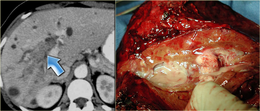 Intraductal CholangiocarcinomaXương ống tuỷ ung thư đường mật
These are very rare tumors.
They present as a intrabiliary mass with biliary dilatation peripheral to the mass.Đây là những khối u rất hiếm gặp.
Chúng biểu hiện dưới dạng một khối trong đường mật với sự giãn đường mật ở ngoại vi.
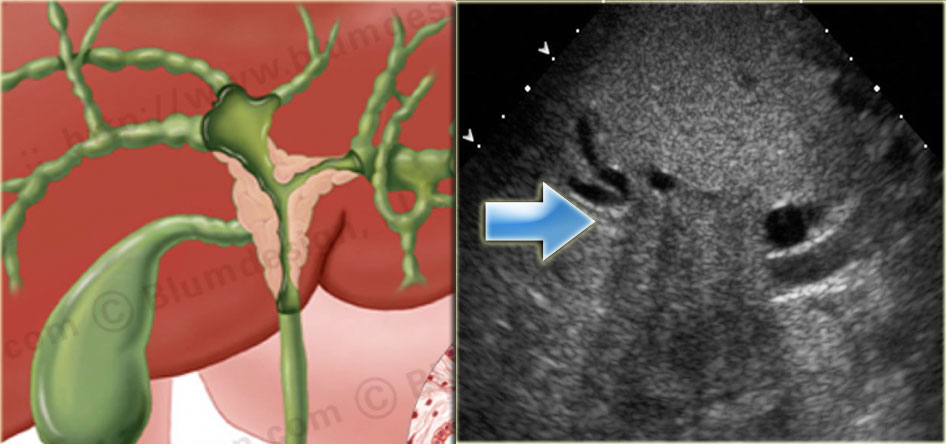 Klatskin Tumor with dilatation of bile ducts in the right and left lobe of the liver Illustration by Heike BlumU Klatskin với giãn ống mật ở gan phải và trái Hình minh hoạ bởi Heike Blum
Klatskin Tumor - Hilar CholangiocarcinomaU Klatskin – Rốn gan ung thư đường mật
The most common site of biliary adenocarcinoma is at or near the confluence of the right and left hepatic ducts.
These tumors are also known as Klatskin tumors.
Klatskin tumors have an aggressive biologic behavior.Vị trí thường gặp nhất của ung thư biểu mô tuyến đường mật là ở hoặc gần nơi hợp lưu của ống gan phải và trái.
Những khối u này còn được gọi là khối u Klatskin.
U Klatskin có hành vi sinh học tích cực.
Imaging features:Đặc điểm hình ảnh:
- Duct dilatationỐng giãn
- Ill-defined massKhối lượng bờ không rõ
- Lobar atrophyTeo thuỳ
- Vascular invasionXâm lấn mạch máu
On the left a nice correlation between an illustration and a sonographic image of a Klatskin tumor.
Notice how ill-defined the tumor is.Bên trái là tương quan giữa hình minh hoạ và hình ảnh siêu âm của một khối u Klatskin.
Lưu ý khối u bờ không rõ như thế nào.
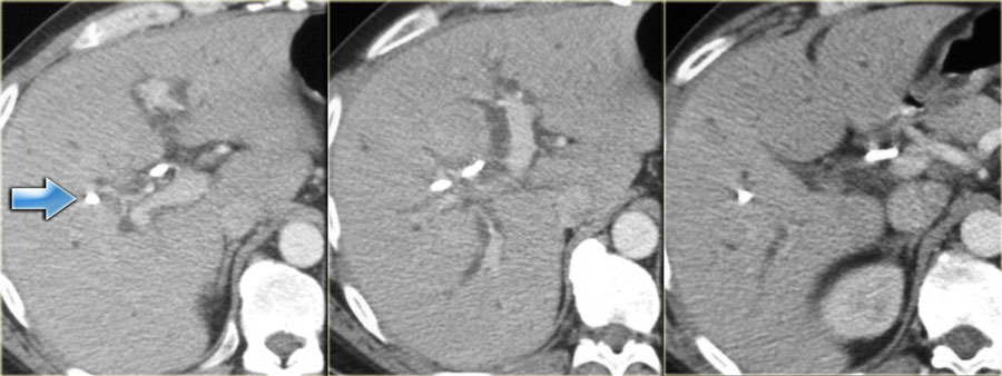 Klatskin Tumor. Barely visible. Stent in situ (arrow)Khối u Klatskin, chỉ nhìn thấy được, đặt stent tại chỗ (mũi tên)
On CT these tumors can be very difficult to visualize.
Many of these patients already have a stent in place when they first come to CT which makes it even harder (figure).
Trên CT, những khối u này có thể rất khó hình dung.
Nhiều bệnh nhân trong số này đã có stent tại chỗ khi họ lần đầu tiên đến CT, điều này càng khiến cho việc chụp CT trở nên khó khăn hơn (hình).
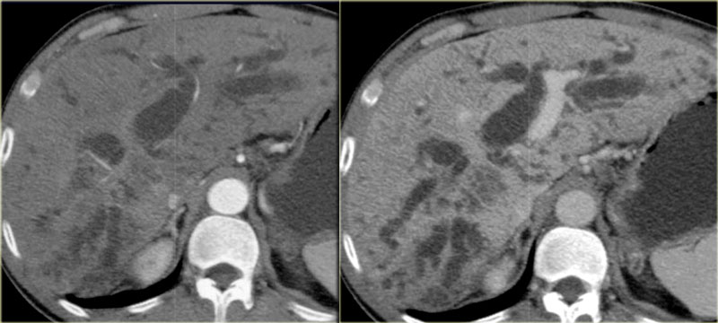 Klatskin Tumor: arterial and portal venous phaseU Klatskin: động mạch và thì tĩnh mạch cửa
Klatskin Tumor (2)
In the case on the left we can identify the tumor at the confluens of the left and right hepatic duct.
The margins of the tumor however are imperceptible because of the infiltrative growth.
Based on the CT it is not possible to stage the tumor correctly.U Klatskin (2)
Trong trường hợp ở bên trái chúng ta có thể xác định được khối u ở hợp lưu của ống gan trái và phải.
Tuy nhiên, bờ của khối u không thể nhận thấy được vì sự phát triển thâm nhiễm.
Dựa vào CT không thể xác định giai đoạn u một cách chính xác.
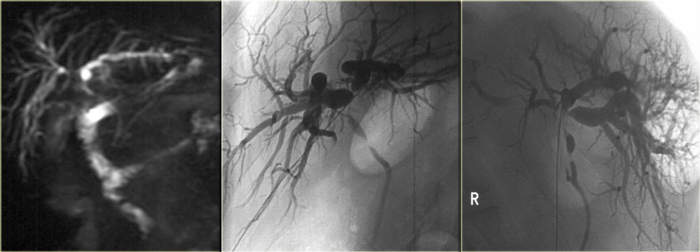 Hiar Cholagiocarcinoma. Notice the superiority of ERCP to MRCP. The image on the right nicely demonstrates the shouldering at the hilum and the multiple strictures.Hiar Ung thư biểu mô sụn. Chú ý ưu thế của ERCP so với MRCP. Hình ảnh bên phải cho thấy rõ ràng vai ở rốn và nhiều chỗ hẹp.
In these tumors it may be difficult to get a definitive diagnosis pre-operatively.
Biopsy is almost impossible and results of endoscopic brushing are commonly negative.
The staging is done with cholangiography and is based on the finding of mass effect (shouldering), irregular margins and abrupt tapering at the obstruction.
The limitations of MRCP in staging are the spatial resolution and the inability in the evaluation of the secondary ducts.
ERCP is superior to MRCP (figure)Trong những khối u này, có thể khó có được chẩn đoán xác định trước phẫu thuật.
Sinh thiết hầu như không thể thực hiện được và kết quả chải răng qua nội soi thường âm tính.
Đánh giá giai đoạn được thực hiện bằng chụp đường mật và dựa trên phát hiện hiệu ứng khối (sốc), bờ không đều và thon đột ngột ở vị trí tắc nghẽn.
Những hạn chế của MRCP trong đánh giá giai đoạn là độ phân giải không gian và không có khả năng đánh giá các ống thứ cấp.
ERCP vượt trội hơn MRCP (hình)
Klatskin Tumor (3) : Resectability
These tumors are unresectable when:Klatskin Tumor (3): Khả năng cắt bỏ
Những khối u này không thể cắt bỏ được khi:
-
Bilateral tumor extensionMở rộng khối u hai bên
- Into secondary ductsVào ống dẫn thứ cấp
- Into hepatic parenchymaVào nhu mô gan
- Hepatic artery or portal veinĐộng mạch gan hoặc tĩnh mạch cửa
- Occlusion main portal veinKhớp cắn chính tĩnh mạch cửa
- N2 nodes (nodes around the pancreas)Các hạch N2 (các hạch quanh tụy)
- Distant metastases Di căn xa
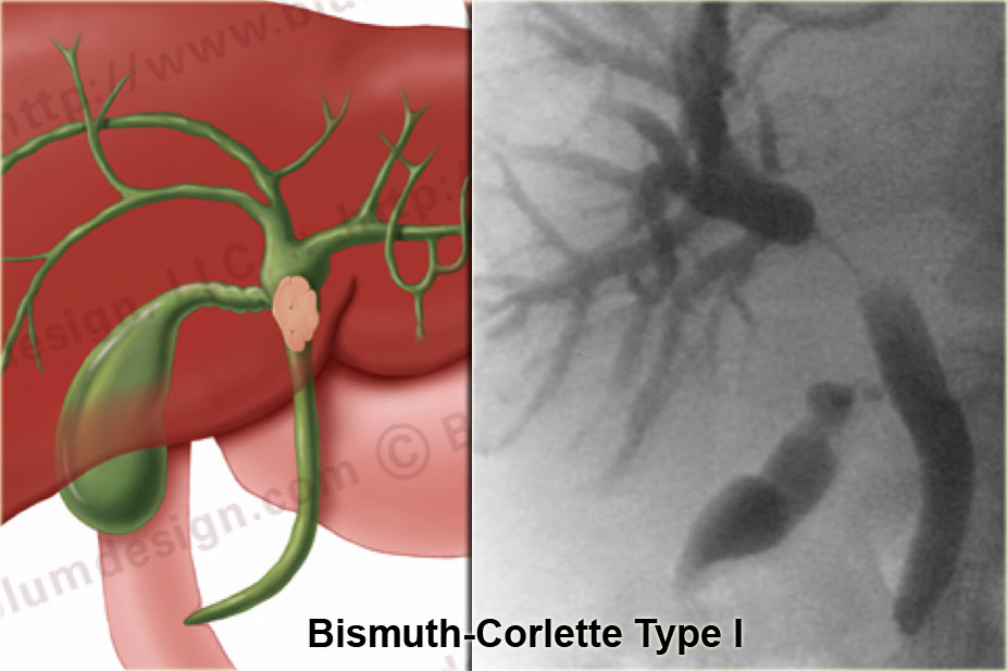
 Bismuth-Corlette type I tumor with abrupt stricture and shouldering below the confluensU Bismuth-Corlette type I có hẹp đột ngột và vai dưới hợp lưu
Klatskin Tumor - Bismuth-classificationU Klatskin - Phân loại Bismuth
Bismuth-Corlette type I
A type I tumor is a lesion limited of the common hepatic duct, i.e. below the confluence.
These patients can undergo resection with bile duct recontruction because the confluence is normal.Bismuth-Corlette type I
U loại I là một tổn thương giới hạn của ống gan chung, tức là dưới hợp lưu.
Những bệnh nhân này có thể được phẫu thuật cắt bỏ với sự tái tạo lại ống mật vì hợp lưu là bình thường.
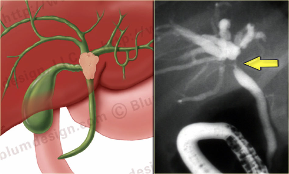 Bismuth-Corlette type II tumor with extention into the origin of the right and left hepatic duct.U Bismuth-Corlette type II với sự lan rộng vào ống gan phải và trái.
Bismuth-Corlette type II
A type II tumor is a lesion that extends to the confluence.
These tumors are potentially resectableBismuth-Corlette type II
U loại II là một tổn thương lan rộng đến hợp lưu.
Những khối u này có khả năng cắt bỏ được
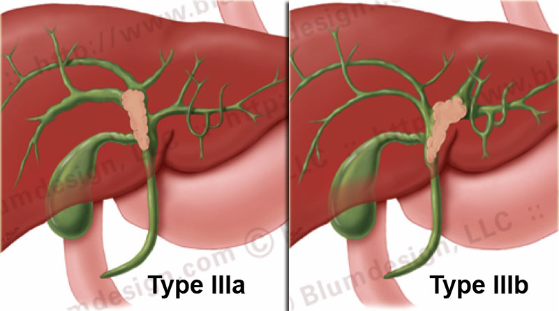
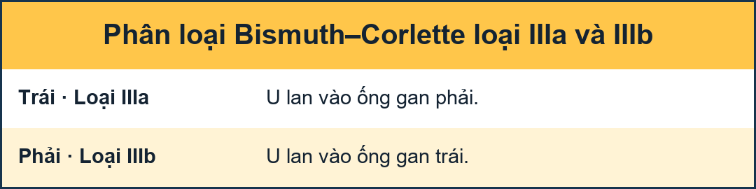 Bismuth-Corlette type III Klatskin-tumorBismuth-Corlette type III Khối u Klatskin
Bismuth-Corlette type IIIa and IIIb
A IIIa-tumor extends into the right and a IIIb-tumor extends into the left hepatic duct.Bismuth-Corlette type IIIa và IIIb
Một khối u IIIa kéo dài sang bên phải và một khối u IIIb kéo dài vào ống gan trái.
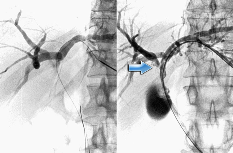 Bismuth-Corlette type IIIa Klatskin-tumorBismuth-Corlette type IIIa Khối u Klatskin
On the left PTC-images of a type IIIa-tumor.
The arrow indicates the extention into the right hepatic duct.
The left duct is normal.
This patient can undergo a resection of the right lobe of the liver.Trên hình ảnh PTC bên trái của một khối u loại IIIa, mũi tên cho thấy sự lan rộng vào ống gan phải.
Ống dẫn lưu bên trái bình thường.
Bệnh nhân này có thể trải qua phẫu thuật cắt bỏ thuỳ phải của gan.
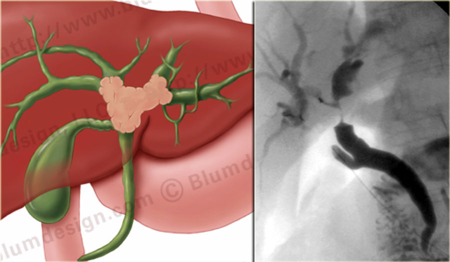 Bismuth-Corlette type IV Klatskin-tumorBismuth-Corlette type IV Khối u Klatskin
Bismuth-Corlette type IV
On the left an illustration and ERCP of a type IV-tumor with extention into the right and left duct.
A type IV tumor is unresectable.Bismuth-Corlette type IV
Bên trái là hình minh hoạ và ERCP của một khối u loại IV có phần mở rộng vào ống phải và trái.
U loại IV không thể cắt bỏ được.