
Liver - Segmental AnatomyGiải phẫu phân thùy gan
Radiology Department of the Alrijne Hospital, Leiderdorp, the Netherlands and University of Virginia Health System, Charlottesville, USA.Khoa X quang của Bệnh viện Alrijne, Leiderdorp, Hà Lan và Đại học Virginia Health System, Charlottesville, Hoa Kỳ.
Ngày đăng / cập nhật: 2006-05-07 / update 2015-10-15 / 2022-06-132006-05-07 / cập nhật 2015-10-15 / 2022-06-13
Bản gốc trên radiologyassistant.nl ↗ · tải về lúc 2026-11-19 20:19 · bản dịch máy có chuẩn hóa thuật ngữ chẩn đoán hình ảnh
The anatomy of the liver can be described using two different aspects: morphological anatomy and functional anatomy.
Giải phẫu gan có thể được mô tả bằng hai khía cạnh khác nhau: giải phẫu hình thái và giải phẫu chức năng.
The traditional morphological anatomy is based on the external appearance of the liver and does not show the internal features of vessels and biliary ducts branching, which are of obvious importance in hepatic surgery.
The French surgeon and anatomist Claude Couinaud was the first to divide the liver into eight functionally indepedent segments allowing resection of segments without damaging other segments.Giải phẫu hình thái truyền thống dựa trên hình dáng bên ngoài của gan và không cho thấy các đặc điểm bên trong của các mạch và ống mật, có tầm quan trọng rõ ràng trong phẫu thuật gan.
Nhà giải phẫu học người Pháp Claude Couinaud là người đầu tiên chia gan thành tám đoạn không độc lập về chức năng cho phép cắt bỏ các đoạn mà không làm tổn thương các đoạn khác.
Segmental anatomyGiải phẫu học
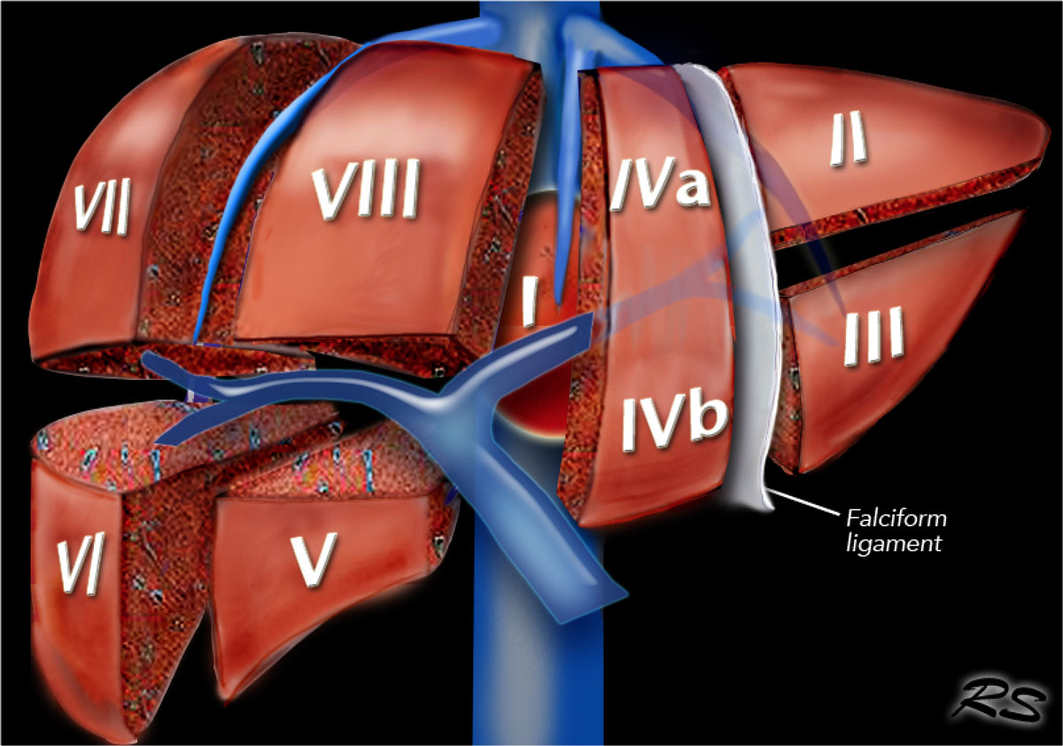
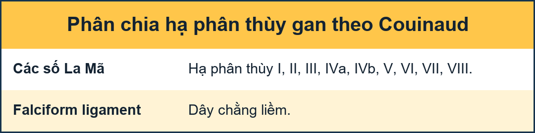 Segmental anatomy according to Couinaud. Click to enlarge.Giải phẫu phân đoạn theo Couinaud. Nhấp để phóng to.
Couinaud classificationPhân loại Couinaud
The Couinaud classification of liver anatomy divides the liver into eight functionally indepedent segments.
Each segment has its own vascular inflow, outflow and biliary drainage.
In the centre of each segment there is a branch of the portal vein, hepatic artery and bile duct.
In the periphery of each segment there is vascular outflow through the hepatic veins.
The liver is divided in three vertical planes:Phân loại giải phẫu gan của Couinaud chia gan thành tám phân đoạn không độc lập về chức năng.
Mỗi đoạn có mạch máu vào, ra và dẫn lưu mật riêng.
Ở trung tâm của mỗi đoạn có một nhánh của tĩnh mạch cửa, động mạch gan và ống mật.
Ở ngoại vi của mỗi đoạn có dòng chảy mạch máu qua các tĩnh mạch gan.
Gan được chia thành ba mặt phẳng thẳng đứng:
- The plane of the right hepatic vein divides the right lobe into anterior and posterior segments.Mặt phẳng tĩnh mạch gan bên phải chia thuỳ phải thành các phần trước và sau.
- The plane of the middle hepatic vein divides the liver into right and left lobes or right and left hemiliver. This plane runs from the inferior vena cava to the gallbladder fossa.Mặt phẳng tĩnh mạch gan giữa chia gan thành thuỳ phải và thuỳ trái hoặc nửa phải và nửa trái, mặt phẳng này chạy từ tĩnh mạch chủ dưới đến hố túi mật.
- The umbilic plane runs from the falciform ligament to the inferior vena cava and divides the left lobe into a medial part, which is segment IV and a lateral part formed by segment II and III. This division is the only vertically oriented plane that is not defined by a hepatic vein. Mặt phẳng rốn chạy từ dây chằng hình thoi đến tĩnh mạch chủ dưới và chia thuỳ trái thành phần trong, là phần IV và phần ngoài được hình thành bởi phần II và III, đây là mặt phẳng định hướng theo chiều dọc duy nhất không được xác định bởi tĩnh mạch gan.
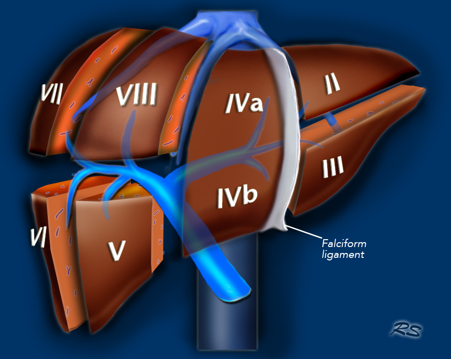

Here another illustration of the functional segmental liver anatomy.Đây là một minh hoạ khác về giải phẫu gan phân đoạn chức năng.
Portal vein
The portal vein divides the liver into upper and lower segments.
The left and right portal veins branch superiorly and inferiorly to project into the center of each segment.
Tĩnh mạch cửa
tĩnh mạch cửa chia gan thành các phân đoạn trên và dưới.
Các tĩnh mạch cửa trái và phải phân nhánh lên trên và xuống dưới để chiếu vào trung tâm của mỗi đoạn.
Left hepatic vein
The significance of the left hepatic vein is somewhat controversial. Some authors have shown it to coincide with the umbilical fissure, but in reality the left hepatic vein courses to the lateral to the umbilical fissure [fig].
While some authors have claimed that the division between segments II and III is formed by the transverse plane of the left portal vein, most investigators feel that it is the plane defined by the left hepatic vein.
In actual practice, when a lesion is located within the lateral segment of the left lobe, both Couinaud segments II and III are usually removed based on the plane formed by the umbilical fissure (i.e. left lateral segmentectomy). Trái tĩnh mạch gan
Ý nghĩa của tĩnh mạch gan bên trái còn nhiều tranh cãi, một số tác giả cho rằng nó trùng với rãnh rốn, nhưng thực tế tĩnh mạch gan bên trái lại đi về phía bên rãnh.
Trong khi một số tác giả cho rằng sự phân chia giữa đoạn II và III được hình thành bởi mặt phẳng ngang của tĩnh mạch cửa bên trái, hầu hết các nhà nghiên cứu cảm thấy rằng đó là mặt phẳng được xác định bởi tĩnh mạch gan bên trái.
Trong thực tế, khi một tổn thương nằm trong phân thuỳ bên của thuỳ trái, cả hai phân thuỳ Couinaud II và III thường được loại bỏ dựa trên mặt phẳng được hình thành bởi khe rốn (tức là cắt phân thuỳ ngoài bên trái).
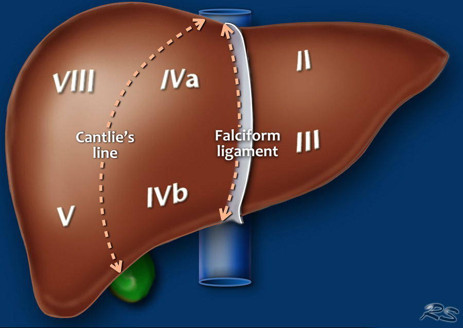
 On a frontal view of the liver the posteriorly located segments VI and VII are not visible.Trên mặt trước của gan, các phân đoạn VI và VII nằm ở phía sau không nhìn thấy được.
The illustration above is a schematic presentation of the liver segments.
In reality however the proportions are different.
On a normal frontal view the segments VI and VII are not visible because they are located more posteriorly.
The right border of the liver is formed by segment V and VIII.
Although segment IV is part of the left hemiliver, it is situated more to the right.Hình minh hoạ trên là sơ đồ trình bày các phân đoạn gan.
Tuy nhiên, trong thực tế tỷ lệ này là khác nhau.
Trên mặt cắt trán bình thường, các phân đoạn VI và VII không nhìn thấy được vì chúng nằm ở phía sau.
Đường viền phải của gan được hình thành bởi đoạn V và VIII.
Mặc dù đoạn IV là một phần của thân gan trái, nó nằm nhiều hơn về bên phải.
Couinaud divided the liver into a functional left and right liver by a main portal scissurae containing the middle hepatic vein.
This is known as Cantlie's line.
Cantlie's line runs from the middle of the gallbladder fossa anteriorly to the inferior vena cava posteriorly.Couinaud chia gan thành gan trái và gan phải bằng một cổng chính có chứa tĩnh mạch gan ở giữa.
Điều này được gọi là dòng Cantlie.
Đường Cantlie chạy từ giữa hố túi mật phía trước đến tĩnh mạch chủ dưới phía sau.
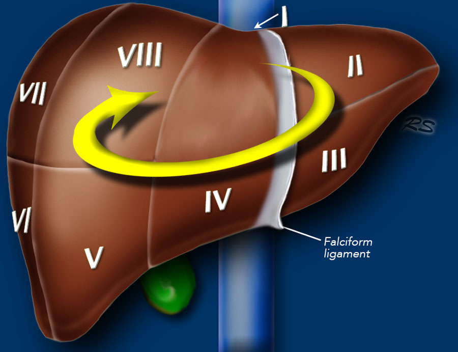
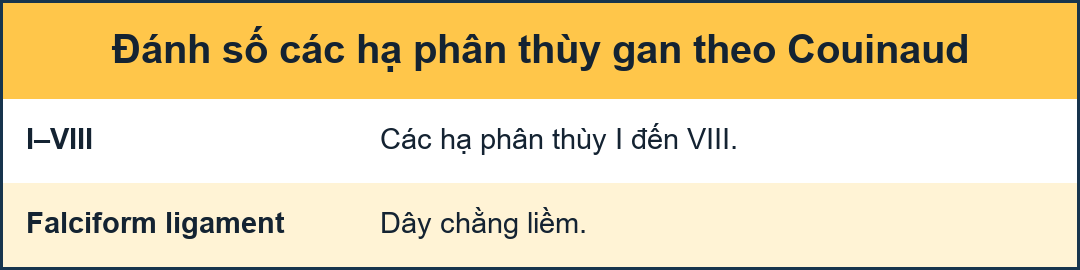 Clockwise numbering of the segmentsĐánh số thứ tự các đoạn theo chiều kim đồng hồ
Segments numberingĐánh số đoạn
There are eight liver segments.
Segment IV is sometimes divided into segment IVa and IVb according to Bismuth.
The numbering of the segments is in a clockwise manner.
Segment I (the caudate lobe) is located posteriorly.
It is not visible on a frontal view.Có tám đoạn gan.
Phân đoạn IV đôi khi được chia thành IVA và IVB theo Bismuth.
Việc đánh số các phân đoạn theo chiều kim đồng hồ.
Phân đoạn I (thùy đuôi) nằm ở phía sau.
Nó không hiển thị trên góc nhìn trực diện.
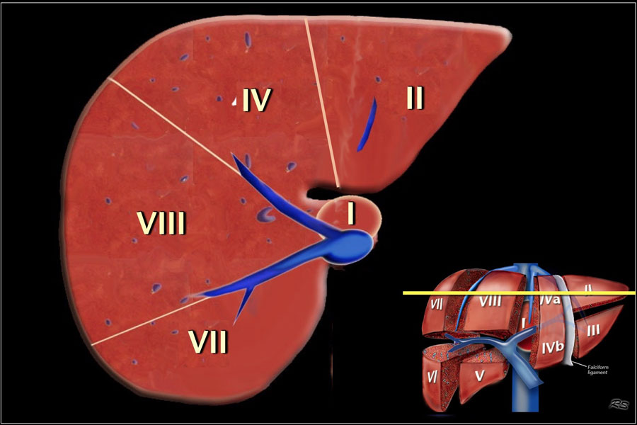
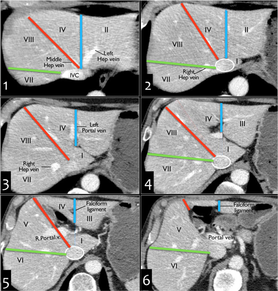
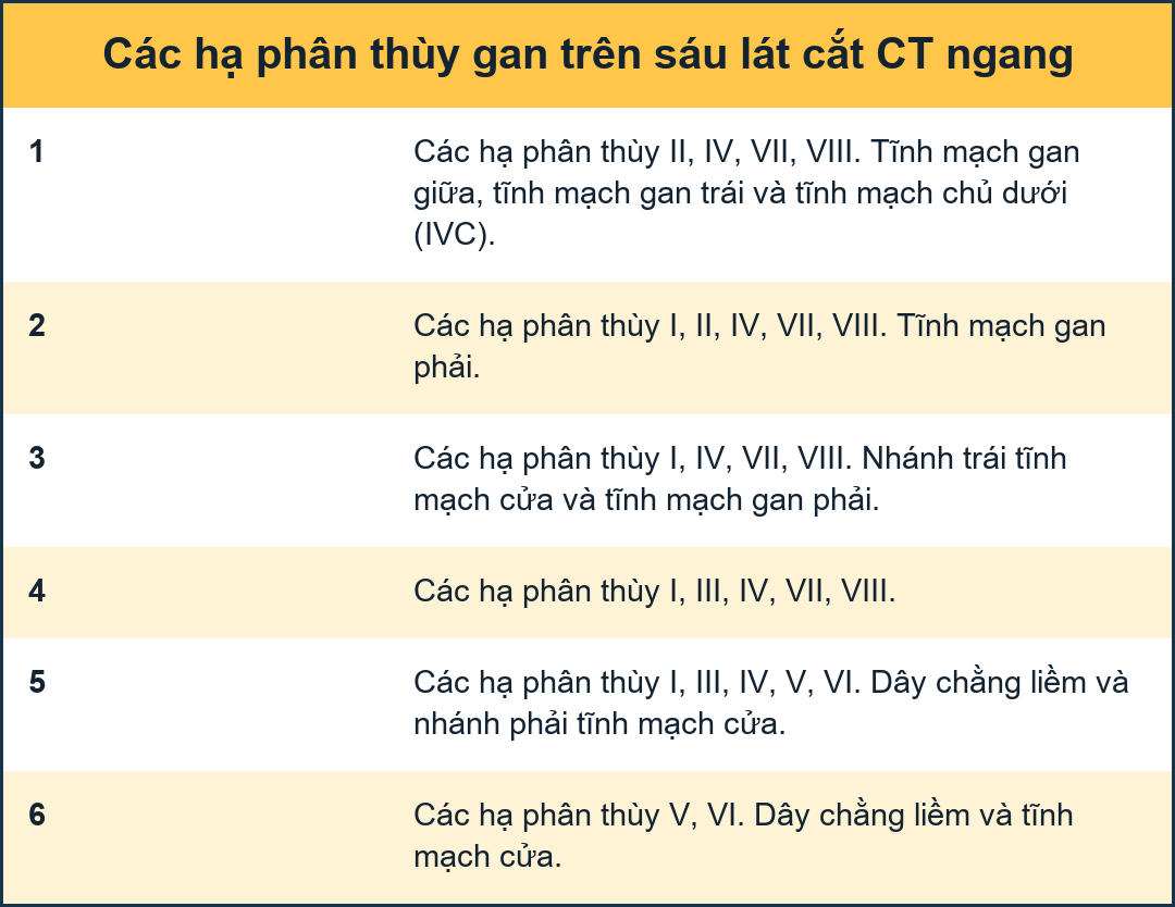
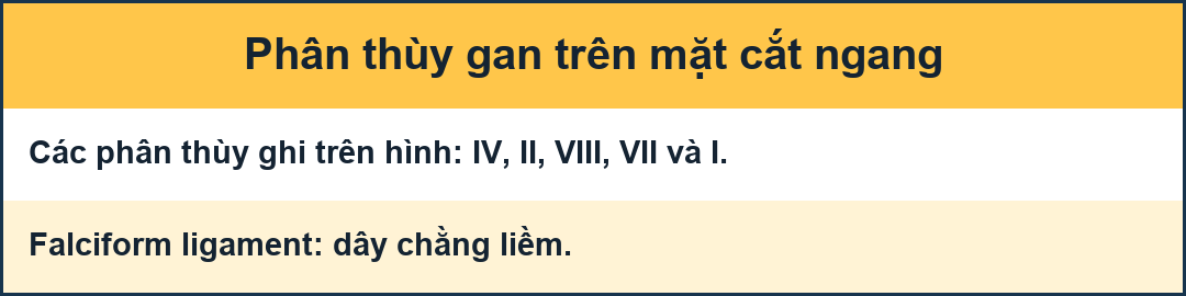 Image at the level of the superior liver segments.Hình ảnh ở mức của các phân đoạn gan trên.
Transverse anatomyGiải phẫu ngang
This figure is a transverse image through the superior liver segments, that are divided by the right and middle hepatic veins and the falciform ligament.Hình này là hình ảnh cắt ngang qua các đoạn gan trên, được phân chia bởi các tĩnh mạch gan phải và giữa và dây chằng hình thoi.
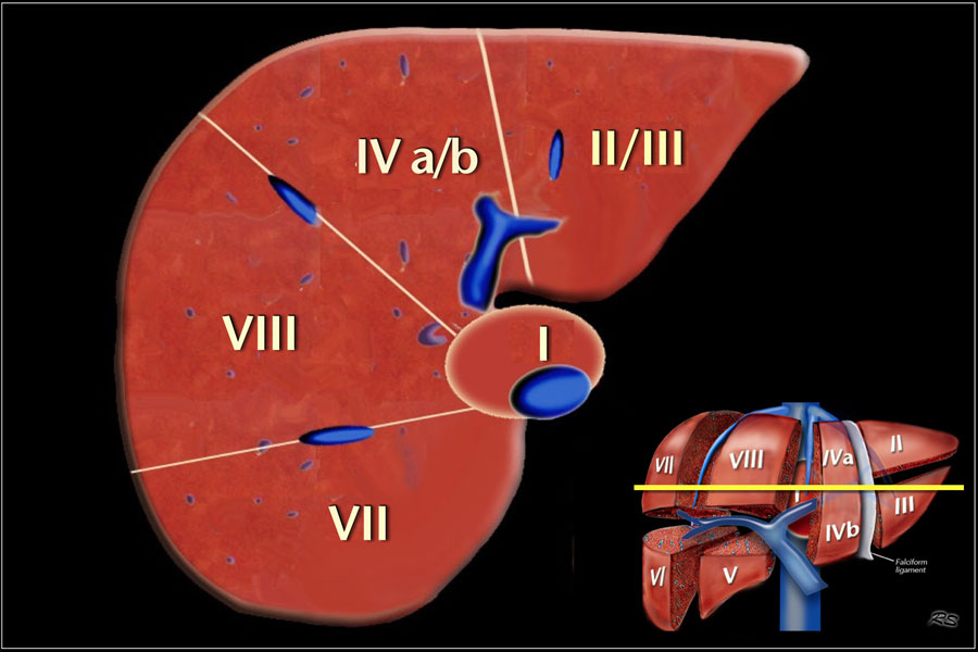
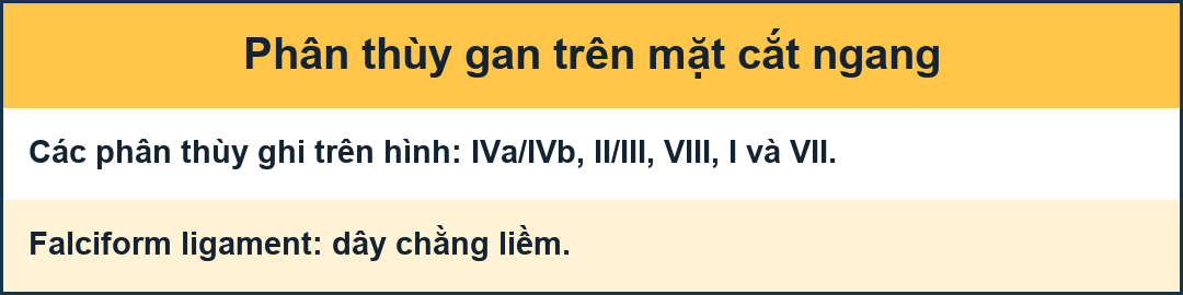 Image at the level of the left portal vein.Hình ảnh ở mức tĩnh mạch cửa bên trái.
This is a transverse image at the level of the left portal vein.
At this level the left portal vein divides the left lobe into the superior segments (II and IVa) and the inferior segments (III and IVb).
The left portal vein is at a higher level than the right portal vein.Đây là hình ảnh ngang ở mức tĩnh mạch cửa bên trái.
Ở mức này, tĩnh mạch cửa bên trái chia thuỳ trái thành các phân thuỳ trên (II và IVa) và các phân thủy dưới (III và IVb).
tĩnh mạch cửa bên trái cao hơn tĩnh mạch cửa bên phải.
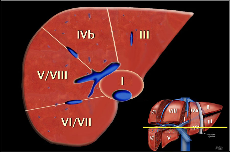
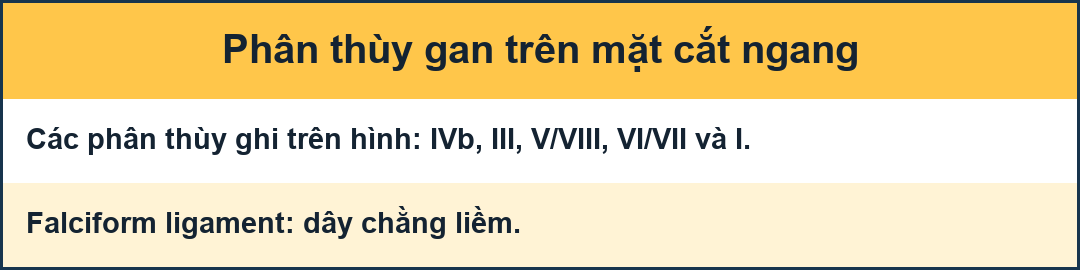 Image at the level of the right portal vein.Hình ảnh ở mức tĩnh mạch cửa bên phải.
This image is at the level of the right portal vein.
At this level the right portal vein divides the right lobe of the liver into superior segments (VII and VIII) and the inferior segments (V and VI).
The level of the right portal vein is inferior to the level of the left portal vein.Hình ảnh này nằm ở mức tĩnh mạch cửa bên phải.
Ở mức này, tĩnh mạch cửa bên phải chia thuỳ phải của gan thành các phân đoạn trên (VII và VIII) và các phân đoạn dưới (V và VI).
Mức tĩnh mạch cửa bên phải thấp hơn mức tĩnh mạch cửa bên trái.
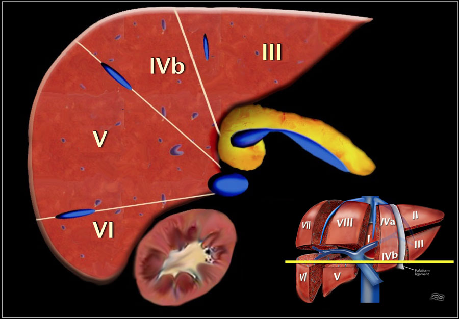
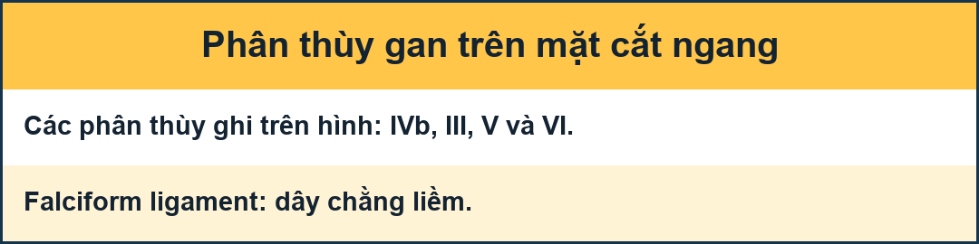 Image at the level of the splenic vein.Hình ảnh ở ngang mức tĩnh mạch lách.
At the level of the splenic vein, which is below the level of the right portal vein, only the inferior segments are visible.Ở mức tĩnh mạch lách, dưới mức tĩnh mạch cửa bên phải, chỉ có thể nhìn thấy các đoạn dưới.
How to separate liver segments on cross sectional imagingCách phân chia các đoạn gan trên hình ảnh cắt ngang
Left liver: lateral(II/III) vs medial segment (IVA/B)
Extrapolate a line along the falciform ligament superiorly to the confluence of the left and middle hepatic veins at the IVC (blue line).
Left vs Right liver: IVA/B vs V/VIII
Extrapolate a line from the gallbladder fossa superiorly along the middle hepatic vein to the IVC (red line).
Right liver: anterior (V/VIII) vs posterior segment (VI/VII)
Extrapolate a line along the right hepatic vein from the IVC inferiorly to the lateral liver margin (green line).Gan trái: gan ngoài (II/III) so với gan trong (IVA/B)
Ngoại suy một đường dọc theo dây chằng hình con chim ưng ở trên điểm hợp lưu của các tĩnh mạch gan trái và giữa tại IVC (đường màu xanh).
Gan trái so với gan phải: IVA / B so với V / VIII
Ngoại suy một đường từ hố túi mật lên trên dọc theo tĩnh mạch gan giữa đến IVC (đường màu đỏ).
Gan phải: trước (V / VIII) so với sau (VI / VII)
Ngoại suy một đường dọc theo tĩnh mạch gan bên phải từ IVC xuống dưới đến bờ gan bên (đường màu xanh lá cây).
Video of MRI anatomyVideo giải phẫu MRI
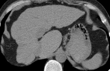 Hypertrophy of caudate lobe in a patient with livercirrhosis. Notice the small lobulated right hemiliver.Phì đại thùy đuôi ở bệnh nhân bị bệnh gan nhiễm mỡ Lưu ý đến phần bán cầu phải có thuỳ nhỏ.
Caudate lobeThùy đuôi
The caudate lobe or segment I is located posteriorly.
The caudate lobe is anatomically different from other lobes in that it often has direct connections to the IVC through hepatic veins, that are separate from the main hepatic veins.
The caudate lobe may be supplied by both right and left branches of the portal vein.Thùy đuôi hoặc đoạn I nằm ở phía sau.
thùy đuôi khác biệt về mặt giải phẫu so với các thuỳ khác ở chỗ nó thường có kết nối trực tiếp với IVC thông qua các tĩnh mạch gan, tách biệt với các tĩnh mạch chính của gan.
thùy đuôi có thể được cung cấp bởi cả hai nhánh phải và trái của tĩnh mạch cửa.
This CT-image is of a patient with liver cirrhosis with extreme atrophy of the right lobe, normal volume of the left lobe and hypertrophy of the caudate lobe.
Due to a different blood supply the caudate lobe is spared from the disease process and hypertrophied to compensate for the loss of normal liverparenchyma.Hình ảnh CT này là của một bệnh nhân bị xơ gan với teo cực độ thuỳ phải, thể tích bình thường của thuỳ trái và phì đại thùy đuôi.
Do nguồn cung cấp máu khác nhau nên thùy đuôi không bị quá tải và phì đại để bù đắp cho sự mất nhu mô sống bình thường.
Liver surgeryPhẫu thuật gan
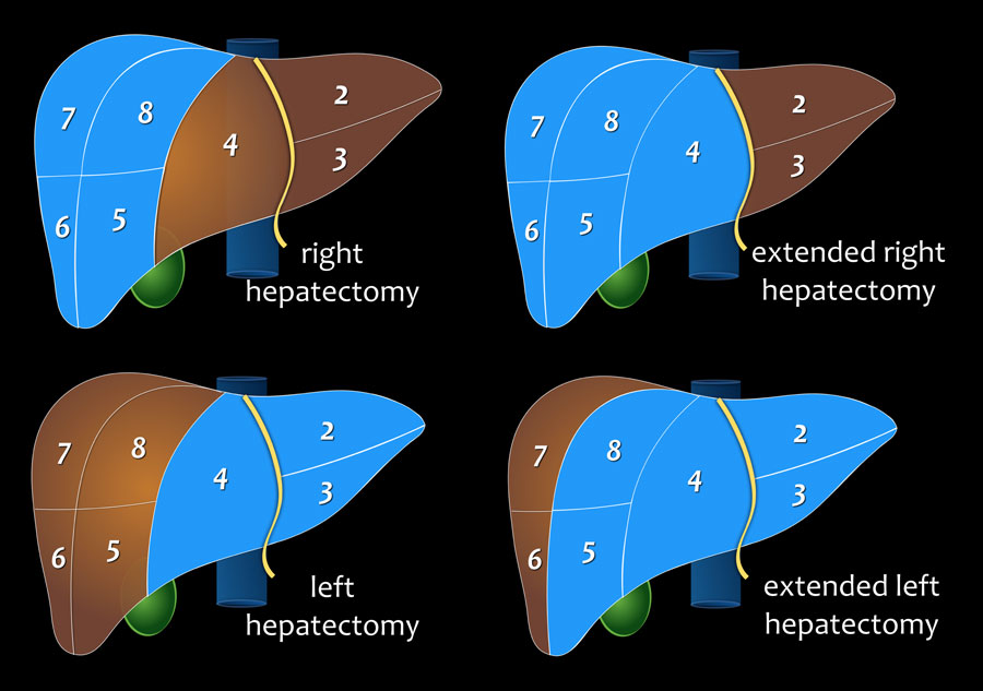
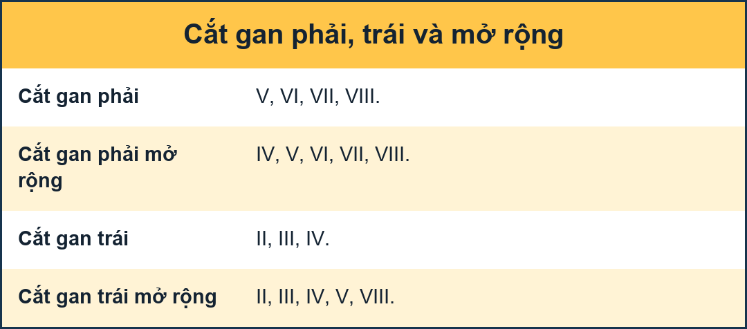
Right hepatectomy
segment V, VI, VII and VIII (± segment I).
Cắt gan phải
phân đoạn V, VI, VII và VIII (± phân đoạn I).
Extended Right or right trisectionectomy
segment IV, V, VI, VII and VIII (± segment I).Cắt ba phần tư mở rộng phải hoặc phải
phân đoạn IV, V, VI, VII và VIII (± phân đoạn I).
Left hepatectomy
segment II, III and IV (± segment I).Cắt gan trái
phân đoạn II, III và IV (± phân đoạn I).
Extended Left or left trisectionectomy
segment II, III, IV, V and VIII (± segment I).Cắt ba phần tư trái hoặc trái mở rộng
phân đoạn II, III, IV, V và VIII (± phân đoạn I).
Many surgeons prefer to use the term "extended" instead of trisectionectomy to indicate that some adjacent tissue of segment 4, or 5/8, as applicable is included rather than the entire segment 4, or 5/8.Nhiều bác sĩ phẫu thuật thích sử dụng thuật ngữ "mở rộng" thay vì cắt ba phần để chỉ ra rằng một số mô lân cận của đoạn 4, hoặc 5/8, nếu có thể, được bao gồm thay vì toàn bộ đoạn 4, hay 5/8.
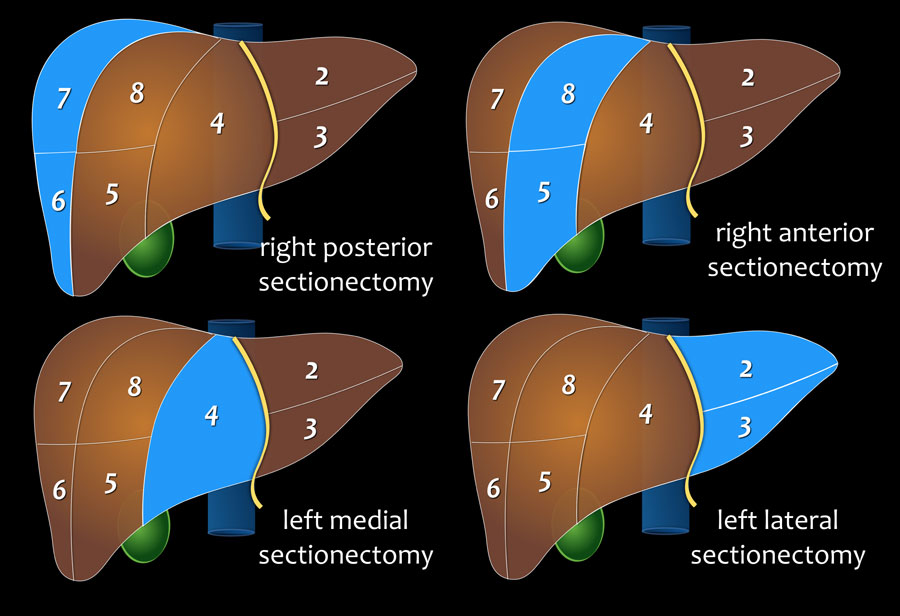
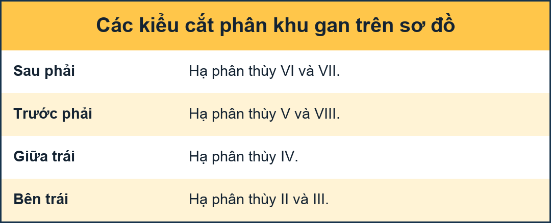
Right posterior sectionectomy
segment VI and VII
Cắt phần sau bên phải
phân đoạn VI và VII
Right anterior sectionectomy
segment V and VIII
Cắt trước phải
phân đoạn V và VIII
Left medial sectionectomy
segment IV
Cắt trong trái
phân đoạn IV
Left lateral sectionectomy
segment II and III
Cắt gan trái
phân đoạn II và III
ReferencesTài liệu tham khảo
-
Portal venous and segmental anatomy of the right hemiliver: observations based on three-dimensional spiral CT renderings
Giải phẫu tĩnh mạch cửa và giải phẫu phân đoạn của bán cầu phải: quan sát dựa trên hình ảnh CT xoắn ốc ba chiều
MS van Leeuwen, J Noordzij, MA Fernandez, A Hennipman, MA Feldberg and EH Dillon
Department of Radiology, University Hospital Utrecht, The Netherlands
MS van Leeuwen, J Noordzij, MA Fernandez, A Hennipman, MA Feldberg và EH Dillon -
Clinical and anatomical basis for the classification of the structural parts of liver
Nghiên cứu cơ sở lâm sàng và giải phẫu để phân loại các bộ phận cấu trúc gan
Saulius Rutkauskas et al.
Clinic of Radiology, Institute of Anatomy, Clinic of Surgery, Kaunas University of Medicine, Lithuania
-
Division of the Left Hemiliver in Man Segments, Sectors, or Sections
Phân chia Hemiliver trái trong phân đoạn Man, phân ngành, hoặc phân đoạn
by Anna C. Botero and Steven M. Strasberg
Liver Transplantation and Surgery, Vol 4, No 3 (May), 1998: pp 226-231
Anna C. Botero và Steven M. Strasberg - Liver Resection Guidelines Hướng dẫn cắt gan