
Anatomy of Peritoneum and MesenteryGiải phẫu phúc mạc và mạc treo
Chief Gastrointestinal Radiology, Department of Radiologic Pathology, Armed Forces Institute of Pathology, Washington DC Associate Professor of Radiology, Uniformed Services University of the Health Sciences, Bethesda, MDTrưởng khoa X quang đường tiêu hóa, Khoa X quang bệnh học, Viện Bệnh học Lực lượng Vũ trang, Washington DC Phó giáo sư X quang, Uniformed Services Đại học Khoa học Y tế, Bethesda, MD
Ngày đăng / cập nhật: 2009-07-05
Bản gốc trên radiologyassistant.nl ↗ · tải về lúc 2026-11-19 20:19 · bản dịch máy có chuẩn hóa thuật ngữ chẩn đoán hình ảnh
This review is based on a presentation given by Angela Levy and adapted for the Radiology Assistant by Robin Smithuis.
We will discuss the normal anatomy and physiology of the peritoneum and peritoneal cavity.
In part II we will discuss peritoneal tumors.
The illustrations are by Heike Blum
Images can be enlarged by clicking on them.Bài tổng quan này dựa trên một bài thuyết trình của Angela Levy và được Robin Smithuis điều chỉnh cho Trợ lý X quang.
Chúng tôi sẽ thảo luận về giải phẫu bình thường và sinh lý của phúc mạc và khoang phúc mạc.
Trong phần II chúng ta sẽ bàn về các khối u phúc mạc.
Các hình minh hoạ là của Heike Blum
Hình ảnh có thể được phóng to bằng cách nhấp vào chúng.
by Angela Levybởi Angela Levy
Definitions and AnatomyĐịnh nghĩa và giải phẫu
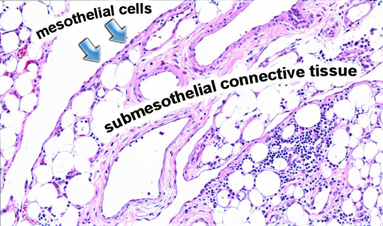

PeritoneumPhúc mạc
The peritoneum is a serosal membrane, which is composed of a single layer of flat mesothelial cells supported by submesothelial connective tissue.
In this subserosal tissue there are fat cells, lymphatics, blood vessels and inflammatory cells like lymphocytes and plasma cells.
Màng phúc mạc là một màng thanh mạc, bao gồm một lớp tế bào trung biểu mô phẳng được hỗ trợ bởi mô liên kết dưới trung biểu mô.
Trong mô dưới da này có các tế bào mỡ, bạch huyết, mạch máu và các tế bào viêm như tế bào lympho và tế bào huyết tương.
MesenteriesMạc treo
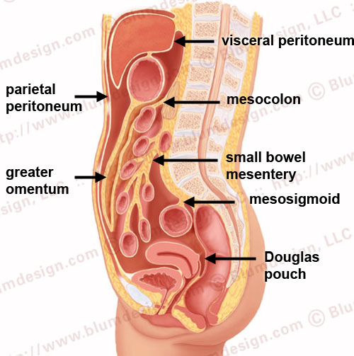
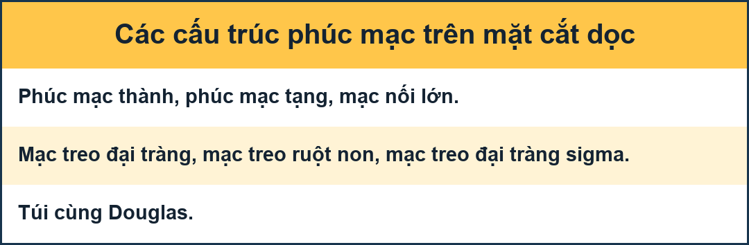
The visceral peritoneum lines all the organs that are intraperitoneal.
The parietal peritoneum lines the anterior, lateral and posterior walls of the peritoneal cavity.
The deepest portion of the peritoneal cavity is the pouch of Douglas in women and the retrovesical space in men, both in the upright and supine position.
The mesentery is a double fold of the peritoneum. Các nội tạng trong phúc mạc xếp hàng tất cả các cơ quan trong phúc mạc.
Lớp thành của phúc mạc nằm dọc theo thành trước, thành bên và thành sau của khoang phúc mạc.
Phần sâu nhất của khoang phúc mạc là túi Douglas ở phụ nữ và khoang sau xương bàn chân ở nam giới, cả ở tư thế thẳng đứng và nằm ngửa.
Mạc treo là một nếp gấp đôi của phúc mạc.
True mesenteries all connect to the posterior peritoneal wall.
These are: Các mạc treo thực sự đều kết nối với thành sau phúc mạc.
Đây là:
- The small bowel mesenteryMắt treo ruột non
- The transverse mesocolonMesocolon ngang
- The sigmoid mesentery (or mesosigmoid)Mạc treo đại tràng sigma (hoặc trung thất sigma)
Specialized mesenteries do not connect to the posterior peritoneal wall.
These are: Các mạc treo chuyên biệt không kết nối với thành sau phúc mạc.
Đây là:
- The greater omentum: connects the stomach to the colonMạc trên: nối dạ dày với đại tràng
- The lesser omentum: connects the stomach to the liver Mối nối mạc nối nhỏ: nối dạ dày với gan
- The mesoappendix: connects the apendix to the ileumLưỡi háng-trụ: nối xương vuông với hồi tràng
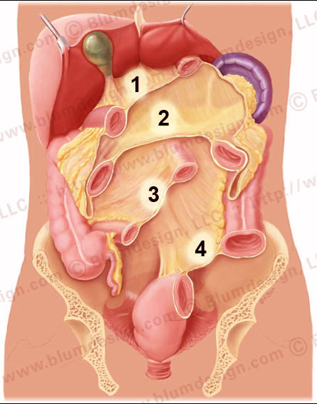
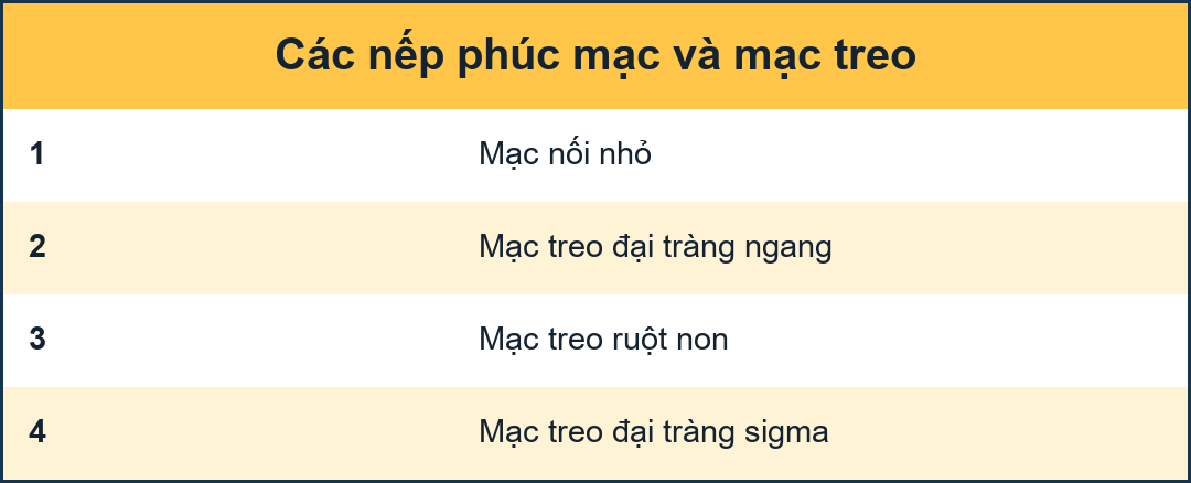 The lesser omentum (1), transverse mesocolon (2), small bowel mesentery (3) and the sigmoid mesentery (4)Mạc nối nhỏ (1), mạc treo ngang (2), mạc treO ruột non (3) và mạc treO đại tràng sigma (4)
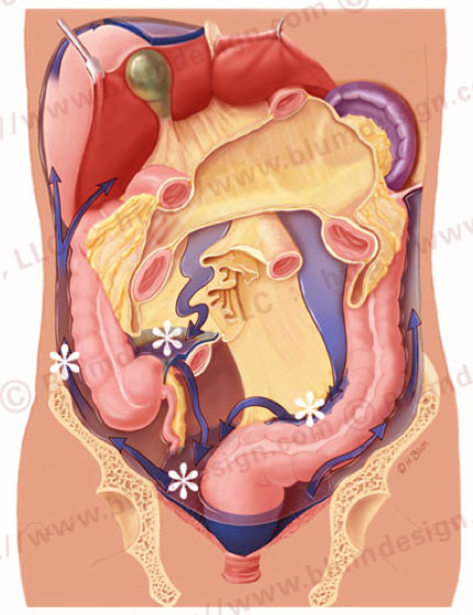
If you remove all of the intraperitoneal bowel, you get a good look at the cut-surface of the mesenteries:Nếu bạn loại bỏ tất cả ruột trong phúc mạc, bạn sẽ có được một cái nhìn tốt về mặt cắt của mạc treo:
- The lesser omentum Mạch mạc nối nhỏ
- Transverse mesocolon Mesocolon ngang
- Small bowel mesentery Mắt treo ruột non
- Sigmoid mesentery Mắc mạc treo đại tràng sigma
Notice that the small bowel mesentery has an oblique orientatien from the ligament of Treitz in the left upper quadrant to the ileocecal junction in the right lower quadrant.Lưu ý rằng mạc treo ruột non có hướng chéo từ dây chằng Treitz ở góc phần tư trên bên trái đến chỗ nối hồi manh tràng ở góc phần trăm dưới bên phải.
 Peritoneal pathwaysĐường dẫn truyền phúc mạc
Peritoneal circulationTuần hoàn phúc mạc
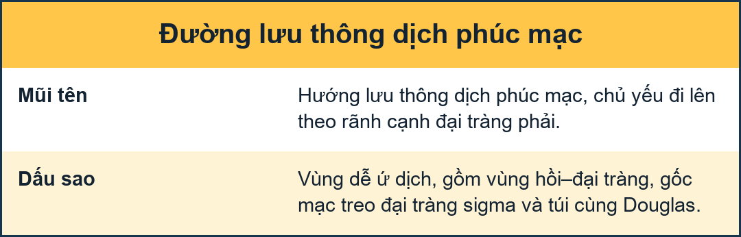
These compartments enable the peritoneal cavity to have a normal circulation for peritoneal fluid.
In the normal abdomen without intraperitoneal disease, there is a small amount of peritoneal fluid that continuously circulates.
The movement of fluid in this circulatory pathway is produced by the movement of the diaphram and peristalsis of bowel.
It predominantly flows up the right paracolic gutter which is deeper and wider than the left and is partially cleared by the subphrenic lymphatics.
There are watershed regions in the peritoneal cavity that are areas of fluid stasis:Những khoang này cho phép khoang phúc mạc có tuần hoàn bình thường cho dịch phúc mạc.
Trong bụng bình thường không có bệnh trong phúc mạc, có một lượng nhỏ dịch phúc mạc liên tục lưu thông.
Sự di chuyển của dịch trong con đường tuần hoàn này được tạo ra bởi sự di chuyển của cơ hoành và nhu động ruột.
Nó chủ yếu chảy lên máng xối paracolic bên phải sâu hơn và rộng hơn bên trái và được làm sạch một phần bởi các bạch huyết dưới thận.
Có những vùng đầu nguồn trong khoang phúc mạc là những vùng ứ dịch:
- Ileocolic region Vùng mắt cá
- Root of the sigmoid mesentery Rễ mạc treo đại tràng sigma
- Pouch of Douglas Túi Douglas
When you are staging a patient for gastrointestinal malignancy you have to look for disease in these areas of stasis.
Clearly the surgeons do better in finding subtle disease in these areas.Khi bạn đang phân giai đoạn bệnh nhân bị bệnh ác tính đường tiêu hóa, bạn phải tìm bệnh ở những khu vực bị ứ đọng này.
Rõ ràng các bác sĩ phẫu thuật làm tốt hơn trong việc tìm ra bệnh lý ở những vùng này.
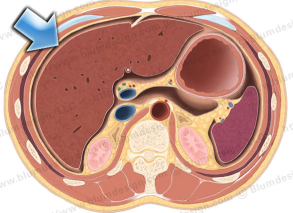 The majority of the peritoneal fluid is cleared at the subphrenic space (arrow) by the submesothelial lymphaticsPhần lớn dịch màng bụng được làm sạch ở khoang dưới màng cứng (mũi tên) bởi các hạch bạch huyết dưới biểu mô
90% of peritoneal fluid is cleared at the subphrenic space by the submesothelial lymphatics.
These lymphatics are connected with lymphatics at the other side of the diafragm.
90% dịch màng bụng được làm sạch ở khoang dưới lách bởi các hạch bạch huyết dưới lớp biểu mô.
Những hạch bạch huyết này được kết nối với các hạch bạch mạch ở phía bên kia của mảnh vỡ.
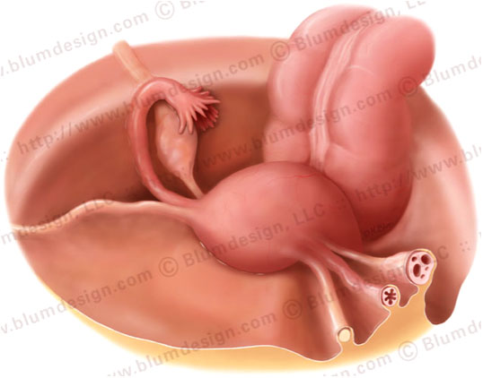
The peritoneum is continuous in the male pelvis.
In women the peritoneum is discontinuous at the ostia of the oviducts.
Through this opening disease can spread from the extraperitoneal pelvis into the peritoneal cavity.
For example, pelvic inflammatory disease (PID). Ổ bụng liên tục ở khung chậu nam.
Ở phụ nữ, phúc mạc không liên tục ở lỗ của ống dẫn trứng.
Thông qua bệnh mở này có thể lan từ khung chậu ngoài phúc mạc vào khoang phúc mạc.
Ví dụ, bệnh viêm vùng chậu (PID).
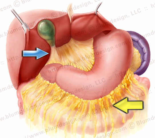 Gastrocolic ligament (yellow arrow), gastrosplenic ligament, gastrohepatic ligament and hepatoduodenal ligament (blue arrowDây chằng ống tiêu hóa (mũi tên màu vàng), dây chằng lách, dây chằng gan dạ dày và dây chằng tá gan (mũI tên màu xanh)
OmentumMạch máu
The omentum is divided into the greater and lesser omentum.
The greater omentum is subdivided into: Mạch mạc nối được chia thành mạc nối lớn và nhỏ.
Mạch máu lớn hơn được chia thành:
- Gastrocolic ligament (yellow arrow): the largest component Dây chằng ống tiêu hóa (mũi tên màu vàng): thành phần lớn nhất
- Gastrosplenic ligament: up to the hilus of the spleenDây chằng lách-tiêu hóa: đến rốn lách
- Gastrophrenic ligament: not shown on this illustration Dây chằng ống tiêu hóa: không được thể hiện trên hình minh hoạ này
The lesser omentum is subdivided into: Mạch mạc nối nhỏ được chia thành:
- Gastrohepatic ligament: connects the left lobe of the liver to the lesser curvature of the stomach.Dây chằng dạ dày-gan: kết nối thuỳ gan trái với độ cong nhỏ hơn của dạ dày.
- Hepatoduodenal ligament (blue arrow): free edge of the omentum, which contains the portal vein, hepatic artery and common bile duct .Dây chằng gan tá tràng (mũi tên màu xanh): cạnh tự do của mạc nối, chứa tĩnh mạch cửa, động mạch gan và ống mật chủ.