
Cardiac AnatomyGiải phẫu tim
Radiology department of the University Medical Centre Groningen and the Medical Centre Alkmaar, the NetherlandsKhoa X quang của Trung tâm Y tế Đại học Groningen và Trung tâm Y khoa Alkmaar, Hà Lan
Ngày đăng / cập nhật: 2009-02-13
Bản gốc trên radiologyassistant.nl ↗ · tải về lúc 2026-11-19 20:19 · bản dịch máy có chuẩn hóa thuật ngữ chẩn đoán hình ảnh
This review is based on a presentation given by Tineke Willems and was adapted for the Radiology Assistant by Marieke Hazewinkel.
We will discuss:Bài đánh giá này dựa trên một bài thuyết trình của Tineke Willems và được Marieke Hazewinkel điều chỉnh cho Trợ lý X quang.
Chúng ta sẽ thảo luận:
- The different cardiac axesCác trục tim khác nhau
- The cardiac anatomy in the order of normal blood flow: from the right to the left.Giải phẫu tim theo thứ tự dòng máu bình thường: từ phải sang trái.
Cardiac AxesTrục tim
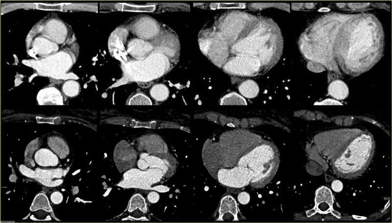 Axial slices through the heartCác lát cắt dọc theo trục xuyên qua tim
Cardiac CT with ECG triggering offers good image quality of the heart when compared to CT performed for other purposes.
As in any other field of radiology, analysis of the acquired images requires a systematic approach.
First of all, it is important to understand that the orientation of the heart in the human body differs from that of other anatomic structures: the right ventricle, for example, does not lie completely on the right, but more anterior.
The left ventricle does not lie on the left, but more posterior.
Also, the heart does not always maintain the same position within the mediastinum - in young people it tends to have a vertical orientation, whereas in older people it tends to rest on the diaphragm, a more horizontal orientation.
Cardiologists analyze the heart using cardiac axes.
These axes are also used in cardiac CT/MR and must be reconstructed in order to assess the heart properly.
Axial slices, such as those imaged on the left, are useful for a global assessment of the morphology of the heart and its relation to the pericardium.
CT tim có kích hoạt ECG cho chất lượng hình ảnh tốt hơn so với CT được thực hiện cho các mục đích khác.
Giống như bất kỳ lĩnh vực X quang nào khác, việc phân tích các hình ảnh thu được đòi hỏi một cách tiếp cận có hệ thống.
Trước hết, điều quan trọng là phải hiểu rằng hướng của tim trong cơ thể con người khác với hướng của các cấu trúc giải phẫu khác: ví dụ, tâm thất phải không nằm hoàn toàn bên phải, mà nằm phía trước nhiều hơn.
Tâm thất trái không nằm bên trái mà nằm phía sau nhiều hơn.
Ngoài ra, tim không phải lúc nào cũng duy trì cùng một vị trí trong trung thất - ở người trẻ tuổi, tim có xu hướng hướng dọc, trong khi ở người cao tuổi, tim thường nằm trên cơ hoành, hướng ngang hơn.
Các bác sĩ tim mạch phân tích tim bằng trục tim.
Những trục này cũng được sử dụng trong CT / MR tim và phải được tái tạo lại để đánh giá tim đúng cách.
Các lát cắt trục, chẳng hạn như các lát cắt được chụp ở bên trái, rất hữu ích để đánh giá toàn diện hình thái của tim và mối liên quan của nó với màng ngoài tim.
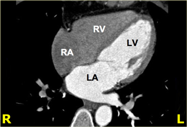
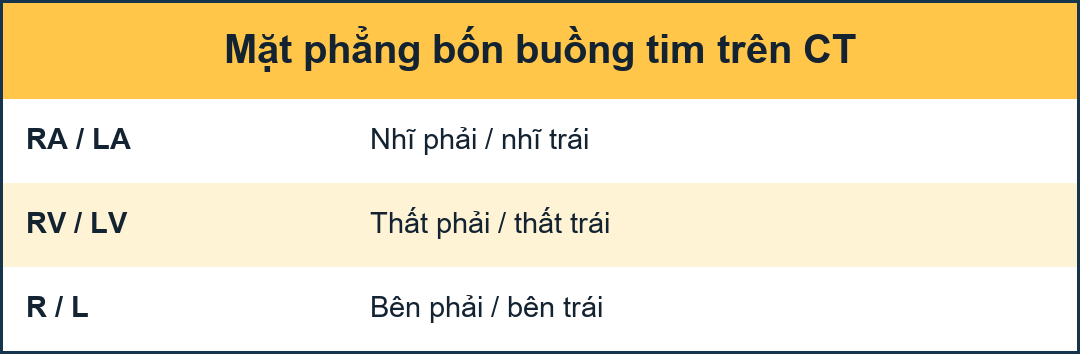 4-chamber view. RA=right atrium, RV=right ventricle, LA=left atrium, LV=left ventricleChế độ xem 4 buồng. RA = nhĩ phải, RV = tâm thất phải, LA = nhĩ trái, LV = tâm thất trái
4-chamber viewChế độ xem 4 buồng
On the left is a 4-chamber view, achieved by rotating upwards from the apex of the heart on the axial slices.
In this axis, the right ventricle is projected next to the right atrium, and the left ventricle next to the left atrium.
The mitral valve comes into view and - depending on the contrast protocol - the tricuspid valve may also be visible.
Another feature of this cardiac axis is that the apex of the heart is well demarcated.
Note that the apex is formed by the left ventricle.Bên trái là hình ảnh 4 buồng, đạt được bằng cách xoay lên trên từ đỉnh của tim trên các lát cắt dọc trục.
Trong trục này, tâm thất phải được chiếu bên cạnh tâm nhĩ phải và tâm thất trái bên cạnh tâm thất trái.
Van hai lá đi vào tầm nhìn và - tuỳ thuộc vào phác đồ thuốc cản quang - van ba lá cũng có thể nhìn thấy được.
Một đặc điểm khác của trục tim này là đỉnh tim được phân định rõ ràng Lưu ý rằng đỉnh tim được hình thành bởi tâm thất trái.
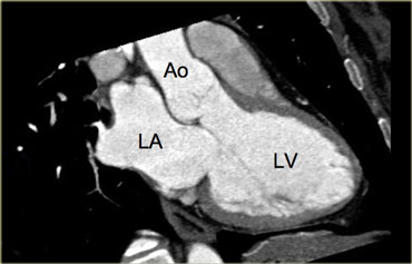
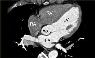
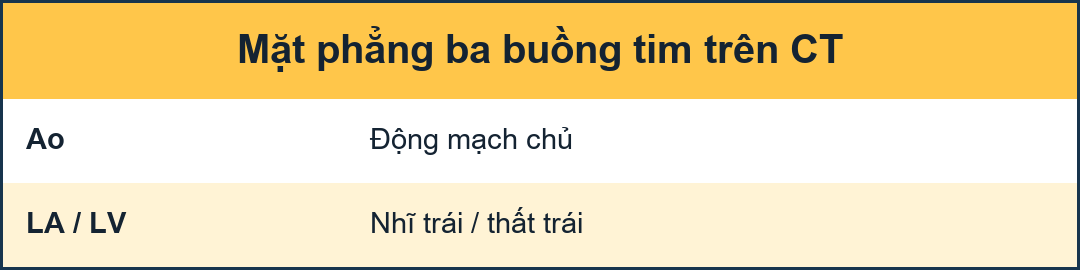 3-chamber view. LA=left atrium, Ao=aorta, LV=left ventricleChế độ xem 3 buồng. LA = nhĩ trái, Ao = động mạch chủ, LV = tâm thất trái
3-chamber viewChế độ xem 3 buồng
When the border between the mitral and aortic valves is localized on the axial slices and the images are rotated from this point, a 3-chamber view like the image on the left can be reconstructed.
On this image, the left atrium, left ventricle, mitral-, aortic valve and proximal aorta ascendens are visible.Khi đường viền giữa van hai lá và van động mạch chủ được định vị trên các lát cắt dọc trục và các hình ảnh được xoay từ điểm này, có thể tái tạo lại chế độ xem 3 buồng giống như hình ảnh bên trái.
Trên hình ảnh này, có thể nhìn thấy tâm nhĩ trái, tâm thất trái, van hai lá, van động mạch chủ và động mạch chủ trên.
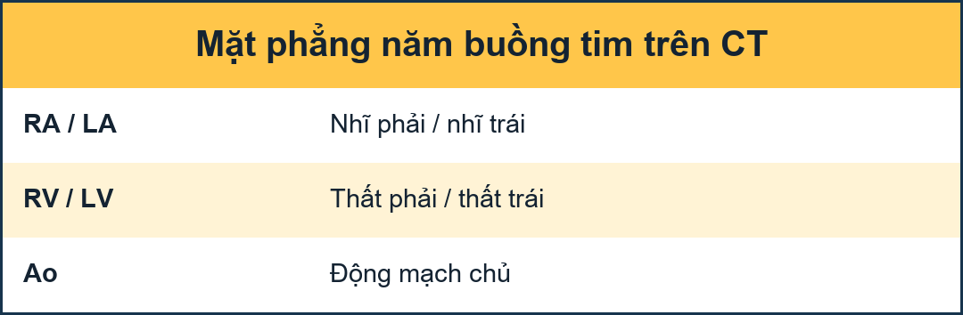 5-chamber view. RA=right atrium, RV=right ventricle, Ao=aorta, LA=left atrium, LV=left ventricleChế độ xem 5 buồng. RA = nhĩ phải, RV = tâm thất phải, Ao = động mạch chủ, LA = nhĩ trái, LV = tâm thất trái
5-chamber viewChế độ xem 5 buồng
On the left is an image of a 5-chamber view, which is similar to the 4-chamber view, but additionally displays the aortic valve and left ventricular outflow tract.
This view is achieved by rotating the 4-chamber view a little more cranially.Ở bên trái là hình ảnh của chế độ xem 5 buồng, tương tự như chế độ xem 4 buồng, nhưng hiển thị thêm van động mạch chủ và đường ra thất trái.
Quan điểm này đạt được bằng cách xoay góc nhìn 4 buồng nhiều hơn một chút.
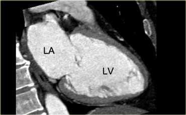
 2-chamber view. LA=left atrium, LV=left ventricleChế độ xem 2 buồng. LA = nhĩ trái, LV = thất trái
2-chamber viewChế độ xem 2 buồng
The 2-chamber view in the image on the left is achieved by rotating the images perpendicularly to the mitral valve and parallel to the cardiac septum.
This axis gives an overview of the left atrium ventricle and mitral valve.
It is a good view for analyzing ventricular function, especially that of the inferior and anterior walls.
For attaining functional data, consecutive short axes must be reconstructed making use of the 3- and 4-chamber views.Chế độ xem 2 buồng trong hình ảnh bên trái đạt được bằng cách xoay các hình ảnh vuông góc với van hai lá và song song với vách tim.
Trục này cho thấy tổng quan về tâm nhĩ trái và van hai lá.
Đây là một điểm quan sát tốt để phân tích chức năng thất trái, đặc biệt là chức năng của thành dưới và thành trước.
Để đạt được dữ liệu chức năng, các trục ngắn liên tiếp phải được xây dựng lại sử dụng chế độ xem 3 và 4 buồng.
Cardiac anatomy from right to leftGiải phẫu tim từ phải sang trái
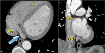
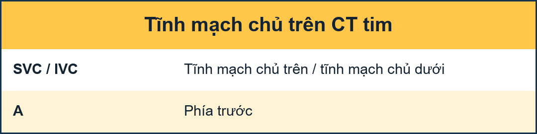 Axial (left) and coronal oblique (right) reconstructions of the heart, depicting the right atrium and its main contributing blood vessels: the coronary sinus (blue arrow) and superior and inferior vena cava. IVC=inferior vena cava, A=anterior, SVC=superior vena cavaHình ảnh tái tạo trục (trái) và chéo vành (phải) của tim, mô tả tâm nhĩ phải và các mạch máu đóng góp chính của nó: xoang vành (mũi tên màu xanh) và xoang trên và tĩnh mạch chủ dưới IVC = tĩnh mạch chủ dưới, A = trước, SVC = tĩnh mạch chủ trên
Right atriumTâm nhĩ phải
The cardiac anatomy will be discussed in the order of normal blood flow: from the right to the left.
In the normal situation, contrast will be injected intravenously - usually in the arm - reaching the right atrium via the superior vena cava.
The right atrium has an anterolateral position in the heart, and lies inferior to the left atrium.
The superior vena cava enters through the roof of the right atrium.
The inferior vena cava enters the right atrium from below near the cardiac septum.
Another structure that carries blood towards the right atrium is the coronary sinus, (venous return of the coronaries) which enters anterior to, and just to the left of the inferior vena cava.Giải phẫu tim sẽ được thảo luận theo thứ tự lưu lượng máu bình thường: từ phải sang trái.
Trong tình huống bình thường, thuốc cản quang sẽ được tiêm tĩnh mạch - thường là ở cánh tay - đến tâm nhĩ phải qua tĩnh mạch chủ trên.
Tâm nhĩ phải có vị trí trước bên trong tim, và nằm dưới tâm nhĩ trái.
Tĩnh mạch chủ trên đi vào qua vòm nhĩ phải.
tĩnh mạch chủ dưới đi vào tâm nhĩ phải từ dưới gần vách ngăn tim.
Một cấu trúc khác mang máu về phía tâm nhĩ phải là xoang vành, (sự trở lại tĩnh mạch của các vành) đi vào phía trước và ngay bên trái của tĩnh mạch chủ dưới.
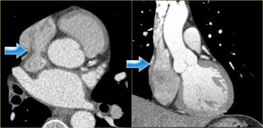 Axial (left) and coronal (right) reconstructions showing the crista terminalis (blue arrows) and its location in the right atriumTái tạo trục (trái) và vành (phải) cho thấy đầu tận cùng của crista (các mũi tên màu xanh) và vị trí của nó ở tâm nhĩ phải
Crista terminalisMào tận cùng
In the right atrium lies the crista terminalis, a muscular ridge that runs from the entrance of the superior- to that of the inferior vena cava.
This structure separates the smooth part of the right atrium - the sinus venosus - from the trabecularized right atrial appendage.
On the images on the left it is visible as a smooth linear structure (blue arrows).
This is not always the case, however, it may simulate a mass.Ở tâm nhĩ phải là crista terminalis, một đường vân cơ chạy từ lối vào của phần trên-đến tĩnh mạch chủ dưới.
Cấu trúc này tách phần nhẵn của tâm nhĩ phải - tĩnh mạch xoang - khỏi phần phụ nhĩ phải có gai, trên hình ảnh bên trái có thể nhìn thấy cấu trúc tuyến tính nhẵn (các mũi tên màu xanh).
Tuy nhiên, không phải lúc nào cũng như vậy, nó có thể mô phỏng một khối.
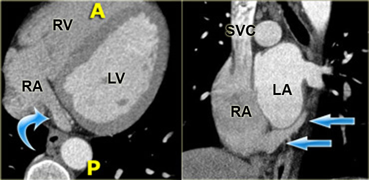
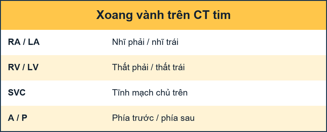 Axial (left) and coronal oblique (right) reconstructions showing the coronary sinus as it enters the right atrium (blue arrows). A=anterior, P=posteriorHình ảnh tái tạo trục (trái) và chéo vành (phải) cho thấy xoang vành khi nó đi vào tâm nhĩ phải (các mũi tên màu xanh). A = trước, P = sau
Coronary sinusXoang vành
The coronary sinus is the main draining vein of the myocardium.
It runs in the atrioventricular groove on the posterior surface of the heart and enters the right atrium in the vicinity of the tricuspid valve.Xoang vành là tĩnh mạch dẫn lưu chính của cơ tim.
Nó chạy trong rãnh nhĩ thất trên mặt sau của tim và đi vào tâm nhĩ phải ở vùng lân cận của van ba lá.
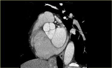
On the left is a reconstruction illustrating the course run by the coronary sinus in the atrioventricular groove on the posterior surface of the heart.Bên trái là hình ảnh tái tạo minh hoạ đường đi của xoang vành trong rãnh nhĩ thất trên mặt sau của tim.
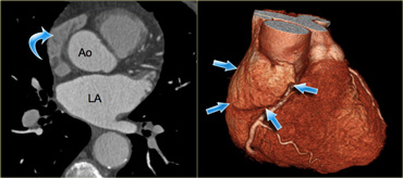
 Axial (left) and 3D-reconstructions (right) of the heart demonstrating the right atrial appendage (blue arrows). Ao=aorta, LA=left atriumHình ảnh tái tạo trục (trái) và 3D (phải) của tim thể hiện phần phụ nhĩ phải (các mũi tên màu xanh). Ao = động mạch chủ, LA = nhĩ trái
Right atrial appendagePhần phụ nhĩ phải
The right atrial appendage is the trabecularized part of the right atrium.
It partially covers the atrioventricular groove and the right coronary artery that runs in it.
Characteristically, it is flat and triangular in shape and contains small muscular bundles which run parallel to the atrium itself.
Phần phụ nhĩ phải là phần có gai của nhĩ phải.
Nó bao phủ một phần rãnh nhĩ thất và động mạch vành phải chạy trong đó.
Đặc trưng, nó có hình dạng phẳng và hình tam giác và chứa các bó cơ nhỏ chạy song song với tâm nhĩ.
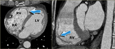
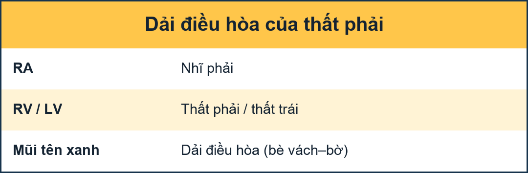 Axial (left) and sagittal oblique (right) reconstructions showing the right ventricle. The blue arrows indicate the moderator band. RA=right atrium, RV=right ventricle, LV=left ventricleHình ảnh tái tạo trục (trái) và dọc dọc (phải) cho thấy tâm thất phải.các mũi tên màu xanh cho biết dải điều tiết.RA = tâm nhĩ phải, RV = tâm thất phải, LV = tâm thất trái
Right ventricleTâm thất phải
Blood leaves the right atrium and enters the right ventricle via the tricuspid valve.
This valve has three leaflets and three papillary muscles, which partially insert on the septum (in contrast to the papillary muscles of the mitral valve, which do not).
The right ventricle is shaped differently to the left ventricle: the left ventricle is cylindrical in shape and the cavity of the right ventricle is effectively wrapped around it.
The right ventricle also has a thinner wall which is more trabecularized, especially towards the apex.
The moderator band is another distinguishing feature of the right ventricle.
It runs from the septum to the lateral wall of the right ventricle, and plays a key role in the electrophysiological conduction of the right ventricle's free wall (blue arrows). Máu rời tâm nhĩ phải và đi vào tâm thất phải qua van ba lá.
Van này có ba lá van và ba cơ nhú, một phần chèn vào vách ngăn (trái ngược với các cơ nhú của van hai lá, không có).
Tâm thất phải có hình dạng khác với tâm thất trái: tâm thất trái có hình trụ và khoang của tâm thất phải được bao quanh nó một cách hiệu quả.
Tâm thất phải cũng có thành mỏng hơn, có nhiều gai hơn, đặc biệt là về phía đỉnh.
Dải điều tiết là một đặc điểm khác biệt của tâm thất phải, nó chạy từ vách ngăn đến thành bên của tâm thất trái và đóng vai trò quan trọng trong dẫn truyền điện sinh lý của thành tự do của tâm thất (các mũi tên màu xanh).
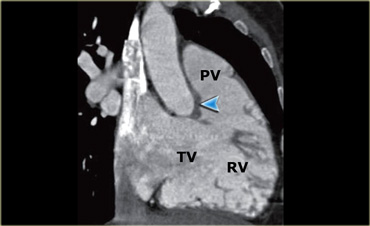
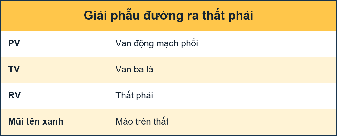 Reconstruction showing the tricuspid (TV) and pulmonary (PV) valves as well as the cavity of the right ventricle (RV). The blue arrow indicates the crista supraventricularisTái tạo cho thấy van ba lá (TV) và van phổi (PV) cũng như khoang của tâm thất phải (RV) mũi tên màu xanh dương cho thấy cơ thắt trên thất
Pulmonary valveVan phổi
Next, blood runs towards the pulmonary valve - first entering the smooth, muscular infundibulum of the right ventricle.
The pulmonary valve has three cusps, and is separated from the tricuspid valve by a thick muscle known as the crista supraventricularis (blue arrow in the image on the left).
This differs from the left ventricular outflow tract, where the mitral and aortic valves lie side by side.
Tiếp theo, máu chảy về phía van động mạch phổi - đầu tiên đi vào phễu cơ trơn của tâm thất phải.
Van động mạch phổi có ba chỏm, và được tách ra khỏi van ba lá bởi một cơ dày được gọi là cơ trên thất (mũi tên màu xanh trong hình bên trái).
Điều này khác với đường ra thất trái, nơi van hai lá và van động mạch chủ nằm cạnh nhau.
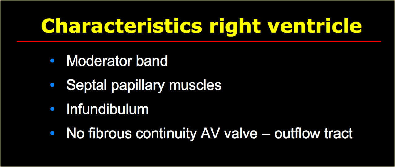
On the left is a summary of the characteristics which are specific for the right ventricle and are useful in distinguishing the left from the right ventricle in cases with complex congenital cardiac anatomy.Bên trái là tóm tắt các đặc điểm đặc trưng cho tâm thất phải và rất hữu ích trong việc phân biệt tâm thất trái với tâm thất phải trong các trường hợp có giải phẫu tim bẩm sinh phức tạp.
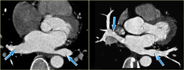 Axial reconstructions showing the pulmonary veins (blue arrows) as they enter the left atriumTạo hình trục cho thấy các tĩnh mạch phổi (các mũi tên màu xanh) khi chúng đi vào tâm nhĩ trái
Pulmonary veinsTĩnh mạch phổi
Oxygen-rich blood enters the left atrium via the pulmonary veins.
In most cases, there are two pulmonary veins on the left and two on the right.
The middle pulmonary veins usually drain into the superior pulmonary vein.Máu giàu oxy đi vào tâm nhĩ trái qua các tĩnh mạch phổi.
Trong hầu hết các trường hợp, có hai tĩnh mạch phổi bên trái và hai tĩnh mạch ở bên phải.
Tĩnh mạch phổi giữa thường dẫn lưu vào tĩnh mạch phổi trên.
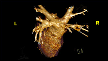 3D-reconstructions showing the pulmonary veins as they enter the left atriumTạo hình 3D cho thấy các tĩnh mạch phổi khi chúng đi vào tâm nhĩ trái
There are frequent variations in pulmonary vein anatomy however, especially on the right, where an anomalous insertion is associated with atrial fibrillation. Tuy nhiên, có nhiều biến thể thường gặp trong giải phẫu tĩnh mạch phổi, đặc biệt là ở bên phải, nơi có sự chèn ép bất thường có liên quan đến rung nhĩ.
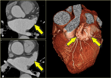 Axial and 3D-reconstructions illustrating the shape and location of the left atrial appendage (yellow arrows).)Hình ảnh trục và 3D mô tả hình dạng và vị trí của phần phụ nhĩ trái (các mũi tên màu vàng).
Left atrial appendagePhần phụ nhĩ trái
The left atrial appendage is a finger like, trabecularized structure which originates supralaterally in the left atrium. It lies over the left atrioventricular groove, and partially covers the left coronary artery in it.
Its small, parallel-running muscles should not be mistaken for thrombus.Nhĩ trái là một cấu trúc dạng ngón tay, dạng bè, bắt nguồn từ trên bên trong nhĩ trái, nằm trên rãnh nhĩ thất trái, và bao phủ một phần động mạch vành trái trong đó.
Không nên nhầm lẫn các cơ nhỏ, chạy song song với huyết khối.
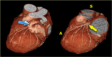
 3D-reconstructions showing the left atrial appendage (blue arrow) and the left coronary artery (yellow arrow) after the left atrial appendage has been removed. A=anterior, S=superior).)Tái tạo 3D cho thấy phần phụ nhĩ trái (mũi tên màu xanh) và động mạch vành trái (mũi tên màu vàng) sau khi phần phụ nhện trái đã được lấy ra.)
When assessing the coronary arteries, the left atrial appendage must be removed, so that the LCX and proximal LAD may be visualized. Khi đánh giá động mạch vành, phần phụ nhĩ trái phải được loại bỏ, để có thể nhìn thấy LCX và LAD gần.
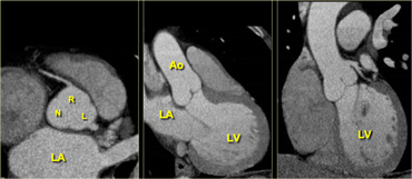
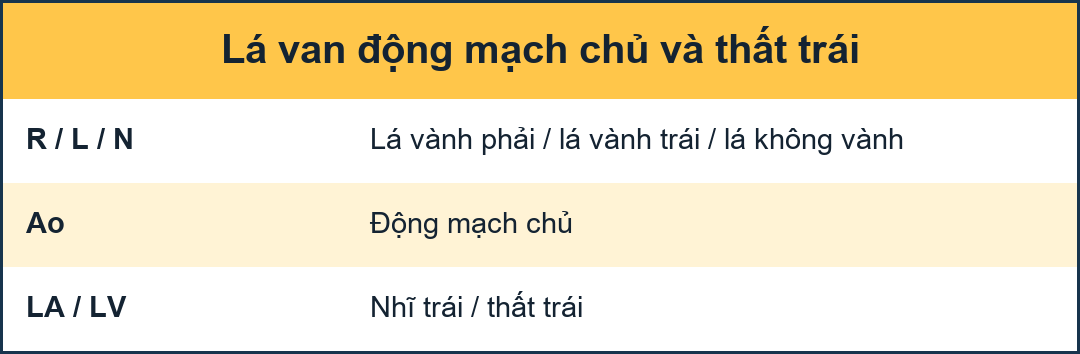 Axial (left) reconstruction, 3-chamber view (middle) and coronal reconstruction (right) of the heart illustrating the relationships between the left atrium, ventricle and aortic root. LA=left atrium, R=right coronary cusp, L=left coronary cusp, N=non-coronary cusp, Ao=aorta, LV=left ventricleHình ảnh 3 buồng (trung) và hình ảnh động mạch vành (phải) của tim, mô tả mối liên quan giữa nhĩ trái, tâm thất và gốc động mạch chủ. LA= nhĩ trái; R= mấu vành phải; L= mấU vành trái; N= mấus không vành; Ao= động mạch chủ; LV= tâm thất trái.
Left ventricleTâm thất trái
Blood enters the left ventricle via the mitral valve.
This is a complex valve, consisting of an annulus and posterior and anterior leaflets.
The leaflets are connected to the papillary muscles via cord-like tendons called chordae tendinae.
The papillary muscles insert into the lateral and posterior walls as well as the apex of the left ventricle.
In normal situations the left ventricle has a uniform thickness, varying end-diastolically from 0.6 to 1.0 cm.
Blood enters the aortic valve via the left ventricular outflow tract.
Note that there appears to be a fibrous connection between the mitral and aortic valve.
Máu đi vào tâm thất trái qua van hai lá.
Đây là một van phức tạp, bao gồm một vòng van và các lá trước và sau.
Các lá được kết nối với các cơ nhú thông qua các gân giống như dây rốn gọi là dây chằng gân.
Các cơ nhú chèn vào thành bên và thành sau cũng như đỉnh của tâm thất trái.
Trong các tình huống bình thường, tâm thất trái có độ dày đồng đều, thay đổi từ 0,6 đến 1,0 cm.
Máu đi vào van động mạch chủ qua đường ra thất trái.
Lưu ý rằng dường như có một kết nối sợi giữa van hai lá và van động mạch chủ.
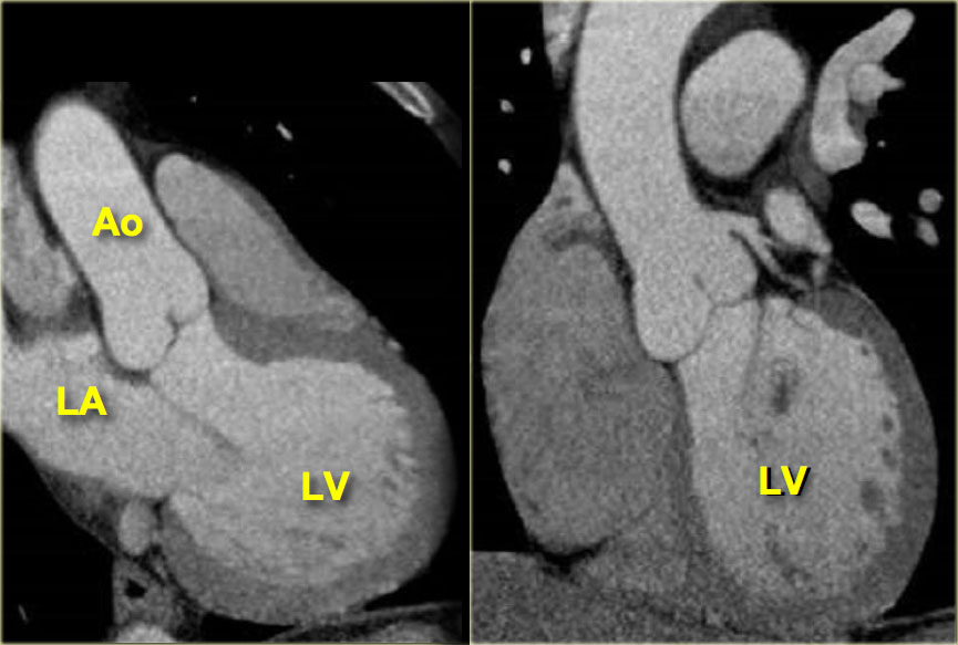
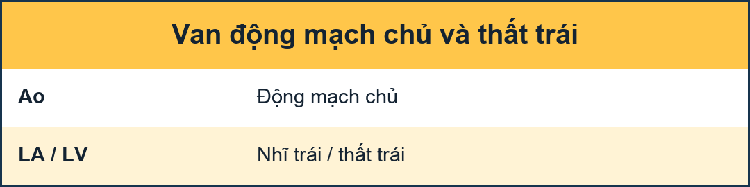 3-chamber (left) and coronal (right) reconstructions, same as aboveTái tạo 3 buồng (trái) và vành (phải), giống như trên
Aortic valveVan động mạch chủ
Like the pulmonary valve, the aortic valve has three cusps.
Just cranially to it there is a slight dilatation of the aortic root.
This is the sinus of Valsalva.
It fills with blood during diastole, supplying the coronary arteries with oxygen-rich blood.
The image on the right shows that the coronary arteries originate fairly cranially, on the border of the ascending aorta.Giống như van động mạch phổi, van động mạch chủ có ba chỏm.
Chỉ cần sọ vào đó có một sự giãn nở nhẹ của gốc động mạch chủ.
Đây là xoang Valsalva.
Nó đổ đầy máu trong thời gian tâm trương, cung cấp máu giàu oxy cho động mạch vành.
Hình ảnh bên phải cho thấy các động mạch vành bắt nguồn khá từ sọ, trên bờ động mạch chủ lên.
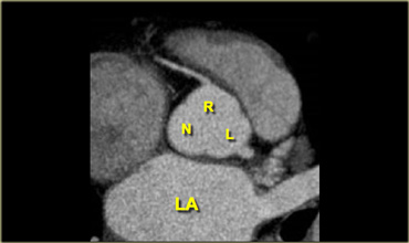
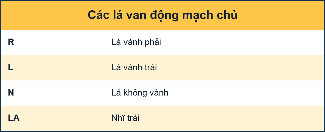 Axial reconstruction depicting the tricuspid aortic valve with its right and left coronary (R and L respectively) and non-coronary cusp (N) Hình ảnh tái tạo trục van động mạch chủ ba lá với nhánh vành phải và trái (lần lượt là R và L) và chỏm không vành (N)
The cusps of the aortic valves are named according to their relationship with the coronary arteries, namely the right coronary, left coronary and non-coronary cusp (R, L and N).Các cusps của van động mạch chủ được đặt tên theo mối quan hệ của chúng với các động mạch vành, cụ thể là vành phải, vành trái và không phải vành (R, L và N).
ReferencesTài liệu tham khảo
-
ECG-Gated Cardiac CT a review
Đánh giá kết quả chụp CT tim qua điện tâm đồ
by Benoit Desjardins and Ella A. Kazerooni
AJR 2004; 182:993-1010
Benoit Desjardins và Ella A. Kazerooni
AJR 2004; 182:993-1010