
Ankle fractures - Weber and Lauge-Hansen ClassificationGãy xương cổ chân – Phân loại Weber và Lauge-Hansen
Radiology Department of the Alrijne Hospital, Leiderdorp, the NetherlandsKhoa Chẩn đoán hình ảnh của Bệnh viện Alrijne, Leiderdorp, Hà Lan
Ngày đăng / cập nhật: 2012-08-23
Bản gốc trên radiologyassistant.nl ↗ · tải về lúc 2026-11-19 20:19 · bản dịch máy có chuẩn hóa thuật ngữ chẩn đoán hình ảnh
Classification of ankle fractures is important in order to estimate the extent of the injury and the stability of the joint.
The Weber classification focuses on the integrity of the fibula and the syndesmosis, which holds the ankle mortise together.
The Lauge-Hansen system focuses on the trauma mechanism.
Adding the stages of Lauge-Hansen to the Weber system will help you to predict ligamentous injury and instability.
This article will help you to correctly stage ankle injuries and to detect fractures, that are not obvious at first sight.Phân loại gãy cổ chân rất quan trọng để ước tính mức độ tổn thương và độ vững của khớp.
Phân loại Weber tập trung vào tính toàn vẹn của xương mác và khớp sợi chày–mác, giữ các mộng cổ chân lại với nhau.
Hệ thống Lauge-Hansen tập trung vào cơ chế chấn thương.
Việc thêm các giai đoạn của Lauge-Hansen vào hệ thống Weber sẽ giúp bạn dự đoán tổn thương dây chằng và mất vững.
Bài viết này sẽ giúp bạn phân loại chính xác các tổn thương cổ chân và phát hiện các loại gãy xương không rõ ràng ngay từ cái nhìn đầu tiên.
Short overviewTổng quan ngắn
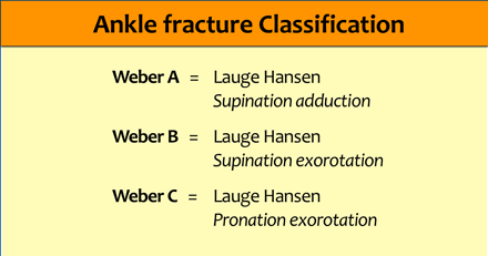
Basically there are three main types of ankle fractures.
Weber classified them as:Về cơ bản có ba loại gãy xương cổ chân chính.
Weber phân loại chúng là:
- type A - infrasyndesmoticloại A - hạ thân nhiệt
- type B - transsyndesmotictype B - chuyển huyết
- type C - suprasyndesmotictype C - tăng huyết áp
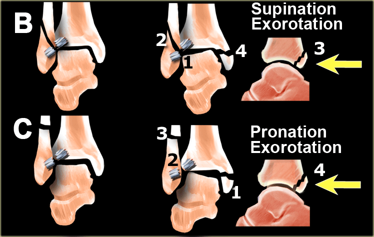
These fractures are identical to the fractures described by Lauge-Hansen as supination-adduction, supination-exorotation and pronation-exorotation.
We will first give a short overview of these fractures and then discuss them in more detail.Những loại gãy này giống với loại gãy được mô tả bởi Lauge-Hansen là gãy ngửa-dẫn, gãy ngả-hướng và gãy sấp-hướng.
Trước tiên chúng tôi sẽ đưa ra một cái nhìn tổng quan ngắn gọn về các loại gãy này và sau đó thảo luận chi tiết hơn về chúng.
Once you understand the trauma mechanism as described by Lauge-Hansen and the sequence of events that take place in stages, then you know where to look for fractures and ligamentous injuries.Một khi bạn hiểu cơ chế chấn thương như được mô tả bởi Lauge-Hansen và chuỗi các sự kiện diễn ra theo các giai đoạn, thì bạn biết nơi để tìm gãy xương và chấn thương dây chằng.
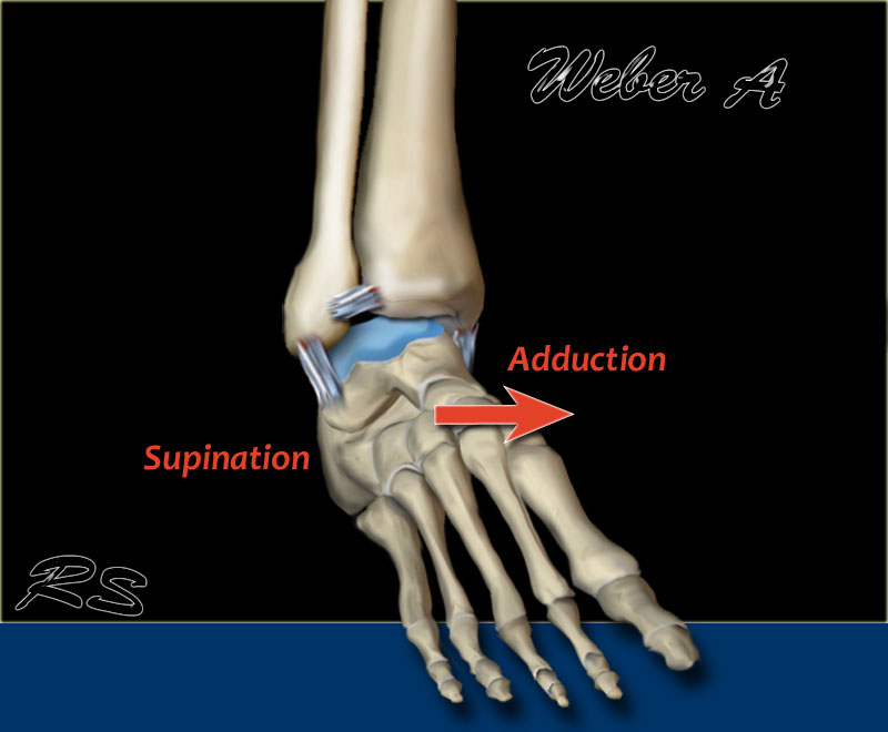
Weber AGia B
Occurs below the syndesmosis, which is intact.
According to Lauge-Hansen, it is the result of an adduction force on the supinated foot.
Xảy ra dưới khớp sợi chày–mác, còn nguyên vẹn.
Theo Lauge-Hansen, đó là kết quả của một lực hút trên bàn chân ngửa.
- Stage 1 - Tension on the lateral collateral ligaments results in rupture of the ligaments
or avulsion of the lateral malleolus below the syndesmosis. Giai đoạn 1 - Căng dây chằng bên ngoài gây đứt dây chằng
hoặc nhổ mắt cá ngoài dưới khớp sợi chày–mác. - Stage 2 - Oblique fracture of the medial malleolus.Giai đoạn 2 - Gãy chéo mắt cá trong.
Scroll through the images.
Notice that the fibular fracture is transverse, because it is an avulsion or pull-off fracture.
The tibial fracture is vertical or oblique, because it is a push-off fracture.Cuộn qua các hình ảnh.
Lưu ý rằng gãy xương mác là gãy ngang, vì nó là một loại gãy nhổ hoặc gãy kéo ra.
Gãy xương chày là gãy dọc hoặc gãy chéo, vì nó là gãy đẩy ra.
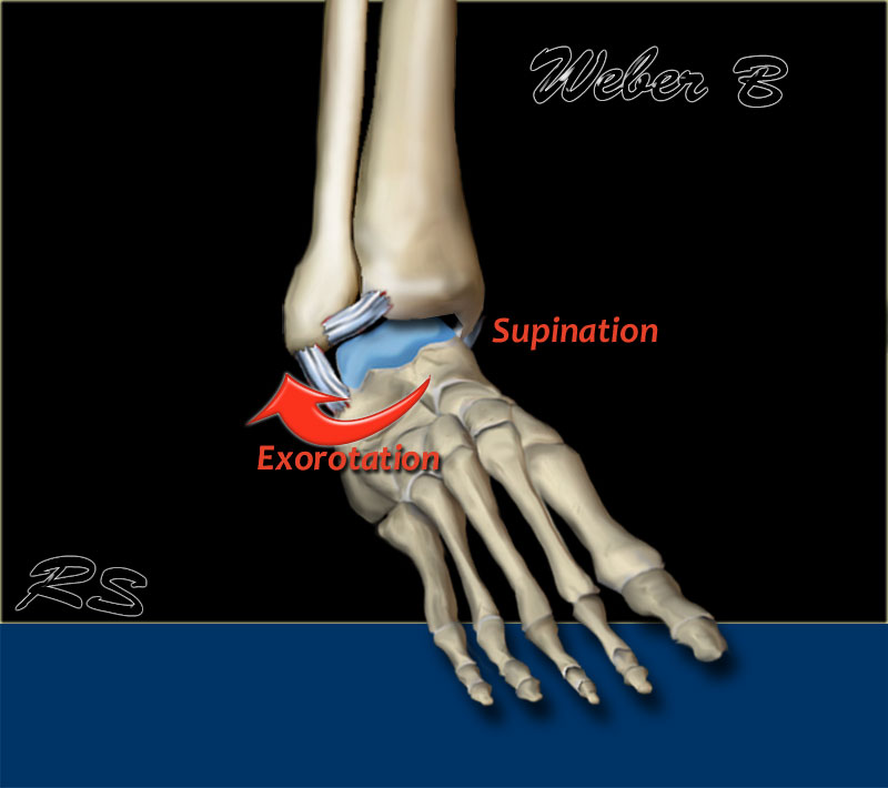
Weber B
This is a transsyndesmotic fracture with usually partial - and less commonly, total - rupture of the syndesmosis.
According to Lauge-Hansen, it is the result of an exorotation force on the supinated foot.Đây là gãy xuyên xương cùng với vỡ một phần xương khớp sợi chày–mác, ít gặp hơn.
Theo Lauge-Hansen, đó là kết quả của một lực đẩy trên bàn chân ngửa.
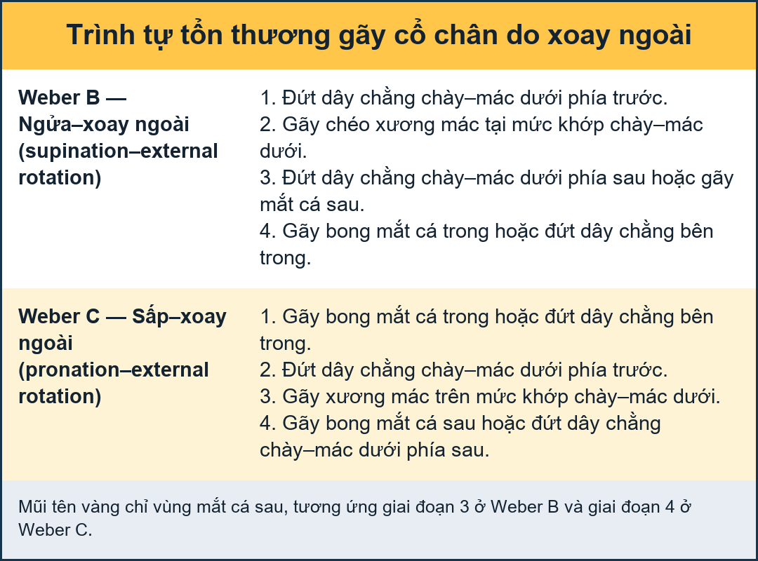
- Stage 1 - Rupture of the anterior syndesmosisGiai đoạn 1 - Vỡ xương phần trước khớp chày–mác dưới
- Stage 2 - Oblique fracture of the fibula (this is the true Weber B fracture)Giai đoạn 2 - Gãy xiên xương mác (đây là gãy Weber B thực sự)
- Stage 3 - Rupture of the posterior syndesmosis
or - fracture of the posterior malleolusGiai đoạn 3 - Vỡ xương sau khớp sợi chày–mác
or - gãy mắt cá sau - Stage 4 - Avulsion of the medial malleolus
or - rupture of the medial collateral bandsGiai đoạn 4 - Nhả mắt cá trong
hoặc - vỡ các đai bên trong
Scroll through the images.
Notice the oblique or vertical orientation of the push-off fibular fracture.Cuộn qua các hình ảnh.
Chú ý hướng chéo hoặc dọc của gãy xương mác đẩy ra.
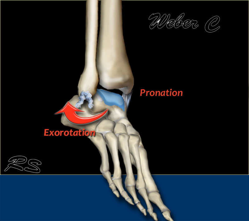
Weber C
This is a fracture above the level of the syndesmosis. Usually there is a total rupture of the syndesmosis with instability of the ankle.
According to Lauge-Hansen, it is the result of an exorotation force on the pronated foot.Đây là một vết gãy trên mức dây chằng dính, thường là đứt dây chằng và mất vững cổ chân.
Theo Lauge-Hansen, đó là kết quả của một lực đẩy ra trên bàn chân sấp.
- Stage 1 - Avulsion of the medial malleolus
or - ligamentous ruptureGiai đoạn 1 - Nhả mắt cá trong
hoặc - đứt dây chằng - Stage 2 - Rupture of the anterior syndesmosisGiai đoạn 2 - Vỡ phần trước khớp chày–mác dưới
- Stage 3 - Fibula fracture above the level of the syndesmosis (this is the true Weber C fracture)Giai đoạn 3 - Gãy xương mác trên mức khớp sợi chày–mác (đây là gãy Weber C thực sự)
- Stage 4 - Avulsion of the posterior malleolus
or - rupture of the posterior syndesmosisGiai đoạn 4 - Nhả mắt cá sau
hoặc - vỡ khớp sợi chày–mác sau
Scroll through the imagesCuộn qua các hình ảnh
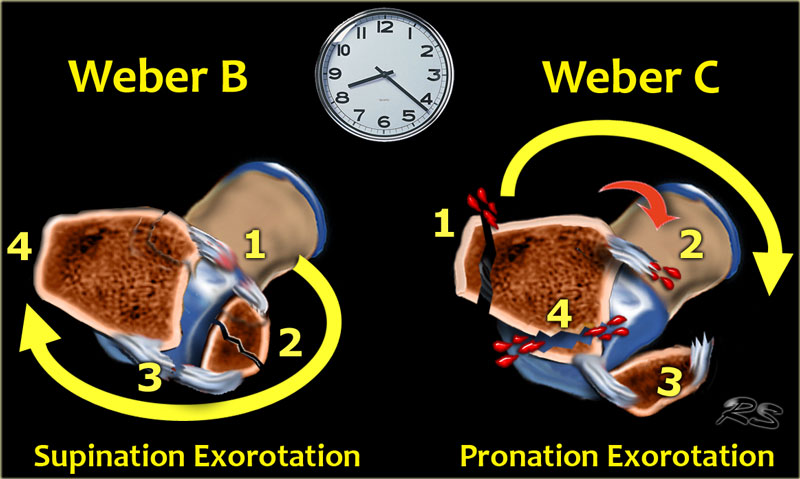
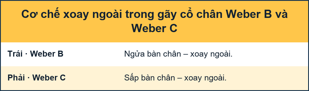
Exorotation injuryChấn thương vùng thắt lưng
Weber A fractures are usually not a problem.
Weber B and C are more difficult and it is essential to understand the sequence of events in these injuries, which are both exorotation injuries.
This implies that 75-80% of ankle injuries are exorotation injuries.Gãy Weber A thường không phải là vấn đề.
Weber B và C khó hơn và cần phải hiểu trình tự các sự kiện trong các chấn thương này, đó là cả hai chấn thương do thở ra.
Điều này ngụ ý rằng 75-80% chấn thương cổ chân là chấn thương vùng cổ chân.
Weber B starts anterolaterally and the sequence is:Weber B bắt đầu từ trước bên và trình tự là:
- Anterior syndesmosis Phần trước khớp chày–mác dưới
- Fibula Tinh vân
- Posterior syndesmosis Phía sau khớp sợi chày–mác
- Medial malleolus Mắt cá trong
Weber C starts medially and the sequence is:Weber C bắt đầu từ giữa và trình tự là:
- Medial malleolus Mắt cá trong
- Anterior syndesmosis Phần trước khớp chày–mác dưới
- Fibula Tinh vân
- Posterior syndesmosis Phía sau khớp sợi chày–mác
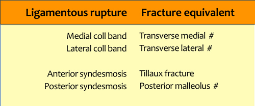
Ligamentous rupture or AvulsionVỡ hoặc bong dây chằng
Another important thing to remember is, that a ligament can rupture or cause an avulsion fracture at the insertion.
Every ligamentous rupture has it's avulsion fracture counterpart.Một điều quan trọng cần nhớ là dây chằng có thể bị đứt hoặc gây ra gãy bong điểm bám khi chọc kim.
Mỗi trường hợp đứt dây chằng đều có đối tác gãy bong điểm bám.
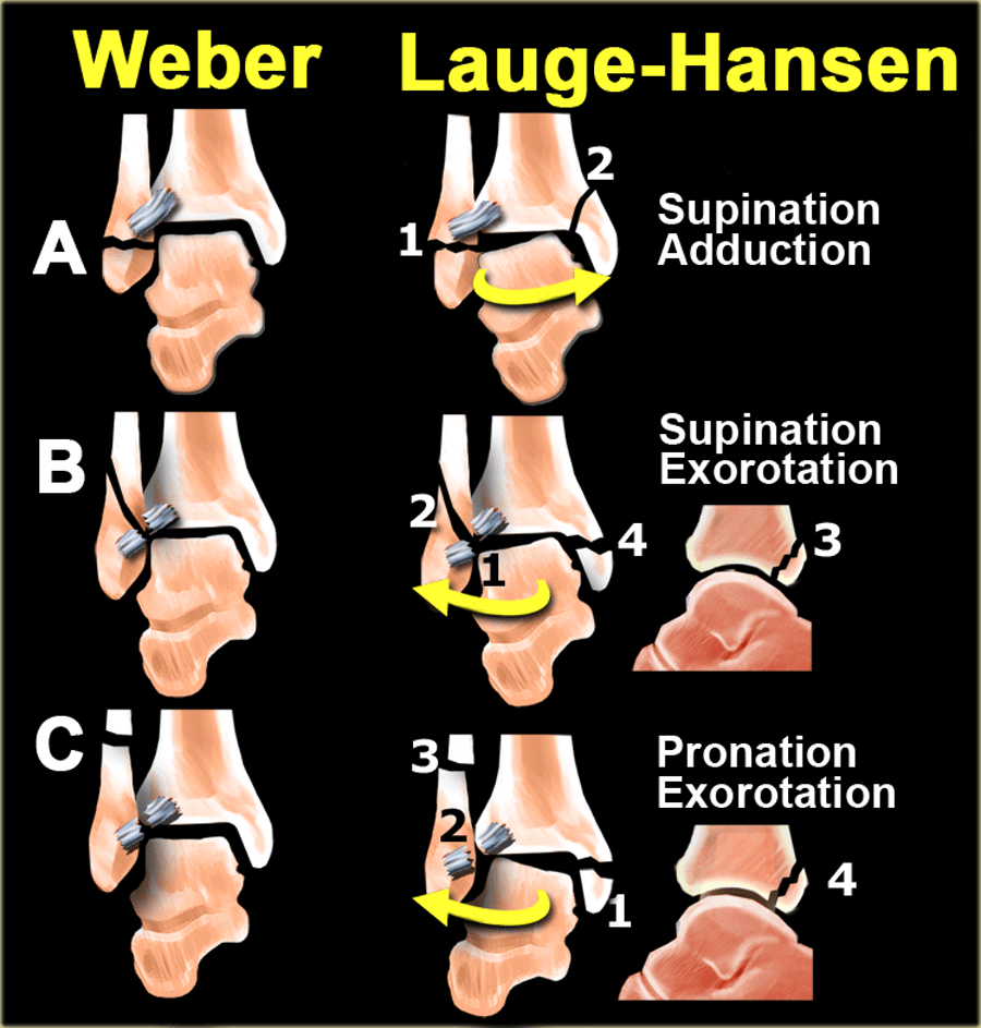
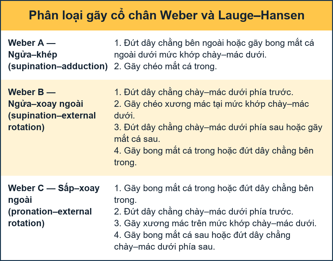
Weber and Lauge-Hansen summaryTóm tắt Weber và Lauge-Hansen
-
Weber A = Infrasyndesmotic
Weber A = Phản ứng hạ đồng bộ
- Avulsion of the lateral malleolusNhảy của mắt cá ngoài
- Oblique fracture of the medial malleolus (uncommon)Gãy chéo mắt cá trong (không phổ biến)
-
Weber B = Transsyndesmotic
Weber B = Chuyển huyết động
- Rupture of the anterior syndesmosisVỡ xương phần trước khớp chày–mác dưới
- Oblique fracture of the fibulaGãy xiên xương mác
- Rupture of the posterior syndesmosis
or - fracture of the posterior malleolusVỡ xương khớp sợi chày–mác sau
or - gãy mắt cá sau - Avulsion of the medial malleolus -
or - rupture of the medial bandsNhảy mắt cá trong -
hoặc - vỡ các dải trong
-
Weber C = Suprasyndesmotic
Weber C = Trên đồng vận
- Avulsion of the medial malleolus
or - ligamentous ruptureNhảy của mắt cá trong
hoặc - đứt dây chằng - Rupture of the anterior syndesmosisVỡ xương phần trước khớp chày–mác dưới
- Fibula fracture above the level of the syndesmosisGãy xương mác trên mức khớp sợi chày–mác
- Avulsion of the posterior malleolus
or - rupture of the posterior syndesmosisNhảy mắt cá sau
hoặc - vỡ khớp sợi chày–mác sau
- Avulsion of the medial malleolus
Instability is seen in:Sự bất ổn định được thấy trong:
- Weber A stage 2Weber A giai đoạn 2
- Weber B stage 3 -4Weber B giai đoạn 3 -4
- Weber C stage 3-4Weber C giai đoạn 3-4
Weber and Lauge-Hansen combinedWeber và Lauge-Hansen kết hợp
In daily practice most use the Weber system, which is easy to memorize, while the Lauge-Hansen seems rather difficult at first glance.
Combining the simplicity of Weber with the explanation of the trauma mechanism given by Lauge-Hansen has the advantage that you still use a simple system, but now you really know what is going on.
Trong thực hành hàng ngày, hầu hết sử dụng hệ thống Weber, dễ nhớ, trong khi Lauge-Hansen có vẻ khá khó khăn ngay từ cái nhìn đầu tiên.
Kết hợp sự đơn giản của Weber với sự giải thích về cơ chế chấn thương do Lauge-Hansen đưa ra có lợi thế là bạn vẫn sử dụng một hệ thống đơn giản, nhưng bây giờ bạn thực sự biết những gì đang xảy ra.
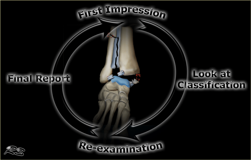
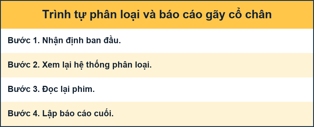
For instance if you see a fracture that is a stage 2 in the Lauge-Hansen system, then you know that there also is a stage 1 injury and you will study the radiographs with a high suspicion for signs of stage 3 and 4.Ví dụ, nếu bạn thấy một vết gãy là giai đoạn 2 trong hệ thống Lauge-Hansen, thì bạn biết rằng cũng có một chấn thương giai đoạn 1 và bạn sẽ nghiên cứu các phim X quang với sự nghi ngờ cao về các dấu hiệu của giai đoạn 3 và 4.
This can best be demonstrated by giving an example.Điều này có thể được chứng minh bằng cách đưa ra một ví dụ.
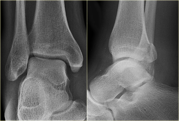 Unstable ankle fractureGãy cổ chân mất vững
- First impression
The radiographs show a fracture of the posterior malleolus.
If you would just report this as - a fracture of the posterior malleolus - you would miss the point.
This is probably an unstable ankle fracture.
A posterior malleolus fracture as an isolated finding is very uncommon.Ấn tượng đầu tiên
X quang cho thấy gãy mắt cá sau.
Nếu bạn chỉ báo cáo điều này như là một vết nứt của mắt cá sau - bạn sẽ bỏ lỡ điểm.
Đây có thể là một gãy xương cổ chân không vững.
Gãy mắt cá sau là một phát hiện đơn độc rất hiếm gặp.
- Looking at the classification system
When we look at the scheme we will notice that a fracture of the posterior malleolus in most cases is part of a Weber B or a Weber C fracture.
A tertius fracture is either Weber B stage 3
or - due to Weber C stage 4 (arrows).
We have to re-examine the films to look for additional findings.
Since we now know where to look, it will be easier to detect additional findings.Nhìn vào hệ thống phân loại
Khi nhìn vào sơ đồ, chúng ta sẽ nhận thấy rằng gãy mắt cá sau trong hầu hết các trường hợp là một phần của gãy Weber B hoặc gãy Webers C.
Gãy xương bánh chè hoặc là Weber B giai đoạn 3
hoặc - do Weber C giai đoạn 4 (các mũi tên).
Chúng tôi phải kiểm tra lại phim để tìm thêm các phát hiện.
Vì bây giờ chúng ta biết nơi để tìm, sẽ dễ dàng hơn để phát hiện các phát hiện bổ sung.
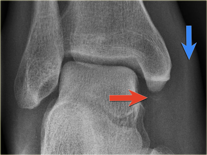 PE stage 1Giai đoạn PE 1
- Re-examination
On the ankle films there was no sign of an oblique fracture of the lateral malleolus, so we can exclude a Weber B fracture.
There is still the possibility of a Weber C fracture stage 4, i.e. medial rupture or avulsion, high fibular fracture and finally a posterior malleolus fracture.
At reexamination you notice the subtle avulsion of the medial malleolus (red arrow), which is stage 1.
Notice also the soft tissue swelling on the medial side (blue arrow)Khám lại
Trên phim cổ chân không có dấu hiệu gãy chéo mắt cá ngoài, do đó có thể loại trừ gãy Weber B.
Vẫn còn khả năng gãy Weber C giai đoạn 4, tức là vỡ trong hoặc bong xương, gãy xương mác cao và cuối cùng là gãy mắt cá sau.
Khi khám lại, bạn nhận thấy hình ảnh bong nhẹ của mắt cá trong (mũi tên màu đỏ), đó là giai đoạn 1.
Chú ý thêm sưng mô mềm ở phía trong (mũi tên màu xanh)
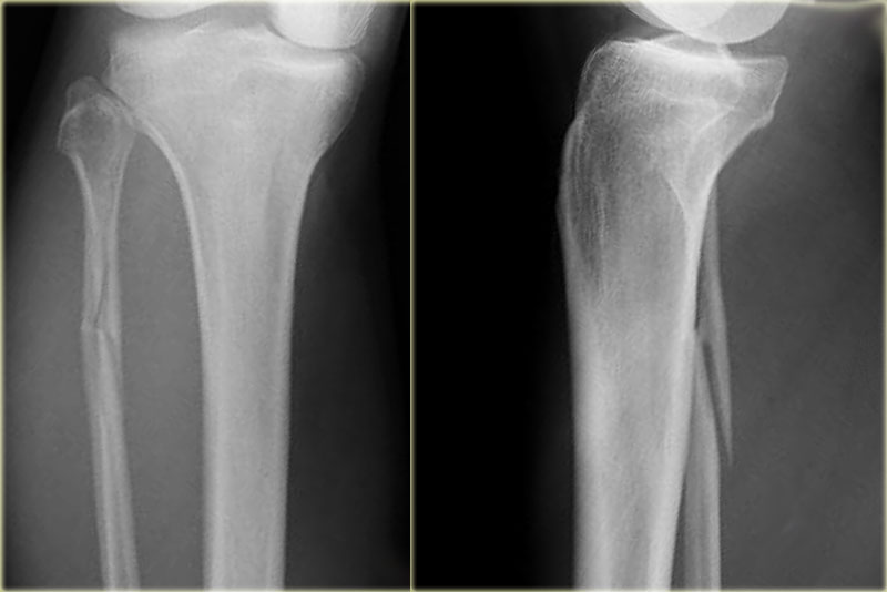 PE stage 3Giai đoạn PE 3
Additional radiographs of the lower extremity were ordered and they demonstrate a high fibular fracture, i.e. Weber C stage 3 also known as a Maisonneuve fracture.Các phim X quang bổ sung của chi dưới được chỉ định và chúng cho thấy gãy xương mác cao, tức là Weber C giai đoạn 3 còn được gọi là gãy Maisonneuve.
-
Final report
Weber C fracture stage 4. Báo cáo tổng kết
Gãy Weber C giai đoạn 4.
This is un unstable ankle injury that needs surgical repair.Đây là tổn thương cổ chân không ổn định cần được phẫu thuật sửa chữa.
This example is an every day case.
The point that I want to make is, that when you understand the sequence of injuries to the ankle, then you know where to look for fractures and soft tissue swelling indicating ligamentous injury.Ví dụ này là một trường hợp hàng ngày.
Điều tôi muốn nói là, khi bạn hiểu được trình tự các chấn thương ở mắt cá chân, thì bạn biết được chỗ nào cần tìm gãy xương và chỗ nào có dấu hiệu sưng mô mềm cho thấy có tổn thương dây chằng.
Weber A in detailWeber A nói chi tiết
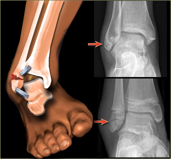
We will now discuss the Weber classification and add the stages of the Lauge-Hansen system.Bây giờ chúng ta sẽ thảo luận về phân loại Weber và thêm các giai đoạn của hệ thống Lauge-Hansen.
Weber A is seen in 20-25% of all ankle fractures.
The diagnosis as well as the treatment is usually no problem.
According to Lauge-Hansen the fracture results from an adduction force on the supinated foot.
The lateral side is under extreme tension with stretch on the ligaments which results in an avulsion fracture.
Almost always the avulsion is seen as a horizontal fracture.
This is called a pull off type of fracture in contrast to a push off type, which is seen as an oblique or vertical fracture.
Weber A gặp ở 20-25% các loại gãy cổ chân.
Chẩn đoán cũng như điều trị thường không có vấn đề gì.
Theo Lauge-Hansen, gãy xương là kết quả của một lực hút trên bàn chân ngửa.
Bên ngoài bị căng thẳng cùng cực với sự căng thẳng trên dây chằng dẫn đến gãy bong điểm bám.
Hầu như luôn luôn bong gân được xem như là một gãy ngang.
Đây được gọi là loại gãy kéo ra, trái ngược với loại gãy đẩy ra, được xem là gãy chéo hoặc gãy dọc.
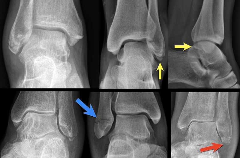 Weber-A stage IWeber-A giai đoạn I
Stage 1Giai đoạn 1
The images show the usual Weber type A fractures.
These are all stage-1 fractures.
Stage-2 is extremely uncommon.Hình cho thấy các kiểu gãy Weber A thường gặp. Tất cả đều ở giai đoạn 1. Giai đoạn 2 cực kỳ hiếm.
Notice the horizontal orientation of the fracture lines.
These are pull off type fractures as a result of avulsion.Chú ý hướng ngang của các đường gãy.
Đây là loại gãy kéo ra do kết quả của sự nhổ.
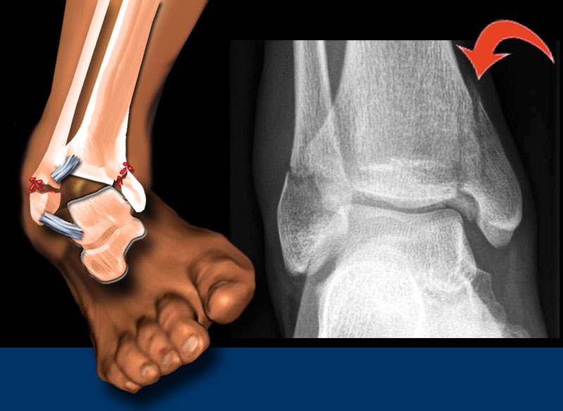 Weber A - stage 2Weber A - giai đoạn 2
Stage 2Giai đoạn 2
Stage 2 is uncommon and easy to detect.
More adduction force results in the medial malleolus being pushed off in a vertical or oblique way.
Stage 2 is unstable because the ring of the ankle is broken in two places.Giai đoạn 2 không phổ biến và dễ phát hiện.
Lực đẩy nhiều hơn dẫn đến mắt cá trong bị đẩy ra theo chiều dọc hoặc xiên.
Giai đoạn 2 không ổn định vì vòng cổ chân bị gãy ở hai nơi.
Notice the horizontal orientation of the lateral malleolus fracture and the vertical orientation of the fracture of the medial malleolus.
Enormous forces must have pushed off the medial malleolus.Lưu ý hướng ngang của gãy mắt cá ngoài và hướng dọc của gãy của mắt cá trong.
Lực lớn đã đẩy ra khỏi mắt cá trong.
More on the ring of the ankle and instabilityThông tin thêm về vòng mắt cá chân và sự mất ổn định
Weber B in detailWeber B chi tiết
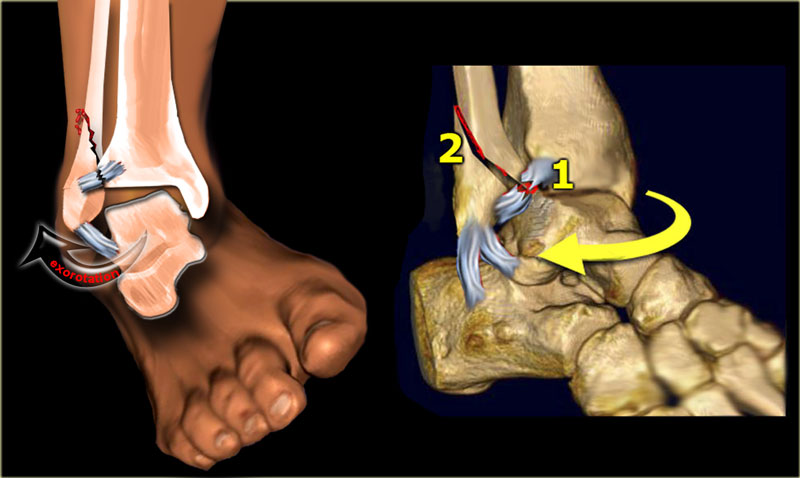 Stage 1: Rupture of anterior tibiofibular ligament - or avulsion fracture (Tilleaux)Giai đoạn 1: Vỡ dây chằng chày mác trước - hoặc gãy bong điểm bám (Tilleaux)
Stage 1-2Giai đoạn 1-2
Weber B is the most common type of ankle fracture and occurs in about 60 %.
According to Lauge-Hansen the fracture results from an exorotation force on the supinated foot. Weber B là loại gãy cổ chân hay gặp nhất và xảy ra trong khoảng 60%.
Theo Lauge-Hansen, gãy xương là kết quả của một lực đẩy ra trên bàn chân ngửa.
Stage 1 is usually not visible on x-rays.
What we normally see is a stage 2 oblique fracture through the syndesmosis and we have to assume that there is also a rupture of the anterior tibiofibular ligament, which is stage 1.
According to Lauge Hansen the first injury is on the lateral side, which is under maximum tension.
In stage 2 the talus exorotates further and since the foot is in supination, the lateral malleolus is held tightly in place by the lateral collateral ligaments.
The lateral malleolus cannot move away without breaking.
As a result more rotation of the talus will fracture the fibula in an oblique or spiral fashion because the lateral malleolus is pushed off from anteromedially to posterolaterally. Giai đoạn 1 thường không nhìn thấy trên phim X-quang.
Những gì chúng ta thường thấy là gãy chéo giai đoạn 2 qua khớp sợi chày–mác và chúng ta phải giả định rằng cũng có một vết rách dây chằng chày mác trước, đó là giai đoạn 1.
Theo Lauge Hansen, chấn thương đầu tiên là ở bên ngoài, dưới sức căng tối đa.
Trong giai đoạn 2, xương sên sẽ mở rộng hơn và vì bàn chân nằm ngửa, mắt cá ngoài được giữ chặt bởi các dây chằng bên ngoài.
mắt cá ngoài không thể di chuyển mà không bị vỡ.
Kết quả là sự xoay của xương sên sẽ làm gãy xương mác theo kiểu chéo hoặc xoắn ốc vì mắt cá ngoài bị đẩy ra từ phía trước trong sang phía sau bên.
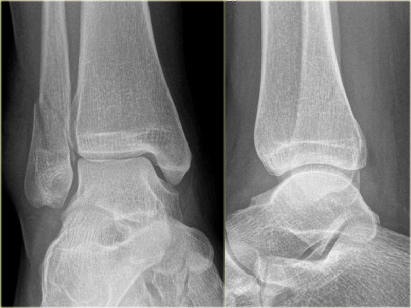
The images show a Weber B fracture.
The oblique course of the fracture is typical for Weber B and results from the exorotation of the talus that pushes against the fixed lateral malleolus.
The malleolar fracture usually starts medially at the level of the talar dome, but can also start a few centimeters above this level.
Hình ảnh cho thấy gãy Weber B.
Đường gãy chéo là điển hình cho Weber B và là kết quả của sự thóat ra của xương sên đẩy vào xương mắt cá ngoài cố định.
Vết gãy mắt cá thường bắt đầu ở giữa ở mức của xương sên, nhưng cũng có thể bắt đầu ở trên mức này vài cm.
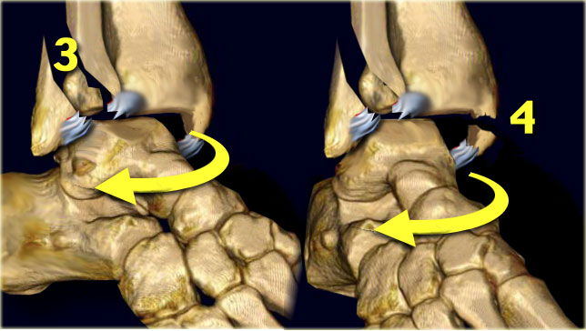 Weber B - stage 3 and 4Weber B - giai đoạn 3 và 4
Stage 3-4Giai đoạn 3-4
Stage 3 More posterior displacement of the lateral malleolus fragment by the talus results in tension on the posterior syndesmosis with rupture or avulsion of the posterior malleolus.Giai đoạn 3 Sự dịch chuyển ra sau của mảnh mắt cá ngoài bởi talus dẫn đến sự căng thẳng trên khớp sợi chày–mác phía sau với vỡ hoặc nhả mắt cá sau.
Stage 4 Further posterior movement of the talus will result in extreme tension on the medial side and the deltoid ligament will either rupture or pull off the medial malleolus in the transverse plane.Giai đoạn 4 Chuyển động sau của xương sên sẽ dẫn đến căng thẳng cực độ ở phía trong và dây chằng delta sẽ đứt hoặc kéo mắt cá trong ra theo mặt phẳng ngang.
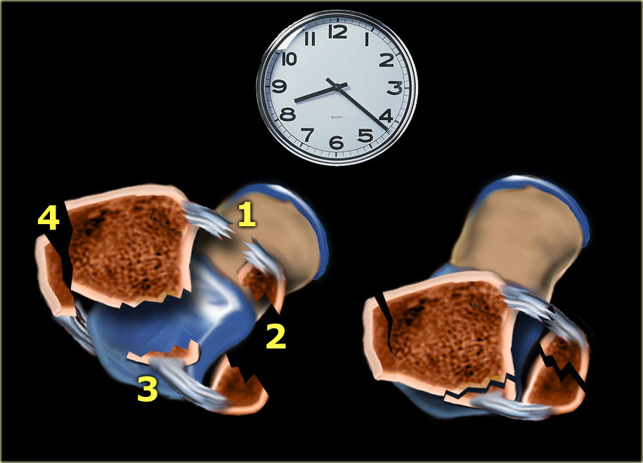
The sequences in a Weber B fracture or Lauge-Hansen supination exorotation injury take place in a clockwise manner:Các trình tự trong gãy Weber B hoặc chấn thương Lauge-Hansen ngửa khi thở ra diễn ra theo chiều kim đồng hồ:
- Rupture of the anterior tibiofibular ligament Vỡ dây chằng chày mác trước
- Oblique fracture of the distal fibula Gãy lệch đầu dưới xương mác
- Avulsion of the posterior malleolus or rupture of the posterior tibiofibular ligament Vỡ mắt cá sau hoặc đứt dây chằng chày mác sau
- Avulsion of the medial malleolus or rupture of the medial collateral ligament Nhổ mắt cá trong hoặc đứt dây chằng bên trong
Immediately after the injury the injured parts may again align, which can make it difficult to detect the injuries.Ngay sau khi bị thương, các bộ phận bị thương có thể thẳng hàng trở lại, điều này có thể gây khó khăn cho việc phát hiện các thương tổn.
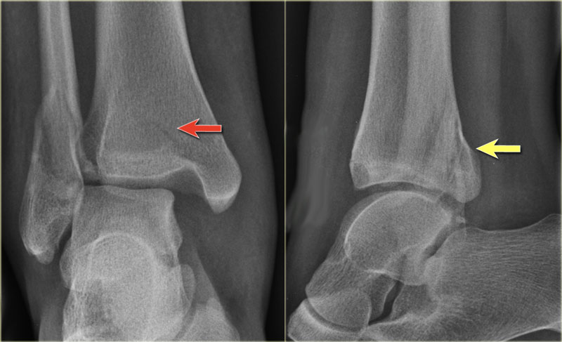 Weber B fractureGãy Weber B
The radiographs show a typical Weber B fracture.
First study the images and then continue reading.
Do you see what stage this is?
X quang cho thấy gãy Weber B điển hình.
Đầu tiên nghiên cứu hình ảnh và sau đó tiếp tục đọc.
Bạn có thấy đây là giai đoạn nào không?
This is a Weber B stage 4 injury.
Notice that all 4 stages are visible:Đây là chấn thương Weber B giai đoạn 4.
Lưu ý rằng tất cả 4 giai đoạn đều có thể nhìn thấy:
- Rupture of the anterior syndesmosis - seen as widening of the space between the distal tibia and fibula (lateral clear space). Vỡ xương phần trước khớp chày–mác dưới - được xem là sự mở rộng của khoảng trống giữa xương chày và xương mác (khoảng trống bên ngoài).
- Oblique fibula fracture at the level of the syndesmosis - i.e. Weber B fracture. Gãy xương mác xiên ở mức của gãy khớp sợi chày–mác - tức là gãy Weber B.
- Tertius fracture - seen on AP view (red arrow) and on lateral view (yellow arrow). Gãy xương Tertius - nhìn thấy trên mặt cắt ngang (mũi tên màu đỏ) và mặt cắt ngang.
- Rupture of the medial collateral ligaments - seen as widening of the space between the medial malleolus and the talus (medial clear space)Vỡ dây chằng bên trong - được xem là sự mở rộng khoảng trống giữa mắt cá trong và xương sên (khoảng trống trong)
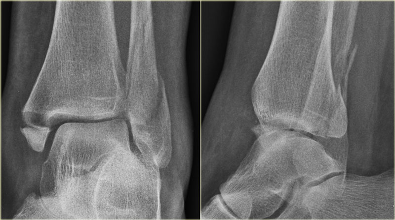
These images show another typical Weber B fracture stage 4.
There is an oblique fracture of the fibula.
There is an avulsion of the posterior malleolus and an avulsion of the medial malleolus.Những hình ảnh này cho thấy một gãy Weber B điển hình khác ở giai đoạn 4.
Có một vết gãy chéo xương mác.
Có sự tách rời mắt cá sau và tách rời của mắt cá trong.
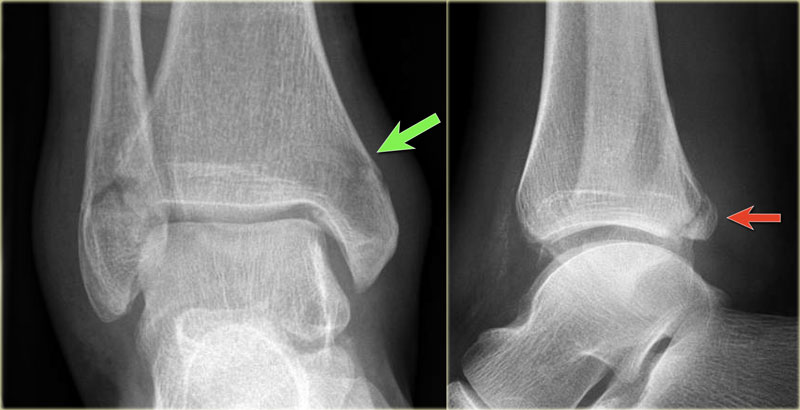
Here another typical Weber B fracture stage 4.
First notice the oblique fibular fracture, which is best seen on the lateral view.
This is stage 2 and we have to assume, that the anterior syndesmosis is ruptured.
On the lateral view a small tertius fragment is seen indicating stage 3.
Now you start looking for stage 4 and you will notice the subtle lucency in the medial malleolus on the AP view (green arrow).
Knowing the stages of Lauge Hansen this must be a fracture.Đây là một gãy Weber B điển hình khác giai đoạn 4.
Đầu tiên, hãy chú ý đến gãy xương mác chéo, nhìn thấy rõ nhất ở mặt nghiêng.
Đây là giai đoạn 2 và chúng ta phải giả định rằng phần trước khớp chày–mác dưới bị vỡ.
Trên mặt cắt ngang, một mảnh tertius nhỏ được nhìn thấy cho thấy giai đoạn 3.
Bây giờ bạn bắt đầu tìm kiếm giai đoạn 4 và bạn sẽ nhận thấy sự sáng tinh tế trong mắt cá trong trên chế độ xem AP (mũi tên màu xanh lá cây).
Biết được các giai đoạn của Lauge Hansen thì đây chắc chắn là một gãy xương.
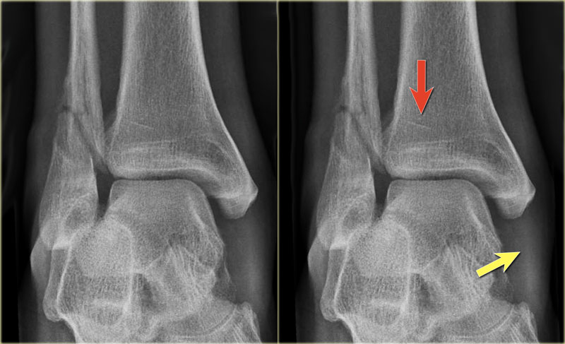
Here a more subtle case.
At first impression there is a Weber B fracture stage 2.
Now we start looking for stage 3, which is a tertius fracture.
The small linear density on the AP-view is enough to diagnose a tertius fracture.
The soft tissue swelling on the medial side is probably a rupture of the medial collateral band , i.e. stage 4. Đây là một trường hợp tinh tế hơn.
Tại thời điểm mổ ban đầu có gãy Weber B giai đoạn 2.
Bây giờ chúng ta bắt đầu tìm kiếm giai đoạn 3, đó là gãy xương tertius.
Mật độ đường thẳng nhỏ trên góc nhìn của AP là đủ để chẩn đoán gãy xương tertius.
Sưng mô mềm ở phía trong có thể là một vết rách của dải băng bên trong, tức là giai đoạn 4.
Weber C in detailWeber C chi tiết
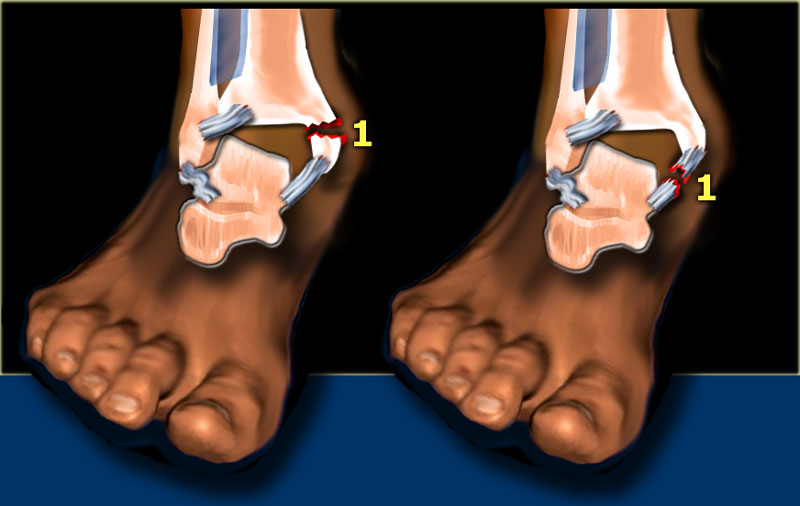
Stage 1Giai đoạn 1
Weber C is seen in approximately 20% of ankle fractures.
It is the most difficult fracture to diagnose and the Lauge-Hansen system will help you to understand the fracture-mechanism, as this will be an enormous help.
According to Lauge-Hansen the fracture results from an exorotation force on the pronated foot. Weber C được thấy trong khoảng 20% các trường hợp gãy cổ chân.
Đây là loại gãy xương khó chẩn đoán nhất và hệ thống Lauge-Hansen sẽ giúp bạn hiểu được cơ chế gãy xương, vì đây sẽ là một trợ giúp rất lớn.
Theo Lauge-Hansen, gãy xương là kết quả của một lực đẩy ra trên bàn chân sấp.
Stage 1 The first injury will occur on the medial side, which is under maximum tension due to the pronation.
It will lead to rupture of the medial collateral ligament or avulsion of the medial malleolus.Giai đoạn 1 Chấn thương đầu tiên sẽ xảy ra ở phía trong, dưới sức căng tối đa do sấp.
Nó sẽ dẫn đến đứt dây chằng bên trong hoặc bong mắt cá trong.
Now the injury can stop and there will only be a rupture of the medial collateral ligaments or avulsion of the medial malleolus.
Lauge Hansen calls this PE stage 1.
We can not cathegorize this in the Weber classification, since there is no fibular fracture.
In many cases the injury progresses to a higher stage.Bây giờ chấn thương có thể dừng lại và sẽ chỉ có một đứt dây chằng bên trong hoặc bong mắt cá trong.
Lauge Hansen gọi đây là giai đoạn PE 1.
Chúng tôi không thể phân loại điều này trong phân loại Weber, vì không có gãy xương mác.
Trong nhiều trường hợp, chấn thương tiến triển đến giai đoạn cao hơn.
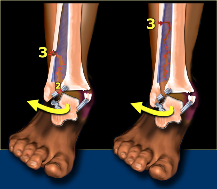
Stage 2-3Giai đoạn 2-3
The talus rotates externally and moves laterally because it is free from its medial attachment.
Due to the pronation, the lateral ligaments are not under tension and the fibula can move away from the tibia.
This causes rupture of the anterior syndesmosis. This is stage 2.Mái taluy xoay ra ngoài và di chuyển theo chiều ngang vì nó không bị dính vào trong.
Do sấp, các dây chằng bên không bị căng và xương mác có thể di chuyển ra khỏi xương chày.
Điều này gây ra vỡ phần trước khớp chày–mác dưới. Đây là giai đoạn 2.
Continuous force will twist the fibula and displace it distally, while proximally it is fixed to the tibia.
Finally the interosseus membrane will rupture up to the point where the fibular shaft fractures. This is stage 3.
This is always above the level of the syndesmosis.
In many cases it is visible on the radiographs of the ankle, but in some cases the fracture is located high and will only be visible on a radiograph of the lower leg.
This last type of fracture is also called Maisonneuve fracture.Lực liên tục sẽ xoắn xương mác và di chuyển nó ra xa, trong khi ở đầu gần nó được cố định vào xương chày.
Cuối cùng màng gian cốt sẽ vỡ đến điểm mà trục xương mác bị gãy, đây là giai đoạn 3.
Giá trị này luôn cao hơn mức khớp sợi chày–mác.
Trong nhiều trường hợp, nó có thể nhìn thấy trên phim X-quang cổ chân, nhưng trong một số trường hợp, gãy xương nằm ở vị trí cao và chỉ có thể nhìn rõ trên phim X quang cẳng chân.
Loại gãy xương cuối cùng này còn được gọi là gãy Maisonneuve.
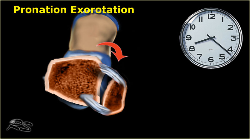
Here we see the different stages in the axial plane.Ở đây chúng ta thấy các giai đoạn khác nhau trong mặt phẳng ngang.
- Medial avulsion fracture or rupture of the collateral bandGãy bong điểm bám trong hoặc vỡ đai bàng hệ
- Rupture of the anterior syndesmosisVỡ xương phần trước khớp chày–mác dưới
- Suprasyndesmotic rupture of the fibula due to rotationVỡ xương mác trên đồng bộ do xoay
- Posterior malleolus fracture or rupture of the posterior syndesmosisGãy mắt cá sau hoặc vỡ mắt cá sau khớp sợi chày–mác
Scroll through the images.Cuộn qua các hình ảnh.
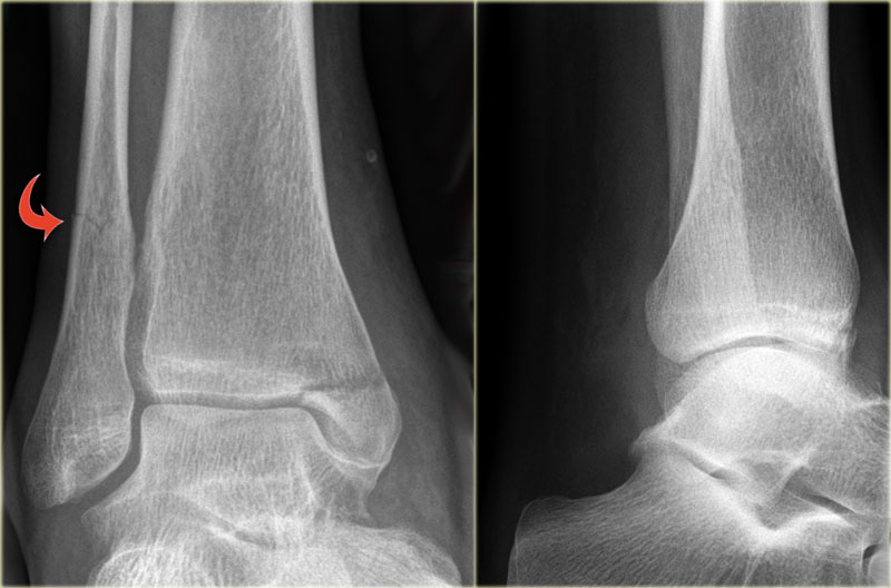 Weber C fracture - stage 3Gãy Weber C – giai đoạn 3
The radiographs shows a Weber C fracture.
There is an avulsion fracture of the medial malleolus and a fibula fracture above the level of the syndesmosis.
According to Lauge-Hansen this is stage 3 pronation exorotation injury and so the anterior syndesmosis (stage 2) must also be ruptured.X quang cho thấy gãy Weber C.
Có một gãy bong điểm bám của mắt cá trong và một gãy xương mác trên mức khớp sợi chày–mác.
Theo Lauge-Hansen đây là chấn thương sấp ngửa giai đoạn 3 và do đó phần trước khớp chày–mác dưới (giai đoạn 2) cũng phải bị vỡ.
We do not see a tertius fracture, which would indicate stage 4, but there may be a rupture of the posterior syndesmosis.Chúng tôi không thấy gãy xương bánh chè, cho thấy giai đoạn 4, nhưng có thể có vỡ xương khớp sợi chày–mác sau.
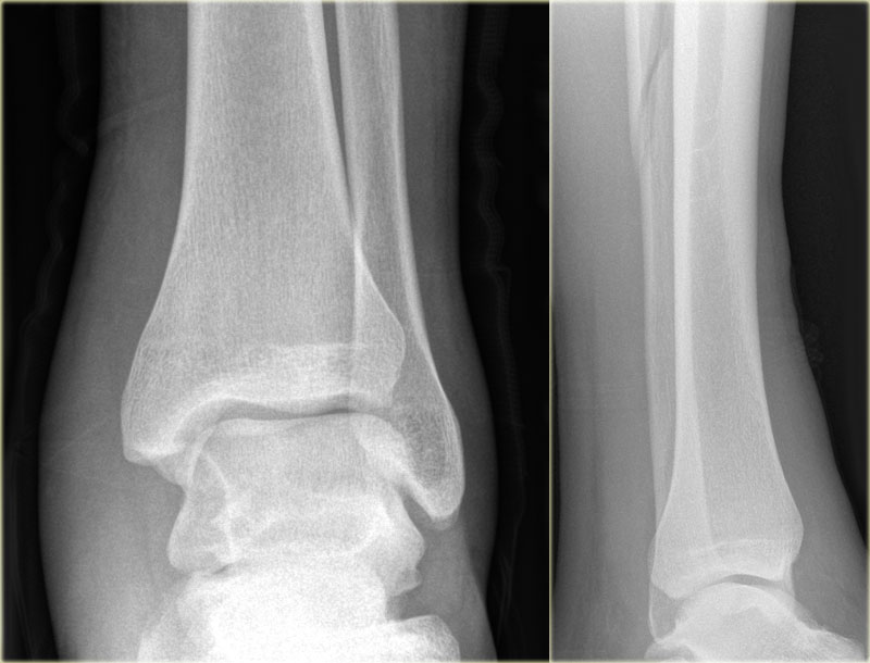 Weber C fracture - at least stage 3Gãy Weber C – ít nhất giai đoạn 3
Here an example of a Weber C fracture with a proximal fibula fracture.
Notice that on the radiograph of the ankle no fracture is seen.
You might misdiagnose this as only some soft tissue swelling.
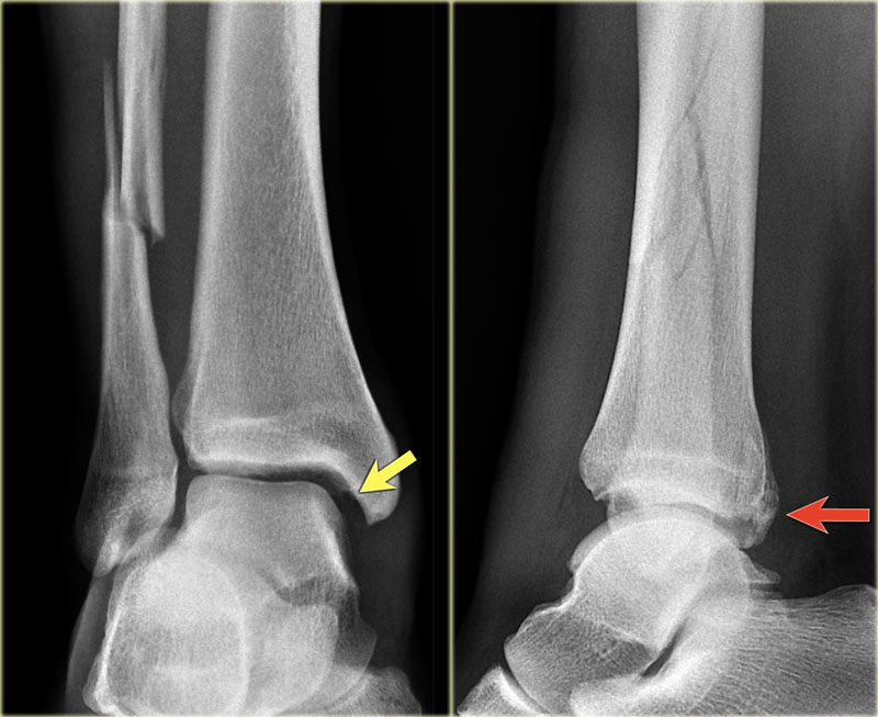
In fact this is an unstable ankle fracture, since there also must be a rupture of the medial collateral ligament (stage 1) , so the ring is broken in two places leading to instability.Đây là một ví dụ về gãy Weber C với gãy xương mác đầu gần.
Lưu ý rằng trên phim X quang của cổ chân không thấy gãy xương.
Bạn có thể chẩn đoán nhầm đây chỉ là một số chỗ sưng mô mềm.
Trên thực tế đây là một loại gãy cổ chân mất vững, vì cũng phải có đứt dây chằng bên trong (giai đoạn 1), do đó vòng bị đứt ở hai nơi dẫn đến mất vững.
According to Lauge Hansen we are probably dealing with: Theo Lauge Hansen, chúng ta có thể đang đối phó với:
- Medial collateral band rupture Vỡ đai bàng hệ trong
- Rupture of the anterior syndesmosis Vỡ xương phần trước khớp chày–mác dưới
- High fibula fracture Gãy xương mác cao
- and possibly a rupture of the posterior syndesmosisvà có thể là vỡ khớp sợi chày–mác sau
Stage 4Giai đoạn 4
Finally the posterior syndesmotic ligament ruptures, or there is an avulsion of the posterior malleolus, also known as posterior malleolus fracture (red arrow).Cuối cùng là đứt dây chằng chày mác sau, hoặc có bong mắt cá sau, còn được gọi là gãy mắt cá sau (mũi tên đỏ).
The medial clear space is only slightly widened, but based on the stages of Lauge Hansen there must be a collateral band rupture.Không gian trong suốt ở giữa chỉ hơi rộng ra, nhưng dựa trên các giai đoạn của Lauge Hansen phải có một dải băng bên bị đứt.
Interpretation and ReportingGiải thích và báo cáo
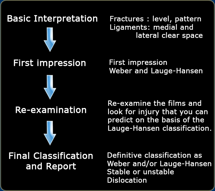
Start with a first impression and look for fractures and signs of ligamentous rupture.
This impression will direct you to both a Weber as well as a Lauge-Hansen classification.
The Lauge-Hansen classification will give you the fracture mechanism and the preliminary stage of the ankle injury.
Bắt đầu với lần khám đầu tiên và tìm các vết nứt và dấu hiệu đứt dây chằng.
Ấn tượng này sẽ hướng bạn đến cả phân loại Weber cũng như phân loại Lauge-Hansen.
Phân loại Lauge-Hansen sẽ cho bạn cơ chế gãy xương và giai đoạn sơ bộ của chấn thương mắt cá chân.
Now re-examine the films to make sure that you do not overlook a higher grade ankle injury.
After this re-examination you can make a final report.
Bây giờ kiểm tra lại các phim để đảm bảo rằng bạn không bỏ qua chấn thương mắt cá chân cấp cao hơn.
Sau khi kiểm tra lại này, bạn có thể làm một báo cáo cuối cùng.
In the final report the fracture is described according to Weber and/or Lauge-Hansen.
Describe the number of malleoli involved and whether there are signs of instability or dislocation.Trong báo cáo cuối cùng, gãy xương được mô tả theo Weber và / hoặc Lauge-Hansen.
Mô tả số lượng malleoli liên quan và có dấu hiệu mất vững hoặc trật khớp hay không.
 Click to enlargeNhấp để phóng to
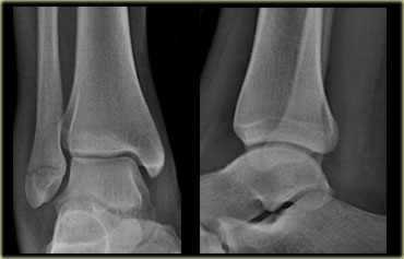
Example 1 Ví dụ 1
- Basic interpretation
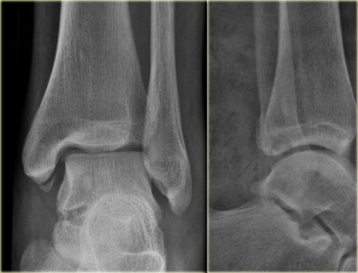
There is a medial malleolar fracture.
You interprete this as an avulsion fracture.
Giải thích cơ bản
Có gãy mắt cá trong.
Bạn giải thích điều này như là một gãy bong điểm bám. - Classification
Not possible to classify according to Weber, but according to Lauge Hansen a medial avulsion fracture indicates that the foot probably was in pronation at the moment of injury.
So this injury is probably a pronation exorotation injury (PER) stage 1 or higher.Phân loại
Không thể phân loại theo Weber, nhưng theo Lauge Hansen, gãy bong điểm bám trong cho thấy bàn chân có thể sấp tại thời điểm bị thương.
Vì vậy, tổn thương này có thể là tổn thương sấp ngửa (PER) giai đoạn 1 hoặc cao hơn.
- Re-examination
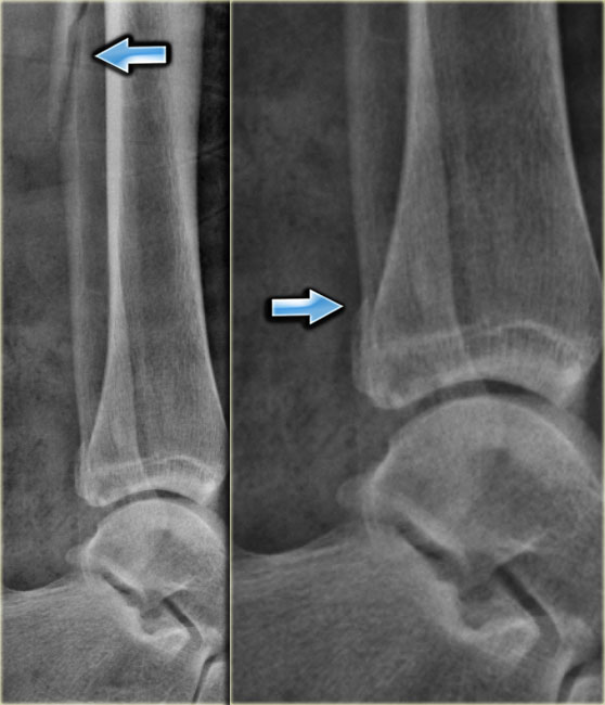
You re-examine the x-rays to look for stage 2 (rupture or avulsion of the anterior syndesmosis), stage 3 (high fibular fracture = Weber C) or even stage 4 (rupture or avulsion of posterior syndesmosis).
So at second look you notice a subtle widening of the lateral clear space on the original films, which could indicate but is definitely no proof of a syndesmotic rupture.
Although the patient is already in a cast you order additional films to look for a possible stage 3.
These films show a high fibular fracture and a subtle posterior malleolus avulsion.Khám lại
Bạn kiểm tra lại phim X-quang để tìm giai đoạn 2 (đứt hoặc nhả phần trước khớp chày–mác dưới), giai đoạn 3 (gãy xương mác cao = Weber C) hoặc thậm chí giai đoạn 4 (đứng hoặc nhả xương khớp sợi chày–mác sau).
Vì vậy, khi nhìn lần thứ hai, bạn nhận thấy một khoảng trống bên trong mở rộng tinh tế trên các phim gốc, có thể chỉ ra nhưng chắc chắn không thấy dấu hiệu vỡ dây chằng dính khớp.
Mặc dù bệnh nhân đã có sẵn dàn diễn viên, bạn vẫn yêu cầu thêm phim để tìm kiếm khả năng ở giai đoạn 3.
Những phim này cho thấy gãy xương mác cao và một chỗ nhô ra mắt cá sau tinh tế. - Final report
Weber C fracture or a PER stage 4 according to Lauge-Hansen. This is an unstable fracture that needs surgical repair.
The ankle circle is interrupted at two places i.e. the medial malleolus and the syndesmosis.
A syndesmotic screw was inserted.Báo cáo tổng kết
Gãy Weber C hoặc PER giai đoạn 4 theo Lauge-Hansen. Đây là loại gãy mất vững cần phẫu thuật sửa chữa.
Vòng cổ chân bị gián đoạn ở hai vị trí, tức là mắt cá trong và khớp sợi chày–mác.
Một vít dính khớp được đưa vào.
Example 2 Ví dụ 2
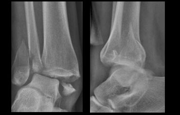
- Basic interpretation
Transverse lateral malleolar fracture.Giải thích cơ bản
Gãy mắt cá bên ngang. -
Classification
Weber A and Supination Adduction stage 1.Phân loại
Weber A và Ngưng thở Giai đoạn dẫn truyền 1. -
Re-examination
No sign of SA stage 2 (medial malleolar fracture)Khám lại
Không có dấu hiệu của SA giai đoạn 2 (gãy mắt cá trong) - Final report
Stable Weber A or SA stage 1 fracture.
Patient will be treated conservatively.Báo cáo tổng kết
Gãy Weber A hoặc gãy xương giai đoạn 1 có độ vững.
Bệnh nhân sẽ được điều trị bảo tồn.
Example 3 Ví dụ 3
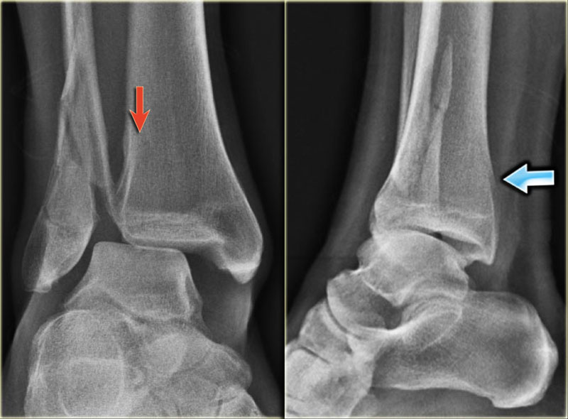
- Basic interpretation
Dislocated bimalleolar fracture. Avulsion fracture of the medial malleolar.
The lateral malleolus is 'pushed off' from anterior to posterior.
Giải thích cơ bản
Gãy lệch xương chè, gãy gãy bong điểm bám xương chè trong.
mắt cá ngoài được đẩy ra từ trước ra sau. - Classification
The fracture starts at the level of the ankle joint and extends proximally, i.e. a Weber B fracture.
According to Lauge Hansen the oblique fibular fracture indicates Supination Exorotation injury stage 2 or higher. Phân loại
Gãy xương bắt đầu ở khớp cổ chân và kéo dài đến đầu gần, tức là gãy Weber B.
Theo Lauge Hansen gãy xương mác chéo cho thấy Chấn thương ngực ngoài giai đoạn 2 hoặc cao hơn. - Re-examination
Look for stage 3 (posterior syndesmotic rupture or avulsion of the posterior malleolus) and stage 4 (rupture of the deltoid ligament or medial malleolar avulsion).
Only now you notice the posterior malleolar fracture on the lateral view. Khám lại
Tìm giai đoạn 3 (vỡ dây chằng chày mác sau hoặc bong mắt cá sau) và giai đoạn 4 (vỡ thắt delta hoặc bong xương mắt cá trong).
Chỉ bây giờ bạn mới thấy gãy mắt cá sau trên phim nghiêng. - Final report
Trimalleolar fracture. Weber B. SER stage 4 (Lauge-Hansen).
This is an unstable fracture with dislocation that needs surgical repair.
The size of the posterior malleolar fragment is probably less than 25% of the articular AP-diameter and will need no separate repair.
Sometimes CT is needed to get a better impression of the size of the fracture fragment of the posterior malleolus. Báo cáo tổng kết
Gãy ba mắt cá Weber B. SER giai đoạn 4 (Lauge-Hansen).
Đây là loại gãy xương mất vững, trật khớp cần phẫu thuật sửa chữa.
Kích thước của mảnh sau mắt cá có thể nhỏ hơn 25% đường kính khớp AP và sẽ không cần sửa chữa riêng biệt.
Đôi khi cần chụp CT để có được ấn tượng tốt hơn về kích thước của mảnh gãy của mắt cá sau.
 Click to enlargeNhấp để phóng to
Example 4 Ví dụ 4
- Basic interpretation
Fracture of the lateral malleolus starting anteriorly at the level of the joint extending proximally posteriorly.Giải thích cơ bản
Gãy mắt cá ngoài bắt đầu từ phía trước ngang mức khớp kéo dài về phía sau. - Classification
The fracture is classified according to Weber as a type B fracture.
According to Lauge Hansen the oblique fibular fracture indicates that this is a Supination Exorotation injury stage 2 or higher.Phân loại
Gãy xương được Weber phân loại là gãy loại B.
Theo Lauge Hansen gãy chéo xương mác cho thấy đây là chấn thương sụn chêm - sụn viền giai đoạn 2 hoặc cao hơn. - Re-examination
Look for stage 3 and stage 4.
There are subtle findings which indicate a fracture of the posterior malleolus. Normally you probably would not have noticed these.
On the lateral view the posterior cortex of the tibia is interrupted indicating a fracture (blue arrow).
Even on the AP-view there are subtle findings that indicate a fracture (red arrows).
There is a widened medial clear space, which indicates a rupture of the medial collateral band, i.e. stage 4.Khám lại
Tìm giai đoạn 3 và giai đoạn 4.
Có những dấu hiệu rất nhỏ cho thấy gãy mắt cá sau, bình thường bạn sẽ không nhận thấy những dấu hiệu này.
Trên phim nghiêng, vỏ sau xương chày bị đứt gãy (mũi tên xanh).
Ngay cả trên phim chụp cắt lớp vi tính có những dấu hiệu tinh tế cho thấy gãy xương (các mũi tên màu đỏ).
Có một khoảng trống trong ở giữa, cho thấy sự đứt gãy của dải bên trong, tức là giai đoạn 4. - Final report
Weber B fracture. According to Lauge-Hansen this is a SER stage 4.
This is an unstable fracture with dislocation that needs surgical repair.Báo cáo tổng kết
Gãy Weber B. Theo Lauge-Hansen đây là gãy giai đoạn 4, không có biến chứng.
Đây là loại gãy xương mất vững, trật khớp cần phẫu thuật sửa chữa.
Charity
Hoạt động từ thiện
All the profits of the Radiology Assistant go to Medical Action Myanmar which is run by Dr. Nini Tun and Dr. Frank Smithuis sr, who is a professor at Oxford university and happens to be the brother of Robin Smithuis.Toàn bộ lợi nhuận của Radiology Assistant được chuyển cho Medical Action Myanmar, do bác sĩ Nini Tun và bác sĩ Frank Smithuis Sr. điều hành. Frank Smithuis Sr. là giáo sư tại Đại học Oxford và là anh em ruột của Robin Smithuis.
Click here or on the image below to watch the video of Medical Action Myanmar and if you like the Radiology Assistant, please support Medical Action Myanmar with a small gift.Nhấp vào đây hoặc vào hình ảnh bên dưới để xem video về Medical Action Myanmar và nếu bạn thích Trợ lý X quang, vui lòng hỗ trợ Medical Action Myanmar bằng một món quà nhỏ.
ReferencesTài liệu tham khảo
-
Anatomy of the distal tibiofibular syndesmosis in adults: a pictorial essay with a multimodality approach
by John J. Hermans, Annechien Beumer, Ton A. W. de Jong and Gert-Jan Kleinrensink.
J. Anat. (2010) 217, pp633-645
Giải phẫu xương chày mác khớp sợi chày–mác ở người lớn: nghiên cứu mô tả bằng hình ảnh với phương pháp tiếp cận đa mô thức - AO-foundation ankle fractures Gãy cổ chân nền AO
-
Introduction to Lauge Hansen & Danis Weber Classifications Ankle Fracture
Giới thiệu về phân loại Lauge Hansen & Danis Weber Gãy cổ chân
Animation on YouTube by Dr Glass. Hoạt hình trên YouTube của Dr Glass.