
Ankle Fracture Mechanism and RadiographyCơ chế gãy xương cổ chân và hình ảnh X-quang
Radiology Department of the Rijnland Hospital, Leiderdorp, the NetherlandsKhoa Chẩn đoán hình ảnh Bệnh viện Rijnland, Leiderdorp, Hà Lan
Ngày đăng / cập nhật: 2010-12-15
Bản gốc trên radiologyassistant.nl ↗ · tải về lúc 2026-11-19 20:19 · bản dịch máy có chuẩn hóa thuật ngữ chẩn đoán hình ảnh
The ankle is the most frequently injured joint.
Management decisions are based on the interpretation of the AP and lateral X-rays.
In this article we will focus on:Mắt cá chân là khớp thường bị tổn thương nhất.
Các quyết định điều trị dựa trên sự giải thích của AP và X-quang bên.
Trong bài viết này, chúng tôi sẽ tập trung vào:
- Trauma mechanism of ankle injuries Cơ chế chấn thương của chấn thương cổ chân
- Ottawa ankle rulesQuy tắc mắt cá chân Ottawa
- Proper positioning of the ankle for radiographyĐịnh vị đúng cổ chân cho X quang
Trauma mechanism in ankle injuriesCơ chế chấn thương trong chấn thương cổ chân
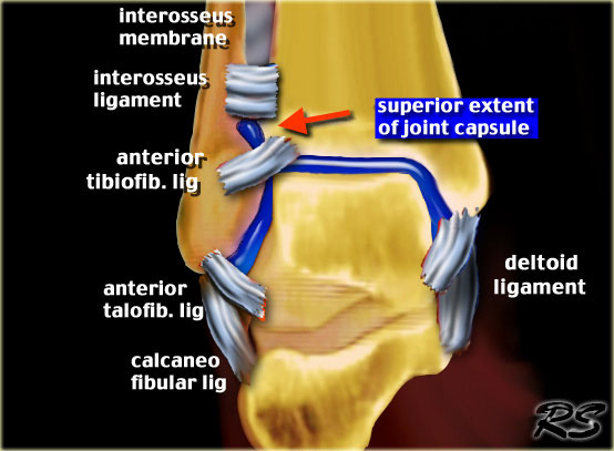
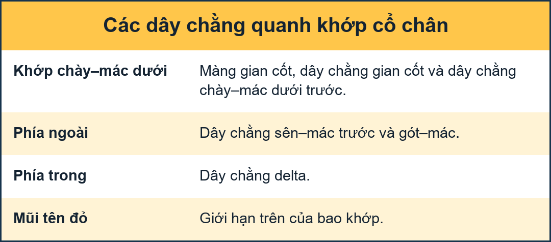
Normal flexibility of the ankleTính linh hoạt bình thường của cổ chân
The ankle joint has to be flexible in order to deal with the enormous forces applied exerted on the talus within the ankle fork. .
The medial side of the joint is quite rigid because the medial malleolus - unlike the lateral malleolus - is attached to the tibia and the medial collateral ligaments are very strong.Khớp cổ chân phải linh hoạt để đối phó với các lực lượng khổng lồ tác động lên xương sên trong khớp cổ chân.
Mặt trong của khớp khá cứng vì mắt cá trong - không giống như mắt cá ngoài - được gắn vào xương chày và dây chằng bên trong rất chắc.
On the lateral side there is a flexible support by the fibula, syndesmosis and lateral collateral ligaments.
This lateral complex allows the talus to move laterally and dorsally in exorotation during forward motion and subsequently pushes it back into its normal position.
The fibula has no weight-bearing function, but merely serves as a flexible lateral support.
The syndesmosis is the fibrous connection between the fibula and tibia formed by the anterior and posterior tibiofibular ligaments - located at the level of the tibial plafond (French for ceiling) - and the interosseus ligament, which is the thickened lower portion of the interosseus membrane.
The anterior and posterior tibiofibular ligaments are often referred to as anterior and posterior syndesmosis.Ở bên ngoài có sự hỗ trợ linh hoạt của xương mác, khớp sợi chày–mác và dây chằng bên ngoài.
Phức hợp bên này cho phép xương sên di chuyển sang bên và ra phía sau trong quá trình thóat vị trong quá trình chuyển động về phía trước và sau đó đẩy nó trở lại vị trí bình thường.
Xương mác không có chức năng chịu lực, nhưng chỉ đơn thuần là một sự hỗ trợ bên linh hoạt.
khớp sợi chày–mác là kết nối sợi giữa xương mác và xương chày được hình thành bởi dây chằng chày mác trước và sau - nằm ngang mức trần xương chày (tiếng Pháp nghĩa là trần) - và dây chằng gian cốt, là phần dưới dày của màng gian cốt.
Dây chằng chày mác trước và sau thường được gọi là khớp sợi chày–mác trước và sau.
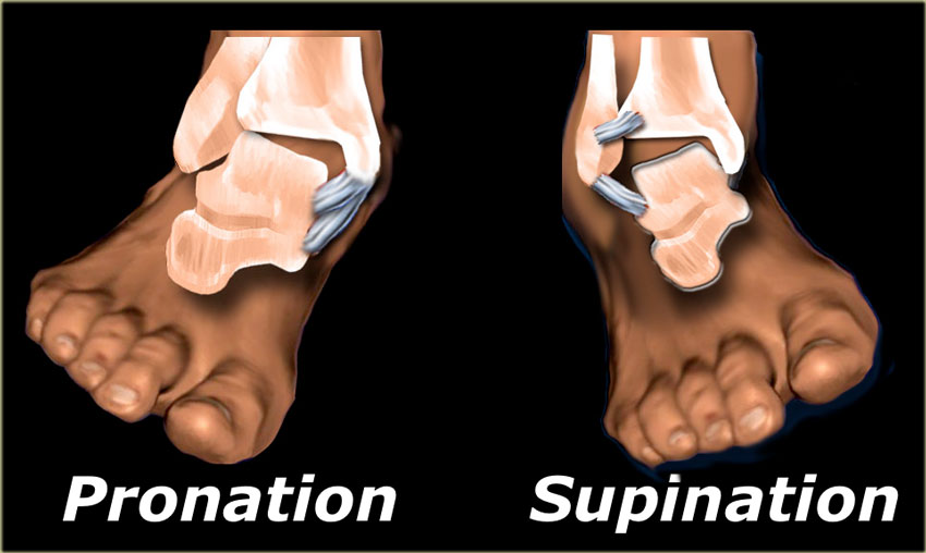
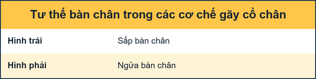
Vulnerable positions of the footVị trí dễ bị tổn thương của bàn chân
There are two positions of the foot in which the flexible ankle joint becomes a rigid and vulnerable system: extreme supination and pronation.
In these positions forces applied to the talus within the ankle mortise can result in fractures of the malleoli and rupture of the ligaments.
Có hai vị trí của bàn chân trong đó khớp cổ chân linh hoạt trở thành một hệ thống cứng và dễ bị tổn thương: ngửa và sấp.
Ở những vị trí này, lực tác động lên xương sên trong lỗ mộng cổ chân có thể dẫn đến gãy xương sên và đứt dây chằng.
In 80% of ankle fractures the foot is in supination.
The injury starts on the lateral side, since that is where the maximum tension is. Trong 80% gãy cổ chân, bàn chân nằm ngửa.
Chấn thương bắt đầu ở bên hông, vì đó là nơi căng tối đa.
In 20% of fractures the foot is in pronation with maximum tension on the medial side.
The injury starts on the medial side with either a rupture of the medial collateral ligaments or an avulsion of the medial malleolus.
Trong 20% các trường hợp gãy xương, bàn chân sấp với lực căng tối đa ở phía trong.
Tổn thương bắt đầu ở phía trong với một trong hai là đứt dây chằng bên trong hoặc bong mắt cá trong.
Pull-off or Push-off fracturesGãy kéo hoặc gãy đẩy
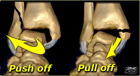
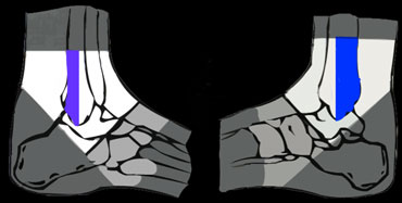
The shape of a fracture indicates which forces were involved.
An oblique or vertically oriented fracture indicates 'push-off'.
A transverse or horizontal fracture is the result of a 'pull-off' or avulsion.
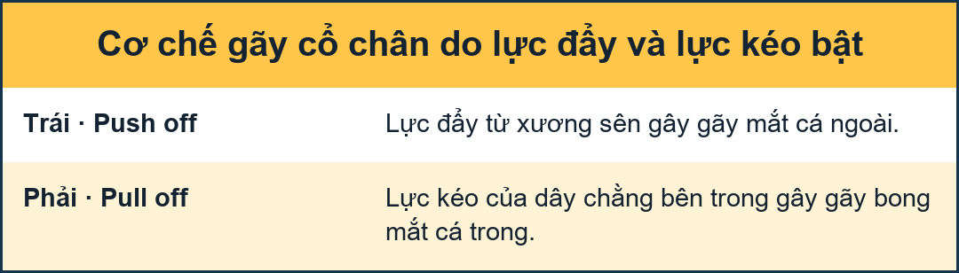
On the left image the lateral malleolus is pushed off by exorotation of the talus.
On the right image the medial malleolus is pulled off by the medial collateral ligament due to pronation of the foot. Hình dạng của một vết gãy cho biết lực nào có liên quan. Một vết gãy chéo hoặc theo chiều dọc cho thấy 'đẩy ra'.
Gãy ngang hoặc gãy ngang là kết quả của một 'kéo' hoặc nhổ.
Trên hình ảnh bên trái, mắt cá ngoài bị đẩy ra bởi sự thóat ra của xương sên.
Trên hình ảnh bên phải, mắt cá trong bị kéo ra bởi dây chằng bên trong do bàn chân sấp.
 Ring of stability in the coronal planeVòng ổn định trong mặt phẳng đứng ngang
StabilityTính ổn định
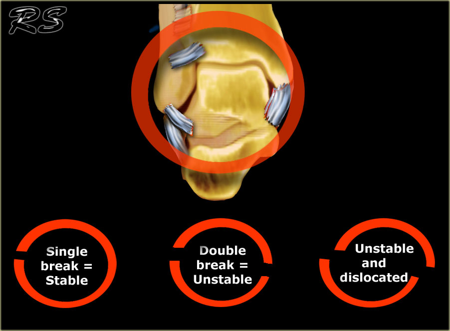
The ankle can be thought of as a ring in which bones as well as ligaments play an equally important role in the maintenance of joint stability.
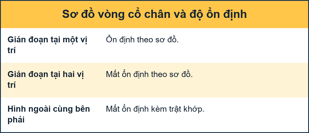
If the ring is broken in one place the ring remains stable.
When it is broken in two places, the ring is unstable and may dislocate.
Now anyone can figure out, that an ankle is unstable when both the medial and the lateral malleoli are fractured.
It becomes more problematic when there is a combination of a fracture and a ligamentous rupture, because the ligamentous rupture is not detectable on the X-ray.
In some fractures there may even be a proximal fibular fracture - which is not visible on the ankle radiographs - in combination with ligamentous ruptures at the level of the ankle.
It is important to realize that in these cases the radiographs of the ankle may be normal, while there still is an unstable ankle injury.Mắt cá chân có thể được coi là một vòng trong đó xương cũng như dây chằng đóng một vai trò quan trọng không kém trong việc duy trì sự ổn định của khớp.
Nếu vòng bị vỡ ở một nơi, vòng vẫn ổn định.
Khi nó bị gãy ở hai nơi, vòng không ổn định và có thể trật khớp.
Giờ thì ai cũng có thể nhận ra rằng, cổ chân bị mất vững khi cả hai mắt cá trong và ngoài bị gãy.
Nó trở nên rắc rối hơn khi có sự kết hợp của gãy xương và đứt dây chằng, bởi vì không thể phát hiện được đứt dải dây chằng trên X-quang.
Trong một số trường hợp gãy xương thậm chí có thể có gãy đầu gần xương mác - không nhìn thấy được trên phim X quang cổ chân - kết hợp với vỡ dây chằng ở mức cổ chân.
Điều quan trọng cần nhận ra là trong những trường hợp này, X quang cổ chân có thể bình thường, trong khi vẫn có tổn thương cổ chân không ổn định.
 Ring of stability in the axial planeVòng ổn định trong mặt phẳng ngang
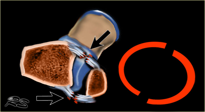
There is also an ring of stability in the axial plane.
When the anterior and posterior syndesmosis rupture or avulse, then the ankle joint is also unstable.
Ngoài ra còn có một vòng ổn định trong mặt phẳng ngang.
Khi khớp sợi chày–mác trước và sau bị đứt hoặc bong thì khớp cổ chân cũng không ổn định.
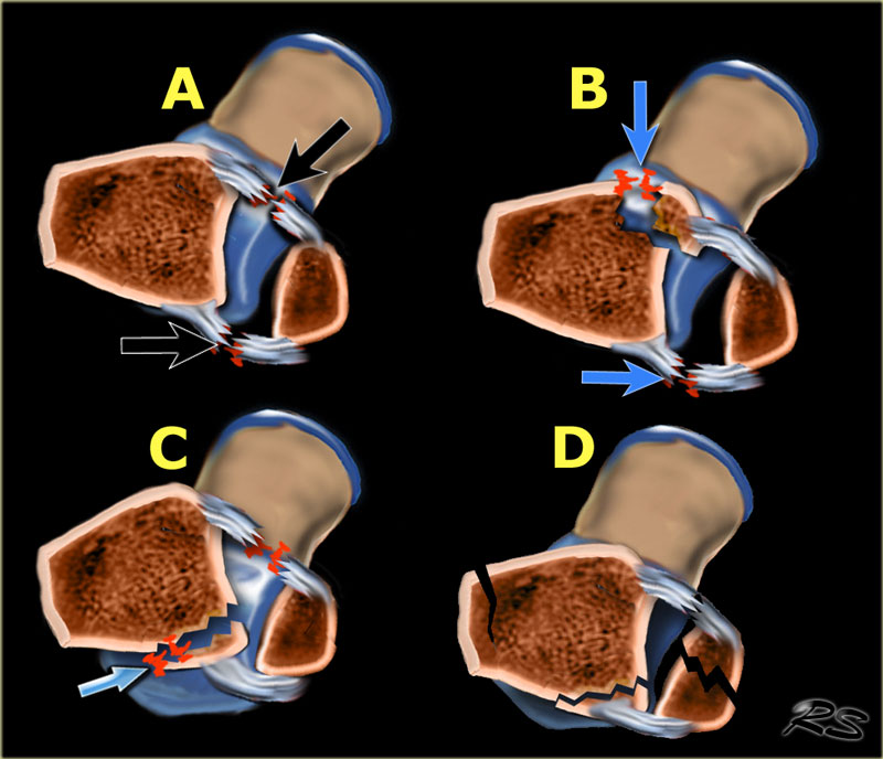
There are many combinations of avulsion fractures and ligamentous ruptures that can produce an unstable ring in the axial plane.Có nhiều kiểu kết hợp của gãy bong gân và đứt dây chằng có thể tạo ra một vòng không ổn định trong mặt phẳng ngang.
A
Anteriorly the anterior syndesmosis is usually one of the first structures to rupture. When the posterior syndesmosis also ruptures, then the ankle is unstable.
B
Less commonly the anterior syndesmosis avulses from the tibial attachment - Tillaux fracture.
C
On the posterior side frequently the posterior malleolus avulses. Sometimes these fractures are difficult to detect, as we will discuss in a moment.
D
After the injury the bones frequently align again, which makes it difficult to detect.A
Trước phần trước khớp chày–mác dưới thường là một trong những cấu trúc đầu tiên bị vỡ, khi khớp sợi chày–mác sau cũng bị vỡ thì cổ chân không ổn định.
B
Ít gặp hơn là gãy phần trước khớp chày–mác dưới bám chày - gãy Tillaux.
C
Ở phía sau thường là các ổ gãy mắt cá sau Đôi khi những gãy xương này rất khó phát hiện, vì chúng ta sẽ thảo luận trong một thời điểm.
D
Sau chấn thương xương thường xuyên thẳng hàng trở lại, điều này gây khó khăn cho việc phát hiện.
Stability (2)
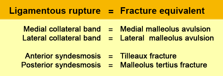
It is important to realize, that for the stability of the ankle it doesn't matter whether there is a rupture of a ligament or an avulsion at the insertion.
Almost every ligamentous rupture has a fracture equivalent.Tính ổn định (2)
Điều quan trọng cần nhận ra là, đối với sự ổn định của cổ chân, không quan trọng là có vỡ dây chằng hay bong gân ở vị trí chọc kim hay không.
Hầu hết các trường hợp đứt dây chằng đều có mức độ gãy tương đương.
Stability (3)
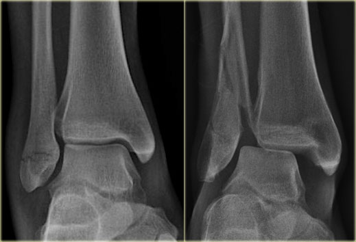
On the left image a Weber A or SA-fracture.
This ankle is stable because there is only an avulsion fracture of the lateral malleolus below the level of the syndesmosis.
The ring is broken in only one place.
On the right image there is an unstable fracture.
The ring of the ankle is broken in two places.
There is a lateral fracture and on the medial side there is a rupture of the collateral ligament allowing the talus to dislocate laterally.Tính ổn định (3)
Bên trái ảnh gãy Weber A hoặc gãy xương SA.
Mắt cá chân này ổn định vì chỉ có một gãy bong điểm bám của mắt cá ngoài dưới mức khớp sợi chày–mác.
Vòng chỉ bị gãy ở một vị trí.
Trên hình ảnh bên phải có một vết gãy không vững.
Vòng cổ chân bị gãy ở hai nơi.
Có một vết gãy bên và ở phía trong có một vết rách dây chằng bên cho phép xương sên trật ra bên.
Stability (4)
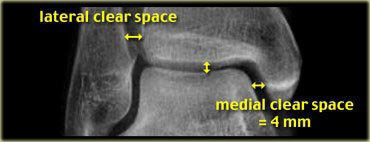
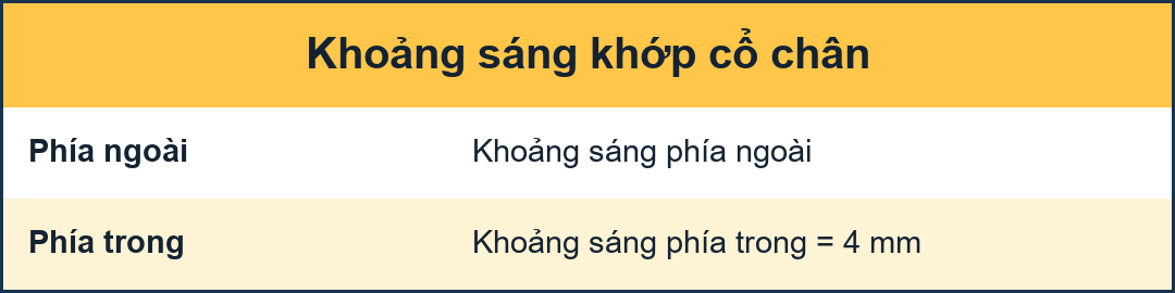
The medial clear space should not exceed 4 mm and is usually equal to the distance between the tibial plafond and the talus.
Widening of the medial joint space up to 6 mm or more requires disruption of the medial collateral ligament. Tính ổn định (4)
Khoảng trống trong không được vượt quá 4 mm và thường bằng khoảng cách giữa tấm che chày và xương sên.
Việc mở rộng khoảng trống khớp trong lên đến 6 mm trở lên đòi hỏi phải đứt dây chằng bên trong.
Stability (5)
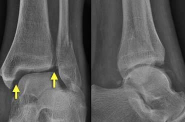
The lateral clear space is measured from the medial border of the fibula to the lateral border of the posterior tibia 1cm above the tibial plafond.
It is less well defined because its width varies with positioning.
Evident widening of the lateral clear space indicates syndesmotic rupture.
Some state that a width of 5.5 mm is abnormal.
It is very important to realize that a normal lateral or medial clear space does not exclude ligamentous rupture.
It simply means that there is no dislocation, but there can still be instability.
The case on the left shows a Weber B fracture.
On these images the ankle fork is normal.
Both the medial and lateral clear spaces are prominent, but within normal limits.
We can conclude that there is no dislocation, but we do not know if there is rupture of the medial collateral ligaments or of the syndesmosis.
Continue with the images post surgery.Tính ổn định (5)
Khoảng trống bên được đo từ bờ trong xương mác đến bờ ngoài xương chày sau 1 cm trên mỏm chày.
Nó ít được xác định rõ ràng hơn vì chiều rộng của nó thay đổi theo vị trí.
Dấu hiệu mở rộng khoảng trống bên cho thấy có đứt dây chằng dính khớp.
Một số trường hợp cho rằng chiều rộng 5,5 mm là bất thường.
Điều rất quan trọng là phải nhận ra rằng một khoảng trống trong bình thường ở bên hoặc trong không loại trừ đứt dây chằng.
Nó đơn giản có nghĩa là không có trật khớp, nhưng vẫn có thể có sự mất vững.
Trường hợp bên trái cho thấy gãy Weber B.
Trên những hình ảnh này, ngã ba cổ chân là bình thường.
Cả hai khoảng trống trong và ngoài đều nổi bật, nhưng trong giới hạn bình thường.
Chúng tôi có thể kết luận rằng không có trật khớp, nhưng chúng tôi không biết liệu có vỡ dây chằng bên trong hay không hoặc là vỡ dây chèn khớp sợi chày–mác.
Tiếp tục với hình ảnh sau phẫu thuật.
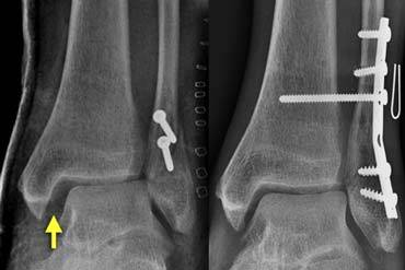
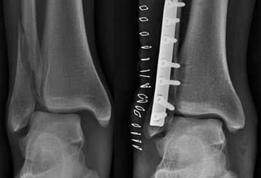
Following osteosynthesis there is obvious widening of the medial and lateral clear spaces (image on the far left).
This indicates that there is a syndesmotic rupture and medial collateral ligament rupture.
The ring is still broken in two places.
The ankle joint is unstable and dislocated.
Resurgery was necessary with placement of a syndesmotic screw to stabilize the ankle joint.Sau quá trình tạo xương, có sự mở rộng rõ ràng các khoảng trống trong (bên trong) và bên ngoài (bên trái).
Điều này cho thấy có một đứt dây chằng dính khớp và đứt một dây chằng bên trong.
Vòng nhẫn vẫn bị vỡ ở hai nơi.
Khớp cổ chân không vững và trật khớp.
Phẫu thuật là cần thiết với việc đặt vít dính để cố định khớp cổ chân.
Stability (6)
On the left another case. There is a Weber B fracture.
Both the medial and lateral clear spaces are widened, indicating instability.
The talus is displaced laterally.
Patient was scheduled for osteosynthesis of the fibular fracture and placement of a syndesmotic screw if necessary.
After osteosynthesis of the fibula, the ankle was tested in the operating room and found to be stable.
There was no indication for a syndesmotic screw.
It was concluded that the syndesmosis was only partially ruptured, as is usually the case in Weber B fractures.
The ring was broken in two places and after repairing one of them, the ring was stable.
Tính ổn định (6)
Bên trái một trường hợp khác, gãy Weber B.
Cả hai khoảng trống trong và ngoài đều được mở rộng, cho thấy sự mất ổn định.
Mái taluy bị dịch chuyển sang bên.
Bệnh nhân được chỉ định phẫu thuật kết hợp xương gãy xương mác và đặt vít dính khớp nếu cần thiết.
Sau khi nắn xương mác, mắt cá chân được kiểm tra trong phòng mổ và thấy ổn định.
Không có chỉ định dùng vít dính.
Kết luận: khớp sợi chày–mác chỉ vỡ một phần, thường gặp trong gãy Weber B.
Vòng bị vỡ ở hai vị trí và sau khi sửa chữa một trong hai vị trí này, vòng đã ổn định.
Ottawa Ankle RulesQuy tắc Ottawa Ankle
These rules are used to determine the need for radiographs in patients with an ankle injury.
Ankle X-ray series are only required in case of:
Pain in the malleolar zone and any one of the following:Những quy tắc này được sử dụng để xác định nhu cầu chụp X-quang ở bệnh nhân bị chấn thương mắt cá chân.
X-quang cổ chân chỉ được yêu cầu trong trường hợp:
Đau ở vùng mắt cá và bất kỳ một trong những điều sau đây:
- Bone tenderness along the distal 6 cm of the posterior edge of the fibula or tip of the lateral malleolus.Đau xương dọc theo đầu xa 6 cm của bờ sau xương mác hoặc đầu mắt cá ngoài.
- Bone tenderness along the distal 6 cm of the posterior edge of the tibia or tip of the medial malleolus.Đau xương dọc theo đầu xa 6 cm của cạnh sau xương chày hoặc đầu mắt cá trong.
- Inability to bear weight for 4 steps both immediately and in the emergency department.Không thể chịu được trọng lượng cho 4 bước cả ngay lập tức và trong khoa cấp cứu.
RadiographyChụp X-quang
Mortise viewChế độ xem tử thi
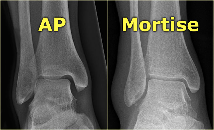
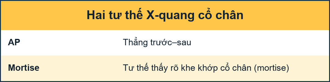
A basic radiographic examination of the injured ankle consists of an AP-view, a Mortise-view and a lateral view.
The Mortise-view is an AP-view taken with a 15-25? endorotation of the foot.
The technologist turns the foot inwards until the lateral malleolus is at the same height as the medial malleolus.
This view visualizes both the lateral and medial joint spaces.
On a true AP-view the talus overlaps a portion of the lateral malleolus, obscuring the lateral aspect of the ankle joint. Khám X quang cơ bản của cổ chân bị thương bao gồm góc nhìn AP, góc nhìn Mortise và góc nhìn bên.
Chế độ xem hình thái là chế độ xem AP được chụp với độ mở rộng 15-25? của bàn chân.
Kỹ thuật viên xoay bàn chân vào trong cho đến khi mắt cá bên ngang bằng với mắt cá trong.
Hình ảnh này cho thấy cả hai khoảng khớp bên và giữa.
Trên góc nhìn thực tế của khớp cổ chân, xương sên chồng lên một phần của mắt cá ngoài, che khuất mặt ngoài của khớp cổ tay.
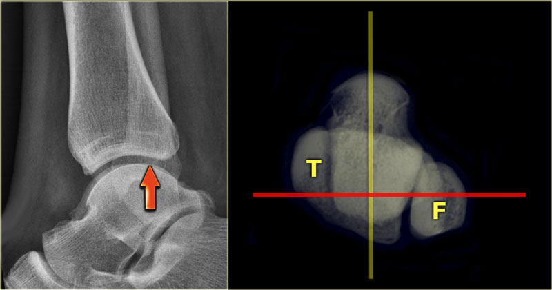 The distal fibula should project on the posterior part of the distal tibiaĐầu xa xương mác phải nằm ở phía sau của đầu xa xương chày.
Lateral viewMặt bên
Many think that for a good lateral view the distal fibula should be in the center of the distal tibia.
However, since the fibula is positioned more dorsally, the fibula should project over the posterior part of the distal tibia (arrow).Nhiều người nghĩ rằng để nhìn bên tốt, xương mác đầu xa nên nằm ở trung tâm của đầu xa xương chày.
Tuy nhiên, vì xương mác được định vị về phía lưng nhiều hơn, xương mác nên nhô ra phía sau của đầu xa xương chày (mũi tên).
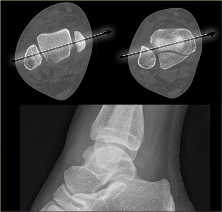 Malpositioning of the lateral viewĐịnh vị sai tư thế nhìn bên
Malpositioning of the Lateral viewSự sai lệch của góc nhìn bên
Malpositioning of the lateral view is the most common mistake in radiography of the injured ankle.
Because the patient is in pain, the technologist is afraid to let the patient turn the ankle fully lateral.
This is one of the reasons why we miss so many fractures of the posterior malleolus.Sai vị trí của mặt bên là sai lầm phổ biến nhất trong chụp X-quang mắt cá chân bị thương.
Vì bệnh nhân đang đau, kỹ thuật viên sợ để bệnh nhân xoay cổ chân hoàn toàn sang bên.
Đây là một trong những lý do tại sao chúng ta bỏ sót rất nhiều vết gãy của mắt cá sau.
The CT demonstrates a large tertius fracture.
On the lateral view and also on the AP- and Mortise views, which will be shown in the paragraph on tertius fractures, this fracture was not visible.
The explanation is that on the lateral radiograph the fibula projects in the middle of the tibia.
The x-ray beam is not parallel to the fracture line.
Since the fracture line of a tertius fracture always has this orientation, we must insist on a true lateral view.CT cho thấy gãy xương lớn.
Trên mặt nghiêng và cả mặt cắt AP- và Mortise, sẽ được trình bày trong đoạn về gãy xương tertius, không thấy gãy xương này.
Giải thích là trên X quang bên xương mác có lồi xương mác ở giữa xương chày.
Chùm tia X không song song với đường gãy.
Vì đường gãy của gãy xương bánh chè luôn có hướng này, chúng ta phải nhấn mạnh vào một góc nhìn bên thực sự.
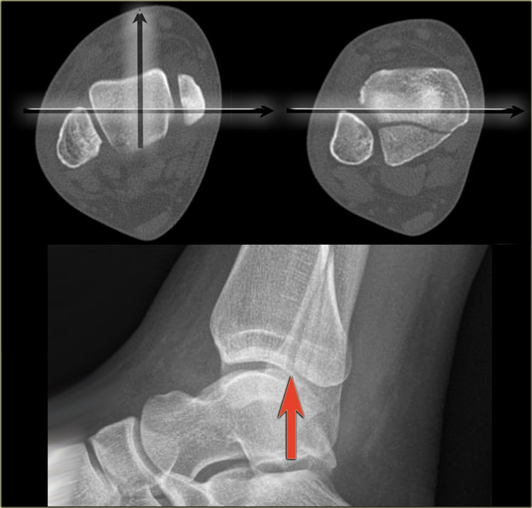 Good positioning of the lateral view - Tertius fractureĐịnh vị tốt mặt ngoài xương gãy Tertius
On a well positioned lateral view the tertius fracture is obvious (red arrow).
This was the only fracture that was seen on the x-rays of the ankle and this patient turned out to have an unstable Weber-C fracture and went for surgery.Trên phim nghiêng, gãy xương bánh chè rõ ràng (mũi tên màu đỏ).
Đây là trường hợp gãy duy nhất được phát hiện trên phim X-quang cổ chân và bệnh nhân này hóa ra bị gãy Weber-C không vững và phải phẫu thuật.
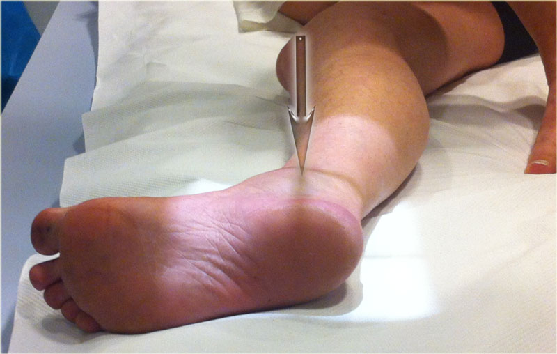
The x-ray beam has to be centered on the malleoli.
Notice the exorotation of the foot for a proper lateral view.Chùm tia X phải tập trung vào các mảnh sụn.
Chú ý sự thóat hơi của bàn chân để có góc nhìn bên thích hợp.
Videos of severe ankle injuriesVideo về chấn thương cổ chân nặng
The forces in ankle injuries can be enormous.
As was discussed in Ankle fractures 1 the injuries usually take place in a logical sequence.
Lực tác động lên chấn thương mắt cá chân có thể rất lớn.
Như đã thảo luận trong Gãy xương cổ chân 1 các chấn thương thường diễn ra theo một trình tự hợp lý.
Ankle - Fractures 1 Weber and Lauge-Hansen classificationMắt cá chân - Gãy xương 1 Phân loại của Weber và Lauge-Hansen
Start the video on the left by clicking on the image.
Notice that at first the foot is in supination with maximal forces on the lateral side.
Subsequently the foot adducts.
The result is an SA or Weber A fracture.
We can assume that this is a Lauge Hansen Supination Adduction injury stage 2.Bắt đầu video bên trái bằng cách nhấp vào hình ảnh.
Lưu ý rằng lúc đầu, bàn chân nằm ngửa với lực tối đa ở phía bên.
Sau đó là các chất phụ trợ ở chân.
Kết quả là gãy xương kiểu SA hoặc Weber A.
Chúng ta có thể giả định đây là giai đoạn chấn thương Lauge Hansen Supination Adduction 2.
Start the video on the left by clicking on the image.
Notice that at first the foot is in pronation, with maximum forces on the medial side.
Subsequently the foot exorotates.
The result is a PER - pronation exorotation injury or Weber C fracture.Bắt đầu video bên trái bằng cách nhấp vào hình ảnh.
Lưu ý rằng lúc đầu, bàn chân sấp, với lực tác động tối đa ở phía trong.
Sau đó bàn chân sẽ tiết mồ hôi.
Kết quả là chấn thương sấp ngửa theo chiều PER hoặc gãy Weber C.
Charity
Hoạt động từ thiện
All the profits of the Radiology Assistant go to Medical Action Myanmar which is run by Dr. Nini Tun and Dr. Frank Smithuis sr, who is a professor at Oxford university and happens to be the brother of Robin Smithuis.Toàn bộ lợi nhuận của Radiology Assistant được chuyển cho Medical Action Myanmar, do bác sĩ Nini Tun và bác sĩ Frank Smithuis Sr. điều hành. Frank Smithuis Sr. là giáo sư tại Đại học Oxford và là anh em ruột của Robin Smithuis.
Click here or on the image below to watch the video of Medical Action Myanmar and if you like the Radiology Assistant, please support Medical Action Myanmar with a small gift.Nhấp vào đây hoặc vào hình ảnh bên dưới để xem video về Medical Action Myanmar và nếu bạn thích Trợ lý X quang, vui lòng hỗ trợ Medical Action Myanmar bằng một món quà nhỏ.
ReferencesTài liệu tham khảo
-
Fractures of the ankle, combined experimental-surgical and experimental-roentgenologic investigationsby N. Lauge-Hansen (1948) Gãy xương cổ chân, kết hợp thực nghiệm-phẫu thuật và thực nghiệm-tổn thương họcbởi N. Lauge-Hansen (1948)
-
Die verletzungen des oberen sprunggelenkesby B.G. Weber (1966)
- Website of the Ottawa Ankle rules Website của Ottawa Ankle rules
-
Ankle fractures
Gãy cổ chân
East Lancashire Foot and Ankle Hyperbook East Lancashire Foot and Ankle Hyperbook (bằng tiếng Anh)
-
Free AO Surgery Reference
Phẫu thuật AO miễn phí
The AO Surgery Reference is a huge online repository of surgical knowledge, consisting of more than 7000 pages. AO Surgery Reference là một kho lưu trữ trực tuyến khổng lồ về kiến thức phẫu thuật, bao gồm hơn 7000 trang.