
Meniscal pathologyBệnh lý sụn chêm
Radiology department of the Washington University School of Medicine, St. Louis, USA and the Rijnland hospital in Leiderdorp, the NetherlandsKhoa X quang của Trường Đại học Y Washington, St. Louis, Hoa Kỳ và Bệnh viện Rijnland ở Leiderdorp, Hà Lan
Ngày đăng / cập nhật: 2005-07-20
Bản gốc trên radiologyassistant.nl ↗ · tải về lúc 2026-11-19 20:19 · bản dịch máy có chuẩn hóa thuật ngữ chẩn đoán hình ảnh
This article is based on a presentation by David Rubin and adapted for the Radiology Assistant by Robin Smithuis.
Interactive cases are presented in the menu bar to test your knowledge.Bài viết này dựa trên bài thuyết trình của David Rubin và được Robin Smithuis điều chỉnh cho Trợ lý X quang.
Các trường hợp tương tác được trình bày trong thanh menu để kiểm tra kiến thức của bạn.
by David Rubin and Robin SmithuisDavid Rubin và Robin Smithuis
Normal Meniscal AnatomyGiải phẫu sụn chêm bình thường
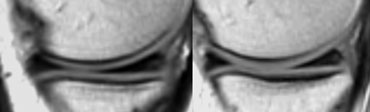 Medial meniscus: The posterior horn is always larger than the anterior horn.Sừng trong sụn chêm: Sừng sau luôn lớn hơn sừng trước.
Medial meniscusTrung gian sụn chêm
Both horns are triangular in shape and have very sharp points.
The posterior horn is always larger than the anterior horn (figure).
If this is not the case, then the shape is abnormal, which can be a sign of a meniscal tear or a partial meniscectomy.Cả hai sừng đều có hình tam giác và có các điểm rất sắc.
Sừng sau luôn lớn hơn sừng trước (hình).
Nếu không phải vậy, thì hình dạng là bất thường, có thể là dấu hiệu của rách sụn chêm hoặc cắt bỏ một phần sụn.
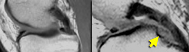 LEFT: normal medial meniscal root immediately anterior to the posterior cruciate ligament. RIGHT: missing posterior root due to meniscal root tear.Trái: Rễ Meniscal trung gian bình thường ngay trước dây chằng chéo sau. Phải: Mất gốc sau do rách chân sụn chêm.
The posterior root is immediately anterior to the posterior cruciate ligament.
If it is missing on the sagittal images, then there is a meniscal root tear (figure).
The anterior horn has an insertion on the tibia and a second portion that travels from medial to lateral to connect to the anterior horn of the lateral meniscus (intermeniscal or transverse ligament).Gốc sau ngay trước dây chằng chéo sau.
Nếu nó bị thiếu trên ảnh mặt phẳng đứng dọc, thì có một rách chân sụn chêm (hình).
Sừng trước có một phần chèn vào xương chày và phần thứ hai đi từ trong ra ngoài để kết nối với sừng trước của sụn chêm bên (dây chằng gian xương hoặc dây chằng ngang).
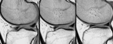 Lateral meniscus. Both horns are about the same size.Sừng bên sụn chêm, cả hai sừng đều có kích thước tương đương nhau.
Lateral meniscusBên sụn chêm
On sagittal images the posterior horn is higher in position than the anterior horn.
Both horns are about the same size.Trên ảnh mặt phẳng đứng dọc sừng sau cao hơn sừng trước.
Cả hai sừng đều có kích thước tương đương nhau.
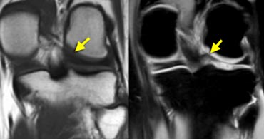 Lateral meniscus: posterior horn and posterior meniscal root.Sụn chêm: sừng sau và rễ sụn chêm sau.
The lateral meniscus posteriorly comes up high over the tibial spine to insert near the posterior cruciate ligament.
This upward position of the posterior horn may be the reason for the higher signal intensity of the posterior horn in all planes due to magic angle effect.Sụn chêm bên phía sau đi lên cao trên xương chày để chèn gần dây chằng chéo sau.
Vị trí sừng sau hướng lên trên này có thể là lý do cho cường độ tín hiệu của sừng sau cao hơn trong tất cả các mặt phẳng do hiệu ứng góc ma thuật.
Meniscal tearsRách sụn chêm
Criteria for tearsTiêu chuẩn nước mắt
The two most important criteria for meniscal tears are an abnormal shape of the meniscus and high signal intensity unequivocally contacting the surface on PD images.Hai tiêu chuẩn quan trọng nhất để chẩn đoán rách sụn chêm là hình dạng bất thường của sụn chêm và cường độ tín hiệu cao tiếp xúc rõ ràng với bề mặt trên ảnh PD.
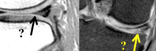 High signal intensity not unequivocally contacting surface. Small black line on inferior margin of the meniscus. At arthroscopy the meniscus was normal.Cường độ tín hiệu cao không tiếp xúc rõ với bề mặt, đường nhỏ màu đen trên bờ dưới của sụn chêm, khi soi khớp sụn chêm bình thường.
It is a misunderstanding that menisci should be homogeneously low in signal intensity on proton-density images.
The meniscus does not have to be black.
Only when the high signal unequivocally reaches the surface of the meniscus you can make the diagnosis of a tear.
If there is doubt whether the high signal touches the surface, look at all the adjacent images.
If there is still doubt, then do not diagnose a tear.
If you have a questionmark in your head, say "meniscus is normal". (figure)Có sự hiểu lầm rằng các sụn chêm phải thấp đồng nhất trong cường độ tín hiệu trên hình ảnh mật độ proton.
sụn chêm không nhất thiết phải có màu đen.
Chỉ khi tín hiệu cao rõ ràng chạm đến bề mặt của sụn chêm, bạn mới có thể chẩn đoán được rách.
Nếu nghi ngờ liệu tín hiệu cao có chạm vào bề mặt hay không, hãy nhìn vào tất cả các hình ảnh liền kề.
Nếu vẫn còn nghi ngờ, thì không chẩn đoán chảy nước mắt.
Nếu bạn có một dấu hỏi trong đầu, hãy nói “sụn chêm là bình thường ”. (hình)
 Basic shapes: Longitudinal, Horizontal and Radial.Hình dạng cơ bản: Dọc, ngang và quay.
Nomenclature of Meniscal TearsDanh mục rách sụn chêm
Shapes. There are 3 basic shapes of meniscal tears: longitudinal, horizontal and radial.
Complex tears are a combination of these basic shapes.Hình dạng Có 3 hình dạng cơ bản của rách sụn chêm: theo chiều dọc, ngang và quay.
Rách phức tạp là sự kết hợp của những hình dạng cơ bản này.
 Bucket handle, Horizontal Flap tear and Parrot beak.Xô cầm tay, vạt ngang rách và mỏ vẹt.
Displaced Tears
Bucket-handle tear = displaced longitudinal tear.
Flap tear = displaced horizontal tear.
Parrot beak = displaced radial tear. Nước mắt di lệch
Rách tay cầm xô = Rách dọc di lệch.
Vạt da bị rách = rách ngang di lệch.
Mỏ vẹt = rách xuyên tâm di lệch.
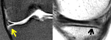
Longitudinal, horizontal and radial tearsRách dọc, ngang và xuyên
Longitudinal tears
Longitudinal tears parallel the long axis of the meniscus dividing the meniscus into an inner and outer part.
Therefore, the distance between the tear and the outer margin of the meniscus is always the same (figure).
The tear never touches the inner margin.Rách dọc
Các đường xé dọc song song với trục dài của sụn chêm chia sụn chêm thành phần trong và phần ngoài.
Do đó, khoảng cách giữa đường rách và bờ ngoài của sụn chêm luôn giống nhau (hình).
Vết rách không bao giờ chạm vào rìa trong.
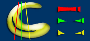 Three sagittal images of a longitudinal tearBa ảnh mặt phẳng đứng dọc của một vết rách dọc
Longitudinal tears follow the collagen bundles that parallel the contour of the meniscus.
If a longitudinal tear has other components (horizontal or radial), then it is a complex tear violating the collagen bundles.
This requires a higher energy trauma.Các vết rách dọc theo các bó collagen song song với đường viền của sụn chêm.
Nếu một vết rách dọc có các thành phần khác (ngang hoặc xuyên tâm), thì đó là một vết rág phức tạp vi phạm các bó collagen.
Điều này đòi hỏi một chấn thương năng lượng cao hơn.
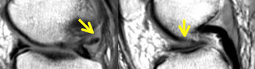 LEFT: abnormal shape of posterior horn. A piece is missing. RIGHT: displaced fragment in the intercondylar fossa.Trái: hình dạng bất thường của sừng sau. một mảnh bị thiếu. Bên phải: Mảnh vỡ bị dịch chuyển trong Fossa Intercondylar.
Longitudinal tear (2)
Bucket handle tear
is a displaced longitudinal tear.Rách dọc (2)
Xô xử lý nước mắt
là rách dọc di lệch.
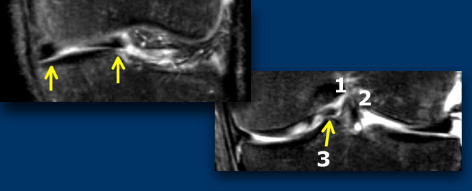 LEFT: meniscus is abnormal in shape and there is a displaced fragment. RIGHT: Three structures in the intercondylar fossa: post cruciate lig (1), ant cruciate lig (2) and displaced fragment (3).Bên trái: sụn chêm có hình dạng bất thường và có một mảnh dịch chuyển. Bên phải: Ba cấu trúc trong Fossa Intercondylar: Post Cruciate Lig (1), Ant Cruciate Lig (2) và mảnh vỡ (3).
On coronal images bucket handle tears are easier to recognize.
Normally there are only two structures in the intercondylar fossa: the anterior and posterior cruciate ligament.
Any other structure in the intercondylar fossa is abnormal and a displaced meniscal fragment is the most likely possibility.Trên ảnh mặt phẳng đứng ngang, nước mắt tay cầm dễ nhận biết hơn.
Thông thường chỉ có hai cấu trúc trong hố liên lồi cầu: trước và dây chằng chéo sau.
Bất kỳ cấu trúc nào khác trong hố liên lồi cầu là bất thường và một mảnh sụn chêm di lệch là khả năng có thể xảy ra nhất.
More on Bucket handle tearsTìm hiểu thêm về rách kiểu quai xô
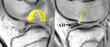
 Flipped meniscus: posterior horn is missing because it is flipped over and located on top of the anterior horn.Lật sụn chêm: mất sừng sau do lật sừng và nằm trên đỉnh sừng trước.
Longitudinal tear (3)
Flipped meniscus is a form of bucket handle tear.
There is a capsular detachment or peripheral tear of the meniscus, usually the posterior horn.
The posterior horn flips over onto the anterior horn.Rách dọc (3)
Lật sụn chêm là một dạng rách tay cầm xô.
Có một bong bao hoặc rách ngoại vi của sụn chêm, thường là sừng sau.
Sừng sau lật lên sừng trước.
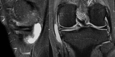 Horizontal tear with a meniscal cystRách sụn chêm với nang sụn
Horizontal tears
Horizontal tears divide the meniscus in a top and bottom part (pita bread).
If horizontal tears go all the way from the apex to the outer margin of the meniscus, they may result in the formation of a meniscal cyst.
The synovial fluid runs peripherally through the horizontal tear and accumulates within the meniscus and finally result in a cyst.
The connection with the joint space is often lost, so they will not fill with contrast on MR-arthrography.
The synovial fluid is absorbed and is replaced by a gelatinous substance. Rách ngang
Rách ngang chia sụn chêm thành phần trên và phần dưới (bánh mìpita).
Nếu các đường rách ngang đi từ đỉnh đến rìa ngoài của sụn chêm, chúng có thể dẫn đến sự hình thành nang sụn chêm.
Dịch khớp chạy xuyên qua đường rách ngang và tích tụ trong sụn chêm và cuối cùng dẫn đến nang.
Sự kết nối với không gian khớp thường bị mất, vì vậy chúng sẽ không được lấp đầy bằng thuốc cản quang trên MR-arthrography.
Dịch khớp được hấp thụ và được thay thế bằng một chất gelatin.
There are 3 criteria for the diagnosis of a meniscal cyst:Có 3 tiêu chuẩn chẩn đoán nang sụn chêm:
- Horizontal tear.Rách ngang.
- Fluid accumulation with bright signal on T2.Tích tụ dịch với tín hiệu sáng trên T 2.
- Flat lining against the periphery of the meniscus.Lớp lót phẳng chống lại ngoại vi của sụn chêm.
The diagnosis of a meniscal cyst is important to the surgeon because it takes one operation on the outside of the knee to remove the cyst and another operation on the inside for the meniscus.Chẩn đoán nang sụn chêm rất quan trọng đối với phẫu thuật viên vì phải thực hiện một phẫu thuật ở bên ngoài khớp gối để loại bỏ nang và một phẫu thuật khác ở bên trong cho sụn chêm.
Radial tears
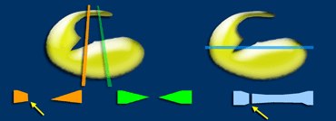
Radial tears are perpendicular to the long axis of the meniscus.
They violate the collagen bundles that parallel the long axis of the meniscus.
These are high energy tears. They start at the inner margin and go either partial or all the way through the meniscus dividing the meniscus into a front and a back piece.
Radial tears are difficult to recognize. You have to combine the findings on sagittal and coronal images to make the diagnosis.Rách xuyên tâm
Rách xuyên tâm vuông góc với trục dài của sụn chêm.
Chúng vi phạm các bó collagen song song với trục dài của sụn chêm.
Đây là những vết rách năng lượng cao. Chúng bắt đầu ở lề trong và đi một phần hoặc toàn bộ đường đi qua sụn chêm chia sụn chêm thành mặt trước và mặt sau.
Rách xuyên tâm rất khó nhận biết, cần phải kết hợp các dấu hiệu trên hình thái đứng dọc và ảnh mặt phẳng đứng ngang để chẩn đoán.
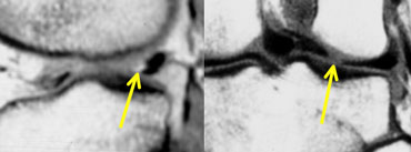 LEFT: triangle missing the tip.RIGHT: disrupted bow tie.Bên trái: Hình tam giác Thiếu đầu. Phải: Bẫy nơ bị gián đoạn.
The following combination of findings is diagnostic:
In one plane: triangle missing the tip and in the other plane: a disrupted bow tie.Sự kết hợp các dấu hiệu sau đây là chẩn đoán:
Trong một mặt phẳng: hình tam giác thiếu đầu và trong mặt phẳng khác: một nơ bị đứt.
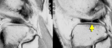 Disrupted bow tie indicating a small radial tear.Mắt bị đứt dây cung cho thấy một vết rách nhỏ xuyên tâm.
Small radial tears are difficult to diagnose.
Sometimes the only sign is a disrupted bow tie.Rách xuyên tâm nhỏ khó chẩn đoán.
Đôi khi dấu hiệu duy nhất là nơ mũi bị đứt.
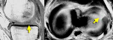 LEFT: Absent or empty meniscus on sagittal image.RIGHT: Axial image shows complete radial tear leading to a defect in the meniscus.Bên trái: Không có hoặc trống sụn chêm trên hình ảnh sagittal. Phải: Hình ảnh trục hiển thị hoàn toàn vết rách xuyên tâm dẫn đến khiếm khuyết trong sụn chêm.
If you image a complete radial tear directly along the length of the tear you will see an absent or empty meniscus.
These complete radial tears open up and give the impression that there is a part missing.
However you will not find a displaced meniscal fragment. It is simply separation of the meniscal parts.Nếu bạn chụp ảnh một vết rách hoàn toàn dọc theo chiều dài của vết rách, bạn sẽ thấy một sụn chêm không có hoặc trống rỗng.
Những vết rách xuyên tâm hoàn toàn này mở ra và tạo ấn tượng rằng có một phần bị thiếu.
Tuy nhiên, bạn sẽ không tìm thấy một mảnh sụn chêm di lệch. Nó chỉ đơn giản là sự tách biệt của các phần sụn.
More on empty meniscus signXem thêm về dấu hiệu hình liềm rỗng
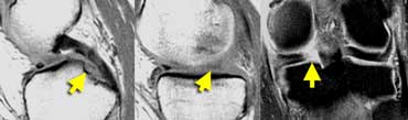 Meniscal root tear: on sagittal images there is an absent or empty meniscus-sign adjacent to the posterior cruciate ligament where the meniscal root should be. On coronal images a meniscal root tear is confirmed.Rách rễ sụn chêm: trên hình ảnh dọc có dấu hiệu sụn san hô không có hoặc trống rỗng liền kề với dây chằng chéo sau, nơi cần có rễ sọ. Trên hình ảnh vành, rách rễ sỏi được xác nhận.
Meniscal root tearRách chân sụn chêm
A meniscal root tear is a radial tear located at the meniscal root.
Normally when you image the posterior cruciate ligament on sagittal images you should see a considerable portion of the posterior horn of the meniscus on that image or the image adjacent to it.
If this is not the case it is an absent or empty meniscus-sign, indicating a radial tear.Rách chân sụn chêm là một vết rách xuyên tâm nằm ở gốc sụn chêm.
Thông thường khi bạn chụp ảnh dây chằng chéo sau trên ảnh mặt phẳng đứng dọc, bạn sẽ thấy một phần đáng kể của sừng sau của sụn chêm trên hình ảnh đó hoặc hình ảnh liền kề với nó.
Nếu không phải vậy thì đó là dấu hiệu sụn chêm vắng mặt hoặc trống rỗng, cho thấy rách xương quay.
Post-operative meniscusSau mổ sụn chêm
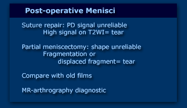
Post-operative menisci are harder to evaluate because the two most important criteria, i.e. abnormal signal and abnormal shape, do not apply.Các sụn chêm sau mổ khó đánh giá hơn vì hai tiêu chí quan trọng nhất là tín hiệu bất thường và hình dạng bất thường không được áp dụng.
Abnormal signal is no longer a reliable sign of a tear, because if there has been a suture repair, this will heal with scar tissue, which also has high signal on PD-images (figure).
Although an uncommon finding, if there is also high signal on T2-weighted images, then you can make the diagnosis of a tear, as this is the result of synovial fluid leaking into a meniscal tear.
This however is an uncommon finding.Tín hiệu bất thường không còn là dấu hiệu đáng tin cậy của rách, bởi vì nếu đã có khâu sửa chữa, vết thương này sẽ lành lại bằng mô sẹo, mô sẹo cũng có tín hiệu cao trên hình ảnh PD (hình).
Mặc dù một phát hiện không phổ biến, nhưng nếu có tín hiệu cao trên hình ảnh xung T2W, thì bạn có thể chẩn đoán rách, vì đây là kết quả của dịch khớp bị rò rỉ thành rách sụn chêm.
Tuy nhiên, đây là một phát hiện không phổ biến.
Abnormal shape can be the result of partial meniscectomy.
So you need to know what procedure was performed during arthroscopy.
Only when comparison is made with prior postoperative images, can you determine, if an abnormal shape is a new finding indicative of a new tear.Hình dạng bất thường có thể là kết quả của phẫu thuật cắt bỏ một phần nội soi.
Vì vậy, bạn cần biết thủ thuật nào đã được thực hiện trong quá trình nội soi khớp.
Chỉ khi so sánh với các hình ảnh sau phẫu thuật trước đó, bạn mới có thể xác định, nếu một hình dạng bất thường là một phát hiện mới cho thấy một vết rách mới.
Sometimes differentiation between normal post-op findings and a re-tear is not possible on conventional MR-images.
In these cases, MR-arthrography with 40cc diluted Gadolinium helps to make the distinction because even small amounts of Gadolinium that leak into a tear are readily visible on fat saturated T1 images.Đôi khi không thể phân biệt giữa các dấu hiệu sau phẫu thuật bình thường và rách tái phát trên hình ảnh MR thông thường.
Trong những trường hợp này, chụp cộng hưởng từ với Gadolinium pha loãng 40cc giúp phân biệt bởi vì ngay cả một lượng nhỏ Gadolinium rò rỉ vào nước mắt cũng có thể nhìn thấy trên hình ảnh T1 bão hoà chất béo.
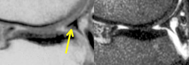 PD and T2W images. Prior partial meniscectomy and suture repair. At arthroscopy, there was no tear.Hình ảnh PD và T2W. Trước đó đã cắt nội soi và khâu lại. Trên nội soi không thấy có chảy nước mắt.
Post-operative Meniscus 1
The case on the left shows a meniscus with an abnormal shape aswell as abnormal signal touching the surface on PD but not on T2W-images.
This patient had a prior partial meniscectomy and a suture repair.
On the basis of these imaging findings, it is impossible to tell if this is a tear or a normal postoperative finding.
This patient had another operation for ACL reconstruction.
The surgeon looked at the meniscus and the meniscus was found to be normal i.e. no tear.Sau mổ sụn chêm 1
Trường hợp bên trái cho thấy một sụn chêm có hình dạng bất thường cũng như tín hiệu bất thường chạm vào bề mặt trên PD nhưng không chạm vào trên T2W-images.
Bệnh nhân này đã được phẫu thuật cắt nội soi một phần và khâu lại.
Trên cơ sở các dấu hiệu hình ảnh này, không thể nói được đây là một vết rách hay một dấu hiệu sau phẫu thuật bình thường.
Bệnh nhân này đã được phẫu thuật tái tạo dây chằng chéo trước.
Bác sĩ phẫu thuật nhìn vào sụn chêm và sụn chêm được phát hiện là bình thường tức là không có nước mắt.
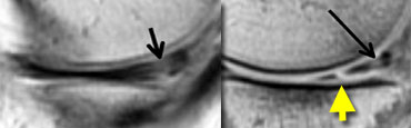 LEFT: Old MR exam with tear. Patient had a suture repair. RIGHT: On new exam, there is a new tear (yellow arrow). It is not possible to tell if the old tear has healed.Trái: MRI cũ cho thấy vết rách. Bệnh nhân đã được khâu phục hồi. Phải: MRI mới có vết rách mới (mũi tên vàng). Chưa thể xác định vết rách cũ đã lành hay chưa.
Post-operative Meniscus 2
This patient had a suture repair for meniscal tear.
There was a new injury.
On the new MR, it is impossible to determine if the old tear had healed.
However a new tear is seen, so this case is easy.Sau mổ sụn chêm 2
Bệnh nhân này được khâu rách sụn chêm.
Có một tổn thương mới.
Trên cộng hưởng từ mới, không thể xác định được liệu vết rách cũ đã lành hay chưa.
Tuy nhiên, có thể thấy một vết rách mới, vì vậy trường hợp này rất dễ.
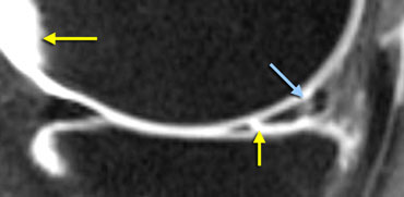 MR-arthrogram: In the new tear the signal is as bright as in the synovial fluid (yellow arrows). In the healed tear the signal is not as bright.MR-arthrogram: Ở vết rách mới, tín hiệu sáng như trong dịch khớp (các mũi tên màu vàng), ở vết rách lành, tín hiệu không sáng như vậy.
On an MR-arthrogram, there was very high signal intensity in the new tear comparable with the synovial fluid, but only moderate signal intensity at the healed old tear.
So comparison with the old films was diagnostic for the new tear, while the arthrogram showed that the old tear has healed.Trên MRI khớp có tiêm thuốc nội khớp, vết rách mới có tín hiệu rất cao, tương đương dịch khớp, còn vết rách cũ đã lành chỉ có tín hiệu trung bình. So sánh với ảnh cũ giúp xác định vết rách mới, còn MRI khớp cho thấy vết rách cũ đã lành.
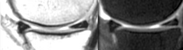 PD and MR-arthrogram after suture repair for meniscal tear: healed tear.Phẫu thuật khâu rách sụn chêm: lành thương sau khâu.
Post-operative Meniscus 3
This patient also had a suture repair for meniscal tear.
After a new injury, the PD-images show high signal unequivocally reaching the surface of the meniscus (seen on the original films, but not clearly seen on the compressed image on the left).
On this image, it is not possible to tell if the tear has healed.
So an MR-arthrogram was performed which showed that the tear has healed.Sau mổ sụn chêm 3
Bệnh nhân này cũng được khâu rách sụn chêm.
Sau một tổn thương mới, ảnh PD cho thấy tín hiệu cao rõ ràng chạm tới bề mặt của sụn chêm (xem trên phim gốc, nhưng không thấy rõ trên ảnh nén bên trái).
Trên hình ảnh này, không thể biết được liệu vết rách đã lành hay chưa.
Vì vậy, chụp cộng hưởng từ khớp đã được thực hiện cho thấy vết rách đã lành.
ReferencesTài liệu tham khảo
- Rubin DA: Magnetic resonance imaging of chondral and osteochondral injuries. Top Magn Reson Imaging 1998; 9:348-359 Rubin DA: Hình ảnh cộng hưởng từ của chấn thương chondral và u xương. Hình ảnh Reson Magn hàng đầu 1998; 9: 348-359
- Rubin DA, Kettering JM, Towers JD, Britton CA: MR imaging of knees having isolated and combined ligament injuries. AJR, 1998; 170:1207-1213 Rubin DA, Kettering JM, Towers JD, Britton CA: Hình ảnh MR của đầu gối bị chấn thương dây chằng bị cô lập và kết hợp. AJR, 1998; 170: 1207-1213