
Meniscus special casesCác trường hợp đặc biệt của sụn chêm
Radiology department of the Rijnland hospital in Leiderdorp, the NetherlandsKhoa Chẩn đoán hình ảnh của bệnh viện Rijnland, Leiderdorp, Hà Lan
Ngày đăng / cập nhật: 2010-04-13
Bản gốc trên radiologyassistant.nl ↗ · tải về lúc 2026-11-19 20:19 · bản dịch máy có chuẩn hóa thuật ngữ chẩn đoán hình ảnh
In this article we will show some examples of special meniscal pathology in more detail.
For the basics of meniscal pathology we advise you first to read the article 'Knee Meniscus - Part 1'.
On most images you can click to get an enlarged view, but this does not work on the iPhone application.Trong bài viết này, chúng tôi sẽ trình bày một số ví dụ về bệnh lý sụn chêm đặc biệt một cách chi tiết hơn.
Để biết những điều cơ bản về bệnh lý sụn chêm, trước tiên chúng tôi khuyên bạn nên đọc bài viết 'Knee sụn chêm - Phần 1'.
Trên hầu hết các hình ảnh, bạn có thể nhấp để có chế độ xem phóng to, nhưng điều này không hoạt động trên ứng dụng iPhone.
Flipped meniscusLật sụn chêm
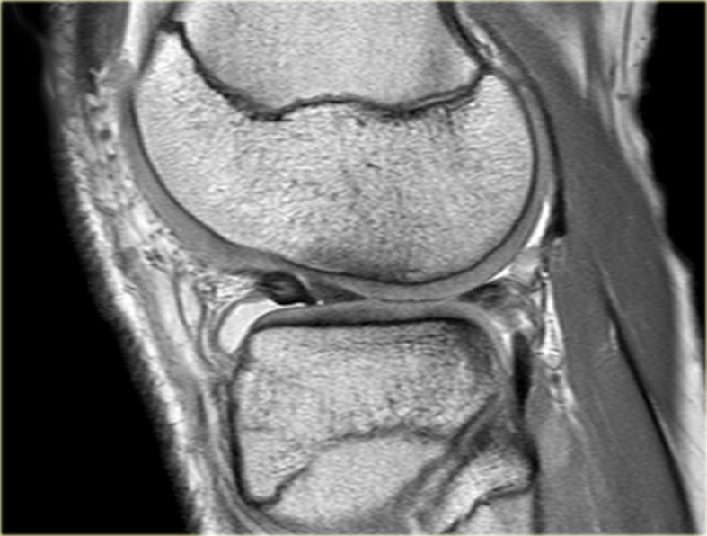
Study the image on the left and try to determine what the problem is with this meniscus.
Then continue with the next consecutive images of the same patient.Nghiên cứu hình ảnh bên trái và cố gắng xác định vấn đề là gì với sụn chêm này.
Sau đó tiếp tục với các hình ảnh liên tiếp tiếp theo của cùng một bệnh nhân.
Go to 'Knee Meniscus - Part 1' for the basics Đi tới 'Khum đầu gối - Phần 1' để biết thông tin cơ bản
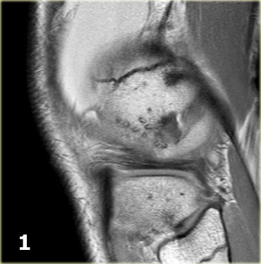
Scroll through the images.
Then continue reading..Cuộn qua các hình ảnh.
Sau đó tiếp tục đọc.
As you already suspected by reading the title of this paragraph, this is a flipped meniscus.
A flipped meniscus is a special form of bucket-handle tear.
A flipped meniscus occurs when the ruptured fragment of the posterior horn is flipped anteriorly so the anterior horn of the meniscus appears to be enlarged.Như bạn đã nghi ngờ bằng cách đọc tiêu đề của đoạn văn này, đây là một sụn chêm lật.
sụn chêm lật là một dạng đặc biệt của rách tay cầm xô.
Một sụn chêm bị lật xảy ra khi mảnh vỡ của sừng sau bị lật ra trước để sừng trước của sụn chêm dường như bị mở rộng.
- Body of lateral meniscus.Thân bên sụn chêm.
- Posterior horn is too small and anterior horn seems enlarged with an irregular contour.Sừng sau quá nhỏ và sừng trước dường như to ra với đường viền không đều.
- At first impression there is a longitudinal tear in the anterior horn, but actually the posterior part of this structure is the flipped part of the ruptured posterior horn.
The anterior structure is the anterior horn.
Also notice the focal bone marrow edema and the cartilage impression.Khi mới ấn sừng, có một vết rách dọc ở sừng trước, nhưng thực ra phần sau của cấu trúc này là phần bị lật của sừng sau bị vỡ.
Cấu trúc trước là sừng trước.
Cũng chú ý đến phù tủy xương khu trú và dấu ấn sụn. - The anterior structure is the anterior horn.Cấu trúc trước là sừng trước.
- Some irregularity of the posterior part.Một số bất thường của phần sau.
- Posterior part moves caudally.Phần sau di chuyển theo đường đuôi.
- In the intercondylar fossa is the connection between the displaced fragment and the other part of the posterior horn.Trong hố liên lồi cầu là sự kết nối giữa mảnh xương di lệch và phần khác của sừng sau.
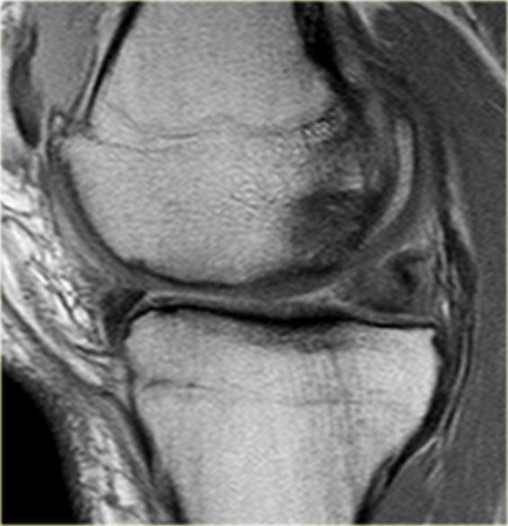
On the left another flipped meniscus.
Now on the medial side.
Part of the anterior horn is flipped posteriorly.
Only a small part of the anterior horn is seen anteriorly.Bên trái một người khác lật sụn chêm.
Bây giờ ở phía trong.
Một phần của sừng trước bị lật ra sau.
Chỉ có một phần nhỏ của sừng trước được nhìn thấy ở phía trước.
Most flipped menisci occur on the lateral side.
The ACL prevents the meniscal fragment from completely migrating into the intercondylar notch. Hầu hết các các sụn chêm lật xảy ra ở bên hông.
ACL ngăn không cho mảnh sụn chêm di chuyển hoàn toàn vào rãnh liên lồi cầu.
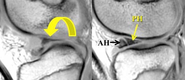

Illustration of the mechanism in a flipped meniscus.Hình minh hoạ cơ chế trong một sụn chêm lật.


On a coronal image you will first see an enlarged bulky anterior horn.
Posteriorly a very small posterior horn will be seen.
See next case.Trên hình ảnh vành, trước tiên bạn sẽ thấy một sừng trước to cồng kềnh.
Sừng sau rất nhỏ sẽ được nhìn thấy.
Xem trường hợp tiếp theo.
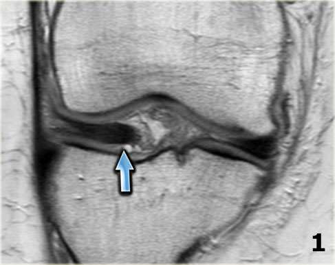
On the left another case of a flipped lateral meniscus.
Scroll through the images.Bên trái là một trường hợp lật sụn chêm.
Cuộn qua các hình ảnh.
- Notice the very large anterior horn.
The medial part is the displaced posterior horn.Chú ý sừng trước rất lớn.
Phần trong là sừng sau di lệch. - Medial part runs into the intercondylar fossa.Phần trong chạy vào hố liên lồi cầu.
- The dislocated part is well seen as 'third structure'
in the intercondylar fossa.Phần trật khớp được xem là “cấu trúc thứ ba ”.
trong hố liên lồi cầu. - Connection to the remnant of the posterior horn.Kết nối với phần còn lại của sừng sau.
- Connection to the remnant of the posterior horn.Kết nối với phần còn lại của sừng sau.
- Very small remnant of the posterior horn.Tàn dư rất nhỏ của sừng sau.
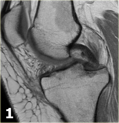
Same case sagittal images.
Notice how the ruptured part of the meniscus runs anteriorly through the intercondylar fossa (arrows)Trường hợp ảnh mặt phẳng đứng dọc.
Lưu ý phần vỡ của sụn chêm chạy trước qua hố liên lồi cầu (các mũi tên)
- Normally only two structures run in the intercondylar fossa. Here we see the normal posterior cruciate ligament. Thông thường chỉ có hai cấu trúc chạy trong hố liên lồi cầu, ở đây chúng ta thấy dây chằng chéo sau bình thường.
- Normal anterior cruciate ligament. Dây chằng chéo trước bình thường.
- Ruptured part of the posterior horn as 'third structure'
in the intercondylar fossa. Vỡ sừng sau là “cấu trúc thứ ba ”
trong hố liên lồi cầu. - Ruptured part runs anteriorly. Phần vỡ chạy về phía trước.
- Ruptured part joins the anterior horn and simulates a longitudinal tear in the anterior horn.
Notice the small remnant of the posterior horn.Phần bị vỡ nối với sừng trước và mô phỏng một vết rách dọc ở sừng trước.
Chú ý đến phần còn lại của sừng sau.
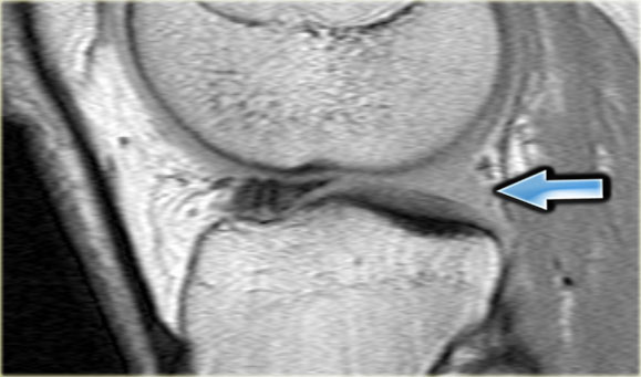
On the left sagittal PD-images of a flipped meniscus.
The whole posterior horn is flipped anteriorly resulting in an empty meniscus sign (arrow).Trên hình ảnh PD dọc bên trái của một sụn chêm lật.
Toàn bộ sừng sau được lật ra trước dẫn đến một dấu hiệu hình liềm trống (mũi tên).
Bucket handle tearXô xử lý nước mắt
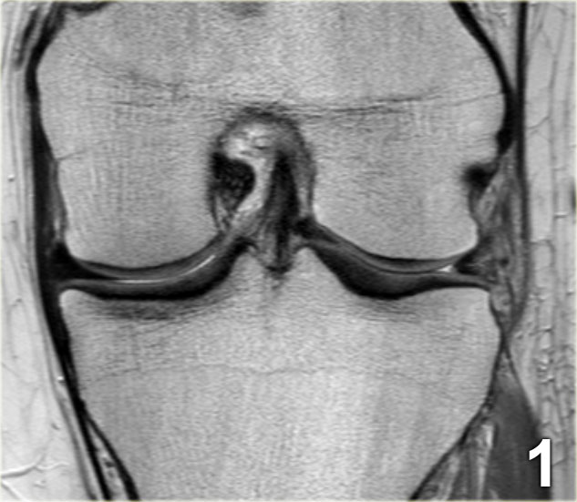
First study the images on the left.
Then continue reading.Đầu tiên nghiên cứu hình ảnh bên trái.
Sau đó tiếp tục đọc.
-
We start with a middle section of the examination.
Notice the small body of the lateral meniscus with an abnormal shape (blue arrow).
The missing inner part is seen just lateral to the anterior cruciate ligament (red arrow). Chúng tôi bắt đầu với phần giữa của cuộc kiểm tra.
Chú ý phần thân nhỏ của sụn chêm bên có hình dạng bất thường (mũi tên màu xanh).
Phần bên trong bị thiếu được nhìn thấy ngay bên cạnh dây chằng chéo trước (mũi tên màu đỏ). - The medially displaced part of the torn meniscus, i.e. the bucket handle can be followed in a posterior direction in the intercondylar fossa. Phần dịch chuyển vào trong của sụn chêm bị rách, tức là tay cầm xô có thể được theo sau theo hướng sau trong hố liên lồi cầu.
- Continue. Tiếp tục.
- Here is the connection between the bucket handle and the posterior horn. Đây là kết nối giữa tay cầm xô và sừng sau.
- Here an anterior slice demonstrating a meniscal cyst. Đây là một lát cắt trước cho thấy một nang sụn chêm.
- Notice the horizontal high signal. Chú ý tín hiệu cao theo chiều ngang.
- Here we can appreciate the complexity of the meniscal tear with a longitudinal and horizontal component (arrows). Ở đây chúng ta có thể đánh giá được độ phức tạp của rách sụn chêm với thành phần theo chiều dọc và chiều ngang (các mũi tên).
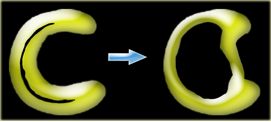
Bucket handle tears are displaced vertical longitudinal tears.
The displaced inner fragment resembles the handle of a bucket.
The remaining larger peripheral portion of the meniscus resembles the bucket.
These tears account for about 10% of all meniscal tears.Xô xử lý nước mắt là những vết rách dọc di dời.
Mảnh vỡ bên trong di lệch giống như tay cầm của một cái xô.
Phần ngoại vi lớn hơn còn lại của sụn chêm giống như cái xô.
Những vết rách này chiếm khoảng 10% tổng số các vết rách sụn chêm.
Double PCL signDấu hiệu PCL kép
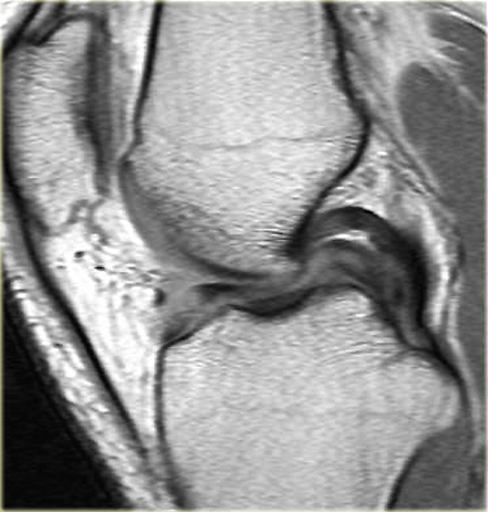
The double posterior cruciate ligament (PCL) sign is a low-signal-intensity band that is parallel and anteroinferior to the PCL on sagittal MR images.
It is a highly specific indicator of a bucket-handle meniscal tear (3).Dấu hiệu dây chằng chéo sau (PCL) kép là một dải cường độ tín hiệu thấp song song và thấp hơn so với PCL trên hình ảnh MR đứng dọc.
Đây là một chỉ số đặc hiệu cao của rách sụn chêm tay cầm xô (3).
Meniscal root tearRách chân sụn chêm
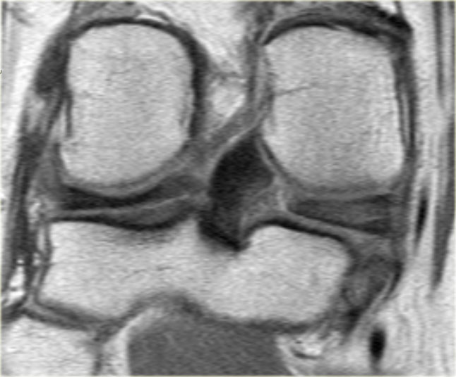
First study the image on the left and try to recognize the meniscal tear.
These tears often go unnoticed.
Then continue with the next images.Đầu tiên nghiên cứu hình ảnh bên trái và cố gắng nhận biết rách sụn chêm.
Những giọt nước mắt này thường không được chú ý.
Sau đó tiếp tục với các hình ảnh tiếp theo.
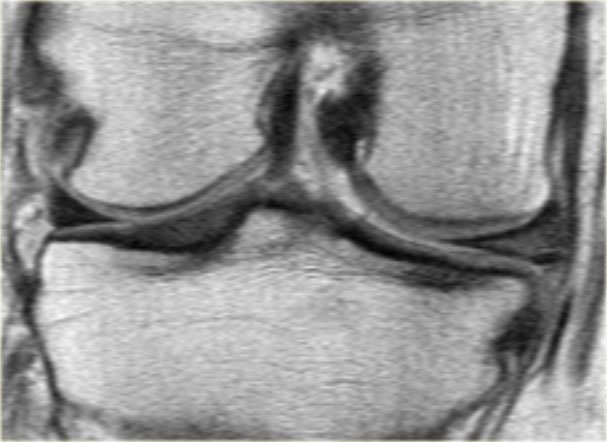
A radial tear is present at the posterior root junction of the medial meniscus which extends through the entire thickness of the meniscus with a cleft of fluid tracking through the defect (red arrows).
Rách xuyên tâm xuất hiện ở chỗ nối chân sau của sụn chêm trong, kéo dài qua toàn bộ độ dày của sụp chêm với một khe dịch theo dõi qua lỗ hổng (mũi tên màu đỏ).
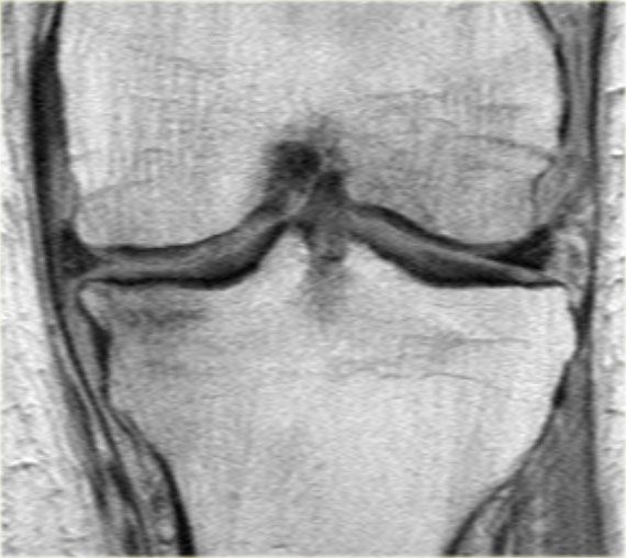
Meniscal root tears are often associated with extrusion of the meniscus beyond the margin of the tibial plateau.
More than 3 mm meniscus extrusion is often associated with tears involving the meniscal root (6).Rách rễ sụn chêm thường liên quan đến sự đùn ra của sụn chêm ngoài rìa của mâm chày.
Trên 3 mm đẩy lồi sụn chêm thường liên quan đến nước mắt liên quan đến rễ sụn (6).
In the case on the left there is a complete radial tear separating the posterior horn from its root (red arrows).
There is also minimal extrusion of the meniscus (image 1/6).
Trường hợp bên trái có rách hoàn toàn xương quay tách sừng sau ra khỏi rễ (các mũi tên màu đỏ).
Ngoài ra còn có sự đùn tối thiểu của sụn chêm (hình 1/6).
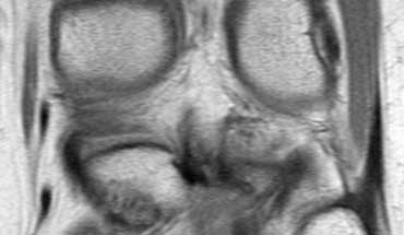
Here another medial meniscal root tear.
Notice that the posterior horn is not attached to the tibia.
Instead there is a gap (curved arrow).Đây là một rách chân sụn chêm khác.
Lưu ý rằng sừng sau không gắn vào xương chày.
Thay vào đó là một khoảng trống (mũi tên cong).
You can easily overlook these tears and think that the posterior horn is normal.Bạn có thể dễ dàng bỏ qua những vết rách này và nghĩ rằng sừng sau là bình thường.
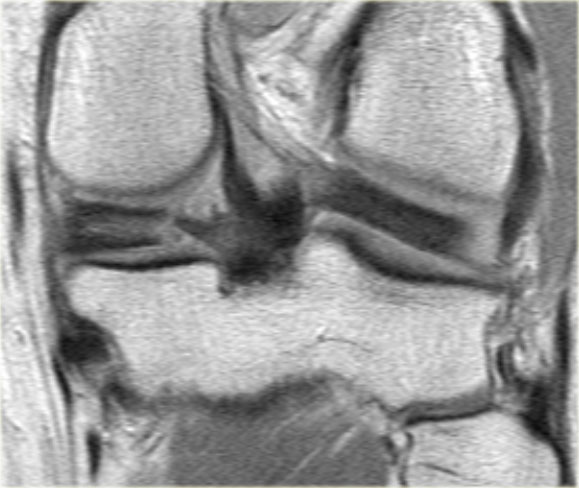
This is another typical case of a medial meniscal root tear.
Notice that there is also a lateral discoid meniscus.Đây là một trường hợp điển hình khác của rách chân sụn chêm trong.
Lưu ý rằng cũng có một đĩa bên sụn chêm.
Empty meniscus signTrống dấu hiệu hình liềm
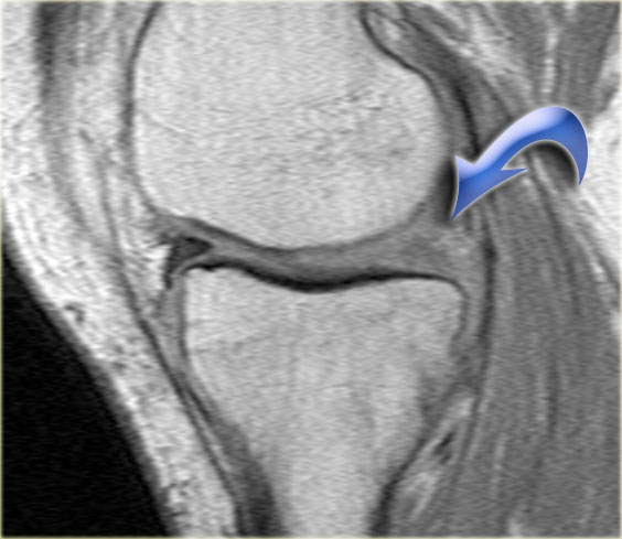
When there is a complete radial tear, the two meniscal fragments can be completely separated.
This can result in an empty meniscal space or empty meniscus sign (arrow).
Khi có rách hoàn toàn xương quay, hai mảnh sụn chêm có thể được tách ra hoàn toàn.
Điều này có thể dẫn đến một khoảng trống sụn chêm hoặc dấu hiệu hình liềm trống rỗng (mũi tên).
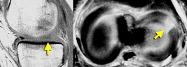 LEFT: Absent or empty meniscus on sagittal image. RIGHT: Axial image shows complete radial tear leading to a defect in the meniscus.Bên trái: vắng mặt hoặc trống sụn chêm trên hình ảnh Sagittal. Bên phải: Hình ảnh hướng trục cho thấy vết rách hoàn toàn dẫn đến khuyết tật trên sụn chêm.
If you image a complete radial tear directly along the length of the tear you will see an absent or empty meniscus.
These complete radial tears open up and give the impression that there is a part missing.
However you will not find a displaced meniscal fragment.
It is simply separation of the meniscal parts.Nếu bạn chụp ảnh một vết rách hoàn toàn dọc theo chiều dài của vết rách, bạn sẽ thấy một sụn chêm không có hoặc trống rỗng.
Những vết rách xuyên tâm hoàn toàn này mở ra và tạo ấn tượng rằng có một phần bị thiếu.
Tuy nhiên, bạn sẽ không tìm thấy một mảnh sụn chêm di lệch.
Nó chỉ đơn giản là sự tách biệt của các phần sụn chêm.
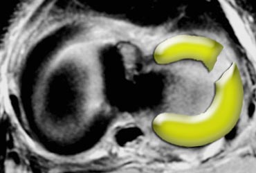
On the left an illustration of a complete radial tear, which can result in an empty meniscus sign.Bên trái là hình minh hoạ của một vết rách xuyên tâm hoàn toàn, có thể dẫn đến một dấu hiệu hình liềm rỗng.
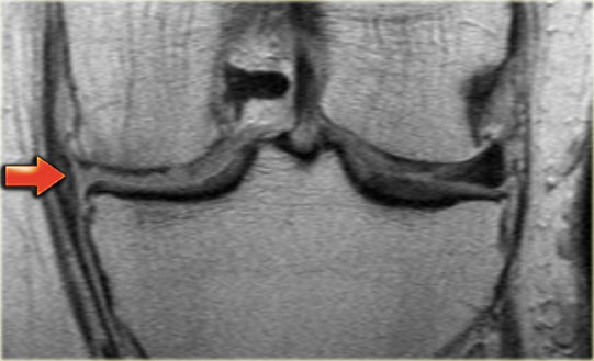
On the left coronal PD-images of a patient with a complete radial tear resulting in an empty meniscus sign. Trên hình ảnh PD động mạch vành trái của một bệnh nhân bị rách hoàn toàn xương quay dẫn đến dấu hiệu hình liềm rỗng.
- 1/7: Empty meniscus sign (horizontal red arrow). 1/7: dấu hiệu hình liềm rỗng (mũi tên màu đỏ nằm ngang).
- 2/7: Small remnant of body is seen.2/7: Tàn tích nhỏ của cơ thể được nhìn thấy.
- 3/7: Normal meniscal root attachment (blue arrow).3/7: Gắn rễ sụn bình thường (mũi tên xanh dương).
- 4/7: Normal posterior horn.4/7: Sừng sau bình thường.
- 5/7: Anteriorly there is extrusion of the meniscus.5/7: Trước có đùn sụn chêm.
- 6/7: Extrusion.6/7: Đùn.
- 7/7: Significant extrusion of more than 3 mm (red vertical arrow).7/7: Đùn nhiều trên 3 mm (mũi tên dọc màu đỏ).
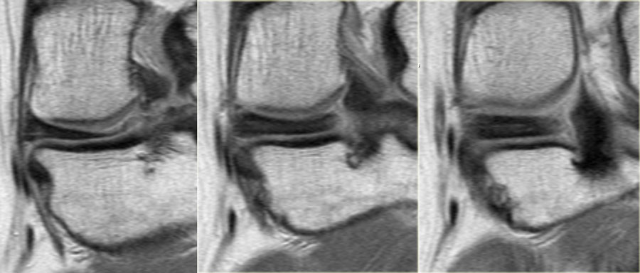
On the left consecutive coronal PD-images of a posterior horn of a meniscus that at first glance might give the impression that it is normal.
Continue with the sagittal images.Trên hình ảnh PD liên tiếp bên trái của sừng sau của sụn chêm thoạt nhìn có thể cho ấn tượng rằng nó là bình thường.
Tiếp tục với ảnh mặt phẳng đứng dọc.
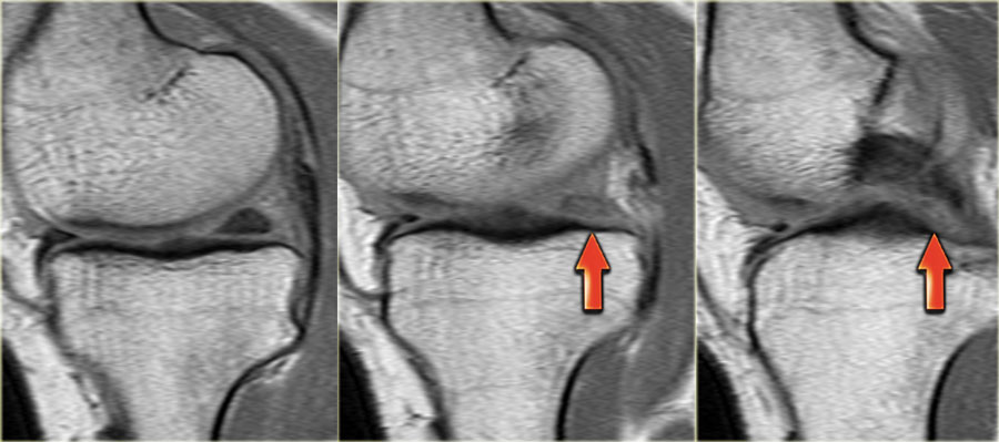
When you take a good look at the sagittal images, you will notice the empty meniscus sign, where normally the meniscal root attaches (red arrows).
This means that we are dealing with a meniscal root tear.
The meniscus posteriorly should come up over the tibial spine to insert near the posterior cruciate ligament. Khi bạn nhìn kỹ ảnh mặt phẳng đứng dọc, bạn sẽ thấy dấu hiệu hình liềm trống rỗng, nơi thường gắn rễ sụn (các mũi tên màu đỏ).
Điều này có nghĩa là chúng ta đang đối phó với một rách chân sụn chêm.
sụn chêm phía sau nên đi lên trên xương chày để chèn gần dây chằng chéo sau.
Vacuum phenomenonHiện tượng chân không
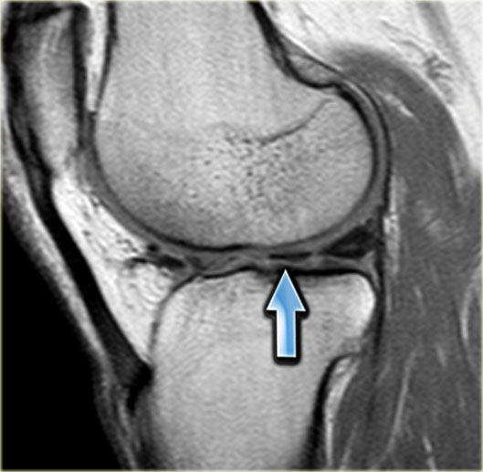
First scroll through the images on the left.
Try to figure out what is going on with this meniscus.
Then continue reading.Đầu tiên, cuộn qua các hình ảnh bên trái.
Cố gắng tìm ra những gì đang xảy ra với sụn chêm này.
Sau đó tiếp tục đọc.
At first impression this looks like a tear within a discoid meniscus.
At closer look you will notice that the horizontal structure has a lower signal intensity than the meniscus and looks funny.
This is a normal variant caused by a vacuum phenomenon.
A vacuum phenomenon is caused by negative pressure within the joint due to the position of the patient, which results in the accumulation of nitrogengas.
Continue with the radiograph of the same patient.Khi nhìn vào ban đầu, nó trông giống như một vết rách trong một đĩa sụn chêm.
Khi nhìn gần hơn, bạn sẽ nhận thấy rằng cấu trúc ngang có cường độ tín hiệu thấp hơn sụn chêm và trông buồn cười.
Đây là một biến thể bình thường gây ra bởi hiện tượng chân không.
Hiện tượng chân không là do áp lực âm trong khớp do vị trí của bệnh nhân, dẫn đến sự tích tụ khí nitơ.
Tiếp tục với phim X-quang của cùng một bệnh nhân.
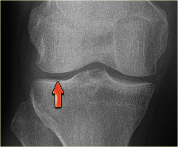
Vacuum phenomenon on the lateral side.Hiện tượng chân không ở mặt bên.
Discoid MeniscusMảng đĩa sụn chêm
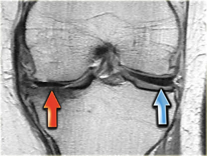
First scroll through the images on the left.
Try to figure out what is going on with this meniscus.
Then continue reading.Đầu tiên, cuộn qua các hình ảnh bên trái.
Cố gắng tìm ra những gì đang xảy ra với sụn chêm này.
Sau đó tiếp tục đọc.
The hypointense structure on the lateral side is a discoid meniscus (blue arrow).
The structure on the medial side is again a vacuum phenomenon.
On an adjacent slice this vacuum phenomenon is not seen any more.Cấu trúc giảm tín hiệu ở mặt bên là một đĩa sụn chêm (mũi tên xanh).
Cấu trúc ở phía trong một lần nữa là một hiện tượng chân không.
Trên một lát cắt liền kề, hiện tượng chân không này không còn được nhìn thấy nữa.
Red zone meniscal tearRách sụn chêm vùng đỏ
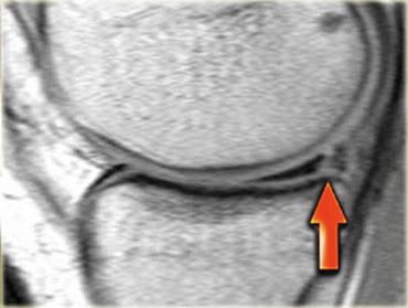
First scroll through the images on the left.
Try to figure out what is going on with this meniscus.
Then continue reading.Đầu tiên, cuộn qua các hình ảnh bên trái.
Cố gắng tìm ra những gì đang xảy ra với sụn chêm này.
Sau đó tiếp tục đọc.
There is a longitudinal tear in the periphery of the meniscus (red arrow).
The outside one-third of the meniscus is called the 'red' zone, because it has a rich blood supply.
The location of a meniscal tear is of importance because tears in this vascular portion of the meniscus are more likely to heal spontaneously than tears in the avascular portion or white zone of the meniscus.Có một vết rách dọc ở ngoại vi của sụn chêm (mũi tên màu đỏ).
Một phần ba bên ngoài của sụn chêm được gọi là vùng 'đỏ', bởi vì nó có nguồn cung cấp máu phong phú.
Vị trí rách sụn chêm có tầm quan trọng vì rách ở phần mạch máu này của sụn khớp có nhiều khả năng tự lành hơn rách ở vùng mạch máu hoặc vùng trắng của sụp chêm.
Meniscus within meniscus signSụn chêm trong dấu hiệu hình liềm
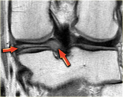
Sometimes extensive triangular or wedge-shaped high signal intensity can be encountered that does not reach te surface of the meniscus.
This is sometimes referred to as meniscus within meniscus sign.
Since this meniscal abnormality does not reach the meniscal surface, it does not fullfill the criteria for a meniscal tear.
It was found that in half of these patients and symptoms warranting arthroscopic follow-up had meniscal tears (4).Đôi khi có thể gặp phải cường độ tín hiệu cao hình tam giác hoặc hình nêm rộng không chạm tới bề mặt te của sụn chêm.
Điều này đôi khi được gọi là sụn chêm trong dấu hiệu hình liềm.
Vì bất thường sụn chêm này không chạm đến bề mặt sụn, nó không đáp ứng đầy đủ các tiêu chuẩn cho rách sụn.
Kết quả cho thấy một nửa số bệnh nhân và các triệu chứng cần thiết cho nội soi khớp theo dõi có rách sụn chêm (4).
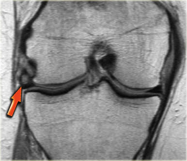
On the left another meniscus with diffuse high signal in the meniscal body.
On other images (not shown) there was no evidence of a tear.
Notice severe extrusion of the meniscus beyond the margin of the tibia plateau.Bên trái là một sụn chêm khác có tín hiệu cao lan toả trong thân sụn chêm.
Trên các hình ảnh khác (không hiển thị) không thấy dấu hiệu một vết rách.
Lưu ý sự đùn ra nghiêm trọng của sụn chêm vượt ra ngoài rìa của mâm chày.
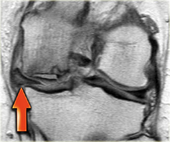
- Meniscus within meniscus sign and extrusion of middle part of meniscus (red arrow).Sụn chêm trong dấu hiệu hình liềm và đùn phần giữa của sụn chêm (mũi tên màu đỏ).
- Complete radial tear in the posterior horn.
Also notice the avascular necrosis.Rách hoàn toàn xương quay ở sừng sau.
Cũng chú ý đến hoại tử vô mạch. - Also notice the avascular necrosis.Cũng chú ý đến hoại tử vô mạch.
- Extrusion of meniscus avascular necrosis.Phẫu thuật đùn hoại tử vô mạch sụn chêm.
Meniscal extrusionĐẩy lồi sụn chêm
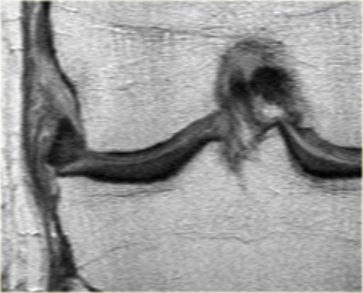
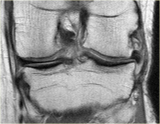
Tears involving the meniscal root (central attachment) are also significantly related to the severity of meniscal extrusion, seen in 3% with minor extrusion and 42% with major extrusion.
With meniscus extrusion, the meniscus is unable to resist hoop stresses and cannot shield the adjacent articular cartilage from excessive axial load.
Over time, this can lead to symptomatic knee osteoarthritis.
Tears of the posterior meniscal root can be easily missed because of inconsistent clinical symptoms and can be overlooked without thorough arthroscopic examination.
Detection of meniscal extrusion is important not only because it is associated with underlying tear but also because meniscal extrusion itself is thought to be related to development of osteoarthritis.Rách liên quan đến rễ sụn chêm (đính kèm trung tâm) cũng có liên quan đáng kể đến mức độ nặng của đẩy lồi sụn chêm, gặp ở 3% với đùn nhỏ và 42% với đùt lớn.
Với đẩy lồi sụn chêm, sụn chêm không thể chống lại ứng suất hoop và không thể che chắn sụn khớp liền kề khỏi tải trọng trục quá mức.
Theo thời gian, điều này có thể dẫn đến thóai hóa khớp gối có triệu chứng.
Rách rễ sụn chêm sau có thể dễ dàng bị bỏ sót do các triệu chứng lâm sàng không nhất quán và có thể bị bỏ qua nếu không được nội soi khớp toàn diện.
Phát hiện đẩy lồi sụn chêm là quan trọng không chỉ vì nó có liên quan đến rách cơ bản mà còn vì đẩy lồi sụn chêm được cho là có liên quan tới sự phát triển của thóai hóa khớp.
Segond fracture and meniscal tearGãy xương sụn chêm và rách sụn giao cảm
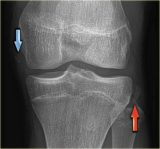
A Segond fracture is an avulsion of the lateral capsular ligament.
The mechanism of injury is internal rotation and varus stress.
On a radiograph it manifests as an elliptic bony fragment off the lateral proximal tibia (figure).
A Segond fracture has a high association with a tear of the anterior cruciate ligament (75-100%) and injuries of the medial and lateral menisci (66-70%).Gãy Segond là tình trạng bong dây chằng bao ngoài.
Cơ chế chấn thương là xoay trong và căng giãn tĩnh mạch chủ.
Trên X quang, nó biểu hiện như một mảnh xương elip ở đầu gần xương chày bên (hình).
Gãy Segond có mối liên quan chặt chẽ với tổn thương rách dây chằng chéo trước (75-100%) và tổn thương các sụn chêm trong và ngoài (66-70%).
On the radiograph you could easily miss the Segond fracture (red arrow).
Notice that there is also an avulsion of the medial collateral ligament.
Continue with the MR-images.Trên phim X-quang bạn có thể dễ dàng bỏ sót gãy Segond (mũi tên màu đỏ).
Lưu ý rằng cũng có một sự nhổ dây chằng bên trong.
Tiếp tục với hình ảnh cộng hưởng từ.
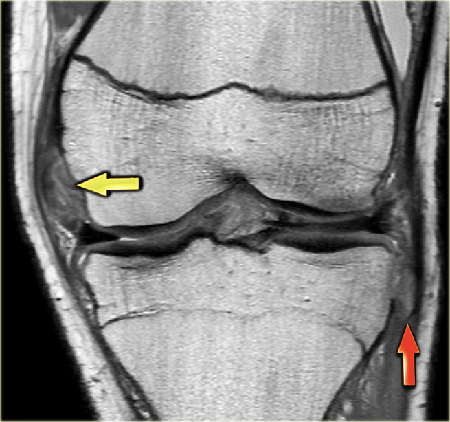
On the left three consecutive coronal PD-images:Bên trái là ba ảnh chụp động mạch vành liên tiếp:
-
Tear of deep medial collateral band (yellow arrow).
Avulsion of bone fragment of lateral proximal tibia, i.e. Segond fracture (red arrow). Rách dải băng bên trong sâu (mũi tên màu vàng).
Xẹp xương của đầu gần xương chày bên, tức là gãy xương Segond (mũi tên màu đỏ). -
Notice displaced inner fragment of the meniscus.
This indicates that there is also a bucket handle tear. Lưu ý mảnh bên trong di lệch của sụn chêm.
Điều này cho thấy cũng có một vết rách tay cầm xô. -
Besides the bucket handle tear (inner blue arrow), there is also a barely visible horizontal tear in the peripheral part of the meniscus (outer blue arrow).
The horizontal tear was better appreciated on other images (not shown).Bên cạnh vết rách tay cầm xô (mũi tên màu xanh bên trong), cũng có một vết rách ngang hầu như không nhìn thấy được ở phần ngoại vi của sụn chêm (mũI tên màu xanh ngoài).
Đường rách ngang được đánh giá tốt hơn trên các hình ảnh khác (không hiển thị).
Continue with the sagittal images.Tiếp tục với ảnh mặt phẳng đứng dọc.
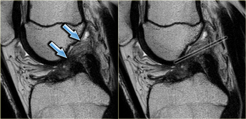
A Segond fracture is almost pathognomonic for an anterior cruciate ligament tear, which was also demonstrated in this patient.Gãy Segond gần như là đặc trưng cho rách dây chằng chéo trước, điều này cũng được chứng minh ở bệnh nhân này.
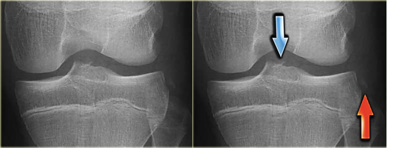
On the left an AP-view of another patient.
In association with a Segond fracture (red arrow), there is also an avulsion fracture of the anterior cruciate ligament (blue arrow).Bên trái là hình ảnh AP của một bệnh nhân khác.
Gãy Segond (mũi tên đỏ), cũng có gãy bong dây chằng chéo trước (mũI tên xanh).
Meniscal cystNang sụn chêm
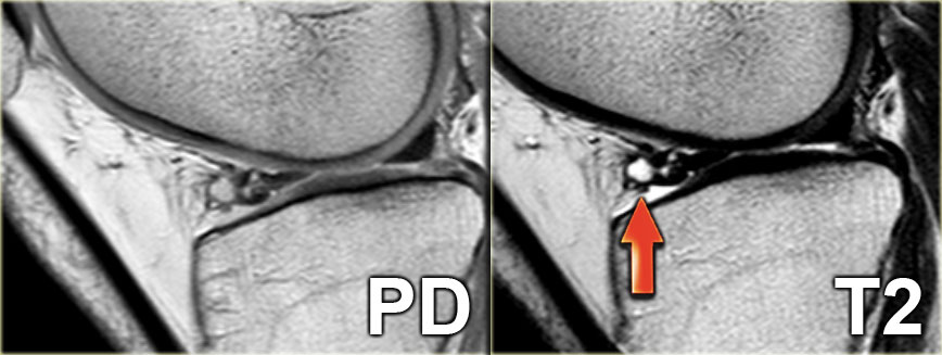
A meniscal cyst results from extrusion of synovial fluid through a peripherally extended horizontal meniscal tear.
Medial meniscal cysts are most commonly located adjacent to the posterior horn and lateral meniscal cysts are most commonly located adjacent to the anterior horn or body.
Nang sụn chêm là kết quả của sự đùn ra của dịch khớp thông qua một vết rách sụn khớp theo chiều ngang mở rộng ra ngoại vi.
Các nang sụn chêm trong thường nằm gần sừng sau và các nang sụN chêm ngoài thường nằm gần với sừng trước hoặc thân trước.
On the left a PD- and a T2-weighted image demonstrating a lateral meniscal cyst are adjacent to the anterior horn as a result of a complex tear.Bên trái là hình ảnh PD- và xung T2W cho thấy một nang sụn chêm bên cạnh sừng trước do rách phức tạp.
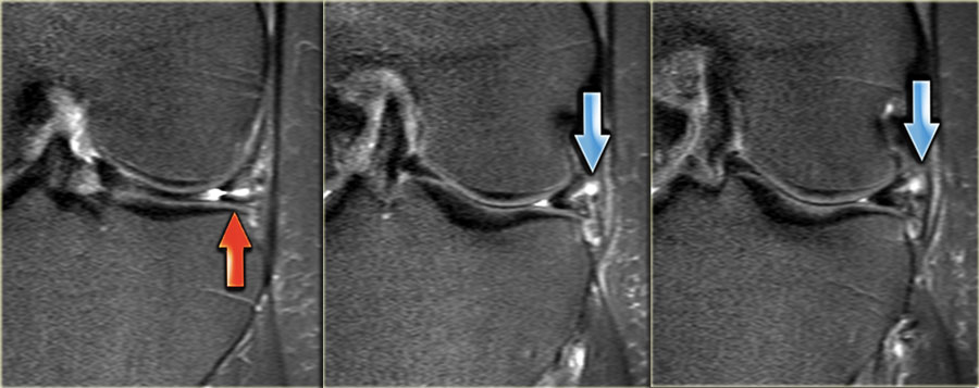
On the left three consecutive images of a small meniscal cyst (blue arrow) as a result of a horizontal tear (red arrow).Ở bên trái, ba hình ảnh liên tiếp của một nang sụn nhỏ (mũi tên màu xanh) là kết quả của một vết rách ngang (mũa tên màu đỏ).
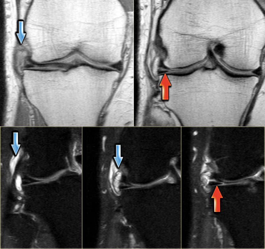
On the left coronal PD-images without fatsat and with fatsat.
A large meniscal cyst is seen in relation to a horizontal tear (red arrow).Ở bên trái là hình ảnh PD vành không có fatsat và có fatsat.
Một nang sụn chêm lớn được nhìn thấy liên quan đến một vết rách ngang (mũi tên màu đỏ).
ReferencesTài liệu tham khảo
-
Radial Meniscal Tears: Significance, Incidence, and MR Appearance
Rách sụn chêm quay: Tầm quan trọng, tỷ lệ mắc và hình thái MR
by Keith W. Harper, Clyde A. Helms, H. Stanley Lambert and Laurence D. Higgins.
AJR 2005; 185:1429-1434
Keith W. Harper, Clyde A. Helms, H. Stanley Lambert và Laurence D. Higgins. -
Bucket-handle tears of the medial and lateral menisci of the knee: value of MR imaging in detecting displaced fragments
Rách xương đòn các sụn chêm giữa và bên khớp gối: giá trị cộng hưởng từ trong phát hiện mảnh xương di lệch
by DH Wright, AA De Smet and M Norris s.
American Journal of Roentgenology, Vol 165, 621-625
bởi DH Wright, AA De Smet và M Norris s. -
The Double Posterior Cruciate Ligament Sign
Dấu hiệu dây chằng chéo sau kép
by Marc A. Camacho
November 2004 Radiology, 233, 503-504.
bởi Marc A. Camacho -
Grade 2C Signal in the Mensicus on MR Imaging of the Knee
Tín hiệu 2C trong phim Mensicus trên cộng hưởng từ hình ảnh khớp gối
by Thomas R. McCauley et al
AJR 2002; 179:645-648
bởi Thomas R. McCauley và cộng sự -
Lateral Meniscus Root Tear and Meniscus Extrusion with Anterior Cruciate Ligament Tear
Rách chân sụn chêm ngoài và đẩy lồi sụn chêm với dây chằng chéo trước Tear
by Jeffrey M. Brody et al bởi Jeffrey M. Brody và cộng sự
-
Medial Meniscus Extrusion on Knee MRI: Is Extent Associated with Severity of Degeneration or Type of Tear?
Mức độ liên quan với mức độ nghiêm trọng của thóai hóa hay loại rách khớp gối trên cộng hưởng từ đẩy lồi sụn chêm trong
by Rosalia Costa et al
AJR 2004; 183:17-23
bởi Rosalia Costa và cộng sự -
Radial Tear of the Medial Meniscal Root: Reliability and Accuracy of MRI for Diagnosis
Rách xuyên tâm rễ sụn chêm trong: Độ tin cậy và độ chính xác của MRI trong chẩn đoán
by So Yeon Lee et al
AJR 2008; 191:81-85
So Yeon Lee và cộng sự -
MR Imaging of Meniscal Cysts: Incidence, Location, and Clinical Significance
Chẩn đoán hình ảnh nang sụn chêm: Tỷ lệ, vị trí và ý nghĩa lâm sàng
by Scot E. Campbell
AJR 2001; 177:409-413
bởi Scot E. Campbell