
Temporal Bone Anatomy 1.0Giải phẫu xương thái dương 1.0
Radiology department of the University Medical Centre of Utrecht and the Rijnland Hospital, Leiderdorp, the NetherlandsKhoa X quang của Trung tâm Y khoa Đại học Utrecht và Bệnh viện Rijnland, Leiderdorp, Hà Lan
Ngày đăng / cập nhật: 2006-07-15
Bản gốc trên radiologyassistant.nl ↗ · tải về lúc 2026-11-19 20:19 · bản dịch máy có chuẩn hóa thuật ngữ chẩn đoán hình ảnh
Updated version: 21-2-2007
In this review we present the normal coronal and axial anatomy of the temporal bone.
Learn the anatomy by scrolling through the images.Cập nhật phiên bản: 21-2-2007
Trong bài tổng quan này, chúng tôi trình bày giải phẫu vành và trục của xương thái dương.
Tìm hiểu giải phẫu bằng cách cuộn qua các hình ảnh.
Temporal boneXương thái dương
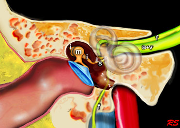
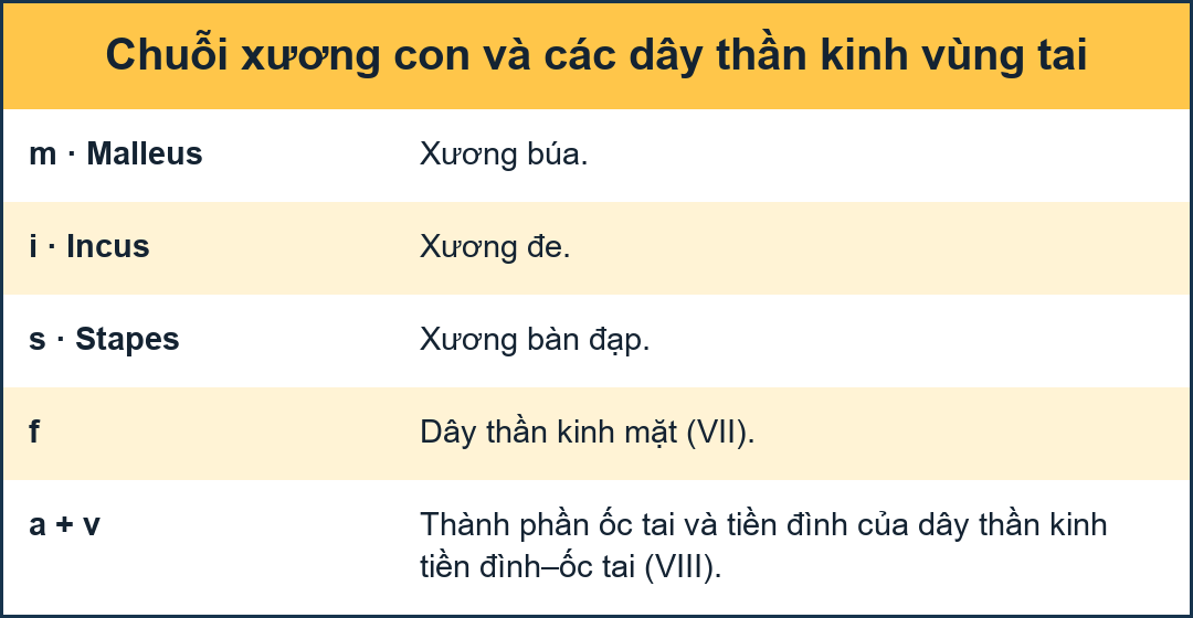
The middle ear consists of the tympanic cavity and the antrum.
The antrum is a large aircell superior and posterior to the tympanic cavity and connected to the tympanic cavity via the aditus ad antrum.
The epitympanum or attic is the upper portion of the tympanic cavity above the tympanic membrane, and contains the head of the malleus and the body of the incus.
The tympanic membrane, the malleus, incus and stapes transfer soundwaves to the stapes footplate, which is attached to the base of the cochlea in the oval window.
Tai giữa bao gồm khoang nhĩ và hang nhĩ.
Phễu nhĩ là một túi khí lớn ở trên và sau khoang nhĩ và kết nối với khoang nhện thông qua sào huyệt.
Lớp ngoài nhĩ là phần trên của khoang nhĩ trên màng nhĩ, và chứa đầu xương búa và thân xương đe.
Màng nhĩ, xương búa, ốc tai và bàn đạp truyền sóng âm đến bàn đạp chân, được gắn vào đáy ốc tai trong cửa sổ hình bầu dục.
Axial anatomyGiải phẫu trục
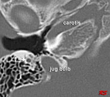
Scroll through the axial anatomy from inferior to superiorCuộn qua giải phẫu trục từ dưới lên trên
- A = Antrum A = hang vị
- f = facial nervef = Dây thần kinh mặt
- genu = first genu of facial nerve at the geniculate ganglion genu = quỳ gối đầu tiên của dây thần kinh mặt ở hạch cằm
- IAC = internal auditory canal IAC = kênh tai trong
- long crus = long crus of the incuslong crus = sụn dài của xương đe
- short crus = short crus of the incus crus ngắn = crus ngắn của xương đe
- U = utricle of vestibuleU = utricle tiền đình
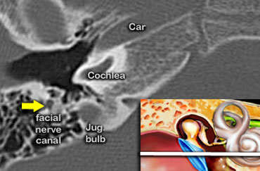
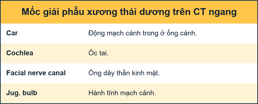
Axial anatomy from inferior to superiorGiải phẫu trục từ dưới lên trên
At the most inferior level we see the facial nerve passing inferiorly to finally reach the stylomastoid foramen (not shown in this image).
The carotid artery is shown within the carotid canal.
Also at this level is the top of the jugular bulb.Ở mức thấp nhất, chúng ta thấy dây thần kinh mặt đi qua dưới để cuối cùng đến lỗ trâm (không được hiển thị trong hình ảnh này).
Động mạch cảnh được thể hiện trong ống động mạch cảnh.
Cũng ở mức này là đỉnh của bóng đèn cảnh.
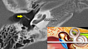

Tympanic membraneMàng nhĩ
The manubrium of the malleus (yellow arrow) is connected to the tympanic membrane.Lỗ ối của xương búa (mũi tên màu vàng) được nối với màng nhĩ.
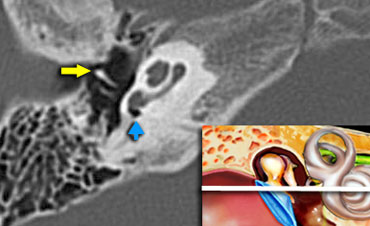 Malleus (yellow arow). Round window (blue arrow)Malleus (hàng màu vàng). Cửa sổ tròn (mũi tên màu xanh)
At this level we can see the manubrium of the malleus (yellow arow) anterior to the long process of the incus.
The round window is indicated by the blue arrow.
The round window dissipates the pressure generated by the fluid vibrations within the cochlea and thus serves as a release valve.Ở mức này, chúng ta có thể thấy xương búa (hào vàng) phía trước của xương đe.
Cửa sổ tròn được biểu thị bằng mũi tên màu xanh.
Cửa sổ tròn làm giảm áp suất do rung động của chất lỏng trong ốc tai và do đó đóng vai trò như một van giải phóng.
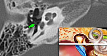 Stapes (green arrow) is seen connecting to the oval window.Dải (mũi tên màu xanh lá cây) được nhìn thấy kết nối với cửa sổ hình bầu dục.
StapesDải
The base of the stapes rocks in and out against the oval window.
The vibrations are transmitted via the endolymph to the hair cells of the organ of Corti of the cochlea.
Đế của bàn đạp đá vào và ra so với cửa sổ hình bầu dục.
Các rung động được truyền qua nội mô đến các tế bào lông của cơ quan Corti của ốc tai.
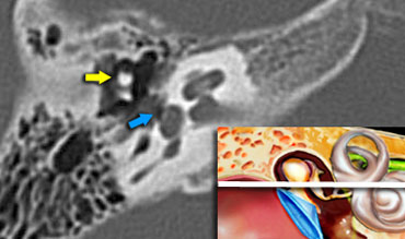
CochleaỐc tai
Within the cochlea the movement of the hair cells convert the sound-vibrations into nerve impulses, that travel over the cochlear nerve to the auditory cortex of the brain, which interprets the impulses as sound. .
The head of the malleus is seen anterior to the head of the incus (yellow arrow).
Trong ốc tai, sự di chuyển của các tế bào lông chuyển đổi các rung động âm thanh thành các xung thần kinh, truyền qua dây thần kinh ốc tai đến vỏ não thính giác, giải thích các xung động là âm thanh.
Đầu xương búa được nhìn thấy phía trước đầu xương đe (mũi tên màu vàng).
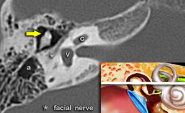

Tympanic segment of the facial nerveĐoạn nhĩ của dây thần kinh mặt
In this image at the level of the internal auditory canal, the tympanic segment of the facial nerve is seen just medial and parallel to the wall of the epitympanum.
The head of the malleus (yellow arrow) is seen anterior to the head and the short process of the incus.
Trong hình ảnh này ở ngang mức ống tai trong, đoạn nhĩ của dây thần kinh mặt được nhìn thấy ngay trong và song song với thành của thượng nhĩ.
Đầu xương búa (mũi tên màu vàng) được nhìn thấy ở phía trước đầu và quá trình ngắn của đe.
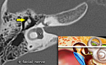

Geniculate ganglion of the facial nerveHạch thần kinh mặt
At this level the aditus ad antrum is seen. This is the connection between the tympanic cavity and the antrum.
The labyrinthine segment of the facial nerve coming from the internal auditory canal angles sharply forward, nearly at right angles to the long axis of the petrous bone, to reach the geniculate ganglion.
At the ganglion the facial nerve makes a U-turn (first genu of the facial nerve) to run posteriorly as the tympanic segment along the medial wall of the epitympanum.
Ở mức này thấy lối vào hang chũm (aditus ad antrum), nối hòm nhĩ với hang chũm. Đoạn mê nhĩ của dây thần kinh mặt đi từ ống tai trong, bẻ gấp ra trước gần vuông góc với trục dài xương đá để tới hạch gối. Tại hạch gối, dây thần kinh mặt đổi hướng ở gối thứ nhất, chạy ra sau thành đoạn nhĩ dọc thành trong của thượng nhĩ.
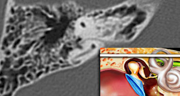
AntrumHang vị
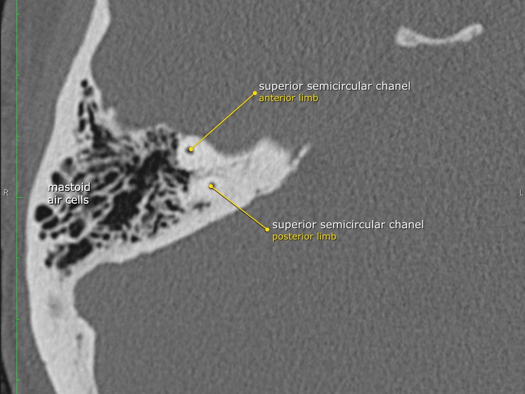
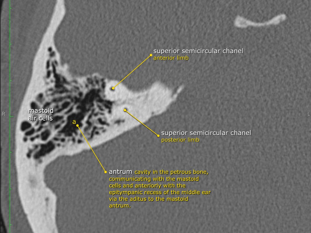
At this level the antrum is seen surrounded by smaller mastoid aircells just lateral to the superior semicircular canals .
The three semicircular canals lie perpendicular to each other to sense acceleration and deceleration movements in each of the 3 spatial planes.
Static head position is sensed by the vestibule, which contains the position hair cells.
Different head positions produce different gravity effects by small calcium carbonate particles (otoliths) on these hair cells.
Ở mức này, có thể thấy ống hang được bao quanh bởi các tế bào mastoid nhỏ hơn ngay bên cạnh các ống bán nguyệt trên.
Ba ống bán nguyệt nằm vuông góc với nhau để cảm nhận chuyển động tăng tốc và giảm tốc trong mỗi mặt phẳng không gian 3.
Vị trí đầu tĩnh được cảm nhận bởi tiền đình, nơi chứa các tế bào tóc vị trí.
Các vị trí đầu khác nhau tạo ra các hiệu ứng trọng lực khác nhau bởi các hạt canxi cacbonat nhỏ (otolith) trên các tế bào tóc này.
Coronal anatomyGiải phẫu vành
The petrous bone is positioned in an oblique orientation from posterolateral to anteromedial.
As a result most structures will be sectioned obliquely on coronal images.
The following coronal images go from anterior to posterior.
First we will see the tympanic membrane with the ossicles, followed by the cochlea, antrum and semicircular canalls.
Finally the most posterior image will show the point where the facial nerve exits the temporal bone at the stylomastoid foramen.Xương đá được định vị theo hướng xiên từ sau bên đến trước trong.
Kết quả là hầu hết các cấu trúc sẽ được cắt chéo trên ảnh mặt phẳng đứng ngang.
ảnh mặt phẳng đứng ngang sau đi từ trước ra sau.
Đầu tiên chúng ta sẽ thấy màng nhĩ với các xương con, tiếp theo là ốc tai, hang và các ống bán khuyên.
Cuối cùng, hình ảnh phía sau nhất sẽ cho thấy điểm mà dây thần kinh mặt thóat ra khỏi xương thái dương tại lỗ trâm.
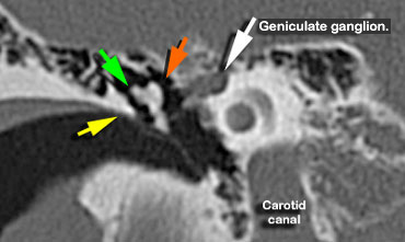
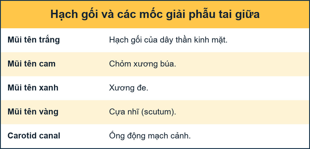 Scutum (yellow arrow). Head of malleus (orange arrow) is seen medial to the incus (green arrow)Đụng (mũi tên màu vàng) Đầu xương búa (mũa tên màu cam) được nhìn thấy ở giữa đe (mũe tên màu xanh lá cây)
ScutumViệt
The scutum (yellow arrow) is a sharp bony spur formed by the lateral wall of the tympanic cavity and the superior wall of the external auditory canal.
It is usually the first bony structure to erode as a result of a cholesteatoma, that is formed by medial retraction of the pars flaccida of the tympanic membrane into the epitympanum.
If the retraction continues it will result in ossicular destruction.
If the cholesteatoma passes posteriorly through the aditus ad antrum into the mastoid itself, erosion of the tegmen mastoideum, with exposure of the dura and erosion of the lateral semicircular canal with deafness and vertigo, may result. Vùng nhĩ (mũi tên màu vàng) là một gai xương sắc nhọn được hình thành bởi thành bên của khoang nhĩ và thành trên của ống tai ngoài.
Đây thường là cấu trúc xương đầu tiên bị bào mòn do cholesteatoma, được hình thành do sự co lại trong của màng nhĩ vào trong thượng nhĩ.
Nếu sự co kéo tiếp tục, nó sẽ dẫn đến sự phá huỷ xương con.
Nếu cholesteatoma đi qua phía sau qua sào huyệt vào xương chũm, có thể dẫn đến sự xói mòn của xương chũM, với sự tiếp xúc của màng cứng và sự xói lở của ống bán nguyệt bên với điếc và chóng mặt.
On the left the most anterior point of the facial nerve is seen (white arrow).
At this point the nerve makes a U-turn.
It is named the genu or geniculum and represents the geniculate ganglion.
The malleus is seen connected to the tympanic membrane and to the incus.Ở bên trái, thấy điểm trước nhất của dây thần kinh mặt (mũi tên màu trắng).
Tại thời điểm này dây thần kinh tạo thành một vòng chữ U.
Nó được đặt tên là khớp gối hoặc mỏ vịt và đại diện cho hạch thần kinh.
Lông tơ được nhìn thấy kết nối với màng nhĩ và đe tai.
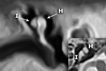
 Coronal reconstruction clearly demonstates that the incus (I) is positioned posterolateral to the malleolar head (H).On the axial image the short crus of the incus is seen pointing posterolaterally.Tái tạo vành nhật hoa cho thấy rõ ràng rằng rãnh (I) được đặt sau bên so với đầu mắt cá (H), trên hình ảnh trục, thấy lớp vỏ ngắn của rãnh hướng về phía sau bên.
In many illustrations you will see the incus connecting medially to the malleus, but this is not correct.
On the coronal reconstruction on the left it is clearly demonstated that the incus is positioned posterolaterally to the malleolar head.
The long crus of the incus subsequently runs inferomedially to the stapes.Trong nhiều hình minh hoạ, bạn sẽ thấy đe kết nối với xương búa, nhưng điều này không đúng.
Trên hình ảnh tái tạo vành bên trái, rõ ràng là đe được đặt ở vị trí sau bên so với đầu mắt cá.
Vảy dài của đe sau đó chạy vào trong đe.
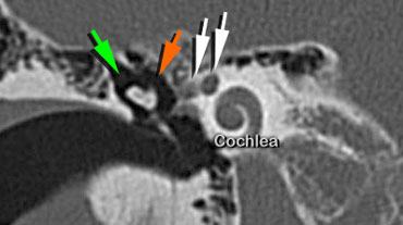
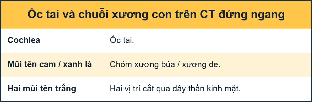 Head of malleus (orange arrow) is seen medial to the Incus (green arrow)Đầu xương búa (mũi tên màu cam) được nhìn thấy ở giữa của xương Inca (mũI tên màu xanh lá cây)
A coronal image slightly more posteriorly will show the facial nerve twice.
The medial portion is the part that exits the internal auditory canal and runs towards the geniculate ganglion (medial white arrow).
The lateral portion is the part that courses in a posterior direction, coming from the U-turn of the first genu. Hình ảnh chụp từ phía sau sẽ cho thấy dây thần kinh mặt hai lần.
Phần trong là phần thóat ra khỏi ống tai trong và chạy về phía hạch có niệu quản (mũi tên màu trắng trong).
Phần bên là phần di chuyển theo hướng sau, đến từ lần xoay chữ U của lần quỳ đầu tiên.
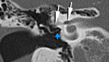 Long crus of the incus is seen connecting to the Stapes (blue arrow).Facial nerve in internal auditory canal and tympanic segment (white arrows).Crus dài của incus được nhìn thấy kết nối với xương bàn chân (mũi tên xanh). Dây thần kinh mặt trong ống thính giác nội và đoạn tympanic (mũi tên trắng).
Facial nerve canalỐng thần kinh mặt
The facial nerve is seen in the internal auditory canal and entering the temporal bone (medial white arrow).
The lateral white arrow represents the tympanic segment of the facial nerve running in the facial canal and curving around the oval window niche.
At this point, the nerve runs in a horizontal plane in a posterior direction superiorly to the oval window .
The incus (orange arrow) is seen connecting to the stapes (blue arrow).Dây thần kinh mặt được nhìn thấy trong ống tai trong và đi vào xương thái dương (mũi tên màu trắng trong).
Dây mũi tên màu trắng bên đại diện cho đoạn nhĩ của dây thần kinh mặt chạy trong ống mặt và cong quanh hốc cửa sổ hình bầu dục.
Tại thời điểm này, dây thần kinh chạy theo một mặt phẳng ngang theo hướng sau lên trên cửa sổ hình bầu dục.
Ốc tai (mũi tên màu cam) được nhìn thấy kết nối với các xương bàn đạp (mũI tên màu xanh).
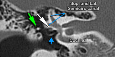
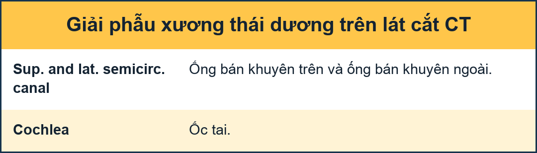
Coronal scan showing the facial nerve (white arrow) above the oval window and below the lateral semicircular canal.Chụp cắt lớp vi tính mặt cắt ngang cho thấy dây thần kinh mặt (mũi tên màu trắng) phía trên cửa sổ hình bầu dục và bên dưới ống bán khuyên bên.
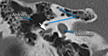

AntrumHang vị
The antrum is a large aircell superior and posterior to the tympanic cavity and connected to the tympanic cavity via the aditus ad antrum.
It is surrounded by smaller mastoid aircells.
Phễu nhĩ là một túi khí lớn ở trên và sau khoang nhĩ và kết nối với khoang nhện thông qua sào huyệt.
Nó được bao quanh bởi các túi khí xương chũm nhỏ hơn.
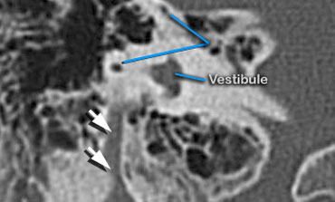

On this last posterior coronal image the facial nerve assumes a vertical position to exit the petrous bone through the stylomastoid foramen. Trên hình ảnh vành sau cuối cùng này, dây thần kinh mặt đảm nhận vị trí thẳng đứng để thóat khỏi xương đá qua lỗ trâm cài.
Test...Kiểm tra
AnatomyGiải phẫu
AnatomyGiải phẫu
ReferencesTài liệu tham khảo
-
Anatomy of the os temporale
Giải phẫu xương thái dương
Interactive Digital Education (part I), F.J.A. Beek, radiologist,
Radiology department of the Wilhelmina Children's Hospital and the University Medical Centre of Utrecht, the Netherlands
Giáo dục kỹ thuật số tương tác (Phần I), F.J.A. Beek, bác sĩ X quang, -
Lemmerling M, Kollias SS, eds. Radiology of the Petrous Bone. Springer 2003. Ch. 1, p. 1-14 Lemmerling M, Kollias SS, eds. X quang của xương petrous. Springer 2003. Ch. 1, p. 1-14