
Carpal instabilityMất vững cổ tay
Mallinckrodt Institute of Radiology Washington University St. Louis, Missouri, USA and the Westeinde hospital the Hague the Netherlands.Mallinckrodt Institute of Radiology Washington University St. Louis, Missouri, USA và bệnh viện Westeinde the Hague, Hà Lan.
Ngày đăng / cập nhật: 2005-08-23
Bản gốc trên radiologyassistant.nl ↗ · tải về lúc 2026-11-19 20:19 · bản dịch máy có chuẩn hóa thuật ngữ chẩn đoán hình ảnh
This article is based on a presentation given by Louis Gilula and adapted for the Radiology Assistant by Ileana Chesaru.
First a systematic analysis of the wrist is presented to look for carpal instability and fracture dislocation.
Secondly cases are presented as examples in the chapter systematic review and diagnosis.Bài viết này dựa trên một bài thuyết trình của Louis Gilula và được Ileana Chesaru điều chỉnh cho Trợ lý X quang.
Đầu tiên, một phân tích hệ thống về cổ tay được trình bày để tìm kiếm sự mất vững và trật khớp cổ tay.
Các trường hợp thứ hai được trình bày như ví dụ trong chương tổng quan hệ thống và chẩn đoán.
Wrist analysisPhân tích cổ tay
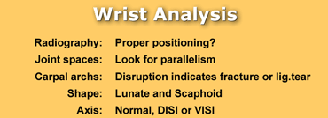
When you analyse the wrist to look for possible carpal instability and fracture dislocation, you should ask yourself the following questions:
- Is there good positioning of the patient. This is essential to be able to make statements about improper alignment or abnormal axes of carpal bones.
- Is there normal alignment between the carpal bones. The carpals should be parallel when profiled. Any overlap indicates abnormal tilting, dislocation or fracture.
- Is there any disruption of the three carpal archs. Disruption indicates ligament tear or a fracture.
- What is the shape and axis of the carpal bones. Give special attention to lunate, scaphoid and capitate. An abnormal shape indicates abnormal tilt with or without dislocation.
Answering these questions will help you find clues to carpal instability, dislocation and fractures.Khi bạn phân tích cổ tay để tìm kiếm sự mất ổn định của cổ tay và trật khớp xương, bạn nên tự hỏi mình những câu hỏi sau:
- Có vị trí tốt của bệnh nhân. Đây là điều cần thiết để có thể đưa ra tuyên bố về sự liên kết không đúng cách hoặc các trục bất thường của xương cổ tay.
- Có sự liên kết bình thường giữa các xương cổ tay. Các ống cổ tay phải song song khi được định hình. Bất kỳ sự chồng chéo nào cho thấy sự nghiêng, trật khớp hoặc gãy bất thường.
- Có bất kỳ sự gián đoạn nào của ba vòm cổ tay. Sự gián đoạn cho biết dây chằng bị rách hoặc gãy.
- Hình dạng và trục của xương cổ tay là gì. Đặc biệt chú ý đến Lunate, Scaphoid và Capitate. Một hình dạng bất thường cho thấy độ nghiêng bất thường có hoặc không có trật khớp.
Trả lời những câu hỏi này sẽ giúp bạn tìm ra manh mối về sự mất ổn định, trật khớp và gãy xương ở cổ tay.
RadiographyChụp X-quang
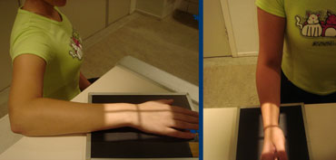
Positioning
PA view should be taken with the wrist and elbow at shoulder height. Only in this position, the radius and the ulna are parallel. Moving the arm down makes the radius cross the ulna and become relatively shorter. So it will be impossible to make any statements on the length of the ulna (plus or minus variant)
Lateral view is taken with the elbow adducted to the side. Shoulder, elbow and wrist are again in one plane. This positioning will make the lateral view exactly perpendicular to the PA view.Định vị
Chế độ xem PA nên được thực hiện với cổ tay và khuỷu tay ở độ cao ngang vai. Chỉ ở vị trí này, bán kính và trụ xương quay song song.Di chuyển cánh tay xuống làm cho bán kính đi qua trụ xương quay và trở nên tương đối ngắn hơn.Vì vậy, sẽ không thể đưa ra bất kỳ tuyên bố nào về chiều dài của trụ xương quay (cộng hoặc trừ biến thể)
Chế độ xem bên được thực hiện với khuỷu tay được đặt sang một bên. Vai, khuỷu và cổ tay lại ở trong một mặt phẳng.Vị trí này sẽ làm cho chế độ xem bên vuông góc chính xác với chế độ xem PA.
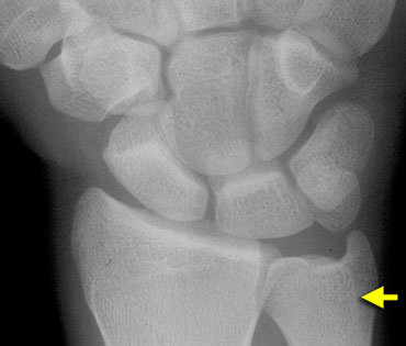 Extensor carpi ulnaris groove (yellow arrow) seen radial to the midportion of the ulnar styloid.Rãnh cơ duỗi cổ tay trụ (mũi tên màu vàng) nhìn thấy theo hướng xuyên tâm đến phần giữa của mỏm trâm trụ.
PA view
A correctly positioned PA view will show the extensor carpi ulnaris groove radial to the midportion of the ulnar styloid.
The PA and lateral view are equally important and thus should both be studied carefully.
The PA view usually shows what is wrong and the lateral view shows in what direction the bones move.
Sometimes an oblique view will also be obtained, especially if you want to look at the trapezium-trapezoid joint in profile.Chế độ xem PA
Chế độ xem PA được định vị chính xác sẽ cho thấy rãnh duỗi cổ tay trụ quay về phía giữa của mỏm trâm trụ.
Góc PA và góc nhìn bên đều quan trọng như nhau và do đó cả hai cần được nghiên cứu cẩn thận.
Phim PA thường cho thấy những gì không đúng và phim bên cho thấy hướng di chuyển của xương.
Đôi khi cũng có thể quan sát theo hướng xiên, đặc biệt nếu bạn muốn quan sát khớp hình thang-hình thang.

Lateral view
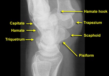
Only on a good positioned lateral view one can see the volar edges of respectively scaphoid, pisiform and capitate separately and lined up as shown on the left.
Lunate is the semilunar bone that fits in the distal radius. Looking through that, one can see the convexity of the scaphoid.
Distally from the scaphoid is the trapezium. The angular shaped bone visible dorsally is the triquetrum.
The square bone that bridges the proximal and distal half of the wrist is the pisiform.
Capitate is the rounded bone fitting inside the distal lunate.
Distally between the metacarpals, one can make out the hook of the hamate.Mặt bên
Chỉ khi nhìn bên có vị trí tốt, người ta mới có thể thấy các cạnh của xương thuyền, xương ức và xương chậu riêng biệt và xếp hàng như thể hiện ở bên trái.
Xương tà là xương bán nguyệt nằm ở đầu xa xương quay, nhìn qua đó, người ta có thể thấy sự lồi của xương thuyền.
Xương hình thang nằm ở mặt trước xương thuyền, xương có hình dạng góc cạnh nhìn thấy được ở mặt sau xương thuyền là xương tam giác.
Xương vuông bắc cầu nửa trên và nửa dưới cổ tay là xương chậu.
Xương chậu là xương tròn nằm bên trong xương nguyệt đầu xa.
Theo chiều dọc giữa các xương bàn tay, người ta có thể nhận ra móc của hamate.
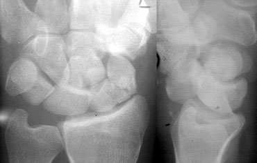 Same projection of ulna and ulnar styloid on PA and lateral view due to malpositioning in a patient with a perilunate fracture dislocation.Hình ảnh lồi trụ và xoắn trâm trụ trên phim XQCLVT và phim nghiêng do bệnh nhân trật khớp quanh xương nguyệt có tư thế sai.
Improper positioning may result in the same view of the ulna on both the PA and lateral view as shown in the case on the left. Nowhere in the body you would accept two views giving you the same image of a bone.Vị trí không đúng có thể dẫn đến cùng một góc nhìn của xương trụ trên cả hai góc PA và góc nhìn bên như trong trường hợp bên trái. Không nơi nào trong cơ thể bạn chấp nhận hai góc nhìn cho bạn cùng một hình ảnh của một xương.
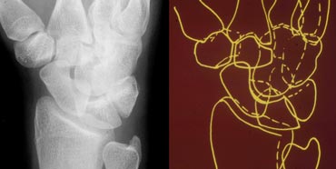 Normal oblique radiograph of the wrist and schematic representationHình ảnh X quang cổ tay và sơ đồ bình thường
Oblique view
An oblique view is not routinely performed. It is however the only view showing the trapezio-trapezoidal joint.Chế độ xem xiên
Chế độ xem chéo không được thực hiện thường xuyên, tuy nhiên đây là chế độ xem duy nhất cho thấy khớp hình thang.
Joint spaces: parallelism and symmetryKhông gian khớp: tính song song và tính đối xứng
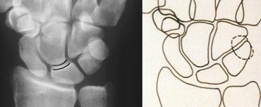
The joint spaces of the wrist have a width of 2 mm or less. Only the radiocarpal joint is slightly wider. The carpometacarpal joints are slightly narrower than the midcarpal joints.
The capitolunate joint is considered the baseline joint width to which other joint spaces can be compared. One should make sure to look at all of them: the radiocarpal, the proximal intercarpal, the midcarpal, the distal intercarpal and the carpometacarpal joint spaces. Khoảng khớp cổ tay có chiều rộng từ 2 mm trở xuống, chỉ có khớp cổ tay quay rộng hơn một chút, các khớp cổ tay - bàn tay hơi hẹp hơn các khớp cổ bàn tay giữa.
Khớp bàn ngón được coi là bề rộng khớp nền mà các khoảng khớp khác có thể được so sánh. Người ta nên chắc chắn rằng phải nhìn vào tất cả các khoảng khớp: cổ tay quay, cổ tay gần, cổ tay giữa, cổ tay xa và các khoảng khớp cổ tay.
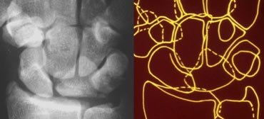
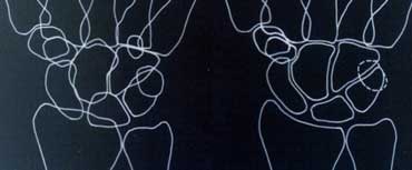 Study the carpal bones as pieces of a jigsaw puzzleLEFT: Schematic representation of the wrist with the outlines tracing the outer margins of the bonesRIGHT: Schematic representation of the wrist with the lines tracing the carpal bones as pieces of a jigsaw puzzleNghiên cứu xương cổ tay như những mảnh ghép hìnhTrái: Biểu diễn theo sơ đồ cổ tay với các đường viền theo dõi các bờ ngoài của xương QUYỀN: Biểu diễn bằng sơ đồ cổ Tay với các đường vẽ theo dõi xương cổ tay là những mảnh ghéP hình
Carpal joints should be symmetrical. Furthermore, when viewed in profile (tangentially), the cortical margins of the bones constituting that joint should be parallel. Bone edges that are not viewed in profile do not display this parallelism, e.g. the distal portion of the scaphoid that articulates with the capitate.
Studying this parallelism is easier when regarding the carpal bones as pieces of a jigsaw puzzle that all fit together, as opposed to tracing carpal bones by their outer cortical margins producing the outlines (figure).
When one bone is not paralleling the others, that is out of place. If the rest of the bones still parallel each other, they have stayed together.Khớp cổ tay phải đối xứng Hơn nữa, khi nhìn theo mặt cắt ngang (theo chiều dọc), các bờ vỏ xương của khớp tạo thành khớp đó phải song song Các cạnh xương không được nhìn theo mặt phẳng không hiển thị sự song song này, ví dụ như phần xa của xương thuyền khớp với xương thuyền.
Nghiên cứu sự song song này dễ dàng hơn khi xem xương cổ tay như những mảnh ghép hình ghép lại với nhau, trái ngược với việc theo dõi xương cổ tay bằng các cạnh vỏ ngoài của chúng tạo ra các đường viền (hình).
Khi một xương không song song với các xương khác, điều đó là không đúng vị trí. Nếu các xương còn lại vẫn song song với nhau, chúng sẽ ở cùng nhau.
 PA radiograph and schematic representation of the wrist, with dislocation of the lunateHình ảnh X-quang phổi và sơ đồ cổ tay, với trật khớp xương nguyệt
The picture on the left shows abnormal overlapping of the lunate with the capitate, hamate and triquetrum. We also see the medial profile surface of the scaphoid, but nothing paralleling it. There is also abnormal widening of the radiolunate space. The other joints are nicely parallel and symmetric. This leads to the conclusion that the lunate is displaced while the other bones have stayed together.Hình bên trái cho thấy sự chồng lấp bất thường của xương ổ chảo với xương ổ chày, xương bàn đạp và xương bàn đạp. Chúng ta cũng thấy bề mặt trong của xương thuyền, nhưng không có gì song song với nó. Ngoài ra còn có sự mở rộng bất thường của không gian xương ổ gà. Các khớp khác song song và đối xứng một cách độc đáo. Điều này dẫn đến kết luận rằng xương ổ gãy bị di dời trong khi các xương khác vẫn ở cùng nhau.
Carpal arcsVòng cung cổ tay
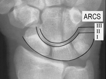
 PA radiograph of the wrist. The three normal carpal arcsX quang phổi cổ tay. Ba cung cổ tay bình thường
The next step is looking at the three carpal arcs: smooth curves joining the surfaces of the carpal bones as shown on the left.
The first arc is a smooth curve outlining the proximal convexities of the scaphoid, lunate and triquetrum.
The second arc traces the distal concave surfaces of the same bones, and the third arc follows the main proximal curvatures of the capitate and hamate. Bước tiếp theo là nhìn vào ba cung xương cổ tay: các đường cong nhẵn nối với bề mặt xương cổ tay như hình bên trái.
Vòng cung đầu tiên là một đường cong nhẵn phác thảo các điểm lồi gần của xương thuyền, xương nguyệt và xương tam giác.
Vòng cung thứ hai theo dõi các bề mặt lõm đầu xa của cùng một xương, và vòng cung thứ ba theo sau các đường cong đầu gần chính của xương ức và xương hàm.
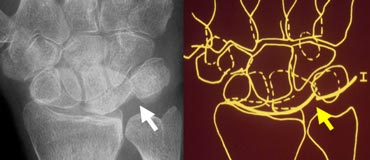 PA radiograph of the wrist and schematic representation showing disruption of the first carpal arcHình ảnh X-quang phổi của cổ tay và sơ đồ cho thấy sự gián đoạn của vòng cung cổ tay đầu tiên
Distruption of carpal arcs
An arc is disrupted if it cannot be traced smoothly. A break in one of the arcs indicates a fracture or the disruption of a ligament leading to a subluxation or dislocation.
On the left one can note the disruption of arc I at the lunotriquetral joint.Chẩn đoán hình ảnh cung cổ tay
Một vòng cung bị gián đoạn nếu nó không thể được theo dõi trơn tru. Việc đứt một trong các vòng cung cho thấy gãy xương hoặc đứt dây chằng dẫn đến trật khớp hoặc trật khớp.
Bên trái có thể thấy sự gián đoạn của cung I ở khớp ngón tay.
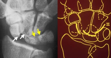 Disruption of the second carpal arcGián đoạn cung cổ tay thứ hai
A disruption of the second carpal arc at the scapholunate joint and the lunotriquetral joint is seen on the left. Although there is a gap in the first arc, it can still be traced by a smooth curve so arc I is considered intact.Sự gián đoạn của cung thứ hai ở khớp vai và khớp cổ tay được nhìn thấy ở bên trái, mặc dù có một khoảng trống trong cung thứ nhất, nó vẫn có thể được theo dõi bởi một đường cong trơn tru để cung I được coi là nguyên vẹn.
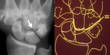 Disruption of the third carpal arc at the capitohamate jointGián đoạn cung cổ tay thứ ba ở khớp Capitohamate
Disruption of the third carpal arc is shown in the next case on the left. There is an abnormal step off at the capitohamate joint.Sự gián đoạn của vòng cung cổ tay thứ ba được thể hiện trong trường hợp tiếp theo bên trái. Có một bước bất thường ở khớp Capitohamate.
Shape of carpal bonesHình dạng xương cổ tay
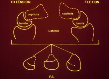
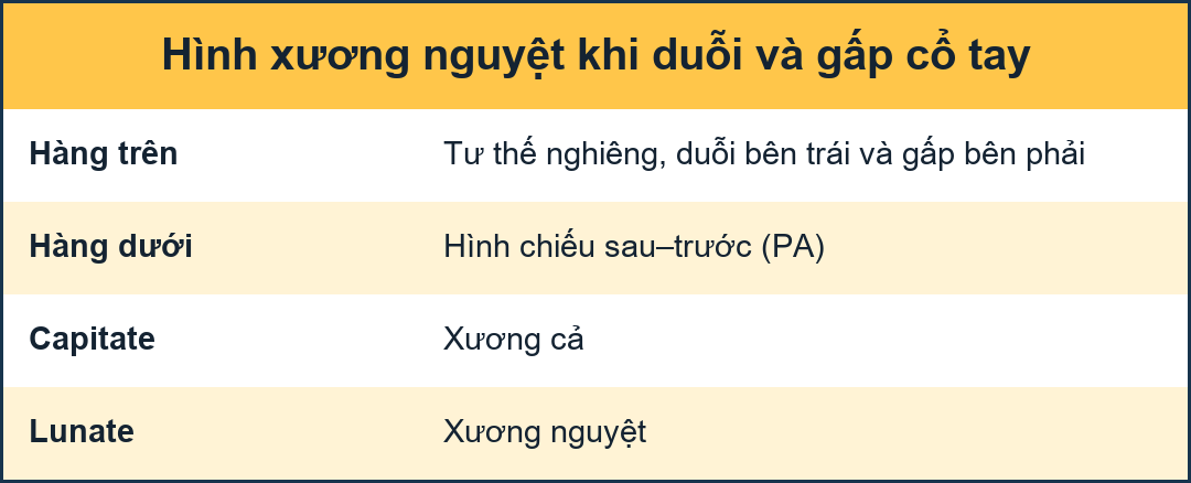 Schematic representation of the lunate shape in different positionsBiểu diễn hình mặt trăng ở các vị trí khác nhau
Lunate shapeHình trăng lưỡi liềm
The lunate has a trapezoidal shape, as the sides converge from the proximal surface to the distal surface, which are grossly parallel. If the lunate is tilted, it becomes triangular in shape. Awareness of this fact prevents thinking the lunate might be dislocated based only on its appearance, that in fact changes with its position.
So it may be dislocated with tilting or just be tilted.Mặt trăng có hình thang, khi các cạnh hội tụ từ bề mặt gần đến bề mặt xa, song song với nhau. Nếu mặt trăng bị nghiêng, nó sẽ trở thành hình tam giác. Nhận thức được thực tế này ngăn cản suy nghĩ mặt trăng có thể bị trật khớp chỉ dựa trên sự xuất hiện của nó, mà trên thực tế thay đổi theo vị trí của nó.
Vì vậy, nó có thể bị trật khớp khi nghiêng hoặc chỉ bị nghiêng.
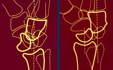 LEFT: Lunate dislocation: capitate is centered over the radius and lunate is tilted out.RIGHT: Perilunate dislocation: lunate is centered over the radius and capitate is tilted out dorsally.Trái, trật xương nguyệt: Xương cả nằm giữa trục xương quay, còn xương nguyệt bị nghiêng ra khỏi trục. Phải, trật quanh xương nguyệt: Xương nguyệt vẫn nằm giữa trục xương quay, còn xương cả nghiêng về phía mu tay.
Lunate vs. perilunate dislocationTrật quanh xương nguyệt so với trật xương nguyệt
Common dislocations of the wrist are the lunate and perilunate dislocations. The key to differentiation between both is what is centered over the radius. If the capitate is centered over the radius and the lunate is tilted out, it is a lunate dislocation. If however the lunate centers over the distal radius and the capitate is dorsal, we are dealing with a perilunate dislocation (figure).Trật khớp cổ tay thường gặp là trật khớp xương nguyệt và quanh xương nguyệt. Chìa khóa để phân biệt giữa hai trật khớp này là trung tâm của trật khớp nằm trên xương quay. Nếu xương quay nằm trên xương xoay và xương nguyệt nghiêng ra ngoài thì đó là trật khớp mặt nguyệt. Tuy nhiên, nếu xương quay nằm ở đầu xa xương quay và xương quay nằm phía lưng thì đó là một trật khớp quanh xương nguyệt (hình).
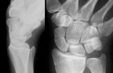 LEFT: Lateral radiograph of the wrist in extention showing scaphoid elongationRIGHT: PA radiograph of the wrist in ulnar deviation showing elongation of the scaphoidTRÁI: X quang cổ tay nghiêng cho thấy xương thuyền kéo dài QUYỀN: X quang PA cổ tay nghiênch cho thấy xương thủy tinh kéo dài
Scaphoid shapeHình dạng xương thuyền
The scaphoid shape changes with movement of the wrist. In ulnar deviation or extension the scaphoid elongates to fill the space between the radial styloid and the base of the thumb (the trapezium).Hình dạng xương thuyền thay đổi theo chuyển động của cổ tay. Trong trường hợp lệch trụ hoặc kéo dài xương thuyền kéo dài để lấp đầy khoảng trống giữa mỏm trâm quay và gốc ngón tay cái (cơ hình thang).
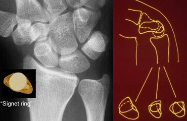
 LEFT: PA radiograph of the wrist in radial deviation showing foreshortening of the scaphoid: signet ring signRIGHT: Schematic representation of the wrist in flexion showing tilting of the scaphoid towards the palmTRÁI: Phim X-quang cổ tay quay nghiêng cho thấy dấu hiệu rút ngắn xương thuyền: dấu hiệu vòng signet QUYỀN: Biểu diễn hình ảnh cổ tay khi gấp cho thấy xương thuyền nghiêng về phía lòng bàn tay
Both with radial deviation aswell as flexion of the wrist the space between the radial styloid and trapezium is reduced. As scaphoid fills this space it will foreshorten and tilt towards the palm. This will give scaphoid a signet ring appearance (figure).Cả hai đều có độ lệch hướng tâm cũng như sự gấp của cổ tay, khoảng trống giữa trâm quay và hình thang đều giảm Khi xương thuyền lấp đầy khoảng trống này, nó sẽ rút ngắn lại và nghiêng về phía lòng bàn tay, điều này sẽ tạo cho xương thuyền một hình vòng tròn (hình).
Axis of the carpal bonesTrục xương cổ tay
Drawing the longitudinal axes of some of the carpal bones on a lateral radiograph and measuring the angles between them is a good method of determining the wrist bones? spatial relationship. The three most important axes are those through the scaphoid, the lunate and the capitate, drawn on the lateral radiograph.Vẽ trục dọc của một số xương cổ tay trên phim X quang nghiêng và đo góc giữa chúng là một phương pháp tốt để xác định mối quan hệ không gian của xương cổ tay, ba trục quan trọng nhất là trục qua xương thuyền, xương nguyệt và xương thuyền, được vẽ trên phim Xquang nghiêng.
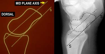
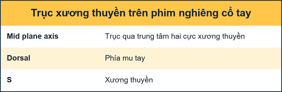
Scaphoid axisTrục vẹo
The true axis of the scaphoid is the line through the midpoints of its proximal and distal poles. Since the midpoint of the proximal pole is often difficult to appreciate, an almost parallel line can be used that is traced along the most ventral points of the proximal and distal poles of the bone (figure). Trục thực sự của xương thuyền là đường thẳng qua các điểm giữa của các cực đầu gần và cực đầu xa của nó. Vì điểm giữa của cực đầu gần thường khó đánh giá, nên có thể sử dụng một đường gần như song song được vẽ dọc theo các điểm trung tâm nhất của các cực gần và cực xa của xương (hình).
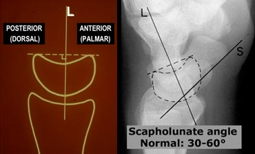
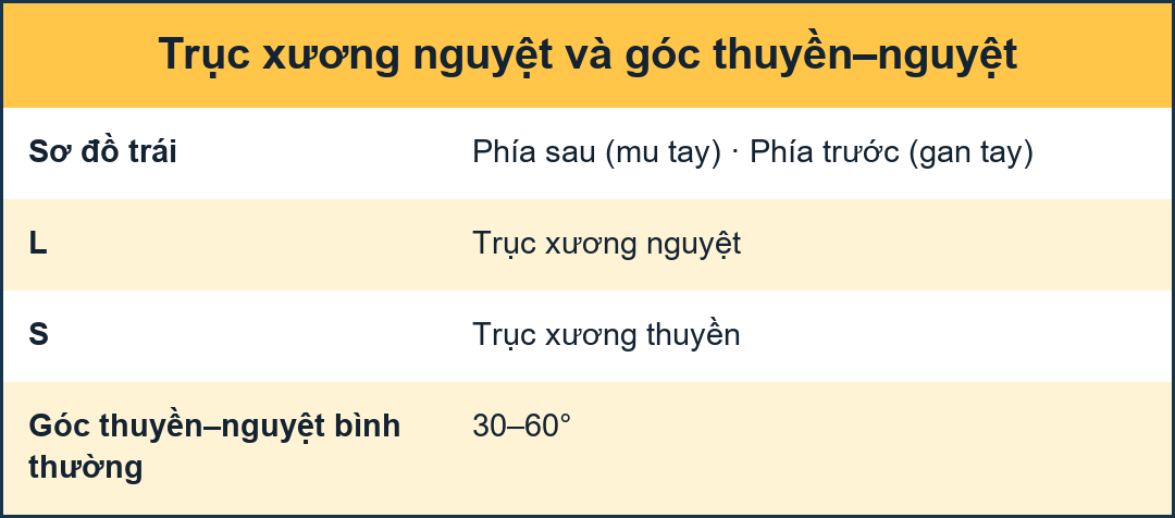 LEFT: Lunate axis drawn perpendicular to a line joining the distal palmar and dorsal bordersRIGHT: Radiograph with lunate and scaphoid axis drawn.TRÁI: Trục mặt trăng được vẽ vuông góc với đường nối giữa bờ xa của lòng bàn tay và bờ mu QUYỀN: X quang với trục mặt trăng và trục thuyền được vẽ.
Lunate axisTrục nguyệt
The axis of the lunate runs through the midpoints of the convex proximal and concave distal joint surfaces and can best be drawn by finding the perpendicular to a line joining the distal palmar and dorsal borders of the bone as demonstrated on the left. Trục của mặt trăng chạy qua các điểm giữa của mặt lồi đầu gần và mặt lõm đầu xa của khớp và có thể được vẽ tốt nhất bằng cách tìm đường vuông góc với đường nối với đường viền đầu xa của xương như được minh hoạ ở bên trái.
Scapholunate angle
Normal: 30 - 60?
Questionably abnormal: 60 - 80?
Abnormal: > 80? This indicates instability of the wrist.
Góc sọ mác
Bình thường: 30-60?
Bất thường đáng ngờ: 60-80?
Bất thường: > 80? Điều này cho thấy sự mất vững của cổ tay.
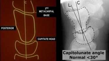
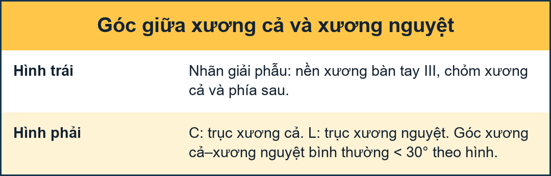 LEFT: Capitate axisRIGHT: Radiograph with lunate and capitate axis drawn.TRÁI: Trục chụp đầu QUÝ: X quang với trục mặt trăng và trục chụp đầu được vẽ.
Capitate axisTrục Capitate
The capitate axis joins the midportion of the proximal convexity of the third metacarpal and that of the proximal surface of the capitate.
Trục đầu xương cùng tham gia vào phần giữa của lồi đầu gần của xương bàn tay thứ ba và của bề mặt đầu gần của đầu xương cùng.
Capitolunate angle
Normal:
Abnormal: > 30?.This indicates instability of the wrist.
Góc vuông góc
Bình thường: Bất thường: > 30?. Điều này cho thấy sự mất vững của cổ tay.
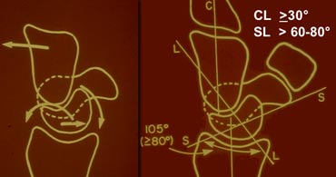
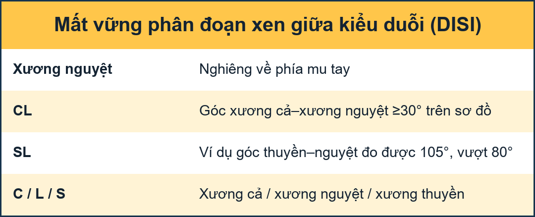 LEFT: Dorsal tilting of the lunate in DISIRIGHT: Scapholunate angle is > 80?TRÁI: Góc nghiêng của mặt trăng ở DISIRIGHT: Góc xương hàm là > 80?
DISI or dorsiflexion instabilityDISI hoặc mất ổn định gấp lưng
DISI is short for dorsal intercalated segmental instability.
The intercalated segment is the proximal carpal row identified by the lunate. The term 'intercalated segment' refers to it being the part in between the proximal segment of the wrist consisting of the radius and the ulna and the distal segment, represented by the distal carpal row and the metacarpals.
So all this means is that in DISI or dorsiflexion instability the lunate is angulated dorsally.
If you think lunate is tilted, measure the scapholunate angle ( 30-60?is normal, 60-80?is questionably abnormal, >80? is abnormal) and the capitolunate angle (
In the figure on the left the scapholunate angle is measured: it is 105 degrees. As mentioned before this angle is considered abnormal if greater then 80 degrees. DISI là viết tắt của bất ổn định phân đoạn có xen kẽ ở mặt lưng.
Đoạn kẽ là hàng cổ tay đầu gần được xác định bởi xương nguyệt Thuật ngữ 'đoạn xen kẽ' đề cập đến nó là phần nằm giữa đoạn gần của cổ tay bao gồm xương quay và xương trụ và đoạn xa, được đại diện bởi hàng cổ tay xa và xương bàn tay.
Vì vậy, tất cả điều này có nghĩa là trong DISI hoặc mất vững do gấp cơ lưng, xương nguyệt được tạo góc về phía lưng.
Nếu bạn nghĩ rằng mặt trăng bị nghiêng, hãy đo góc scapholunate (30-60? là bình thường, 60-80? là bất thường, > 80? là bất bình thường) và góc capitolunate ( Trong hình bên trái, góc scaphoun được đo: nó là 105 độ Như đã đề cập trước đây, góc này được coi là bất thường nếu lớn hơn 80 độ.
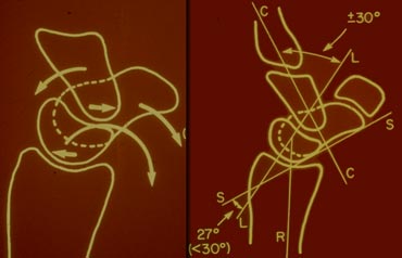
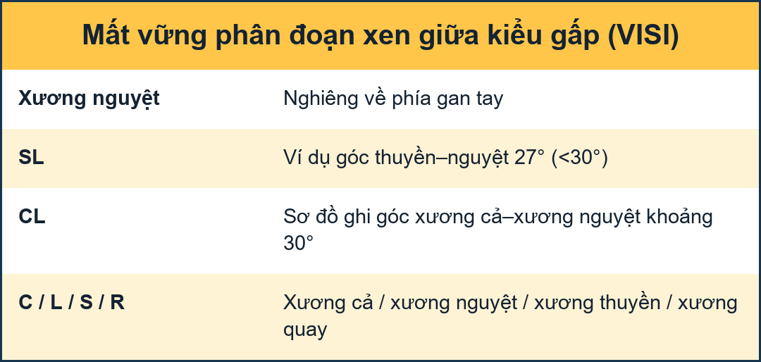 LEFT: Volar tilting of the lunate in VISIRIGHT: Scapholunate angle isTRÁI: Góc nghiêng của mặt trăng trong VISIRIGHT: Góc xương hàm là
VISI or volarflexion instabilityVISI hoặc mất ổn định volarflexion
Volar intercalated segmental instability or palmar flexion instability is when the lunate is tilted palmarly too much.
While most DISI is abnormal, in many cases VISI is a normal variant, especially if the wrist is very lax.Mất vững phân đoạn gan bàn tay hoặc mất vững gấp gan tay là khi mặt trăng nghiêng quá nhiều.
Trong khi hầu hết DISI là bất thường, trong nhiều trường hợp VISI là một biến thể bình thường, đặc biệt nếu cổ tay rất lỏng lẻo.
Systematic review and diagnosisTổng quan hệ thống và chẩn đoán
In the next cases we advise you to first look at the images on the left and give a full description of the radiographs.
Look for symmetry, parallelism, and the shape and axis of the carpal bones. Find out if there are any fractures and then try to make the diagnosis
Then read the text on the right to see if you're right.Trong các trường hợp tiếp theo, chúng tôi khuyên bạn nên xem hình ảnh bên trái và mô tả đầy đủ về phim X-quang.
Tìm sự đối xứng, song song, hình dạng và trục của xương cổ tay. Tìm hiểu xem có gãy xương nào không và sau đó cố gắng chẩn đoán.
Sau đó đọc văn bản bên phải để xem bạn có đúng không.
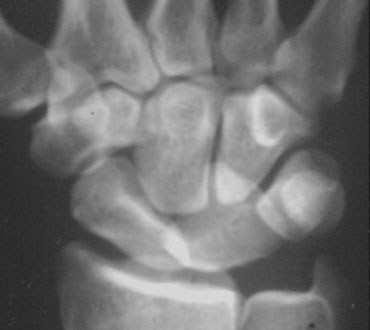 PA and lateral view of the wristPA và mặt bên của cổ tay
Case 1
Systematic interpretation of the case on the left shows us the following:
1. On the PA-view all the carpal bones parallel each other except for the lunate.
2. The carpal arcs I and II are disrupted at the LT and SL joints.
3. Triangular shaped lunate
So by just looking at the PA view we can make the diagnosis of lunate dislocation.Trường hợp 1
Giải thích hệ thống về trường hợp bên trái cho chúng ta thấy những điều sau:
1. Trên mặt phẳng PA-view, tất cả các xương cổ tay song song với nhau ngoại trừ xương nguyệt.
2. Các cung I và II của cổ tay bị phá vỡ ở các khớp CTG và CTG.
3. Mặt trăng hình tam giác
Vì vậy, chỉ cần nhìn vào hình ảnh của xương đòn ta có thể chẩn đoán trật khớp xương nguyệt.
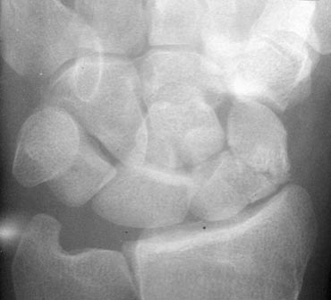
Case 2
Analysis:
1. No parallelism at the TL joint since there is overlapping of the triquetrum and the lunate.
2. Also overlapping of the hamate and the lunate.
3. There is parallelism between radius, lunate, proximal pole of scaphoid and proximal pole of capitate. So these bones form a unit.
4. Also parallelism between triquetrum, hamate, distal pole of capitate, trapezium and distal pole of scaphoid.
5. Fracture of capitate and scaphoid
So these findings indicate that this is a transscaphoid, transcapitate perilunate fracture-dislocation.Trường hợp 2
Phân tích:
1. Không có sự song song ở khớp TL vì có sự chồng chéo của triquetrum và mặt trăng.
2. Cũng chồng chéo của hamate và lunate.
3. Có sự song song giữa xương quay, xương nguyệt, xương đầu gần xương thuyền và xương đầu gần thân thuyền, do đó các xương này tạo thành một đơn vị.
4. Cũng song song giữa triquetrum, hamate, cực xa của capitate, trapezium và cực xa của scaphoid.
5. Gãy xương thuyền và xương thuyền
Như vậy, đây là một trường hợp gãy xương quanh nguyệt, xuyên xương thuyền.
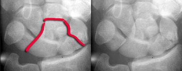
On the left you see the same case with a line indicating the fracture-dislocation line.Ở bên trái, bạn thấy cùng một trường hợp với một đường chỉ ra đường gãy xương.
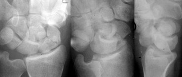
Same case with additional oblique and lateral view showing the dorsal dislocation.Cùng một trường hợp với góc nghiêng và nghiêng cho thấy trật khớp lưng.
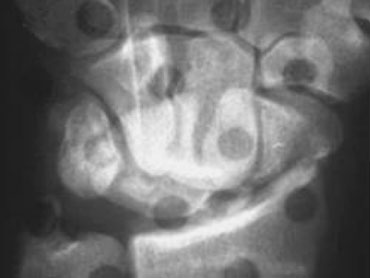
Case 3
Analysis:
1. Fracture of scaphoid and ulnar styloid process.
2. Broken arcs I and II at LT joint.
3. Some parallelism between lunate and proximal pole of scaphoid with the radius.
4. Scaphoid is foreshortened so it is tilted and has moved towards the palm.
5. All the other carpals show parallelism exept for lunate, the proximal pole of scaphoid and the radius.
Although this probably is a perilunate dislocation, based on the PA-view alone it is very difficult to say if this is a lunate or perilunate dislocation. The triangular shape of the lunate could be the result of just tilting or dislocation with tilting.Trường hợp 3
Phân tích:
1. Gãy xương thuyền và trụ trâm.
2. Gãy cung I và II ở khớp CTG.
3. Một số điểm song song giữa mặt trăng và cực gần của xương thuyền với xương quay.
4. Lách được rút ngắn lại để nó nghiêng và đã di chuyển về phía lòng bàn tay.
5. Tất cả các xương cổ tay khác đều có hình dạng song song với xương nguyệt, cực gần xương thuyền và xương quay.
Mặc dù đây có thể là trật khớp quanh xương nguyệt, nhưng chỉ dựa vào góc nhìn PA thì rất khó để nói đây là trật khớp nguyệt hay trật quanh xương nguyệt. Hình dạng tam giác của xương nguyệt có thể là kết quả của việc nghiêng hoặc trật khớp với nghiêng.
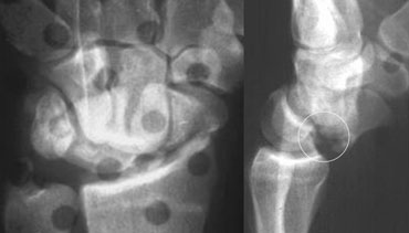 Perilunate dislocation with fracture of scaphoid and ulnar styloid processThe volar tip of lunate is also broken (see circle).Trật khớp quanh xương nguyệt kèm theo gãy xương thuyền và xương trụ mỏm trâm Đầu xương của xương nguyệt cũng bị gãy (xem hình tròn).
Same case with the lateral view also shown.
Now we see that there definitely is a perilunate dislocation.
So the triangular shape of the lunate is the result of just tilting.
On the lateral view a fracture of the volar tip of lunate is seen. So this patient is at risk for recurrent dislocation.Trường hợp tương tự với góc nhìn bên cũng được hiển thị.
Bây giờ chúng ta thấy rằng chắc chắn có trật khớp quanh xương nguyệt.
Vì vậy, hình dạng tam giác của mặt trăng là kết quả của việc nghiêng.
Trên phim nghiêng có thể thấy gãy đầu xương ức của xương nguyệt, do đó bệnh nhân này có nguy cơ bị trật khớp tái phát.
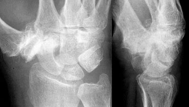
Case 4
Analysis:
The case on the left shows severe arthrosis at the STT joint and CMC1 joint with subluxation. Carpal arcs are normal and there is normal paralelism.The scaphoid is elongated which means it is dorsally tilted.
On the lateral view we can see that the lunate is also tilted dorsally. The proximal carpal row has moved as a unit, so there is no dissociation.
Final diagnosis: non-dissociated DISI with arthrosis and subluxation of STT joints.Trường hợp 4
Phân tích:
Trường hợp bên trái cho thấy khớp STT và CMC1 bị viêm nặng, bán trật khớp, cung cổ tay bình thường, khớp song song bình thường, xương thuyền kéo dài, có nghĩa là xương cổ tay nghiêng về phía lưng.
Mặt bên xương quay cũng nghiêng về phía lưng, hàng cổ tay gần di chuyển như một đơn vị, do đó không có sự phân ly.
Chẩn đoán cuối cùng: DISI không phân ly với khớp dạng thấp và khớp dạng thấp.
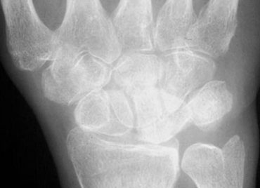
Case 5
Analysis:
1. Loss of parallelism at LT joint resulting in broken arc I and II.
2. Lunate and scaphoid are parallel to each other but not to the other carpals.
3. Scaphoid is foreshortened due to palmar tilting.
4. Lunate is parallel to scaphoid. So the triangular shape must be the result of palmar tilting.
5. The proximal carpal row is not a unit since arc I is broken.
Trường hợp 5
Phân tích:
1. Mất tính song song ở khớp CTG gây gãy cung I và II.
2. Mặt trăng và xương thuyền song song với nhau nhưng không song song với các xương bàn tay khác.
3. Lách to bị rút ngắn do lòng bàn tay nghiêng.
4. Mặt trăng song song với xương thuyền, do đó hình tam giác phải là kết quả của sự nghiêng lòng bàn tay.
5. Hàng cổ tay gần không phải là một đơn vị kể từ khi vòng cung I bị phá vỡ.
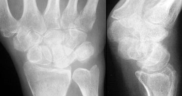 Lateral view demonstrates the volar tilting of lunate which was already suspected on the PA view.Mặt cắt ngang cho thấy sự nghiêng của mặt trăng đã bị nghi ngờ trên mặt cắt PA.
Final diagnosis:
VISI with dissociation at the LT joint.Chẩn đoán cuối cùng:
VISI có phân ly tại khớp CTG.
Case 6
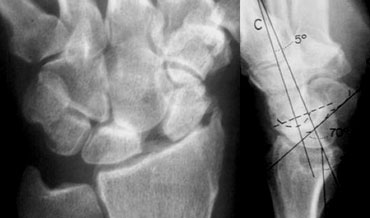
Analysis:
1. Widened and narrowed joints, but there is normal parallelism , so there is no dislocation.
2. Scapholunate dissociation with widening of the SL joint and foreshortening of the scaphoid due to palmar tilt.
3. Arthrosis of the Radioscaphoid and Capitolunate joint due to the abnormal movements of scaphoid and lunate.
4. Dorsal tilt of lunate indicating DISI.
This condition is known as SLAC. In this case it is post-traumatic due to the SL-ligament tear.
Trường hợp 6
Phân tích:
1. Khớp rộng và hẹp, nhưng có sự song song bình thường, do đó không có trật khớp.
2. Phân ly xương thuyền với sự mở rộng của khớp SL và rút ngắn xương thuyền do nghiêng lòng bàn tay.
3. Khớp cổ xương thuyền và xương chũm do sự di chuyển bất thường của xương thuyền và mặt trăng. 4. Độ nghiêng về phía sau của mặt trăng cho thấy DISI.
Tình trạng này được gọi là SLAC, trong trường hợp này là sau chấn thương do rách dây chằng SL.
SLAC (scapholunate advanced collapse) refers to a specific pattern of osteoarthritis and subluxation which results from untreated chronic scapholunate dissociation or from chronic scaphoid non-union
The degenerative changes occur in areas of abnormal loading, which is the radial-scaphoid joint, followed by degeneration in the unstable lunatocapitate joint, as capitate subluxates dorsally on lunate.
SLAC (sụp xương thuyền tiến triển) đề cập đến một mô hình cụ thể của thóai hóa khớp và bán trật khớp là kết quả của sự phân ly xương thuyền mạn tính không được điều trị hoặc từ sự không liên kết xương thuyền mạn
Những thay đổi thóai hóa xảy ra ở các khu vực có tải trọng bất thường, đó là khớp xương quay-khớp vai, tiếp theo là thóai hóa ở khớp xương mặt trăng không ổn định, khi khớp xương quay dưới khớp lưng.
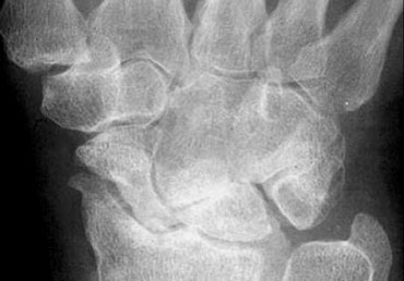 SLAC of CPPD TYPESLAC của CPPD TYPE
On the left another case of SLAC. This case is due to CPPD.
Characteristics of CPPD with SLAC are:
- Decreased size of proximal scaphoid due to erosion and resorption.
- Scaphoid fossa erosionBên trái là một trường hợp khác của SLAC, trường hợp này là do CPPD.
Đặc điểm của CPPD với SLAC là:
- Giảm kích thước xương thuyền đầu trên do ăn mòn và tiêu xương.
- Xói mòn hố Scaphoid