
FracturesGãy xương
Radiology department of the Rijnland Hospital in Leiderdorp, the NetherlandsKhoa Chẩn đoán hình ảnh của bệnh viện Rijnland, Leiderdorp, Hà Lan
Ngày đăng / cập nhật: 2008-01-12
Bản gốc trên radiologyassistant.nl ↗ · tải về lúc 2026-11-19 20:19 · bản dịch máy có chuẩn hóa thuật ngữ chẩn đoán hình ảnh
Fractures of the distal radius account for one-sixth of all fractures seen in the emergency department.
The radiologist must possess an understanding of the factors that alter clinical decision making and patient treatment.
In this review we will discuss:Gãy đầu dưới xương quay chiếm một phần sáu số trường hợp gãy xương được phát hiện ở khoa cấp cứu.
Bác sĩ chẩn đoán hình ảnh phải hiểu rõ các yếu tố làm thay đổi quyết định lâm sàng và điều trị cho bệnh nhân.
Trong bài tổng quan này, chúng ta sẽ thảo luận:
- Importance of standard radiography of the wristTầm quan trọng của X quang cổ tay chuẩn
- Proper description of fractures with knowledge of injury mechanisms, consequent care and complicationsMô tả đúng gãy xương với kiến thức về cơ chế chấn thương, hậu quả chăm sóc và biến chứng
- Commonly used fracture eponyms like Colles', Smith's, Barton's etc.Các tên gọi gãy xương thường được sử dụng như Colles ’, Smith ’ s, Barton ’ s v.v.
- AO- and Fernandez classificationPhân loại AO- và Fernandez
- TreatmentĐiều trị
- ComplicationsCác biến chứng
ImagingHình ảnh
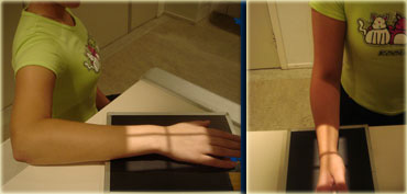 PA view: wrist and elbow at shoulder heightLateral view: shoulder, elbow and wrist in sagittal planeChế độ xem PA: cổ tay và khuỷu tay ở độ cao vai Chế độ xem bên: vai, khuỷu và cổ tay ở mặt phẳng đứng dọc
PositioningĐịnh vị
PA view should be taken with the wrist and elbow at shoulder height.
This means that the wrist, elbow and shoulder are all in the transverse plane, perpendicular to the x-ray beam.
Only in this position, the radius and the ulna are parallel.
Lowering the arm makes the radius cross the ulna and become relatively shorter resulting in improper measurement of the length of the radius.
Nên chụp phim tư thế nằm sấp ở cổ tay và khuỷu tay.
Điều này có nghĩa là cổ tay, khuỷu tay và vai đều nằm trên mặt phẳng ngang, vuông góc với chùm tia X.
Chỉ ở vị trí này, xương quay và xương trụ song song.
Hạ thấp cánh tay làm cho bán kính đi qua xương trụ và trở nên tương đối ngắn hơn dẫn đến việc đo chiều dài của bán kính không đúng cách.
Lateral view is taken with the elbow adducted to the side.
Shoulder, elbow and wrist are again in one plane, i.e. the sagittal plane.
This positioning will make the lateral view exactly perpendicular to the PA view.
Chụp nghiêng với khuỷu tay khép sang một bên.
Vai, khuỷu tay và cổ tay lại nằm trong một mặt phẳng, tức là mặt phẳng đứng dọc.
Vị trí này sẽ làm cho mặt bên vuông góc chính xác với mặt PA.
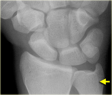
On a correctly positioned PA view the extensor carpi ulnaris tendon groove (arrow) can be seen.
The extensor carpi ulnaris tendon groove should be at the level of or radial to the base of the ulnar styloid.
Trên màn hình PA được định vị chính xác, có thể nhìn thấy rãnh gân cơ duỗi cổ tay trụ (mũi tên).
Rãnh gân cơ duỗi cổ tay trụ phải ngang mức hoặc hướng ra ngoài với nền của trụ trụ.
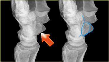 True lateral: The palmar cortex of pisiform (red arrow) lies between the palmar cortex of scaphoid and capitate.Bên thật: Vỏ gan tay hình quả lê (mũi tên màu đỏ) nằm giữa vỏ gan tay hình thuyền và hình đầu người.
A true lateral view is defined by the relationship between the pisiforme, capitate and scaphoid bones.
On a standard lateral view, the palmar cortex of the pisiform bone should overlie the central third of the interval between the palmar cortices of the distal scaphoid pole and the capitate head.Một mặt cắt thực sự được xác định bởi mối quan hệ giữa xương bàn chân, xương đùi và xương thuyền.
Trên mặt cắt ngang tiêu chuẩn, vỏ gan tay của xương đáy phải nằm trên một phần ba trung tâm của khoảng cách giữa các vỏ gan tay ở đầu xa xương thuyền và đầu thuyền.
Apparent volar tilt of the surface of the distal radius, as measured on the lateral view, increases with supination and decreases with pronation of the wrist (5).
A change of 10 degrees rotation between two consecutive control lateral radiographs is not uncommon during clinical follow-up and results in 5 degrees change in apparent tilt.Độ nghiêng rõ ràng của bề mặt của đầu xa xương quay, được đo ở mặt bên, tăng khi ngửa và giảm khi sấp cổ tay (5).
Sự thay đổi độ xoay 10 độ giữa hai phim X quang bên đối chứng liên tiếp không phải là hiếm trong quá trình theo dõi lâm sàng và dẫn đến sự thay đổi độ nghiêng rõ ràng là 5 độ.
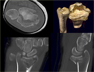
CT should be performed if conventional radiographs provide insufficient detail about radiocarpal articular step-off and gap displacement.
On the left a patient with a communitive intraarticular fracture of the distal radius with displacement of the volar rim of the radius together with the carpus (i.e. a volar Barton's).
There is an axial CT image with 3D-, coronal and sagittal reconstructiosn.
CT nên được thực hiện nếu X quang thông thường không cung cấp đủ chi tiết về sự di lệch và di lệch khớp cổ tay.
Bên trái là một bệnh nhân bị gãy đầu dưới xương quay kèm theo di lệch bờ xương quay cùng với xương đùi (tức là một xương Barton).
Hình ảnh CT dọc với cấu trúc 3D-, vành và dọc.
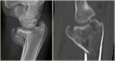
On the left sagittal reconstructions of 1mm axial CT slices.
Scroll through the images and notice how well CT demonstrates the fracture components and the displacement.Trên hình ảnh tái tạo dọc trái của các lát cắt CT dọc trục 1 mm.
Cuộn qua các hình ảnh và chú ý đến mức độ CT thể hiện các thành phần gãy xương và sự di lệch.
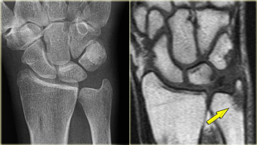
Magnetic resonance (MR) imaging is of benefit when concomitant injuries of ligaments and triangular fibrocartilage complex (TFCC) are suspected or if a fracture is suspected but not demonstrated on routine radiographs.
Cộng hưởng từ (MR) có lợi khi nghi ngờ có tổn thương đồng thời dây chằng và phức hợp xơ vữa hình tam giác (TFCC) hoặc nếu nghi ngờ gãy xương nhưng không được chứng minh trên X quang thường quy.
On the left a fracture of the ulnar styloid process not visible on standard radiography, but clearly demonstrated with MR.Bên trái gãy mỏm trâm trụ không thấy trên phim X quang chuẩn, nhưng được chứng minh rõ ràng bằng cộng hưởng từ.
MeasurementsĐo lường
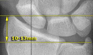
Radial length or height
Radial length is measured on the PA radiograph as the distance between one line perpendicular to the long axis of the radius passing through the distal tip of the radial styloid.
A second line intersects distal articular surface of ulnar head.
This measurement averages 10-13 mm.
Chiều dài hoặc chiều cao xuyên tâm
Chiều dài xương quay được đo trên phim PA là khoảng cách giữa một đường thẳng vuông góc với trục dài của xương quay đi qua đầu xa của mỏm trâm quay.
Đường thứ hai cắt ngang mặt khớp xa của đầu trụ.
Kích thước này trung bình 10-13 mm.
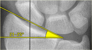
Radial inclination or angle
Radial inclination represents the angle between one line connecting the radial styloid tip and the ulnar aspect of the distal radius and a second line perpendicular to the longitudinal axis of the radius.
The radial inclination ranges between 21 and 25 degrees .
Loss of radial inclination will increase the load across the lunate.Độ nghiêng hoặc góc quay
Độ nghiêng của xương quay thể hiện góc giữa một đường nối đầu trâm quay và mặt trụ của đầu xa xương quay và đường thứ hai vuông góc với trục dọc của xương quay.
Độ nghiêng xuyên tâm dao động từ 21 đến 25 độ.
Mất độ nghiêng hướng tâm sẽ làm tăng tải trọng trên mặt trăng.
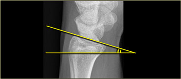
Radial tilt
Radial tilt is measured on a lateral radiograph.
The radial tilt represents the angle between a line along the distal radial articular surface and the line perpendicular to the longitudinal axis of the radius at the joint margin.
The normal volar tilt averages 11 degrees and has a range of 2-20 degrees .Nghiêng xuyên tâm
Độ nghiêng quay được đo trên phim X quang.
Độ nghiêng của khớp quay thể hiện góc giữa đường thẳng dọc theo mặt khớp quay đầu xa và đường thẳng vuông góc với trục dọc của xương quay ở rìa khớp.
Độ nghiêng bình thường trung bình 11 độ và có phạm vi từ 2-20 độ.
Radiological InterpretationGiải thích X quang
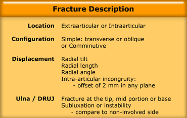
There are many ways to describe distal radial fractures and there are several classification systems.
In clinical practice however frequently eponyms like Colles' and Barton's are used.
When these epomyms are used, an accurate description of the fracture characteristics should always be included in the report(5).
In addition it should also be noted if there is osteoporosis or additional findings such as ligamentous injuries.
Có nhiều cách mô tả gãy đầu dưới xương quay và có nhiều hệ thống phân loại.
Tuy nhiên, trong thực hành lâm sàng, các từ như Colles 'và Barton' s thường được sử dụng.
Khi sử dụng các epomyms này, mô tả chính xác các đặc điểm gãy xương phải luôn được đưa vào báo cáo (5).
Ngoài ra, cần lưu ý nếu có loãng xương hoặc các dấu hiệu bổ sung như chấn thương dây chằng.
We will discuss the following: Chúng ta sẽ thảo luận về những điều sau:
- Description of the fracture characteristics (5) Mô tả đặc điểm gãy xương (5)
- Desciption of the commonly used fracture eponyms Mô tả các tên gọi thường dùng trong gãy xương
- Most popular classification systemsCác hệ thống phân loại phổ biến nhất
LocationVị trí
One of the most important characteristics is whether a fracture is extraarticular or intraarticular.
Extraarticular fractures are usually less complicated, unless they are comminutive.
Intraarticular fractures either involve the radiocarpal joint, distal radioulnar joint, or both.
Một trong những đặc điểm quan trọng nhất là gãy ngoài khớp hay gãy trong khớp.
Gãy ngoài khớp thường ít phức tạp hơn, trừ khi chúng là loại nhỏ.
Gãy nội khớp có thể liên quan đến khớp quay cổ tay, khớp quay đầu xa hoặc cả hai.
ConfigurationCấu hìnhName
Always mention whether the fracture is transverse (good prognosis), oblique or comminuted (multifragmented).
When a fracture is oblique or when it is comminuted with crossing of the mid axial line, it can be unstable.
Luôn đề cập đến gãy xương ngang (tiên lượng tốt), gãy chéo hoặc gãy vụn (gãy nhiều mảnh).
Khi gãy chéo hoặc khi nó bị gãy với đường ngang trục giữa, nó có thể không ổn định.
On the left a patient with an extraarticular distal radius fracture.
Notice the oblique course on the lateral view.
Fractures with this configuration frequently show loss of reduction at follow up and need surgical treatment.
Bên trái là một bệnh nhân gãy đầu dưới xương quay ngoài khớp.
Chú ý hướng nghiêng trên mặt nghiêng.
Gãy xương có cấu hình này thường mất độ nắn chỉnh khi theo dõi và cần điều trị phẫu thuật.
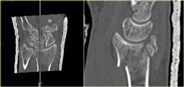
On the left a sagittal reconstruction of an oblique intraarticular fracture of the distal radius.
This is a volar Barton's type fracture.
Even in a cast the volar fragment will show progressive displacement at follow-up and a volar buttress plate is needed to hold the volar rim in place.Bên trái là hình ảnh tái tạo dọc của gãy chéo trong khớp đầu dưới xương quay.
Đây là loại gãy xương Barton dạng xoắn đỉnh.
Ngay cả trong quá trình bó bột, mảnh xương ức sẽ cho thấy sự dịch chuyển tiến triển khi theo dõi và cần có tấm trụ xương ức để giữ cố định vành xương ức.
DisplacementDịch chuyển
Fractures are either displaced or nondisplaced.
A fracture with an offset of 2 mm or more in any plane or 2 mm offset involving the articular surface is considered displaced.
Displacement can be dorsal, volar, radial or proximal.
Axial shortening, radial inclination and radio-ulnar displacement can be measured on the routine posterior/anterior film.
Dorsal tilt and dorsal or palmar displacement can be measured on the routine lateral X-ray.
Fragment displacement and rotation may be further determined using CT.Gãy xương có thể di lệch hoặc không di lệch.
Gãy xương có độ lệch từ 2 mm trở lên ở bất kỳ mặt phẳng nào hoặc độ lệch 2 mm liên quan đến bề mặt khớp được coi là di lệch.
Di lệch có thể là ở mặt lưng, mặt lưng, hướng tâm hoặc đầu gần.
Có thể đo ngắn trục, nghiêng trục và chuyển vị quay-trụ trên phim chụp sau / trước thường quy.
Nghiêng lưng và di lệch lưng hoặc lòng bàn tay có thể được đo trên X-quang thường quy.
Sự dịch chuyển và xoay của các mảnh có thể được xác định thêm bằng CT.
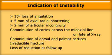
InstabilityMất ổn định
Instability is defined as a high risk of secondary displacement after initial adequate reduction.
Radiographic signs that favor instability are displacement and an oblique or comminuted configuration (as mentioned above).
These signs are listed in the table on the left.
Although the initial x-ray after reduction may look good, always look for loss of reduction at follow up.
Articular incongruity is the most important factor in the development of posttraumatic osteoarthritis of the wrist.
Mất vững được định nghĩa là nguy cơ cao của di lệch thứ phát sau khi nắn chỉnh đầy đủ ban đầu.
Các dấu hiệu X quang ủng hộ sự mất ổn định là sự dịch chuyển và cấu hình xiên hoặc phức tạp (như đã đề cập ở trên).
Những dấu hiệu này được liệt kê trong bảng bên trái.
Mặc dù chụp X-quang ban đầu sau khi nắn chỉnh có thể nhìn tốt, luôn tìm kiếm sự mất nắn giảm khi theo dõi.
Sự bất thường khớp là yếu tố quan trọng nhất trong sự phát triển của thóai hóa khớp cổ tay sau chấn thương.
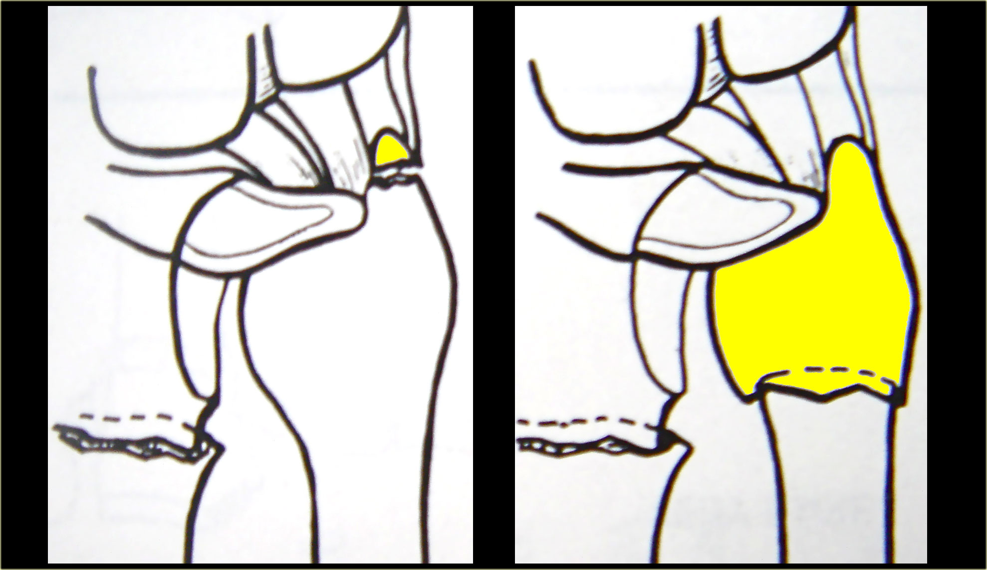 Avulsion of the tip or stable fractures of the ulnar neckNhổ đầu xương trụ hoặc gãy ổn định cổ trụ
Ulna and Distal radioulnar joint (DRUJ)Khớp trụ ngoài và khớp trụ ngoài xa (DRUJ)
Assessment of a wrist fracture must also include a description of the distal ulna and distal radioulnar joint (9).
The distal ulna articulates with the sigmoid notch of the radius.
Đánh giá gãy xương cổ tay cũng phải bao gồm mô tả về khớp trụ xa và khớp quay xa (9).
Đầu xa xương trụ khớp với rãnh sigma của xương quay.
Type I: stable
Avulsion fractures of the tip of the ulnar styloid and stable fractures of the ulnar neck have a good prognosis.
Following reduction of the radius the DRUJ is congruent and stable.
Extraarticular unstable fractures however, require plate fixation.Loại I: ổn định
Gãy nhô đầu trụ và gãy ổn định cổ trụ có tiên lượng tốt.
Sau khi giảm bán kính, DRUJ đồng đều và ổn định.
Tuy nhiên, gãy không vững ngoài khớp cần phải được cố định bằng nẹp vít.
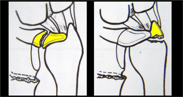 Tear of the TFCC or avulsion of the base of the ulnar styloidRách TFCC hoặc bong tróc nền sụn trâm trụ
Type II: unstable
There is subluxation or dislocation of the ulnar head as a result of avulsion of the base of the ulnar styloid or tear of the TFCC and/or capsular ligaments.
The subluxation has to be reduced with closed or operative treatment to avoid chronic instability and arthosis.
Loại II: không ổn định
Có trật khớp hoặc trật khớp đầu trụ do nhổ bỏ nền của mỏm trâm trụ hoặc rách TFCC và / hoặc dây chằng bao khớp.
Mất vững khớp phải được giảm bớt khi điều trị kín hoặc phẫu thuật để tránh mất vững và khớp mạn tính.
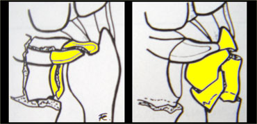 Intraarticular fracture of sigmoid notch or ulnar headModified and reprinted with permission from reference (9)Gãy nội khớp chỏm xương chũm hoặc chỏm trụ Sửa đổi và in lại với sự cho phép của tài liệu tham khảo (9)
Type III: potentially unstable
Intraarticular fractures of the sigmoid notch and intraarticular fractures of the ulnar head are potentially unstable because the incongruity of the DRUJ.
Subluxation is possible.Loại III: có khả năng không ổn định
Gãy trong khớp chỏm xương cùng và gãy trong khớp đầu trụ có khả năng không ổn định do sự không tương đồng của DRUJ.
Có thể có trật khớp dưới.
Common Fracture EponymsCác ngoại danh gãy xương thường gặp
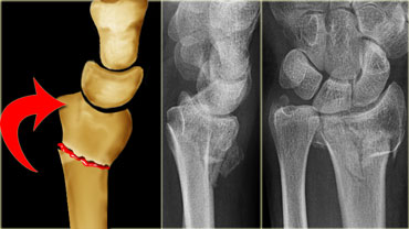
Colles' fractureGãy Colles
A Colles' fracture is a fracture of the distal metaphysis of the radius with dorsal angulation and displacement leading to a 'silver fork deformity'.
Colles fractures are seen more frequently with advancing age and in women with osteoporosis.
Gãy Colles là gãy xương đầu xa xương quay với góc xương lưng và di lệch dẫn đến 'biến dạng chĩa bạc'.
Gãy Colles gặp nhiều hơn ở tuổi cao và ở phụ nữ bị loãng xương.
In many cases a Colles' fracture is an extraarticular, uncomplicated and stable fracture, but it can be intraarticular.
So look for signs of instability in all Colles' fractures, especially:Trong nhiều trường hợp, gãy Colles là gãy ngoài khớp, không biến chứng và ổn định, nhưng có thể gãy trong khớp.
Vì vậy, hãy tìm kiếm các dấu hiệu mất vững trong tất cả các loại gãy Colles, đặc biệt là:
- Intraarticular radiocarpal or DRUJ extension of the fracture Xạ đốt trong khớp cổ tay hoặc DRUJ mở rộng của gãy xương
- Radial shortening Rút ngắn xuyên tâm
- Loss of radial inclinationMất độ nghiêng quay
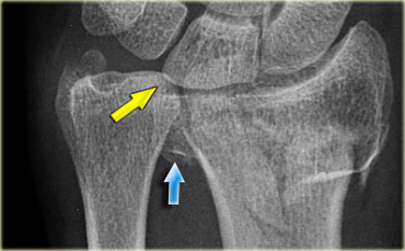
On the left a detailed AP view of the same patient as above.
In addition to the dorsal angulation seen on the lateral view, notice the following:Bên trái là hình ảnh chi tiết của cùng một bệnh nhân như trên.
Ngoài góc lưng nhìn từ mặt bên, hãy chú ý những điểm sau:
- Extension into the radiocarpal joint and the distal radioulnar joint Mở rộng vào khớp cổ tay quay và khớp quay xa
- Displacement of the articular fragments (blue arrow) Sự dịch chuyển của các mảnh khớp (mũi tên màu xanh)
- Radial shortening and loss of radial inclination resulting in distal ulna abutting the lunate (yellow arrow) Rút ngắn xương quay và mất độ nghiêng xương quay dẫn đến xương trụ ở đầu xa xương nguyệt (mũi tên màu vàng)
- Fracture of ulnar styloidGãy xương trụ xoắn trâm
Just calling this fracture a Colles' fracture would be insufficient.
All the characteristics have to be mentioned in the radiology report to convey the full extent of the injury, possible complications and treatment.Chỉ gọi gãy này là gãy Colles là không đủ.
Tất cả các đặc điểm cần được đề cập trong báo cáo X quang để truyền đạt đầy đủ mức độ tổn thương, các biến chứng có thể xảy ra và điều trị.
Smith's fractureGãy Smith
Smith's fractures occur in younger patients and are the result of high energy trauma on the volar flexed wrist.
Volar comminution and intraarticular extension are more common.Gãy Smith xảy ra ở những bệnh nhân trẻ tuổi và là kết quả của chấn thương năng lượng cao trên cổ tay gấp.
Mất vững và lan rộng trong khớp là phổ biến hơn.
On the left an extraarticular Smith's fracture with palmar and radial angulation and displacement.
There is also an avulsion of the ulnar styloid process.Bên trái gãy Smith ngoài khớp với góc gan tay và quay và di lệch.
Ngoài ra còn có một chỗ nhổ của mỏm trâm trụ.
Barton's fractureGãy Barton
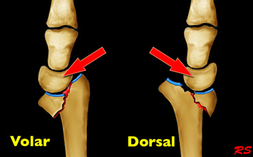
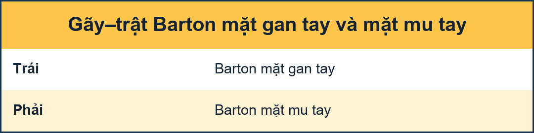
Volar-type Barton's is a fracture-dislocation of the volar rim of the radius.
This type is the most common.
Barton kiểu xương bánh chè là một dạng gãy xương-trật khớp của vành xương bánh rào.
Loại này là phổ biến nhất.
Dorsal-type Barton's is a fracture-dislocation of the dorsal rim of the radius.
Barton kiểu lưng là một dạng gãy xương-trật khớp của vành lưng xương quay.
Dislocation of the radiocarpal joint is the hallmark of Barton's fractures.
These are shear type fractures of the distal articular surface of the radius with translation of the distal radial fragment and the carpus.
These fractures have a great tendency for redislocation and malunion.
They usually require operative treatment.
Trật khớp quay cổ tay là dấu hiệu đặc trưng của gãy xương Barton.
Đây là loại gãy xương kiểu cắt ở mặt khớp đầu xa của xương quay với sự dịch chuyển của mảnh xương đầu xa và xương ức.
Những gãy này có xu hướng tái trật khớp và không liền xương.
Chúng thường cần điều trị phẫu thuật.
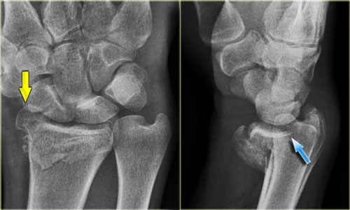
On the left a volar-type Barton's fracture.
Bên trái gãy kiểu Barton.
The radiographic findings are the following:Các dấu hiệu trên X-quang như sau:
- Comminuted intraarticular fracture of the distal radius Gãy đầu dưới xương quay nhiều mảnh
- Volar rim maintains relationship with the carpus and both are displaced proximally (blue arrow) Vành Volar duy trì mối quan hệ với carpus và cả hai đều di chuyển về phía gần (mũi tên màu xanh)
- Fracture of radial styloid process with loss of radial inclination (yellow arrow) Gãy mỏm trâm quay mất độ nghiêng quay (mũi tên màu vàng)
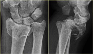
On the left a dorsal-type Barton's fracture.
Bên trái gãy Barton kiểu lưng.
The radiographic findings are the following:Các dấu hiệu trên X-quang như sau:
- Comminuted intraarticular fracture of the distal radius Gãy đầu dưới xương quay nhiều mảnh
- Dorsal rim and carpus are displaced dorsally and proximally Vành lưng và carpus bị di dời về phía lưng và phía gần
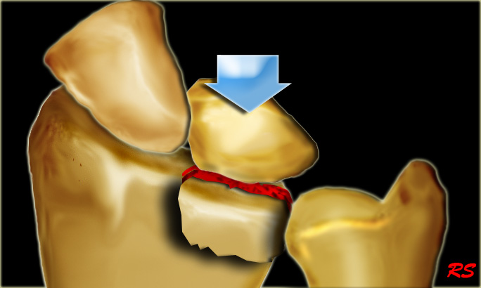
Die-punch fractureGãy xương Die-punch
A die-punch fracture is a depression fracture of the lunate fossa of the distal radius.
It is the result of a transverse load through the lunate.
Gãy xương đấm chết là gãy lõm hố mặt trăng ở đầu dưới xương quay.
Nó là kết quả của một tải trọng ngang qua mặt trăng.
The radiographic findings can be very subtle.
In many cases there is also a subtle proximal displacement of lunate, seen as a break in carpal arc I.
(see the article Wrist - Carpal instability).Các dấu hiệu trên X-quang có thể rất tinh tế.
Trong nhiều trường hợp cũng có một sự dịch chuyển gần tinh tế của mặt trăng, được xem như là một sự phá vỡ trong vòng cung cổ tay I.
(xem bài viết Mất vững cổ tay - cổ tay).
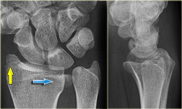 Die punch fractureDie đấm gãy xương
On the left a typical die-punch fracture.
The blue arrow indicates the depressed fragment of the lunate fossa.
Notice the articular step-off.
The yellow arrow indicates a subtle fracture of the radial styloid process.
There is no disruption of carpal arc I.
Notice that you can easily overlook such a fracture.Bên trái là một gãy xương điển hình.
mũi tên màu xanh dương chỉ ra đoạn lõm của hố mặt trăng.
Chú ý dấu hiệu khép khớp.
mũi tên màu vàng cho thấy một vết nứt tinh tế của mỏm trâm quay.
Không có tổn thương xương cổ tay I.
Lưu ý rằng bạn có thể dễ dàng bỏ qua một vết gãy như vậy.
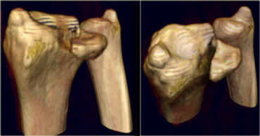
On the left two 3D-reconstructions of the same fracture as above.Bên trái hai hình ảnh tái tạo 3D của cùng một loại gãy xương như trên.
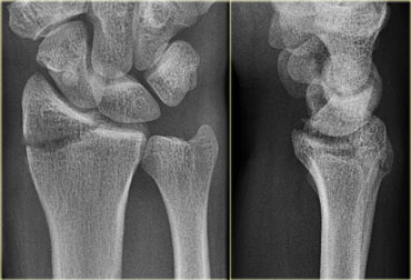
Chauffeur's fractureGãy Chauffeur
An isolated fracture of the radial styloid process is also called a Hutchinson's or chauffeur's fracture.
Displacement of the fragment is uncommon.
There can be associated injury to the scapholunate ligament.
Một gãy đơn độc của mỏm trâm quay cũng được gọi là gãy Hutchinson hoặc gãy người lái xe.
Sự dịch chuyển của mảnh vỡ là không phổ biến.
Có thể có tổn thương dây chằng scapholunate.
In most cases a fracture of the radial styloid process is part of a comminutive intraarticular fracture.Trong hầu hết các trường hợp, gãy mỏm trâm quay là một phần của gãy nội khớp nhỏ.
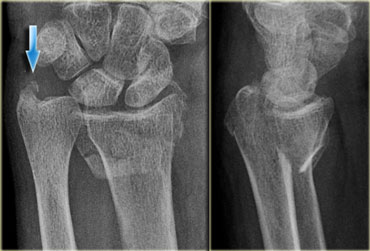
Ulnar styloid process fracture
An ulnar styloid process fracture is usually associated with radial fractures and rarely isolated.
An isolated fracture of the tip is clinically insignificant.
Displaced fractures of the base are usually associated with TFC tears and can be associated with instability of the distal radioulnar joint (DRUJ).
Gãy quá trình xoắn trụ
Gãy trụ xoắn trâm thường đi kèm với gãy xương quay và hiếm khi đơn độc.
Gãy đầu xương cụt đơn độc không có ý nghĩa lâm sàng.
Gãy di lệch nền thường liên quan đến rách TFC và có thể liên quan đến mất vững khớp quay xa (DRUJ).
On the left a subtle fracture of the tip of the ulnar styloid process (blue arrow) in a patient with a volar Barton's fracture.
Notice the depression of the volar rim.Bên trái là một vết gãy tinh tế của chóp mỏm trâm trụ (mũi tên màu xanh) ở một bệnh nhân gãy xương bánh chè Barton.
Chú ý sự sụp đổ của vành xương bánh chè.
Fractures in ChildrenGãy xương ở trẻ em
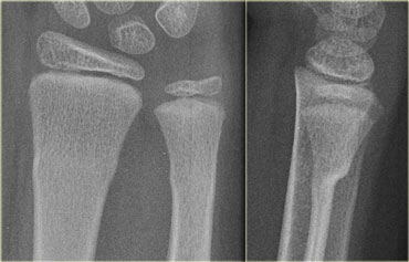
Torus fractureGãy thân đốt sống
Torus fractures, or buckle fractures, are extremely common injuries in children.
Because children have softer bones, one side of the bone may buckle.
The word torus is derived from the Latin word 'Tori' meaning swelling or protuberance.
These injuries tend to heal much more quickly than the similar greenstick fractures.
Gãy xương Torus, hoặc gãy xương khóa, là những chấn thương rất phổ biến ở trẻ em.
Bởi vì trẻ em có xương mềm hơn, một bên xương có thể bị cong.
Từ torus có nguồn gốc từ tiếng Latin 'Tori' có nghĩa là sưng hoặc nhô ra.
Những tổn thương này có xu hướng lành nhanh hơn nhiều so với các loại gãy greenstick tương tự.
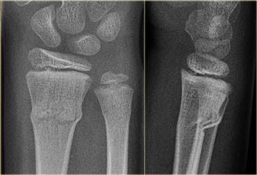
Green stick fractureGãy thanh xanh
These are partial fractures, since only one part of the bone is broken and the other side is bent.
The name is derived from an analogy of breaking a young, fresh tree branch.
Most often the greenstick fracture must be bent back into the proper position.
Greenstick fractures can take a long time to heal because they tend to occur in the middle, more slowly growing parts of bone.
Đây là những gãy xương bán phần, vì chỉ có một phần xương bị gãy và phần còn lại bị cong.
Tên gọi này bắt nguồn từ một sự tương tự của việc bẻ gãy một cành cây non, tươi.
Thông thường gãy greenstick phải được uốn cong trở lại vị trí thích hợp.
Gãy Greenstick có thể mất nhiều thời gian để lành vì gãy thường xảy ra ở phần giữa xương, phần xương phát triển chậm hơn.
Epiphysiolysis fractureGãy đầu trên xương rốn
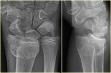
These are usually Salter Harris type II epiphysiolysis fractures.
Restorage of the anatomical situation is necessary to prevent growth disturbances.
Redislocation is common after closed reduction.
In many cases they need percutaneous pinning.
Đây thường là gãy xương tầng sinh môn loại II Salter Harris.
Việc giữ lại các vị trí giải phẫu là cần thiết để ngăn ngừa sự tăng trưởng bất thường.
Tái định vị là phổ biến sau khi nắn kín.
Trong nhiều trường hợp, cần phải kẹp qua da.
Classification systemsHệ thống phân loại
Muller AO-classificationPhân loại Muller AO
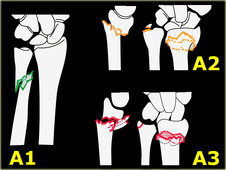
The Muller AO-classification is adapted by the Orthopaedic Trauma Association.
In reference (6) a link is provided to download the illustrations of the Muller AO Classification of Fractures.Phân loại Muller AO được Hiệp hội Chấn thương Chỉnh hình điều chỉnh.
Để tham khảo (6), một liên kết được cung cấp để tải xuống các hình minh hoạ của Phân loại gãy xương Muller AO.
A = extra-articular fracture A = gãy ngoài khớp
- A1 = ulna, radius intactA1 = trụ, bán kính nguyên vẹn
- A2 = radius, simple and impactedA2 = bán kính, đơn giản và bị ảnh hưởng
- A3 = radius, multifragmentaryA3 = bán kính, đa tầng
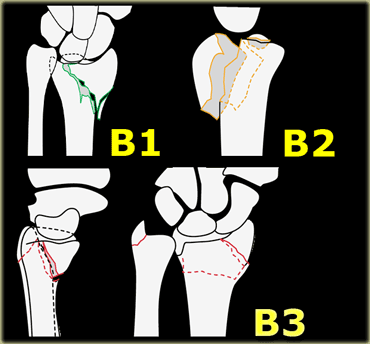
B = partial articular fracture
B = gãy một phần khớp
- B1 = radius, sagittal B1 = bán kính, dọc
- B2 = radius, frontal, dorsal rim B2 = bán kính, trán, vành lưng
- B3 = radius, frontal, volar rim B3 = bán kính, trán, vành
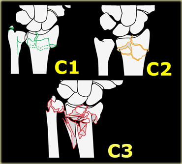
C = complete articular fracture of radius
C = gãy hoàn toàn khớp quay
- C1 = articular simple, metaphyseal simple C1 = khớp đơn, khớp liên thân đốt đơn
- C2 = articular simple, metaphyseal multifragmentary C2 = khớp đơn giản, đa mảnh
- C3 = articular multifragmentary C3 = đa mảnh khớp
Fernandez ClassificationPhân loại Fernandez
This classification is popular, since it addresses the mechanism of injury and the consequent treatment options.
Instability is defined as a high risk of secondary displacement after initial adequate reduction.
Associated traumatic lesions are ligamentous rupture, nerve compression and compartment syndrome.Phân loại này rất phổ biến, vì nó đề cập đến cơ chế chấn thương và các lựa chọn điều trị.
Mất vững được định nghĩa là nguy cơ cao của di lệch thứ phát sau khi nắn chỉnh đầy đủ ban đầu.
Tổn thương chấn thương kèm theo là đứt dây chằng, chèn ép dây thần kinh và hội chứng chèn ép khoang.
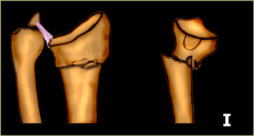
Type 1 : Bending fracture Loại 1: Gãy xương gập
- Mechanism: one cortex of the metaphysis fails due to tensile stress and the opposite undergoes a certain degree of comminution. Cơ chế: một vỏ não của xương chuyển vị bị hỏng do căng thẳng và đối diện trải qua một mức độ nhỏ nhất định.
- Stability: can be stable or unstable Tính ổn định: có thể ổn định hoặc không ổn định
- Displacement pattern: non-displaced, dorsal (Colles') or volar (Smith's fracture) Mô hình di lệch: không di lệch, mặt lưng (Colles ') hoặc mặt lưng (gãy Smith)
- Number of fragments: two main fragments Số lượng mảnh ghép: hai mảnh ghép chính
- Associated lesions: uncommon Tổn thương liên quan: ít gặp
- Recommended treatment: usually conservative, when unstable:percutaneous pinning or external fixationĐiều trị khuyến nghị: thường là bảo tồn, khi không ổn định: kẹp qua da hoặc cố định bên ngoài
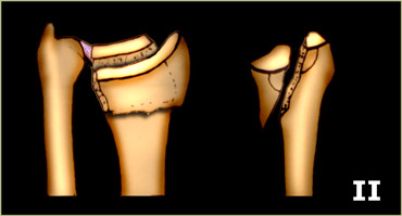
Type 2 : Shearing fracture Loại 2: Gãy xương
- Mechanism: fracture of the joint surface (i.e. volar and dorsal Barton's, radial styloid process fractures, simple articular fracture) Cơ chế: gãy mặt khớp (ví dụ gãy mặt lưng và mặt lưng Barton, gãy mỏm trâm quay, gãy khớp đơn thuần)
- Stability: unstable Tính ổn định: không ổn định
- Displacement pattern: dorsal, volar or radial Mô hình dịch chuyển: lưng, lưng hoặc quay
- Number of fragments: two-part, three-part or comminuted Số lượng đoạn: hai phần, ba phần hoặc
- Associated lesions: less common Tổn thương liên quan: ít gặp hơn
- Recommended treatment: open reduction with screw- or plate fixation Điều trị khuyến nghị: nắn mở bằng nẹp vít hoặc nẹp cố định
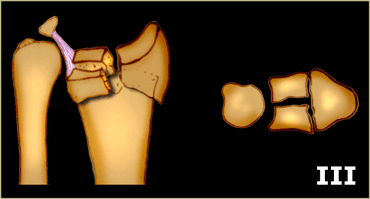
Type 3 : Compression fracture Loại 3: gãy lún
- Mechanism: compression fracture of the joint surface with impaction of subchondral and metaphyseal bone (i.e. die-punch fracture or intraarticular comminuted fracture Cơ chế: gãy lún của bề mặt khớp với sự tác động của xương dưới sụn và xương bàn ngón (tức là gãy xương đấm hoặc gãy xương trong khớp
- Stability: stable or unstable Tính ổn định: ổn định hoặc không ổn định
- Displacement pattern: non-displaced, dorsal, volar or radial Mô hình dịch chuyển: không dịch chuyển, lưng, lưng hoặc quay
- Number of fragments: 3- or 4-part or comminuted Số lượng mảnh: 3 hoặc 4 phần hoặc comminuted
- Associated lesions: common Tổn thương kèm theo: thường gặp
- Recommended treatment: Điều trị khuyến cáo:
- closed đóng
- limited arthroscopic assistednội soi khớp vai có hỗ trợ
- extensile open reductionMở rộng nắn chỉnh
- percutaneous pins combined with external and internal fixationkết hợp xương bên trong và bên ngoài với chốt qua da
- bone graftghép xương
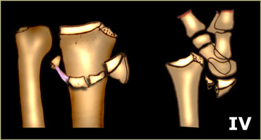
Type 4 : Avulsion fracture Loại 4: gãy bong điểm bám
- Mechanism: avulsion fracture usually associated with ligamentous injury Cơ chế: gãy bong điểm bám thường liên quan đến tổn thương dây chằng
- Stability: unstable Tính ổn định: không ổn định
- Displacement pattern: dorsal, volar, radial or proximal Mô hình dịch chuyển: lưng, lưng, quay hoặc đầu gần
- Number of fragments: Số lượng mảnh:
- 2-part: radial styloid, ulnar styloid2 phần: trâm quay, trâm trụ
- 3-part: volar, dorsal margin3 phần: phần lưng, phần lưng
- comminuted
- Associated lesions: frequent ligamentous injury Tổn thương kèm theo: Tổn thương dây chằng thường gặp
- Recommended treatment: closed or open reduction with pin or screw fixation or tension wiring Điều trị khuyến nghị: nắn kín hoặc mở bằng cố định bằng pin hoặc vít hoặc dây căng
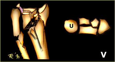 Fernandez classification. Modified and reprinted with permission from reference (9)Phân loại Fernandez. Đã sửa đổi và in lại với sự cho phép của tài liệu tham khảo (9)
Type 5 : Combined fractures Loại 5: Gãy xương phối hợp
- Mechanism: combination of forces of type I-IV as a result of high velocity injuries Cơ chế: phối hợp các lực loại I-IV do chấn thương vận tốc cao
- Stability: unstable Tính ổn định: không ổn định
- Displacement pattern: dorsal, volar, radial or proximal Mô hình dịch chuyển: lưng, lưng, quay hoặc đầu gần
- Number of fragments: comminuted Số lượng mảnh: comminuted
- Associated lesions: always present Tổn thương liên quan: luôn có mặt
- Recommended treatment: combined method Điều trị khuyến nghị: phương pháp phối hợp
TreatmentĐiều trị
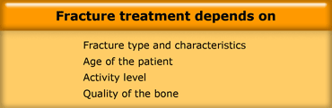
The treatment decision of a distal radius fracture is complex and depends on the type of the fracture, the age and activeness of the patient and the quality of the bone.
At one extreme a stable, undisplaced extra-articular fracture has an excellent prognosis.
At the other an unstable, displaced intra-articular fracture is difficult to treat and has a poor prognosis.
If the alignment of the bones is not acceptable, they need to be reduced by closed or open reduction.Quyết định điều trị gãy đầu dưới xương quay phức tạp và phụ thuộc vào loại gãy, tuổi, mức độ hoạt động của bệnh nhân và chất lượng xương.
Ở một mức độ, gãy xương ngoài khớp ổn định, không di lệch có tiên lượng tốt.
Gãy nội khớp mất vững, di lệch là một bệnh lý khó điều trị và tiên lượng xấu.
Nếu sự sắp xếp của xương không được chấp nhận, cần phải nắn chỉnh bằng nắn kín hoặc mở.
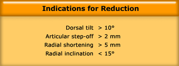
Indications for Reduction in Distal Radius FracturesChỉ định nắn chỉnh gãy đầu dưới xương quay
Many authors suggest that distal radial fractures be reduced anatomically, but the real question is 'what is acceptable and what is not?'.
Based on basic science and clincal studies some of the recommendations of the International Distal Radius Fracture Study Group are presented in the table on the left, although these recommendations are still the subject of ongoing debate (5).Nhiều tác giả cho rằng gãy đầu dưới xương quay cần được nắn chỉnh về mặt giải phẫu, nhưng câu hỏi thực sự là 'cái gì có thể chấp nhận và cái gì không?'.
Dựa trên các nghiên cứu khoa học cơ bản và nghiên cứu lâm sàng, một số khuyến nghị của Nhóm nghiên cứu gãy xương đầu xa quốc tế được trình bày trong bảng bên trái, mặc dù những khuyến nghị này vẫn là chủ đề của cuộc tranh luận đang diễn ra (5).
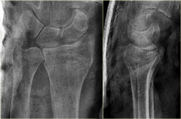 Good alignment after closed recuction for extraarticular Colles fractureĐánh giá kết quả nắn chỉnh khớp gối sau phẫu thuật nắn kín trong điều trị gãy Colles ngoài khớp
Closed ReductionGiảm đóng
The initial treatment for most radius fractures is closed reduction and plaster immobilization.
A displaced fracture is reduced under regional or general anaesthetic.
First the arm is placed under traction to unlock the fragments.
The deformity is then reduced with appropriate closed reduction, depending on the type of deformity.
A splint or cast is placed in such a way that the risk of re-displacement is minimized.
X-rays are taken to ensure that the reduction was successful.
The cast is usually maintained for about 6 weeks.
Điều trị ban đầu cho hầu hết các loại gãy xương quay là nắn kín và cố định bằng nẹp vít.
Gãy xương di lệch được nắn chỉnh dưới gây tê vùng hoặc gây mê toàn thân.
Đầu tiên cánh tay được đặt dưới lực kéo để mở khóa các mảnh vỡ.
Sau đó, độ biến dạng được nắn chỉnh với nắn kín phù hợp, tuỳ thuộc vào loại biến dạng.
Nẹp hoặc bó bột được đặt theo cách sao cho nguy cơ di lệch được giảm thiểu.
X-quang được thực hiện để đảm bảo rằng việc nắn chỉnh đã thành công.
Bó bột thường được duy trì trong khoảng 6 tuần.
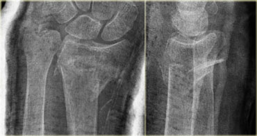 X-ray in cast. Unsuccesful reduction.X-quang trong bó bột, nắn chỉnh không thành công.
Guidelines for non-acceptable reduction are (8):Hướng dẫn cho việc giảm không thể chấp nhận được là (8):
- Radial shortening > 5 mmRút ngắn xương quay > 5 mm
- Radial inclination Tilt on lateral projection > 10 degrees dorsal tilt and > 20 degrees volar tiltĐộ nghiêng xuyên tâm Nghiêng khi chiếu bên > 10 độ nghiêng lưng và > 20 độ nghiêng volar
- Intra-articular step-off 2 mm or moreBước ra trong khớp từ 2 mm trở lên
- Articular incongruity 2 mm or more of the sigmoid notch (articular surface of distal radius in DRUJ).Khớp bất thường 2 mm trở lên của rãnh sigma (bề mặt khớp của đầu xa của bán kính trong DRUJ).
On the left a control radiograph made after reduction.
The reduction was unsuccessful, because there is a dorsal tilt > 10 degrees , loss of inclination and radial shortening.Bên trái là phim X quang đối chứng sau khi nắn chỉnh.
Việc nắn chỉnh không thành công, vì có độ nghiêng lưng > 10 độ, mất độ nghiêng và ngắn xương quay.
Surgical treatmentĐiều trị ngoại khoa
Although in most cases closed reduction is attempted, surgical intervention is required when there is failure to obtain or maintain closed reduction.
40% of distal radial fractures are considered to be unstable and require surgical fixation.
Many techniques of fixation are now available, including percutaneous pinning, intramedullary pinning, external fixation, and internal fixation with customized implants, including the Distal Volar Radius (DVR) system.
Surgical fixation allows almost immediate mobility.
Ultimately less stiffness and greater function is possible.
Mặc dù trong hầu hết các trường hợp, nắn kín được cố gắng thực hiện, can thiệp phẫu thuật là cần thiết khi không thể lấy được hoặc duy trì nắn đóng.
40% gãy đầu dưới xương quay được coi là không vững và cần phẫu thuật cố định.
Hiện nay, có nhiều kỹ thuật cố định, bao gồm kẹp qua da, kẹp trong tuỷ, cố định ngoài và cố định trong với các implant tuỳ chỉnh, bao gồm hệ thống DVR.
Phẫu thuật cố định cho phép di chuyển gần như ngay lập tức.
Cuối cùng là có thể giảm độ cứng và tăng chức năng.
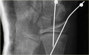
On the left a post-operative image of a Salter-Harris II fracture, which is held in place with two pins after closed reduction.Bên trái là hình ảnh sau mổ gãy xương Salter-Harris II, được giữ cố định bằng hai chốt sau khi nắn kín.
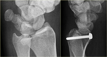
On the left a patient with a die-punch fracture, nicely shown on an oblique radiograph.
The fracture fragment of the lunate fossa was replaced and fixated with a screw.Bên trái là một bệnh nhân gãy xương đấm, được thể hiện rõ trên phim X quang nghiêng.
Mảnh vỡ xương hố mặt trăng được thay thế và cố định bằng vít.
Volar buttress plate
Barton's fractures are rarely successfully treated with closed reduction due to the shearing nature of the injury.
A volar buttress plate is the treatment of choice.
Tấm chắn trụ Volar
Gãy Barton hiếm khi được điều trị thành công bằng nắn kín do tính chất đứt gãy của chấn thương.
Tấm nẹp vít là phương pháp điều trị được lựa chọn.
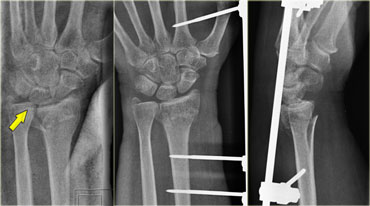
Comminution or osteoporotic bone make external fixation the preferred surgical treatment option.Xương nhỏ hoặc loãng xương làm cho cố định ngoài là lựa chọn điều trị phẫu thuật được ưa thích.
On the left an intraarticular fracture of the distal radius with shortening of the radius.
The ulna abutts the lunate.
External fixation was used to lengthen the radius.Ở bên trái, gãy nội khớp đầu dưới xương quay với sự rút ngắn của xương quay.
Trụ trụ tiếp giáp với xương nguyệt.
Kết hợp xương bên ngoài để kéo dài xương quay.
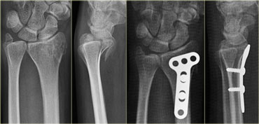
On the left a patient with a dorsal Barton's fracture (shown before).
Bên trái là một bệnh nhân gãy xương Barton lưng (hình trước).
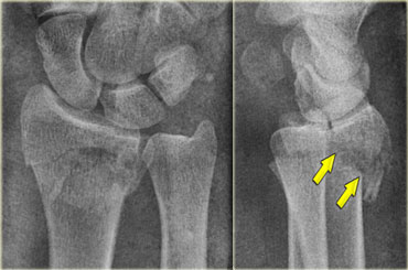
After closed reduction the position of the dorsal rim is better, but this still is an unstable situation.
Sau khi nắn kín vị trí bờ lưng tốt hơn, tuy nhiên đây vẫn là tình trạng không ổn định.
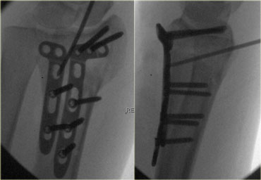
Volar plates were used with screws to lock the dorsal rim.
The volar approach was chosen, because this is an easier approach.
Tấm đệm được sử dụng với vít để khóa mép lưng.
Phương pháp volar được chọn vì đây là cách tiếp cận dễ dàng hơn.
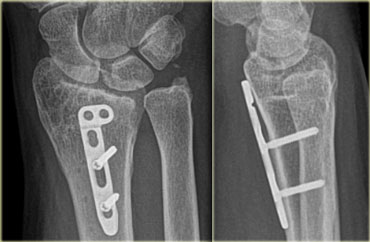
Final result after one of the plates has been removed.Kết quả cuối cùng sau khi một trong các đĩa đệm đã được lấy ra.
ComplicationsCác biến chứng
MalunionMalunionName
Non-union is uncommon in distal radial fractures, since there is excellent vascularisation of this region.
Malunion however is a common complication and is related to radial shortening, angulation and incongruity of the articular surface.
This results in malfunction and early osteoarthritis.
More than 2 mm incongruity of articular surface is the most important factor in the development of posttraumatic osteoarthritis of the wrist.
Không liền xương là không phổ biến ở gãy đầu dưới xương quay, vì có sự hình thành mạch máu tuyệt vời ở vùng này.
Tuy nhiên, tật khúc xạ là một biến chứng thường gặp và có liên quan đến sự ngắn lại của khớp quay, góc cạnh và sự không đồng nhất của bề mặt khớp.
Điều này dẫn đến sự cố và thóai hóa khớp sớm.
Sự không phù hợp của bề mặt khớp trên 2 mm là yếu tố quan trọng nhất trong sự phát triển của thóai hóa khớp cổ tay sau chấn thương.
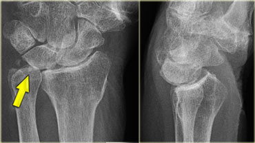
On the left a patient with malunion.
The radial shortening results in the ulna abutting the lunate.
Notice the loss of radiocarpal joint space indicating osteoarthritis.Bên trái một bệnh nhân bị mất liền.
Sự ngắn lại của xương quay dẫn đến xương trụ nằm sát xương nguyệt.
Lưu ý sự mất khoảng trống của khớp cổ tay cho thấy thóai hóa khớp.
 MalunionMalunionName
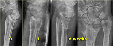
Closed reduction is frequently unsuccessful when the fracture has an oblique course or when the fracture is comminutive.
Nắn kín thường không thành công khi gãy xương có đường gãy chéo hoặc khi gãy nhỏ.
On the left a patient with an intraarticular fracture with dorsal tilt (i.e. intraarticular Colles' fracture).
On the lateral radiograph at presentation there is an extreme dorsal tilt.
After closed reduction and at follow up after one week, there is an acceptable tilt.
Finally at 6 weeks follow-up, there is malunion with extreme dorsal tilt, radial shortening and loss of inclination.
The ulna abutts the lunate.
The final result will be malfunction, radiocarpal and distal radioulnar osteoarthritis. Bên trái là một bệnh nhân bị gãy nội khớp với góc nghiêng lưng (tức là gãy Colles nội khớp).
Trên phim X-quang bên có một góc nghiêng lưng cực lớn.
Sau khi nắn kín và theo dõi sau một tuần, có thể chấp nhận được độ nghiêng.
Cuối cùng sau 6 tuần theo dõi, có sự bất thường về khớp với độ nghiêng lưng cực lớn, ngắn xương quay và mất độ nghiêng.
Trụ trụ tiếp giáp với xương nguyệt.
Kết quả cuối cùng là thóai hóa khớp cổ tay và khớp quay xa.
Risks specific to cast treatment relate to the potential for compression of the swollen arm causing compartment syndrome or carpal tunnel syndrome.
Reflex sympathetic dystrophy and median nerve injury are uncommon complications.
Complications associated with plating include tendon irritation or rupture and the need for plate removal.Các nguy cơ cụ thể khi điều trị liên quan đến khả năng chèn ép cánh tay bị sưng gây ra hội chứng khoang hoặc hội chứng ống cổ tay.
Loạn dưỡng phản xạ giao cảm và tổn thương thần kinh giữa là những biến chứng không phổ biến.
Các biến chứng liên quan đến việc mạ bao gồm kích thích hoặc đứt gân và cần phải loại bỏ tấm.
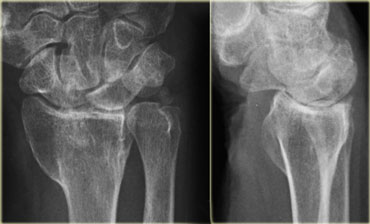
On the left another patient with malunion and osteoarthritis.
There is also scapholunate dissociation as a result of associated ligamentous rupture with volar tilt of lunate indicating volar flexion instability (VISI). Bên trái một bệnh nhân khác bị khớp cắn và thóai hóa khớp.
Ngoài ra còn có sự phân ly scapholunate do đứt dây chằng liên quan với độ nghiêng của mặt trăng cho thấy sự mất ổn định của mặt trăng (VISI).
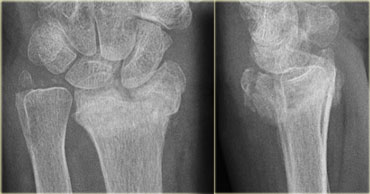
On the left another patient after unsuccessful treatment.
There is loss of radial inclination and radial shortening, dorsal tilt and an articular step-off.Bên trái là một bệnh nhân khác sau điều trị không thành công.
Có mất độ nghiêng quay và ngắn quay, nghiêng lưng và mất khớp nối.
ReferencesTài liệu tham khảo
-
Wrist Fractures: What the Clinician Wants to Know
Gãy xương cổ tay: Bác sĩ lâm sàng muốn biết gì
by Charles A. Goldfarb, MD, Yuming Yin, MD, Louis A. Gilula, MD, Andrew J. Fisher, MD and Martin I. Boyer, MDRadiology. 2001;219:11-28. bởi Charles A. Goldfarb, MD, Yuming Yin, MD, Louis A. Gilula, MD, Andrew J. Fisher, MD và Martin I. Boyer, MDRadiology. 2001; 219: 11-28.
-
Treatment of Unstable Distal Radial Fractures with the Volar Locking Plating System
Điều trị gãy đầu dưới xương quay không vững bằng hệ thống mạ khóa Volar
by Kevin C. Chung et al
The Journal of Bone and Joint Surgery (American). 2006;88:2687-2694.
bởi Kevin C. Chung và cộng sự - Trauma and Fractures in Wheeless' Textbook of Orthopaedics online Chấn thương và gãy xương trong sách giáo khoa của Wheeless về chỉnh hình trực tuyến
-
Palmar Tilt of the Distal Radius: Influence of Off-lateral Projection Initial Observations
Nghiêng gan tay của bán kính xa: Ảnh hưởng của việc chiếu ngoài bên
by Marco Zanetti, MD, Louis A. Gilula, MD, Hilaire A. C. Jacob, PhD and Juerg Hodler, MD
Radiology. 2001;220:594-600
bởi Marco Zanetti, MD, Louis A. Gilula, MD, Hilaire A. C. Jacob, Tiến sĩ và Juerg Hodler, MD -
HOW TO CLASSIFY DISTAL RADIAL FRACTURES
Cách phân loại gãy đầu xa xương quay
A Report by the
IFSSH BONE AND JOINT COMMITTEE
Báo cáo của Viện -
Muller AO Classification of Fractures
Phân loại gãy xương Muller AO
Download the illustrations as packages: Radius/Ulna: ZIP File (351 KB) Tải xuống các hình minh hoạ dưới dạng gói: Radius / Ultrana: ZIP File (351 KB)
-
Unstable extra-articular fractures of the distal radius
Gãy ngoài khớp không vững ở đầu dưới xương quay
A PROSPECTIVE, RANDOMISED STUDY OF IMMOBILISATION IN A CAST VERSUS SUPPLEMENTARY PERCUTANEOUS PINNING
by T. Azzopardi et al. J Bone Joint Surg Br 2005; 87-B: 837-840
Nghiên cứu tiền cứu, giả định về sự bất động trong phẫu thuật tạo hình răng vĩnh viễn -
Indications for Reduction in Distal Radius Fractures
Chỉ định nắn chỉnh gãy đầu dưới xương quay
by David L. Nelson, MD of the International distal radius fracture study group David L. Nelson, MD nhóm nghiên cứu gãy đầu dưới xương quay quốc tế
-
by Diego Fernandez, Jesse Jupiter
Springer, New York, Second Edition, 2002, ISBN 0-387-95195-4.
của Diego Fernandez, Jesse Jupiter