
...
Bản gốc trên radiologyassistant.nl ↗ · tải về lúc 2026-11-19 20:19 · bản dịch máy có chuẩn hóa thuật ngữ chẩn đoán hình ảnh
Answers to ankle and foot casesĐáp ứng với các trường hợp mắt cá chân và bàn chân
FootChân
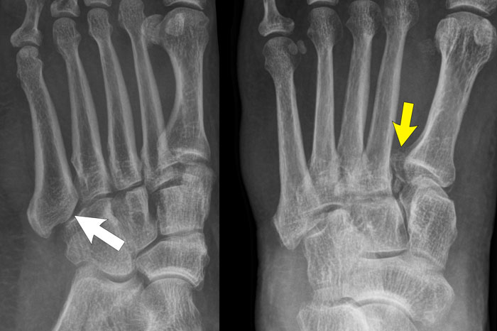
case 1 - distortiontrường hợp 1 - biến dạng
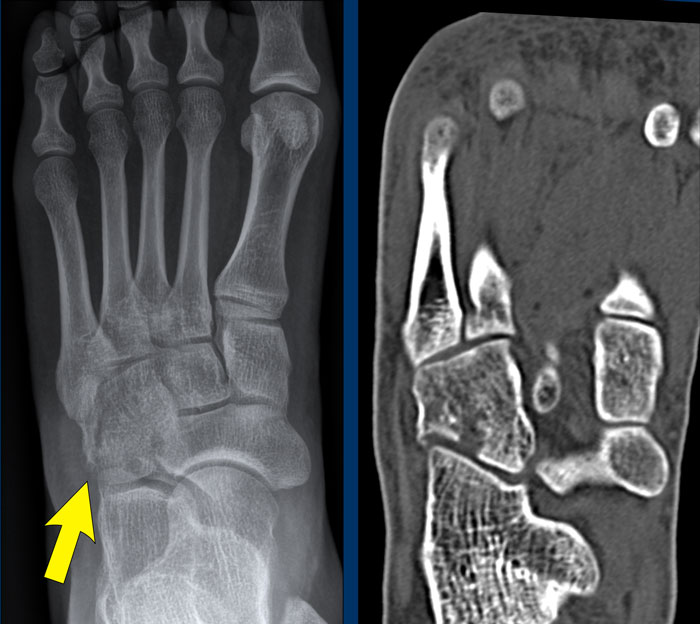
The findings are:
Kết quả nghiên cứu:
- Large distance between the base of MT1 and MT2 (yellow arrow).Khoảng cách lớn giữa đáy MT1 và MT2 (mũi tên màu vàng).
- Subluxation of the joint between cuboid and the base of MT5 (white arrow).Sự bán trật khớp giữa khối lập phương và nền MT5 (mũi tên trắng).
This means that there is a Lis Franck dislocation fracture.Điều này có nghĩa là có gãy trật khớp Lis Franck.
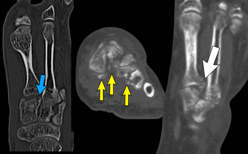
CT confirms multiple fractures at the level of the TMT Joints.CT xác nhận nhiều gãy xương ở mức khớp TMT.
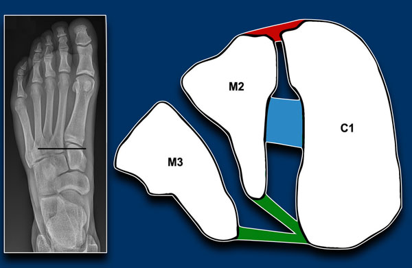
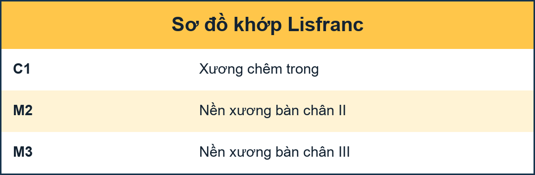
The Lisfranc ligament complex runs between the medial cuneiform bone (C1) and the base of MT2 and MT3.Phức hợp dây chằng Lisfranc chạy giữa xương hình nêm trong (C 1) và nền của MT2 và MT 3.
It consists of 3 components: Nó bao gồm 3 thành phần:
- a dorsal ligamentdây chằng lưng
- interosseous ligamentdây chằng gian cốt
- plantar ligament, which has insertions on the base of MT2 and MT3. dây chằng gan bàn chân, có chèn vào trên nền của MT2 và MT 3.
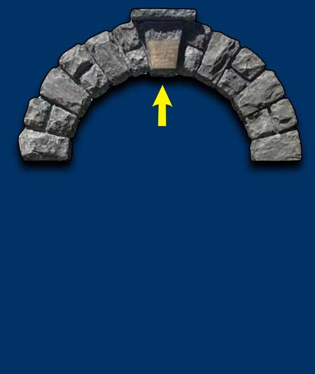
The base of the metatarsals form an arch shape which provides stability to the mid foot.
Just like old window arches, a keystone is neccessary at the top to close the arch.
If this stone is removed, the whole arch collapses.Phần đáy của xương bàn chân tạo thành một hình dạng vòm cung cấp sự ổn định cho bàn chân giữa.
Cũng giống như các vòm cửa sổ cũ, một keystone là cần thiết ở phía trên để đóng vòm.
Nếu loại bỏ sỏi này, toàn bộ cung sỏi sẽ sụp đổ.
In the foot the base of MT2 is the keystone of the foot arch.
If it looses its connection with the other metatarsals, the foot arch will collapse.
This will also result in multiple avulsions of the ligaments joining the bases of the metatarsals. Trong bàn chân, nền của MT2 là đáy của cung bàn chân.
Nếu nó mất kết nối với các xương bàn chân khác, cung bàn chân sẽ sụp đổ.
Điều này cũng sẽ dẫn đến nhiều dây chằng bị bong ra khi nối với nền xương bàn chân.
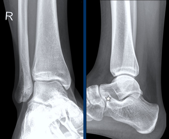
case 2 - chronic pain post ankle sprainsTrường hợp 2 - đau mạn tính sau chấn thương mắt cá chân
The findings are:
Kết quả nghiên cứu:
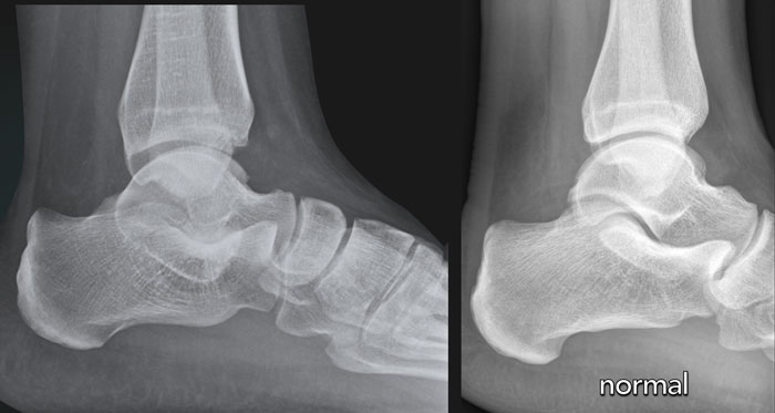
- elongated anterior process of the calcaneus that articulates with the navicular bone.thon dài xương gót khớp với xương đòn.
- the diagnosis is calcaneonavicular coalition.
chẩn đoán là liên kết xương gót.
The calcaneus and navicular bone do not normally articulate.
With osseous coalition, a bony bar that bridges the two bones is seen.Xương gót và xương đe thường không có khớp.
Với sự kết hợp xương, một thanh xương nối hai xương được nhìn thấy.
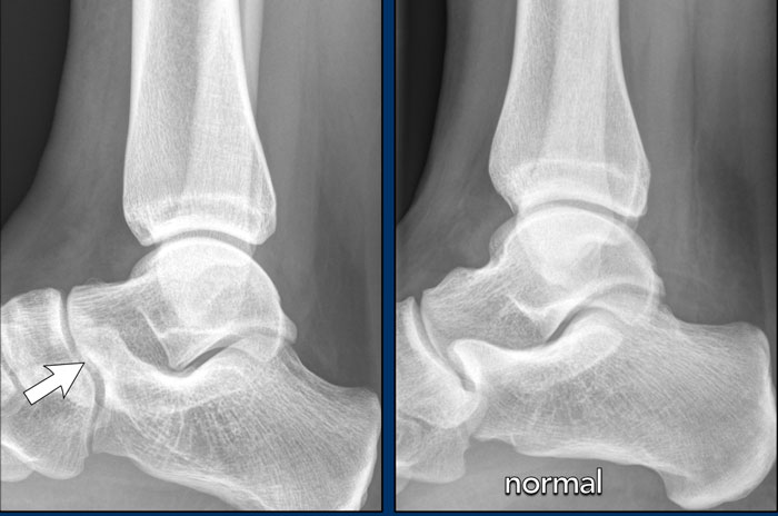
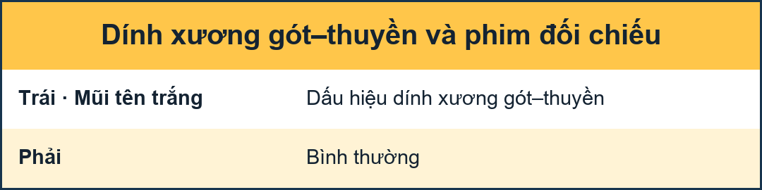
Compare to the normal situation.So sánh với tình hình bình thường.
With fibrous or cartilaginous coalition, the bones are in close proximity, both have irregular surfaces, and the anteromedial calcaneus is abnormally widened or flattened.
On lateral radiographs, elongation of the anterior dorsal calcaneus may simulate an anteater’s nose.
Hypoplasia of the talus is sometimes observed in calcaneonavicular coalition.
Với liên kết xơ hoặc sụn, xương ở gần nhau, cả hai đều có bề mặt không đều, và xương gót trước trong được mở rộng hoặc dẹt bất thường.
Trên phim X quang bên, sự kéo dài của xương gót trước có thể mô phỏng mũi của một con thú ăn kiến.
Giảm sản xương sên đôi khi được quan sát thấy trong liên kết xương gót.
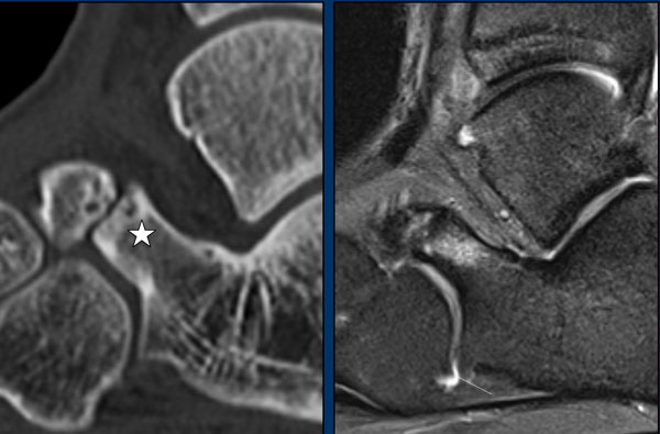
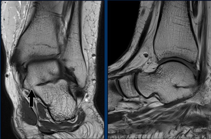
On the CT we see a coalition between the calcaneus and the os naviculare.
The CT shows sclerosis and subchondral cysts as a sign of ‘friction’ due to the neoarticulation.
The MRI shows bone marrow edema as a sign of a symptomatic coalition.Trên CT, chúng ta thấy một liên kết giữa xương gót và xương mu.
CT cho thấy xơ cứng và nang dưới sụn là dấu hiệu của “ma sát ” do tân tạo mạch máu.
MRI cho thấy phù tủy xương là một dấu hiệu của một liên kết triệu chứng.
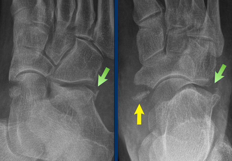
case 3 - distortiontrường hợp 3 - biến dạng
The findings are:
Kết quả nghiên cứu:
- Avulsion of the medial pole of navicular boneNhổ xương đòn cột trong
- Fracture on the lateral side of the calcaneusGãy mặt ngoài xương gót
This means that there is an injury to the Chopart's joint, which has an S-shape and is formed by the articulation of the calcaneus with the cuboid and the articulation of the talus with the navicular.
Điều này có nghĩa là có tổn thương khớp Chopart, có hình chữ S và được hình thành bởi khớp xương gót với xương hình khối và khớp xương sên với xương đòn.
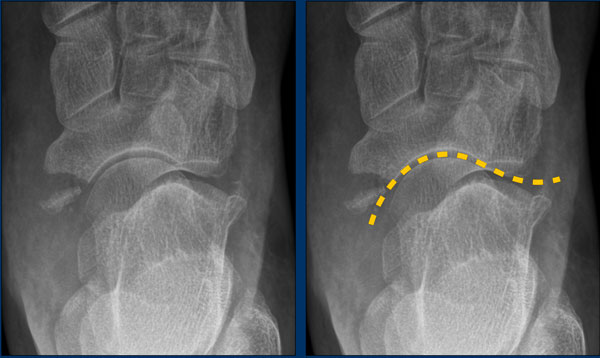
The movements in this joint are more extensive than in other tarsal joints and consists of a sort of rotation with the foot being carried medially (inversion) or laterally (eversion).Các chuyển động trong khớp này rộng hơn các khớp cổ chân khác và bao gồm một kiểu xoay với bàn chân được đưa vào trong (đảo ngược) hoặc ra ngoài (đảO ngược).
Inversion leads to distraction calcaneocuboidal en compression talonavicular often with plantarflexion, which can lead also to avulsion on the dorsal side of the talonavicular joint.Đảo ngược dẫn đến sự phân tâm xương gót chân trong nén khớp vuốt vuốt thường có gấp bàn chân, cũng có thể dẫn đến nhổ ở mặt lưng của khớp vuốt.
Eversion leads to distraction on the talonavicular side with either an avulsion of the tibial posterior tendon or an avulsion fracture and compression on the calcaneocuboidal side (nutcracker fracture).Lật ra sau dẫn đến mất tập trung ở phía xương móng vuốt với một trong hai là nhổ gân chày sau hoặc gãy bong điểm bám và chèn ép ở phía xương gót chân (gãy xương chẻ).
This means that when you see one fracture, there probably are more fractures in combination with ligamentous rupture and small avulsions.Điều này có nghĩa là khi bạn nhìn thấy một vết gãy, có thể có nhiều vết gãy kết hợp với đứt dây chằng và các chỗ bong nhỏ.
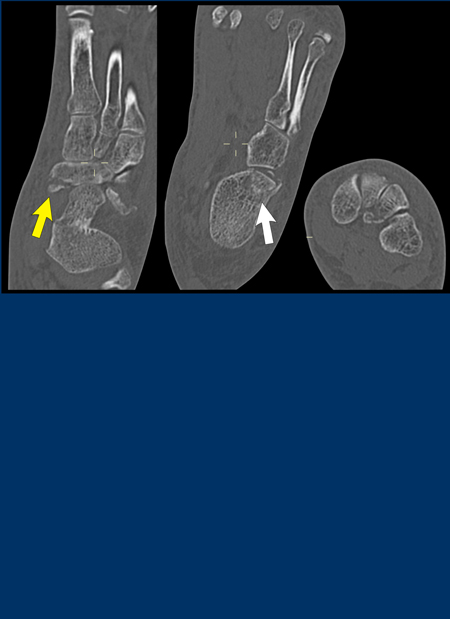
In this case there was an eversion with distraction on the talonavicular side with an avulsion of the medial pole of navicular bone (yellow arrow) and compression on the calcaneocuboidal side leading to a nutcracker fracture (white arrow).Trong trường hợp này có một sự di lệch với sự phân tâm ở phía xương móng vuốt với sự nhổ bỏ cực trong của xương đòn (mũi tên màu vàng) và chèn ép ở phía xương gót chân dẫn đến gãy xương gãy (mũI tên màu trắng).
case 4 - distortiontrường hợp 4 - biến dạng
This is another case of a nutcracker fracture as a result of compression on the lateral side of the tarsus due to eversion in Chopart's joint.
Đây là một trường hợp gãy đai ốc do chèn ép bên ngoài của sụn khớp do trật khớp Chopart.
The eversion can also lead to distraction on the talonavicular side with either avulsion of the posterior tibial tendon or avulsion of the medial pole of navicular bone, where the tendon attaches, as was seen in case 2. Sự lộn ngược cũng có thể dẫn đến sự phân tâm ở phía xương móng vuốt với sự nhổ của gân chày sau hoặc nhổ cực trong của xương đòn, nơi gân bám vào, như đã thấy trong trường hợp 2.
case 5 - chronic painca 5 - đau mạn tính
The findings are:
Kết quả nghiên cứu:
- narrowing of the posterior subtalar joint with sclerosishẹp khớp dưới cẳng chân sau do xơ cứng
- C signDấu hiệu C
The subtalar joint consists of the anterior, middle, and posterior facets. Talocalcaneal fusion most commonly involves the middle facet at the level of the sustentaculum tali.
Talocalcaneal coalitions may be difficult to visualize on the standard radiographic views of the foot because of the complex orientation of the subtalar joint.Khớp dưới xương hàm bao gồm các mặt trước, giữa và sau. Phẫu thuật hợp nhất khớp bàn ngón thường liên quan đến mặt giữa ở mức độ của mỏm móc.
Liên kết khớp bàn chân có thể khó hình dung trên hình ảnh X quang tiêu chuẩn của bàn chân vì sự định hướng phức tạp của khớp dưới xương hàm.
A number of secondary radiographic signs of talocalcaneal coalition have been described, including:Một số dấu hiệu X quang thứ phát của liên kết ống tuỷ đã được mô tả, bao gồm:
- Talar beakMỏ vịt
- Narrowing of the posterior subtalar jointHẹp khớp dưới cẳng chân sau
- Rounding of the lateral talar processLàm tròn mặt sên
- Lack of depiction of the middle facets on lateral radiographs.Thiếu sự mô tả các khía cạnh ở giữa trên phim X quang.
- C-sign
A talar beak occurs because of impaired subtalar joint motion, which results in the navicular overriding the talus. Periosteal elevation occurs at the insertion of the talonavicular ligament, and, ultimately, a cycle of osseous repair results in formation of the talar beak.Mỏ sên xuất hiện do sự suy giảm vận động khớp dưới xương hàm, dẫn đến xương đòn đè lên xương sên.Sự nâng cao màng xương xảy ra khi chèn dây chằng móng vuốt, và cuối cùng, một chu kỳ sửa chữa xương con dẫn đến sự hình thành mỏ sên.
The “C sign,” is a C-shaped line on the lateral view, that outlines the medial talar dome and posteroinferior sustentaculum. The C sign results from bone bridging between the talar dome and sustentaculum, as well as the prominent inferior outline of the sustentaculum.
As the x-ray beam strikes the posteroinferior outline of the abnormal sustentaculum tangentially over a long distance, an uninterrupted “C” results. The C sign may be observed in both osseous and nonosseous coalitions. Dấu hiệu “C" là một đường hình chữ C trên mặt cắt ngang, phác thảo vòm xương sên trong và mỏm sên sau. Dấu Hiệu C là kết quả của sự bắc cầu xương giữa vòm sên và mỏn sên, cũng như đường viền dưới nổi bật của mỏn.
Khi chùm tia X chiếu vào đường viền sau dưới của mỏ vịt bất thường một cách tiếp tuyến trên một khoảng cách dài, kết quả là “C” không bị gián đoạn. Dấu hiệu C có thể được quan sát thấy ở cả xương và không xương.
Images
On the T1W-image there is a bony coalition and ankylosis of the talus with the calcaneus.Hình ảnh
Trên hình ảnh T1W có một liên kết xương và khớp xương sên với xương gót.
AnkleCổ chân
case 1 - distortiontrường hợp 1 - biến dạng
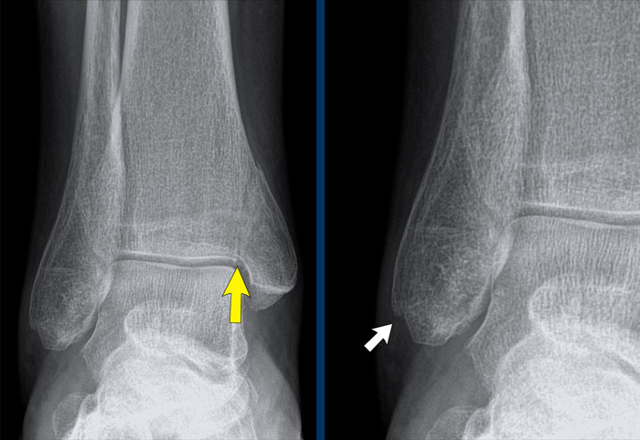
The findings are:
Kết quả nghiên cứu:
- vertical fracture of the medial malleolus (yellow arrow).
This is not an avulsion or pull-off fracture, but a push-off fracture.Gãy mắt cá trong (mũi tên màu vàng).
Đây không phải là gãy nhổ hay gãy kéo, mà là gãy đẩy ra. - Maybe there is an avulsion of the lateral malleolus (white arrow), but that is not sure.
However this does not matter as we will show in a moment.Có thể có một sự nhả của mắt cá ngoài (mũi tên trắng), nhưng điều đó không chắc chắn.
Tuy nhiên, điều này không quan trọng vì chúng tôi sẽ trình bày trong một thời điểm.
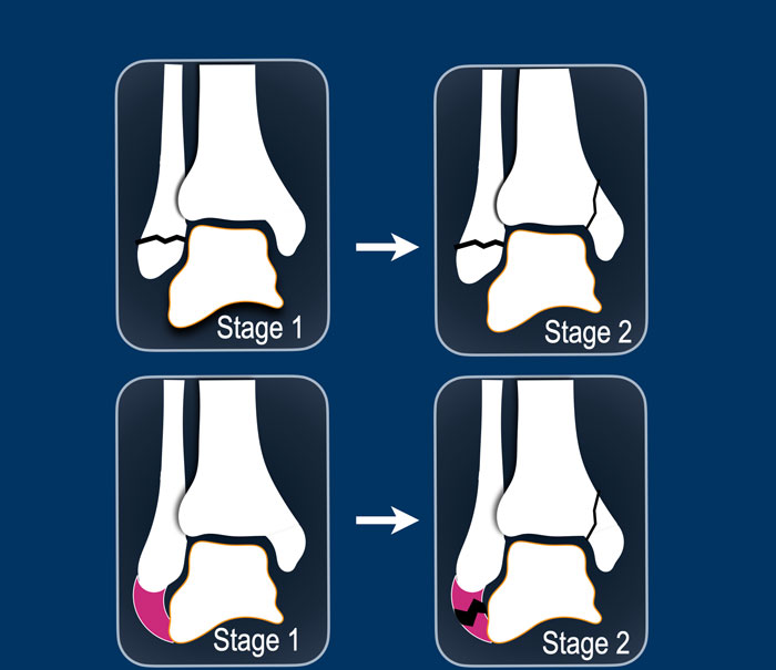
A vertical fracture of the medial malleolus is a push-off fracture.
Gãy dọc mắt cá trong là gãy đẩy ra.
According to Lauge-Hansen the fracture results from an adduction force on the supinated foot.
The lateral side is under extreme tension with stretch on the lateral collateral band.
Theo Lauge-Hansen, gãy xương là kết quả của một lực hút trên bàn chân ngửa.
Bên ngoài bị căng quá mức với sự căng ra trên dải bên.
In stage 1 there is either a rupture of the lateral collateral band or an avulsion fracture (which we also know as Weber A). Giai đoạn 1 có thể có đứt dây bên hoặc gãy bong điểm bám (mà chúng ta cũng biết là Weber A).
In stage 2 there is always a vertical fracture of the medial malleolus and there has to be either an avulsion of the lateral malleolus or a rupture of the collateral band as these sequences always are in this order with first stage 1 and then stage 2.Trong giai đoạn 2 luôn có gãy dọc mắt cá trong và phải có một trong hai là bong mắt cá bên hoặc vỡ dải băng bên vì các trình tự này luôn theo thứ tự này với giai đoạn 1 và sau đó là giai đoạn 2.
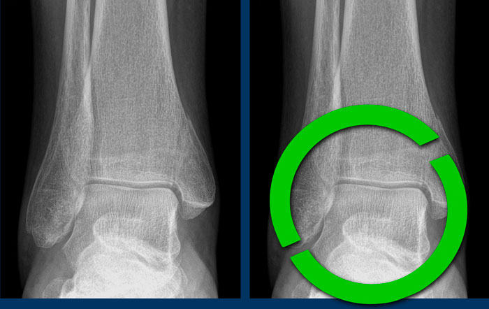
This means, that whether we see a fracture on the lateral side or not, it doesn't matter.
There must be an injury to both the medial aswell as the lateral side and we now know that the ankle is unstable, because the ring of stability is broken in two places.Điều này có nghĩa là, dù chúng ta có thấy gãy xương ở bên ngoài hay không, cũng không quan trọng.
Phải có tổn thương cả bên trong và bên ngoài và hiện nay chúng ta biết rằng cổ chân không ổn định, vì vòng ổn định bị gãy ở hai nơi.
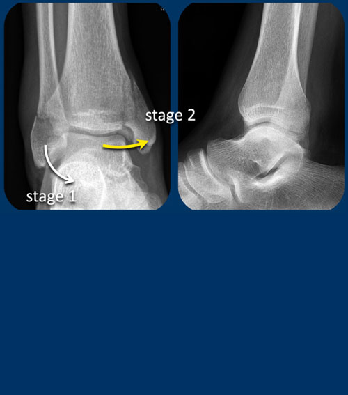
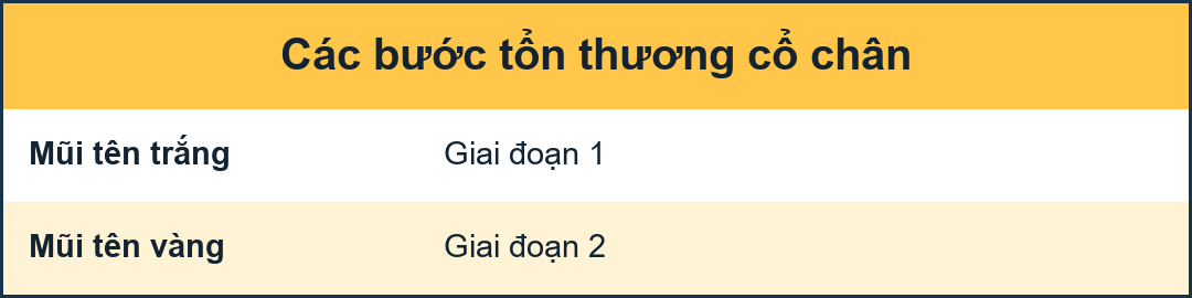
Here another unstable ankle fracture.Đây là một trường hợp gãy cổ chân không vững.
According to Lauge-Hansen this is SA stage 2.
According to Weber this is Weber A with additional fracture to the medial malleolus. Theo Lauge-Hansen đây là giai đoạn SA 2.
Theo Weber đây là Weber A có thêm gãy mắt cá trong.
case 2 - distortiontrường hợp 2 - biến dạng
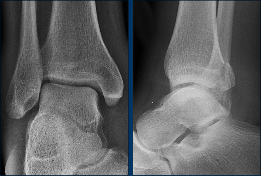
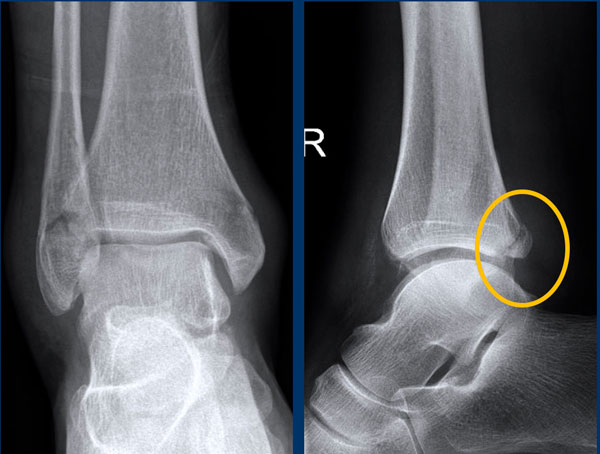
In this case the most obvious finding is a fracture of the posterior malleolus.
This should urge you to look for other findings, since an isolated fracture of the posterior malleolus is extremely uncommon and probably does not exists.Trong trường hợp này, phát hiện rõ ràng nhất là gãy mắt cá sau.
Điều này sẽ thúc giục bạn tìm kiếm các dấu hiệu khác, vì gãy xương mắt cá sau đơn độc là cực kỳ hiếm gặp và có thể không tồn tại.
You need to look at the algoritm for ankle fractures..Anh cần phải nhìn vào cơ tim để tìm gãy cổ chân.
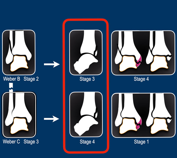
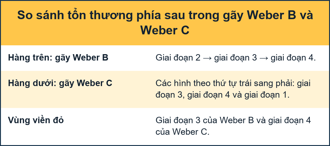
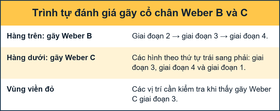
Algoritm for ankle fracturesThuật toán điều trị gãy cổ chân
In the algoritm a fracture of the posterior malleolus is either: Trong bệnh lý cơ tim, gãy mắt cá sau có thể là:
- Stage 3 of a Weber B fracture
(also called Supination Exorotation according to Lauge-Hansen) orGiai đoạn 3 của gãy Weber B
(còn được gọi là Supination Exorotation theo Lauge-Hansen) hoặc - Stage 4 of a Weber C fracture
(also called Pronation Exorotation according to Lauge-Hansen)Giai đoạn 4 của gãy Weber C
(còn được gọi là Pronation Exorotation theo Lauge-Hansen)
This means that we have to look for other signs.
Since there is no sign of a Weber B fracture, this must be a Weber C fracture.
Điều này có nghĩa là chúng ta phải tìm kiếm các dấu hiệu khác.
Vì không có dấu hiệu gãy Weber B nên đây phải là gãy Webers C.
Are there any other signs that are in favor of a Weber C fracure?Có bất kỳ dấu hiệu nào khác có lợi cho việc làm sạch Weber C không?
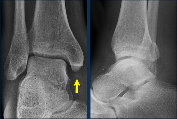
Yes there is.
Now we recognize the soft tissue swelling on the medial side and a small avulsion, which is stage 1 of this injury.Có.
Bây giờ chúng ta nhận ra mô mềm sưng ở phía trong và một chỗ nhổ nhỏ, đó là giai đoạn 1 của tổn thương này.
We now can predict a high fibula fracture, which was confirmed by a radiograph of the whole lower leg.
Bây giờ chúng ta có thể dự đoán được gãy xương mác cao, được xác nhận bằng X quang toàn bộ cẳng chân.
case 3 - distortiontrường hợp 3 - biến dạng
This is another case of a fracture of the posterior malleolus.Đây là một trường hợp gãy mắt cá sau khác.
Again we have to look at the algoritm for ankle fractures...Một lần nữa chúng ta phải xem xét các chỉ số của xương cổ chân...
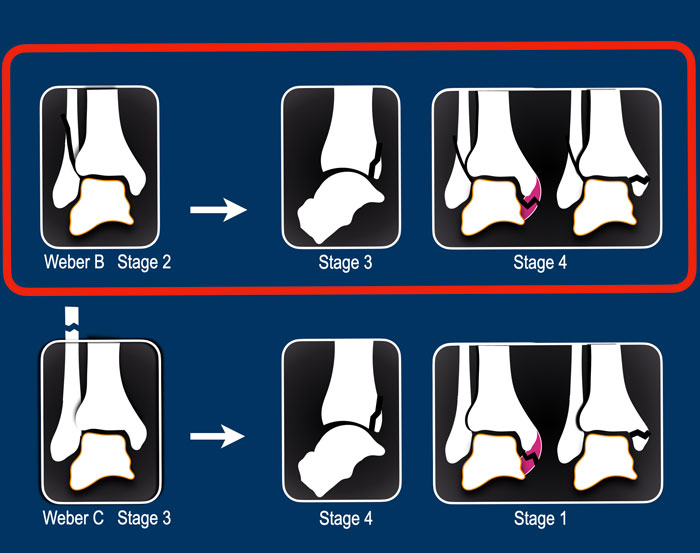
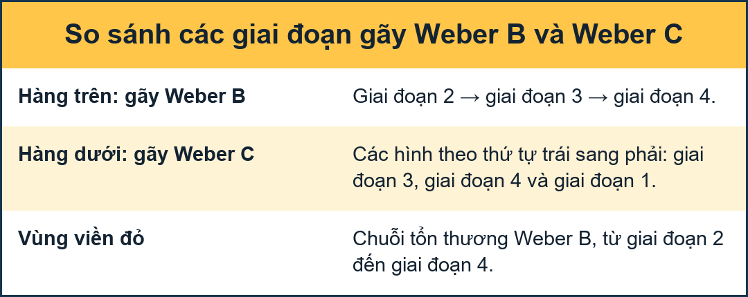
A fracture of the posterior malleolus is seen in a Weber B or a Weber C fracture.Gãy mắt cá sau được thấy trong gãy Weber B hoặc Weber C.
In this case we have already seen an oblique fracture of the lateral malleolus, which means that we are dealing with a Weber b fracture.
Now we only need to determine which stage it is.
The most common stage 2 is stable, but stage 3 and 4 are unstable.Trong trường hợp này chúng ta đã thấy một gãy chéo của mắt cá ngoài, có nghĩa là chúng ta đang đối phó với gãy Weber b.
Bây giờ chúng ta chỉ cần xác định giai đoạn đó là gì.
Giai đoạn 2 thường gặp nhất là ổn định, nhưng giai đoạn 3 và 4 không ổn định.
In this case we already in stage 3 and we wanna look for signs of stage 4.
A painful soft tissue swelling at examination would already indicate stage 4, but in this case there is more.Trong trường hợp này chúng ta đã ở giai đoạn 3 và chúng ta muốn tìm kiếm các dấu hiệu của giai đoạn 4.
Triệu chứng sưng mô mềm đau khi khám đã cho thấy giai đoạn 4, nhưng trong trường hợp này có nhiều hơn.
Maybe you want to go back to the radiograph before you continue reading...Có lẽ anh muốn quay lại chụp X - quang trước khi tiếp tục đọc...
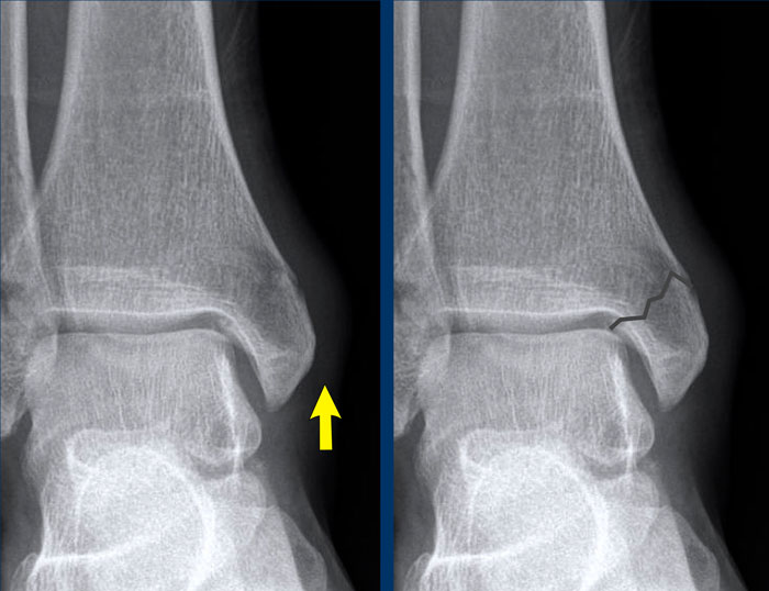
In this case we are looking for a stage 4.Trong trường hợp này chúng tôi đang tìm kiếm một giai đoạn 4.
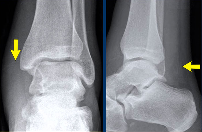
Now you notice the soft tissue swelling on the medial side (arrow).
The most important finding however is the irregularity and radiolucency of the upper part of the medial malleolus.
If you use your imagination, you can identify a fracure line here, which you would not have seen if you did not use the algoritm.Bây giờ bạn nhận thấy sưng mô mềm ở phía trong (mũi tên).
Tuy nhiên, dấu hiệu quan trọng nhất là sự bất thường và mờ của phần trên của mắt cá trong.
Nếu bạn sử dụng trí tưởng tượng của mình, bạn có thể xác định một đường gãy ở đây, mà bạn sẽ không nhìn thấy nếu bạn không sử dụng thuật toán.
case 4 - distortiontrường hợp 4 - biến dạng
The only thing that we notice is soft tissue swelling especially on the medial side.Chỉ có một điểm chúng tôi nhận thấy là sưng mô mềm đặc biệt là ở phía trong.
Continue with the ankle injury algoritm...Tiếp tục với chấn thương cổ chân...
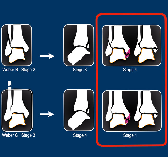
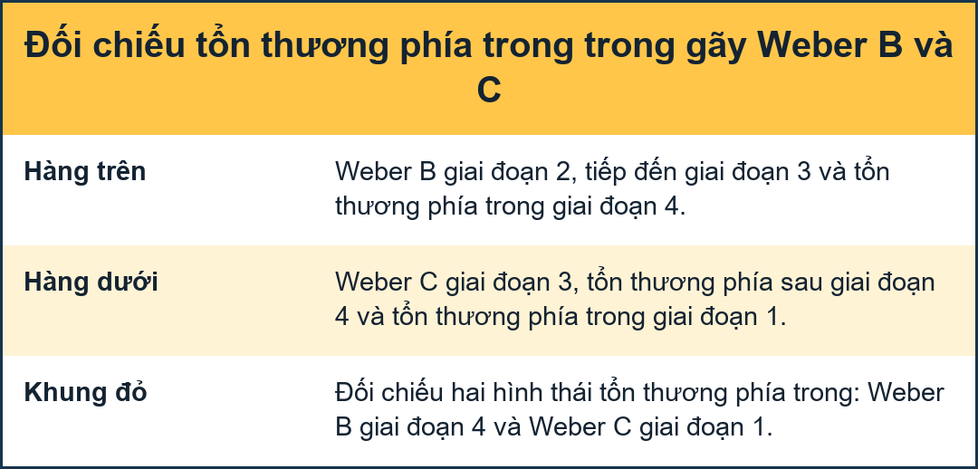
Injury to the medial collateral bands can be seen in Weber B fracture stage 4.
However there are no signs of a WeberB fracture.Tổn thương các dải băng bên trong có thể được nhìn thấy trong gãy Weber B giai đoạn 4.
Tuy nhiên không có dấu hiệu gãy WeberB.
The other possibility is that there is a Weber C fracture with a high fibula fracture, that is not visible on the x-rays of the ankle.
The medial band rupture is stage 1 and the injury may stop there or continue to stage 2,3 or even 4.
We need additional x-rays of the entire lower leg to find out which stage we are dealing with.Một khả năng khác là có gãy Weber C với xương mác cao, không thể nhìn thấy trên phim X-quang cổ chân.
Vỡ dải trong là giai đoạn 1 và chấn thương có thể dừng lại ở đó hoặc tiếp tục giai đoạn 2,3 hoặc thậm chí 4.
Chúng tôi cần chụp X-quang thêm toàn bộ cẳng chân để tìm ra giai đoạn nào chúng tôi đang xử lý.
Continue with the x-rays of the lower leg...Tiếp tục với phim X-quang cẳng chân...
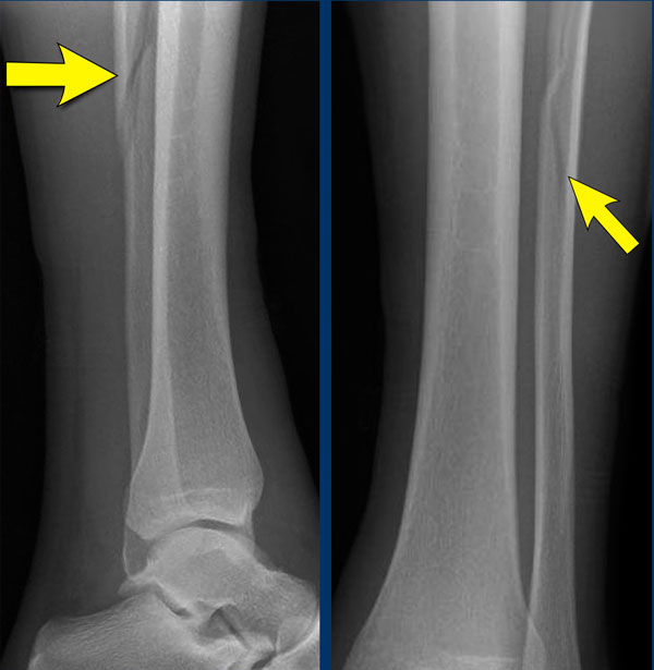
A high fibula fracture is seen (Maisonneuve).
Usually the patient will only feel pain at the level of the ankle since ligament ruptures are very painful and not notice the fibula fracture.Có thể thấy gãy xương mác cao (Maisonneuve).
Thông thường bệnh nhân sẽ chỉ cảm thấy đau ở mức cổ chân vì đứt dây chằng rất đau và không nhận thấy gãy xương mác.
case 5 - distortiontrường hợp 5 - biến dạng
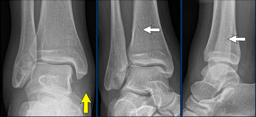
There is a subtle fracture visible on the Morrison's and the lateral view.
This is a large tertius fracture.
There is no sign of a Weber B type of fibula fracture.Có một vết nứt nhỏ có thể nhìn thấy trên mặt Morrison và mặt nghiêng.
Đây là một gãy xương lớn.
Không có dấu hiệu của gãy xương mác Weber B.
The combination of this fracture and the soft tissue swelling on the medial side indicate that we are dealing with a Weber C type of injury (or pronation exorotation injury according to Lauge-Hansen).Sự kết hợp của gãy xương này và sưng mô mềm ở bên trong cho thấy chúng ta đang đối mặt với loại chấn thương Weber C (hoặc chấn thương sấp ngửa theo Lauge-Hansen).
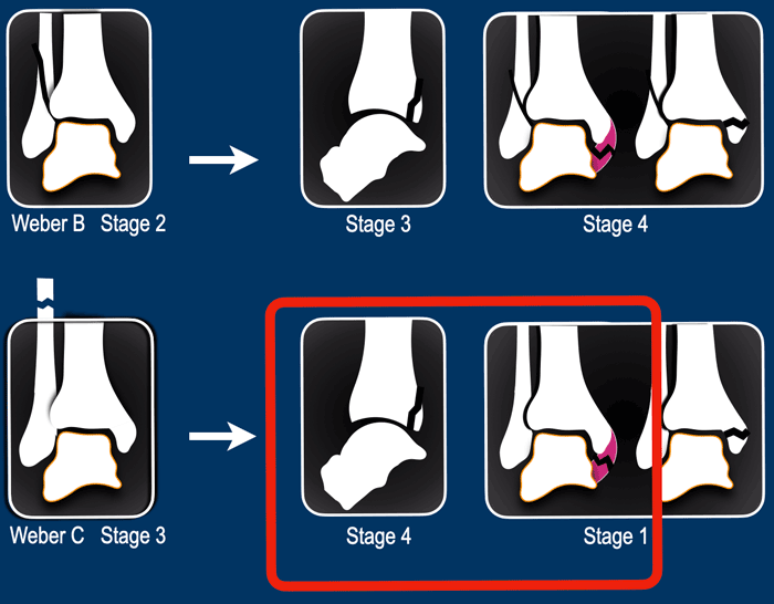
The medial collateral band injury is stage 1 and the tertius fracture is stage 4.
This means that this is an unstable fracture.
There must be a high fibula fracture.Tổn thương dây bên trong là giai đoạn 1 và gãy xương bánh chè là giai đoạn 4.
Điều này có nghĩa đây là một loại gãy xương không vững.
Có thể gãy xương mác cao.