
Brain AnatomyGiải phẫu não
Radiology department, Alrijne Hospital Leiden, the Netherlands.Khoa Chẩn đoán hình ảnh, Bệnh viện Alrijne Leiden, Hà Lan.
Ngày đăng / cập nhật: 2008-11-24
Bản gốc trên radiologyassistant.nl ↗ · tải về lúc 2026-11-19 20:19 · bản dịch máy có chuẩn hóa thuật ngữ chẩn đoán hình ảnh
Axial anatomyGiải phẫu trục
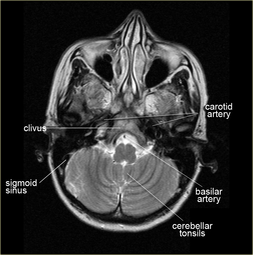
Scroll through the images on the left.Cuộn qua các hình ảnh bên trái.
Specific regionsCác vùng cụ thể
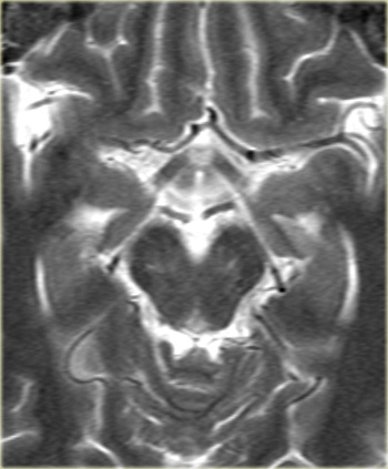
Circle of WillisVòng tròn Willis
- A1-segment
Anterior cerebral artery from carotid bifurcation to anterior communicating artery gives rise to the medial lenticulostriate arteries.A1 đoạn
Động mạch não trước từ chỗ chia đôi động mạch cảnh đến động mạch thông trước làm phát sinh các động mạch dạng thấu kính trong. - A2-segment
Part of anterior cerebral artery distal to the anterior communicating artery.A2 - đoạn
Một phần động mạch não trước ở xa động mạch thông trước. - P1-segment
Part of the posterior cerebral artery proximal to the posterior communicating artery.
The posterior communicating artery is between the carotid bifurcation and the posterior cerebral artery) P1 - đoạn
Một phần của động mạch não sau gần với động mạch thông sau.
Động mạch thông sau nằm giữa chỗ chia đôi động mạch cảnh và động mạch não sau) - P2-segment
Part of the posterior cerebral artery distal to the posterior communicating artery P2 - đoạn
Một phần động mạch não sau ở xa động mạch thông sau - M1-segment
Horizontal part of the middle cerebral artery which gives rise to the lateral lenticulostriate arteries which supply most of the basal ganglia.
The M2-segment is the part in the sylvian fissure and the M3-segment is the cortical segment. M1 - đoạn
Phần ngang của động mạch não giữa tạo ra các động mạch thấu kính bên cung cấp phần lớn hạch nền.
Đoạn M2 là một phần trong rãnh sylvian và đoạn M3 là đoạn vỏ não. - Cisterna ambiens
Also called ambient cistern is a cistern of the subarachnoid space between the posterior end of the corpus callosum and the superior surface of the cerebellum.
It is sometimes defined as including the quadrigerminal cistern. Cisterna ambiens
Cũng được gọi là bể chứa xung quanh là một bể chứa của khoang dưới nhện giữa đầu sau của thể chai và bề mặt trên của tiểu não.
Đôi khi nó được định nghĩa là bao gồm bể chứa nước tiểu.
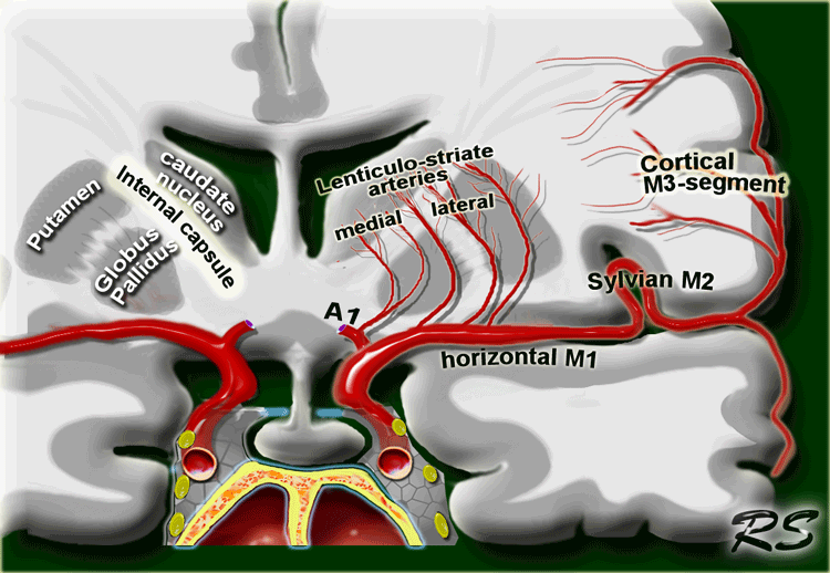
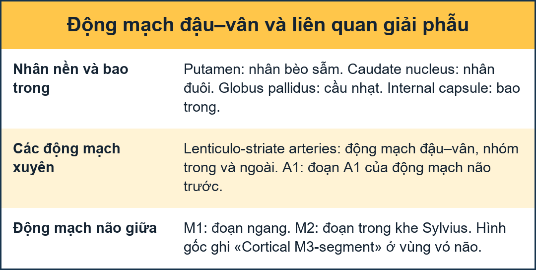
On the left a coronal view of the segments of the middle cerebral artery.
Bên trái là hình ảnh mặt cắt của các đoạn của động mạch não giữa.
- Horizontal M1-segment
gives rise to the lateral lenticulostriate arteries which supply part of head and body of caudate, globus pallidus, putamen and the posterior limb of the internal capsule.
Notice that the medial lenticulostriate arteries arise from the A1-segment of the anterior cerebral artery.Phân đoạn M1 nằm ngang
làm phát sinh các động mạch thấu kính bên cung cấp một phần đầu và thân của xương đuôi, cầu nhạt, nhân hầm và chi sau của bao trong.
Lưu ý rằng các động mạch thấu kính trong phát sinh từ đoạn A1 của động mạch não trước. - Sylvian M2-segment
Branches supply the temporal lobe and insular cortex (sensory language area of Wernicke), parietal lobe (sensory cortical areas) and inferolateral frontal lobe Đoạn M2 Sylvian
Các nhánh cung cấp cho thuỳ thái dương và vỏ đảo (vùng ngôn ngữ cảm giác của Wernicke), thuỳ đỉnh (vùng vỏ não cảm giác) và thuỳ trán dưới bên. - Cortical M3-segment
Branches supply the lateral cerebral cortexĐoạn M3 vỏ não
Các nhánh cung cấp cho vỏ não bên
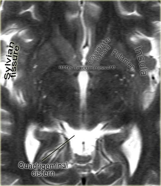
Anterior commissureMép trước
The anterior commissure is a bundle of white fibers that connects the two cerebral hemispheres across the middle line.
At this level frequently perivascular CSF-spaces of Virchow-Robin are seen.Nếp gấp trước là một bó các sợi trắng kết nối hai bán cầu não qua đường giữa.
Ở mức này, thường thấy các khoảng CSF quanh mạch của Virchow-Robin.
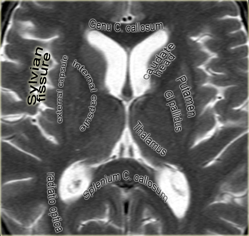
Thalamic levelMức độ thalassemia
At this level the basal ganglia are seen.
The two dark lines medially of the thalamus are the internal cerebral veins.Ở mức này, hạch nền được nhìn thấy.
Hai đường tối ở giữa đồi thị là các tĩnh mạch não trong.
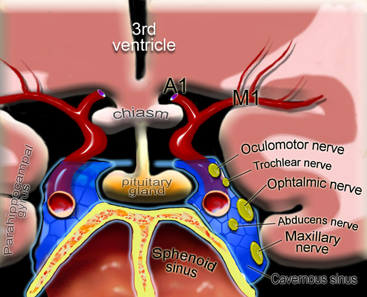
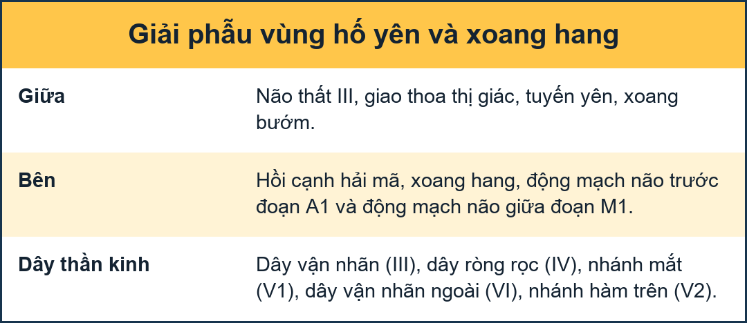
Pituitary glandTuyến yên
On the left a coronal illustration of the anatomy of the pituitary gland and the surrounding structures.Bên trái là hình minh hoạ của vành của giải phẫu tuyến yên và các cấu trúc xung quanh.
Read more about the pituitary gland in the article on sellar and parasellar tumorsĐọc thêm về tuyến yên trong bài viết về khối u yên và cận yên
HippocampusHông cá
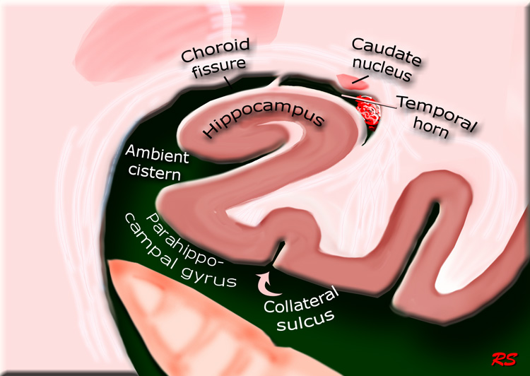
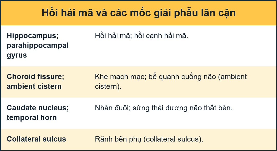
On the left a coronal illustration of the area of the hippocampus.Bên trái là hình minh hoạ của vùng hải mã.
Read more about the hippocampus in the article on the role of MRI in dementiaĐọc thêm về vùng hải mã trong bài viết về vai trò của MRI trong chứng mất trí nhớ
ReferencesTài liệu tham khảo
-
The whole brain atlas
Bản đồ toàn bộ não
Neuroimaging Primer - Harvard Medical School lecture notes: Introduction to Neuroimaging
by Keith Johnson and Alex Becker
NeuroImaging Primer - Bài giảng của Trường Y Harvard: Giới thiệu về hình ảnh thần kinh -
the Brain Module
Mô-đun não
Developed by Jeffrey E. Zapawa and Anthony L. Alcantara, MD Phát triển bởi Jeffrey E. Zapawa và Anthony L. Alcantara, MD