
Renal Tumors in ChildrenKhối u thận ở trẻ em
Department of Radiology and Nuclear Medicine, University Medical Center Utrecht and Princess Máxima Center for Pediatric Oncology in UtrechtKhoa Chẩn đoán hình ảnh và Y học hạt nhân, Trung tâm Y khoa Đại học Utrecht và Trung tâm Ung thư Nhi khoa Princess Máxima ở Utrecht
Bản gốc trên radiologyassistant.nl ↗ · tải về lúc 2026-11-19 20:19 · bản dịch máy có chuẩn hóa thuật ngữ chẩn đoán hình ảnh
Wilms tumor comprises the vast majority of renal neoplasms in young children and resection with neoadjuvant chemotherapy is the treatment of choice.
U Wilms chiếm đa số trong các u thận ở trẻ nhỏ và điều trị bằng hóa trị tân bổ trợ là lựa chọn điều trị.
Most other renal tumors cannot be reliably distinguished from Wilms tumor at imaging, but some features may suggest an alternative diagnosis, which in some cases can lead to the decision to adjust the treatment or in some cases to perform a biopsy before start of pre-operative treatment. Hầu hết các khối u thận khác không thể phân biệt được với khối u Wilms trên hình ảnh, nhưng một số đặc điểm có thể gợi ý chẩn đoán khác, trong một số trường hợp có thể dẫn đến quyết định điều chỉnh điều trị hoặc trong một số Trường hợp phải sinh thiết trước khi bắt đầu điều trị trước phẫu thuật.
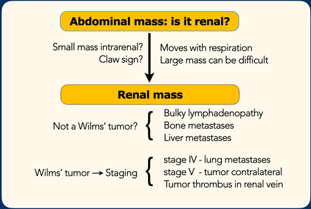
The role of imaging in children with a possible renal tumor is first to determine whether the mass originates in the kidney.
The next step is to look for signs that would make you consider an alternative diagnosis instead of a Wilms tumor.
The final step is staging.Vai trò của chẩn đoán hình ảnh ở trẻ em có khối u thận có thể là đầu tiên để xác định xem khối u có nguồn gốc từ thận hay không.
Bước tiếp theo là tìm kiếm các dấu hiệu khiến bạn xem xét chẩn đoán khác thay vì khối u Wilms.
Bước cuối cùng là phân giai đoạn.
In this article we will discuss common and some less common renal tumors and include some malignancies that can appear as a primary renal tumor, like lymphoma, neuroblastoma and leukemia.
Trong bài viết này, chúng tôi sẽ thảo luận về các loại u thận phổ biến và ít phổ biến hơn, bao gồm một số loại u ác tính có thể xuất hiện như u nguyên phát ở thận, như u lympho, u nguyên bào thần kinh và bệnh bạch cầu.
IntroductionMở đầu
Role of ImagingVai trò của hình ảnh
- Determine whether a mass is a renal mass.
In most cases ultrasound can determine whether a mass originates from the kidney. Especially look for the claw sign.
In large masses it can be difficult and MRI or CT can solve the problem.
A large neuroblastoma, that compresses or invades the kidney can look like a renal tumor.
Xác định khối là khối thận hay không.
Trong hầu hết các trường hợp, siêu âm có thể xác định xem một khối có nguồn gốc từ thận hay không, đặc biệt là tìm dấu hiệu móng vuốt.
Ở những khối lớn, có thể khó khăn và MRI hoặc CT có thể giải quyết vấn đề.
Một u nguyên bào thần kinh lớn, chèn ép hoặc xâm lấn thận có thể trông giống như một khối u thận. - Is it a Wilms' tumor or could it be something else?
If it is a renal mass, then in most cases it will be a Wilms' tumor, especially in the age group 6 month to nine years. For the differential diagnosis, look for signs that are uncommon in a Wilms' tumor, like bulky lymphadenopathy (RCC or lymphoma) or liver and bone metastases (Clear cell sarcoma).
Đó là khối u Wilms hay là một thứ gì khác?
Nếu đó là một khối u ở thận thì trong hầu hết các trường hợp sẽ là khối u Wilms, đặc biệt là ở nhóm tuổi 6 tháng đến chín tuổi Đối với chẩn đoán phân biệt, hãy tìm các dấu hiệu không phổ biến trong khối u của Wilms, như bệnh hạch (RCC hoặc u lympho) hoặc gan và di căn xương ( sarcoma tế bào rõ). - Staging
When a Wilms' tumor is the most likely diagnosis, but also in other tumors staging is the next step.
Especially look for tumor thrombus, lung metastases (stage IV) and tumor in the contralateral kidney (stage V)
Phân giai đoạn
Khi một khối u Wilms là chẩn đoán có khả năng nhất, nhưng cũng có trong các khối u khác, giai đoạn là bước tiếp theo.
Đặc biệt là tìm huyết khối u, di căn phổi (giai đoạn IV) và khối u ở thận đối bên (giai đoạn V)
 Claw signDấu hiệu móng vuốt
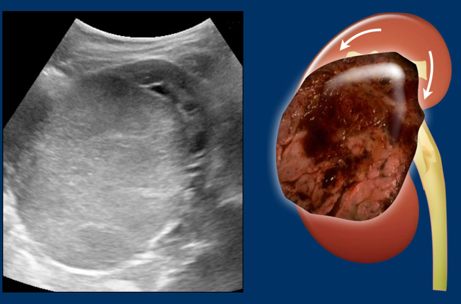
This sign in renal tumors is useful in determining that the tumor arises from the kidney rather than is located adjacent to the kidney and distorts the outline.Dấu hiệu này trong các khối u thận rất hữu ích trong việc xác định rằng khối u phát sinh từ thận chứ không phải nằm liền kề với thận và làm biến dạng đường viền.
The claw sign refers to the sharp angles on either side of the mass, which the surrounding normal parenchyma forms when the mass has arisen from the kidney. Dấu hiệu móng vuốt đề cập đến các góc nhọn ở hai bên của khối u, mà nhu mô bình thường xung quanh hình thành khi khối u phát sinh từ thận.
Images
Left sided Wilms' tumor in a one-year-old girl.
The remnant of the kidney is draped over the tumor, the “claw sign”.Hình ảnh
Khối u bên trái của Wilms ở một bé gái một tuổi.
Phần còn lại của thận được phủ lên khối u, “dấu hiệu móng vuốt”.
ManagementQuản lý
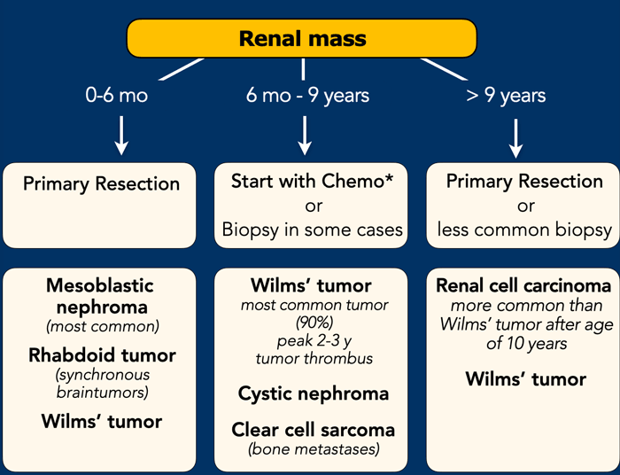
Imaging plays a limited role in determining the exact nature and management of a renal tumor in children, since almost all pediatric renal tumors will be resected and most tumors will be a Wilms tumor.
Age at presentation is a far more important predictor of tumor type. Chẩn đoán hình ảnh đóng một vai trò hạn chế trong việc xác định bản chất chính xác và quản lý khối u thận ở trẻ em, vì hầu hết các khối u thận trẻ em sẽ được cắt bỏ và hầu hết các u sẽ là khối u Wilms.
Tuổi là yếu tố tiên lượng quan trọng hơn nhiều về loại khối u.
0-6 months-old
0-6 tháng tuổi
Besides Wilms tumor, the differential diagnosis comprises two tumors that occur predominantly in infants: congenital mesoblastic nephroma and rhabdoid tumor of the kidney (MRTK). Both tumors are treated with primary resection. Therefore, renal tumors in infants below six months are primarily resected if possible. Bên cạnh khối u Wilms, chẩn đoán phân biệt bao gồm hai khối u xảy ra chủ yếu ở trẻ sơ sinh: u thận bẩm sinh nguyên bào trung gian và u thận dạng rhabdoid (MRTK). Cả hai khối u đều được điều trị bằng phẫu thuật cắt bỏ ban đầu, do đó, khối u thận ở trẻ sơ sanh dưới sáu tháng được cắt bỏ ban đât nếu có thể.
6-months – 9 years
A renal tumor in children in this age group is considered a Wilms tumor without histological confirmation.6 tháng – 9 năm
U thận ở trẻ em trong độ tuổi này được coi là u Wilms mà không có xác nhận mô học.
>9 years
In children above 9 years-old in most cases a primary nephrectomy is performed.
A renal cell carcinoma (RCC) is more common in this age group than a Wilms tumor. >9 năm
Ở trẻ em trên 9 tuổi trong hầu hết các trường hợp, phẫu thuật cắt thận được thực hiện.
Ung thư biểu mô tế bào thận (RCC) phổ biến hơn ở nhóm tuổi này so với khối u Wilms.
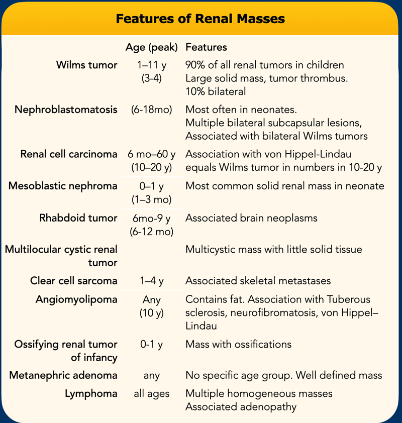
This table summarizes the features of pediatric renal tumors.Bảng này tóm tắt các đặc điểm của u thận ở trẻ em.
Some of these pediatric renal masses may be differentiated from a Wilms tumor on the basis of their clinical and imaging features.
Một số khối u thận ở trẻ em này có thể được phân biệt với khối u Wilms dựa trên các đặc điểm lâm sàng và hình ảnh của chúng.
- Wilms tumor is usually very large at presentation. It is the most likely diagnosis, especially when there are bilateral tumors, venous invasion, or pulmonary
metastases.
U Wilms thường có kích thước rất lớn, là chẩn đoán có khả năng nhất, đặc biệt khi có u hai bên, xâm lấn tĩnh mạch, hoặc di căn phổi. - Nephroblastomatosis occurs most often in neonates and is characterized by multiple bilateral subcapsular masses, often associated with Wilms tumors.
Bệnh nguyên bào thận thường xảy ra ở trẻ sơ sinh và được đặc trưng bởi nhiều khối dưới bao hai bên, thường liên quan đến khối u Wilms. - Renal cell carcinoma is unusual in children except in association with von Hippel–Lindau syndrome and typically occurs in the 2nd decade.
Ung thư biểu mô tế bào thận là bất thường ở trẻ em ngoại trừ kết hợp với hội chứng von Hippel-Lindau và thường xảy ra trong thập kỷ thứ 2. - Congenital mesoblastic nephroma (CMN) is the most common renal tumor in the neonatal period and the most frequent benign renal tumor in childhood. A whirlpool aspect of the tumor on ultrasound
may suggest a congenital mesoblastic nephroma.
U thận nguyên bào trung gian bẩm sinh (Congenital mesoblastic nephroma - CMN) là khối u thận thường gặp nhất ở giai đoạn sơ sinh và là khối u lành tính thường gặp nhất trong thời thơ ấu, hình ảnh xoáy nước của khối u trên siêu âm có thể gợi ý u thận nguyên bào Trung gian bẩM sinh. - Rhabdoid tumor is distinguished by its association with synchronous intracranial tumors.
U Rhabdoid được phân biệt bởi sự liên quan của nó với các khối u nội sọ đồng bộ. - Multilocular cystic renal
tumors include cystic nephroma and cystic partially differentiated
nephroblastoma (CPDN). The diagnosis is suggested by a large mass constiting of
multiple cysts and no nodular solid tissue. The treatment is resection.
U nang thận đa thùy bao gồm u thận dạng nang và u nguyên bào thận biệt hóa một phần dạng nang (CPDN). Chẩn đoán được gợi ý bởi một khối u lớn bao gồm nhiều nang và không có mô đặc dạng nốt. Điều trị là cắt bỏ. - Clear cell sarcoma of the kidney (CCSK) is known for its propensity to metastasize to bone, a rare location for Wilms tumor metastasis.
Sarcoma tế bào sáng của thận (CCSK) được biết đến với xu hướng di căn đến xương, một vị trí hiếm gặp đối với di căn khối u Wilms.
- Angiomyolipoma contains fat and is associated with tuberous sclerosis.
U cơ mỡ có chứa mỡ và có liên quan đến xơ cứng củ. - Renal medullary carcinoma occurs in patients with sickle cell trait or hemoglobin SC disease and manifests as an infiltrative mass with metastases.
Ung thư biểu mô tuỷ thận xảy ra ở những bệnh nhân có đặc điểm hồng cầu hình liềm hoặc bệnh hemoglobin SC và biểu hiện như một khối thâm nhiễm với di căn. - Ossifying renal tumor of infancy (ORTI) is differentiated from mesoblastic nephroma by the presence of ossified elements. It herniates into the renal pelvis and mimics a staghorn calculus in appearance.
U thận do xương hóa ở trẻ sơ sinh (ORTI) được phân biệt với u thận nguyên bào trung bình bởi sự hiện diện của các yếu tố bị xương hóa, nó thóat vị vào bể thận và giống như một viên sỏi san hô. - Metanephric adenoma lacks specific features but is always well defined.
U tuyến thiếu các đặc điểm cụ thể nhưng luôn được xác định rõ. - Renal lymphoma is characterized by multiple homogeneous masses, often with associated lymph adenopathy.U lympho thận được đặc trưng bởi nhiều khối đồng nhất, thường có hạch bạch huyết liên quan.
Wilms tumorKhối u Wilms
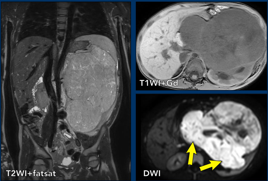 Nephroblastoma in a five-year-old boy.U nguyên bào thận ở một cậu bé năm tuổi.
More than 90% of renal tumors in children are Wilms tumors, also called nephroblastomas. Wilms tumor presents mostly in children between 6 month and nine years of age with a peak incidence from 2 to 3 years.Hơn 90% khối u thận ở trẻ em là khối u Wilms, còn được gọi là u nguyên bào thận. Khối u Wilms biểu hiện chủ yếu ở trẻ em từ 6 tháng đến chín tuổi với tỷ lệ mắc cao nhất từ 2 đến 3 tuổi.
Wilms tumors are generally very large at initial diagnosis and these children present with a distended abdomen.
Children with Wilms tumor are generally not ill.Các khối u Wilms thường rất lớn khi chẩn đoán ban đầu và những đứa trẻ này có bụng phình to.
Trẻ em bị khối u Wilms thường không bị bệnh.
case 1
The coronal T2W-image shows an inhomogeneous tumor in the left kidney with small cystic
components.
The tumor enhances less than the peripheral remnant
of normal renal tissue.
The solid parts of the tumor show strong diffusion
restriction (arrows).
This was a Wilms tumor in a five-year-old boy. Pre-operative chemotherapy was given according to the Umbrella protocol of the SIOP-RTSG after which a nephrectomy
was performed.Ca 1
Ảnh T2W mặt phẳng đứng ngang cho thấy u thận trái không đồng nhất với các thành phần dạng nang nhỏ. U ngấm thuốc kém hơn phần nhu mô thận bình thường còn lại ở ngoại vi. Phần đặc hạn chế khuếch tán rõ (mũi tên). Đây là u Wilms ở bé trai 5 tuổi. Bệnh nhi được hóa trị trước mổ theo phác đồ Umbrella của SIOP-RTSG rồi cắt thận.
Beckwith-Wiedemann syndromeHội chứng Beckwith-Wiedemann
Smaller tumors can be detected when haematuria, abdominal pain, or hypertension with headache is the presenting symptom or during sonographic screening in children with syndromes which predispose to nephroblastoma, like Beckwith Wiedemann syndrome, WAGR, Perlman
syndrome, DICER 1 and Li-Fraumeni syndrome.
Các khối u nhỏ hơn có thể được phát hiện khi xuất hiện triệu chứng tiểu máu, đau bụng, hoặc tăng huyết áp kèm đau đầu hoặc trong quá trình sàng lọc siêu âm ở trẻ em có hội chứng có khuynh hướng u nguyên bào thận, như hội chứng Beckwith Wiedemann, WAGR, hội chứng Perlman, DICER 1 và hội chứng Li-Fraumeni.
Bilateral nephroblastomas are often genetic or syndrome-related.
U nguyên bào thận hai bên thường liên quan đến di truyền hoặc hội chứng.
Small tumors are usually homogeneous echogenic.
Larger tumors are more inhomogeneous with cystic necrotic parts and hemorrhage.
10% of nephroblastomas have fine calcifications.Các khối u nhỏ thường có tiếng vang đồng nhất.
Các khối u lớn hơn không đồng nhất với các phần hoại tử dạng nang và xuất huyết.
10% u nguyên bào thận có vôi hóa.
The lungs are the most frequent site of metastases.
Liver and bone metastasis are rare.Phổi là vị trí di căn thường gặp nhất.
Di căn gan và xương hiếm gặp.
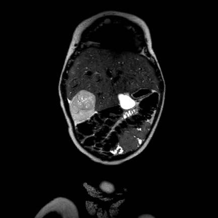
case 2
This is a two-year-old boy with Beckwith-Wiedemann syndrome.
A screening ultrasound showed a small homogeneous tumor in the left part of a horseshoe kidney. Trường hợp 2
Đây là một cậu bé hai tuổi mắc hội chứng Beckwith-Wiedemann.
Siêu âm sàng lọc cho thấy một khối u nhỏ đồng nhất ở phần bên trái của thận móng ngựa.
MR findingsKết quả cộng hưởng từ
Wilms tumors are mostly inhomogeneous, with decreased signal intensity on T1 and increased signal intensity on T2.
Necrotic cystic parts are often present.
Enhancement after Gadolinium is inhomogeneous and less than the enhancement of normal renal parenchyma. U Wilms chủ yếu là không đồng nhất, với cường độ tín hiệu giảm ở T1 và tăng cường độ tín hiệu ở T 2.
Các phần nang hoại tử thường có mặt.
Sự tăng cường sau khi Gadolinium không đồng nhất và ít hơn sự tăng cường của nhu mô thận bình thường.
The solid parts of the tumor will show restricted diffusion.
Signs of hemorrhage are often present. Hemorrhagic areas will also show restricted diffusion, so look at the T1 for signs of bleeding. Phần đặc của khối u sẽ hiển thị hạn chế khuếch tán.
Dấu hiệu xuất huyết thường có mặt. Vùng xuất huyết cũng sẽ hiển thị hạn chế khuếch tán, vì vậy hãy nhìn vào T1 để tìm dấu hiệu chảy máu.
MRI can nicely demonstrate tumor thrombus in the renal vein and inferior caval vein, as well as lymph node enlargement. It allows accurate and repeatable measurements of the tumor 3-dimensional, on the original and follow-up examinations.MRI có thể chứng minh rõ ràng huyết khối u trong tĩnh mạch thận và tĩnh mạch chủ dưới, cũng như hạch bạch huyết mở rộng, cho phép đo chính xác và lặp lại khối u 3 chiều, trên các lần khám ban đầu và theo dõi.
case 3
This MRI is of a three-year-old
girl with a tumor of the right kidney.
Scroll through the coronal T2W-images.
There is a large tumor thrombus extending from the renal vein into the inferior
caval vein (arrows). trường hợp 3
MRI này là của một bé gái ba tuổi với một khối u ở thận phải.
Cuộn qua hình ảnh T2W vành.
Có một huyết khối u lớn kéo dài từ tĩnh mạch thận vào tĩnh mạch chủ dưới (các mũi tên).
Continue with the ultrasound...Tiếp tục với siêu âm...
This is the initial ultrasound of the same patient showing the tumor and tumor thrombus. Đây là hình ảnh siêu âm ban đầu của cùng một bệnh nhân cho thấy khối u và huyết khối u.
case 4
This video is of a three-year-old
girl with a large mass in the right flank. trường hợp 4
Video này là của một bé gái ba tuổi với một khối lớn ở sườn phải.
Ultrasound with a high frequency
transducer shows that the tumor originates from the right kidney.
The remnant
of the collecting system is dilated, as is often the case. Siêu âm với đầu dò tần số cao cho thấy khối u bắt nguồn từ thận phải.
Phần còn lại của hệ thống thu thập được giãn ra, như thường xảy ra.
Continue with the MRI...
Tiếp tục chụp MRI...
The MRi shows the extent of the tumor.
Notice the tumor thrombus in the inferior caval
vein.Chụp cộng hưởng từ cho thấy mức độ rộng của khối u.
Lưu ý huyết khối u trong tĩnh mạch chủ dưới.
Once you are sure of the renal origin of the tumor, scrutinize the other kidney for tumor or nephroblastomatosis (see below).
Search for enlarged para-aortal lymph nodes.
Search with color Doppler for patency of the renal vein and of the inferior caval vein, as a Wilms tumor tends to grow into the renal vein and inferior caval vein.Một khi bạn chắc chắn về nguồn gốc thận của khối u, hãy xem xét thận khác để tìm khối u hoặc bệnh nguyên bào thận (xem bên dưới).
Tìm kiếm hạch bạch huyết cạnh động mạch chủ to.
Tìm kiếm bằng Doppler màu cho sự thông thóang của tĩnh mạch thận và tĩnh mạch chủ dưới, vì khối u Wilms có xu hướng phát triển vào tĩnh mạch thận.
The finding of a liver metastasis should urge you to consider an alternative diagnosis.Việc phát hiện di căn gan nên khuyến khích bạn xem xét một chẩn đoán khác.
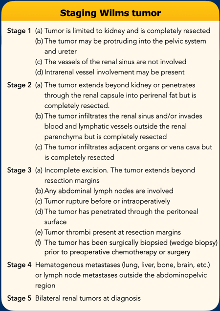
Staging and surgeryPhân giai đoạn và phẫu thuật
Surgery is the cornerstone for the treatment of WT.
The Children’s Oncology Group (COG) from North America recommends surgery before chemotherapy, whereas SIOP in Europe suggests preoperative chemotherapy.
As the SIOP group, the National Wilms Tumor Study Group (NWTSG) has concerns about performing a biopsy first because of the risk of tumor upstaging. Phẫu thuật là nền tảng cho điều trị WT.
Nhóm Ung thư Trẻ em (COG) từ Bắc Mỹ khuyến cáo phẫu thuật trước khi hóa trị, trong khi SIOP ở Châu Âu đề nghị hóa trị trước phẫu thuật.
Là nhóm SIOP, Nhóm nghiên cứu khối u Wilms quốc gia (NWTSG) có lo ngại về việc thực hiện sinh thiết trước vì nguy cơ khối u tăng cao.
The SIOP recommends preoperative chemotherapy to decrease the risk of intraoperative rupture, downstage the tumor, and to reduce the need for irradiation.
The advantage of preoperative chemotherapy is the identification of chemoresistant high-risk blastemal predominant subtype that benefits from treatment intensification.SIOP khuyến cáo hóa trị trước phẫu thuật để giảm nguy cơ vỡ trong phẫu thuật, giảm mức độ của khối u và giảm nhu cầu chiếu xạ.
Ưu điểm của hóa trị trước mổ là xác định được nhóm ưu thế nguy cơ cao do hóa trị liệu có lợi ích từ việc tăng cường điều trị.
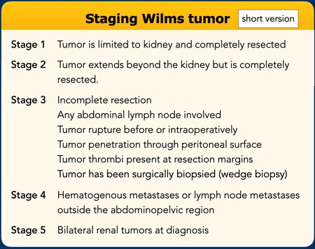
This is the short version of Wilms tumor staging.Đây là phiên bản ngắn của phân giai đoạn khối u Wilms.
NephroblastomatosisBệnh lý nguyên bào thận
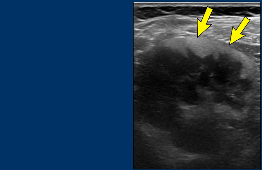
A
kidney can harbor a nephrogenic rest, which is persisting embryologic tissue.
If
multiple rests are present it is called nephroblastomatosis.
Most nephrogenic
rests resolve spontaneously, but they may transform into a Wilms tumor.Thận có thể chứa một phần còn lại của thận, đó là mô phôi thai tồn tại dai dẳng.
Nếu có nhiều lần nghỉ, nó được gọi là bệnh nguyên bào thận.
Hầu hết các trường hợp nghỉ ngơi do thận tự khỏi, nhưng chúng có thể biến thành khối u Wilms.
On ultrasound Wilms tumor is difficult to detect as these lesions tend to show similar echogenicity compared to the renal cortex. Trên siêu âm, u Wilms khó phát hiện vì những tổn thương này có xu hướng cho thấy tính gây tiếng vang tương tự so với vỏ thận.
Image
Persistent
nephroblastomatosis in a seven-year-old boy who was treated five years ago for
a Wilms tumor. A peripheral rim of echogenic tissue is seen. These
residual lesions are followed up with regular ultrasound untill 5 years after
treatment.Hình ảnh
Bệnh nguyên bào thận dai dẳng ở một cậu bé bảy tuổi được điều trị khối u Wilms năm năm trước. Một vành ngoại vi của mô echogenic được nhìn thấy. Những tổn thương còn sót lại này được theo dõi bằng siêu âm thường xuyên cho đến 5 năm sau khi điều trị.
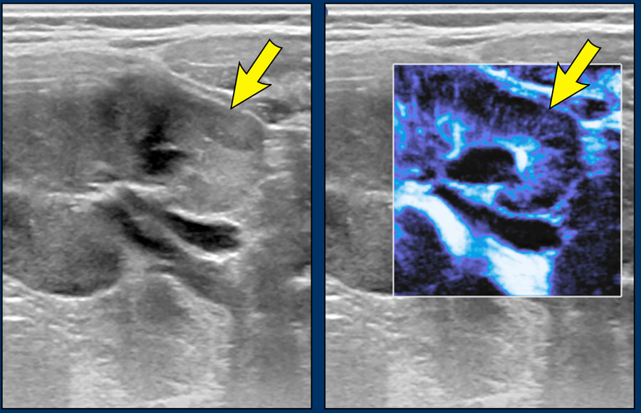
Microflow imaging can help to identify hypoperfused areas with nephroblastomatosis.Chụp vi dòng có thể giúp xác định các vùng giảm tưới máu có bệnh lý nguyên bào thận.
Images
A two-year-old
boy with a Wilms tumor in the left kidney and bilateral nephroblastomatosis.
With microflow color imaging it is less perfused than normal renal tissue.
Continue with the MR images... Hình ảnh
Một cậu bé hai tuổi bị khối u Wilms ở thận trái và bệnh nguyên bào thận hai bên. Với hình ảnh màu vi dòng, nó ít được tưới máu hơn mô thận bình thường.
Tiếp tục với hình ảnh cộng hưởng từ...
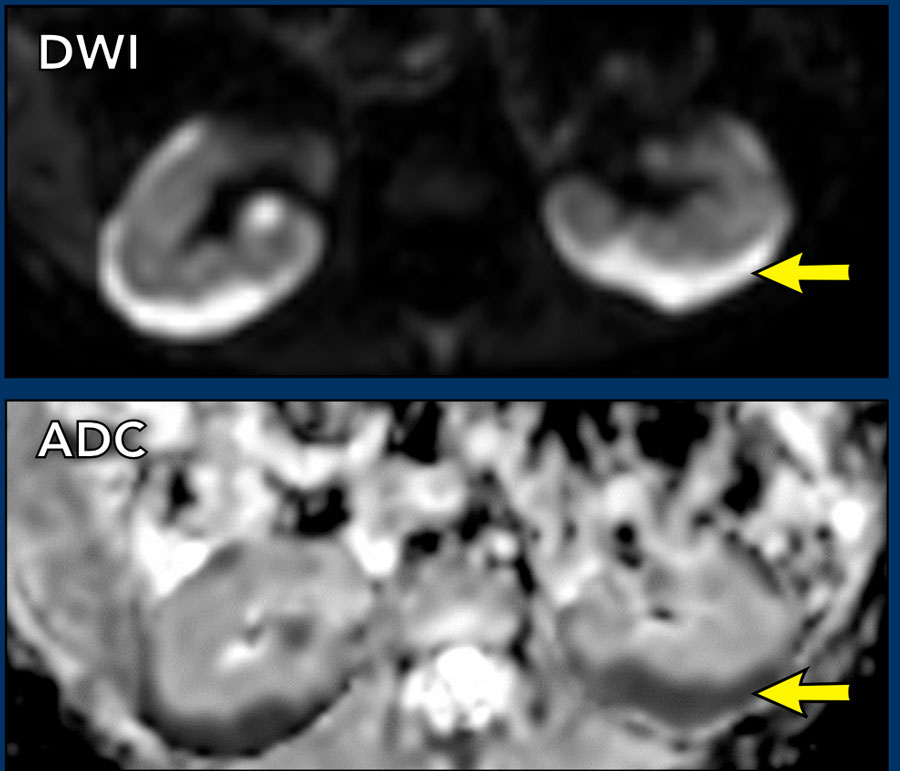
On MRI nephroblastomatosis is best seen on the post contrast images and diffusion-weighted images.Trên MRI, bệnh nguyên bào thận được thấy rõ nhất trên các hình ảnh sau cản quang và hình ảnh khuếch tán.
Images
MRI
images of the same patient show the hypoperfusion of the pathologic tissue and
the strong diffusion restriction with low ADC values.
Patient
was treated with pre-operative chemotherapy after which the tumor on the left
side was resected through partial nephrectomy. The diffuse
nephroblastomatosis was treated with monthly gift of AV (Actinmycin D and
Vincristin) chemotherapy for one year.Hình ảnh
MRI của cùng bệnh nhân cho thấy mô bệnh lý giảm tưới máu và hạn chế khuếch tán rõ với ADC thấp. Sau hóa trị trước mổ, u bên trái được cắt bằng phẫu thuật cắt thận bán phần. Bệnh lý tồn lưu nguyên bào thận lan tỏa được điều trị hóa chất AV (actinomycin D và vincristine) mỗi tháng trong một năm.
Congenital mesoblastic nephromaU thận nguyên bào trung mô bẩm sinh
Congenital mesoblastic nephroma is almost always a benign renal tumor that can be detected antenatally.
It is the most common neonatal renal tumor and most are detected before the age of one year.
The young age of the patient will strongly suggest this tumor. Nephroma trung bì bẩm sinh hầu như luôn luôn là một khối u thận lành tính có thể được phát hiện trước khi sinh.
Đây là khối u thận trẻ sơ sinh phổ biến nhất và hầu hết được phát hiện trước tuổi một tuổi.
Tuổi trẻ của bệnh nhân sẽ mạnh mẽ gợi ý khối u này.
On imaging there are no clear characteristics to distinguish it from a Wilms tumor, although a whirlpool aspect of the tumor on ultrasound may suggest a congenital mesoblastic nephroma.
The tumor is treated with nephrectomy.Trên hình ảnh không có đặc điểm rõ ràng để phân biệt với khối u Wilms, mặc dù khía cạnh xoáy nước của khối u trên siêu âm có thể gợi ý một u thận nguyên bào trung gian bẩm sinh.
Khối u được điều trị bằng phẫu thuật cắt thận.
video
In
a newborn boy the obstetrician palpated a tumor in the abdomen.
Ultrasound
showed a large mass in the upper pole of the right kidney. The video suggests a
whirled aspect of parts of the tumor.video
Ở một bé trai sơ sinh, bác sĩ sản khoa sờ thấy một khối u ở bụng.
Siêu âm cho thấy một khối lớn ở cực trên của thận phải, video cho thấy một khía cạnh xoắn của các phần của khối u.
Continue with the MRI.Tiếp tục với MRI.
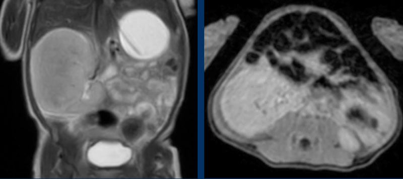
MRI
The tumor has similar signal intensity as normal
renal tissue on all pulse sequences.
It enhances also equal to the remnant of the kidney.MRI
Khối u có cường độ tín hiệu tương tự như mô thận bình thường trên tất cả các chuỗi xung.
Nó cũng tăng cường tương đương với phần còn lại của thận.
The kidney was excised and contained a mesoblastic nephroma.Thận được cắt bỏ và chứa một nephroma mesoblastic.
Multilocular cystic renal tumorU thận đa nang
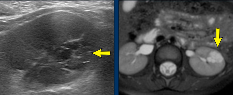 Cystic nephromaU nang thận
A
multilocular cystic renal tumor is a confusing lesion.
It can be a benign cystic nephroma, or a malignant cystic partially differentiated
nephroblastoma.
Only the pathologist can make the difference.U thận dạng nang đa ổ là một tổn thương khó hiểu.
Nó có thể là u nang thận lành tính, hoặc u nguyên bào thận biệt hóa một phần ác tính.
Chỉ có nhà giải phẫu bệnh mới có thể tạo ra sự khác biệt.
The radiologist has to distinguish these multilocular cystic tumors which arise from the kidney with preservation of some normal renal tissue from a multicystic dysplastic kidney where no normal renal tissue is visible. Bác sĩ chẩn đoán hình ảnh phải phân biệt các khối u nang đa nang phát sinh từ thận với sự bảo tồn một số mô thận bình thường với một thận loạn sản đa nang không có mô thận bình Thường.
Images
Ultrasound and MRI of a four-year-old boy with a multilocular cystic lesion
in the interpolar region of the left kidney (arrow).
It was detected as an incidental
finding at the age of nine months. On follow-up no change was seen. The
lesion is consistent with a cystic nephroma, a benign condition.
The same lesion is seen on the T2 weighted transverse
image.Hình ảnh
Siêu âm và MRI của bé trai 4 tuổi có tổn thương dạng nang nhiều thùy ở vùng giữa hai cực thận trái (mũi tên). Tổn thương được phát hiện tình cờ khi bé 9 tháng tuổi và ổn định khi theo dõi, phù hợp với u thận dạng nang (cystic nephroma), một tổn thương lành tính. Có thể thấy cùng tổn thương trên ảnh T2W mặt phẳng ngang.
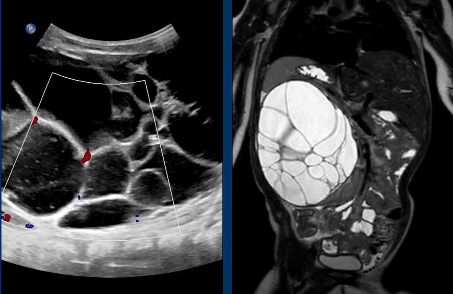 Cystic nephromaU nang thận
These images are of a one-year-old girl.
On ultrasound a multicystic tumor was seen with some perfusion of
septa.Những hình ảnh này là của một cô gái một tuổi.
Trên siêu âm, một khối u đa nang được phát hiện với một số tưới máu của vách ngăn.
T2 weigthed image better shows the extent of the lesion. A remnant of the normal renal tissue of the lower pole is visible, distinguishing it from a multicystic dysplastic kidney.Hình ảnh T2 cho thấy rõ hơn mức độ tổn thương, có thể nhìn thấy phần còn lại của mô thận bình thường ở cực dưới, phân biệt nó với thận loạn sản đa nang.
This tumor was diagnosed as a cystic nephroma for which right nephrectomy was performed.U được chẩn đoán là u nang thận và được cắt thận phải.
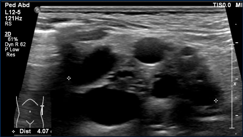
Multicystic dysplastic kidneyThận đa nang loạn sản
An 8-months-old boy with a multicystic dysplastic
kidney.
On ultrasound multiple cysts are present and no
normal renal tissue is seen.
No additional imaging is needed.
Often follow-up
ultrasound is performed to ascertain regression of the MCDK over time and to
diagnose contralateral abnormalities (in 30% of cases).Một bé trai 8 tháng tuổi bị bệnh thận đa nang.
Trên siêu âm có nhiều nang và không thấy mô thận bình thường.
Không cần thêm chẩn đoán hình ảnh.
Siêu âm thường được thực hiện để xác định sự hồi quy của MCDK theo thời gian và để chẩn đoán các bất thường đối bên (trong 30% trường hợp).
Malignant rhabdoid tumor of the kidneyU ác tính dạng rhabdoid ở thận
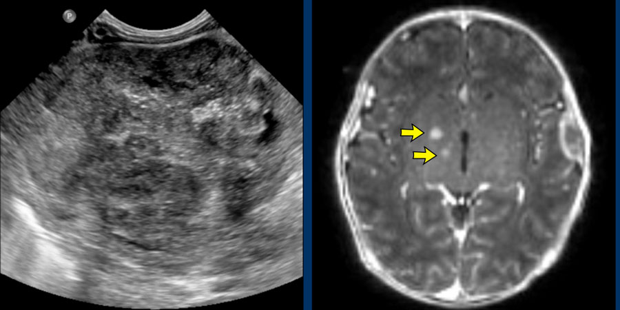 Malignant rhabdoid tumor of the kidney with brain (arrows) and bone metastasesU thận ác tính có bộ não (các mũi tên) và di căn xương
Malignant rhabdoid tumor of the kidney is a highly
malignant tumor with a poor
prognosis.
Distant metastases at the time of diagnosis of the tumor are
common, to the lungs, liver, brain, lymph nodes, and skeleton.
Rhabdoid renal tumors can occur in conjunction with a brain tumor, which can
be a primary atypical teratoid rhabdoid brain tumor (ATRT), but also other tumors
like medulloblastoma and ependymoma are described.U cơ vân ác tính của thận là một khối u ác tính cao với tiên lượng xấu.
Di căn xa tại thời điểm chẩn đoán u là phổ biến, đến phổi, gan, não, hạch bạch huyết, và xương.
U thận dạng rhabdoid có thể xảy ra cùng với u não, có thể là u não dạng rhab doid không điển hình nguyên phát (ATRT), nhưng cũng có thể mô tả các khối u khác như u nguyên bào tuỷ và u màng não thất.
There are no distinguishing features from a Wilms tumor, but one should think of a rhabdoid tumor if the tumor is relatively small with an infiltrative growth pattern, low T2 signal and diffusion restriction. Không có đặc điểm phân biệt với khối u Wilms, nhưng người ta nên nghĩ đến khối u dạng rhabdoid nếu khối u tương đối nhỏ với mô hình tăng trưởng thâm nhiễm, tín hiệu T2 thấp và hạn chế khuếch tán.
Although rhabdoid tumors are reported to demonstrate a subcapsular fluid collection, this can occur also as an uncommon manifestation in Wilms tumor, but these collections occur in absolute numbers more in Wilms tumor than in rhabdoid tumors.Mặc dù các khối u dạng rhabdoid được báo cáo là có sự tích tụ dịch dưới bao, nhưng điều này cũng có thể xảy ra như một biểu hiện không phổ biến trong khối u Wilms, nhưng những sự tích tụ này xảy ra với số lượng tuyệt đối nhiều hơn trong khối u của Wilms so với trong khối u dạng Rhabdoid.
Images
Two-week-old girl presenting with a mass in the
left kidney.
A MRI of the brain at the time of initial diagnosis
showed tiny lesions, possibly metastases (arrows).
An MRI four weeks later
showed multiple brain and skull metastases.
The patient died two weeks later.Hình ảnh
Bé gái được 2 tuần tuổi có khối u thận trái. MRI não lúc chẩn đoán ban đầu cho thấy các tổn thương rất nhỏ, có thể là ổ di căn (mũi tên). MRI sau 4 tuần cho thấy nhiều ổ di căn não và xương sọ. Bệnh nhi tử vong sau đó 2 tuần.
Clear cell sarcoma of the kidneySarcom tế bào sáng của thận
Clear
cell sarcoma of the kidney is rare, approximately 3% of pediatric renal tumors.
The 5-year survival rate is around 80% but it is worse in children below one
year of age.
The tumor has a propensity to metastasize to the bone.
Sarcoma tế bào sáng của thận là hiếm, khoảng 3% khối u thận ở trẻ em.
Tỷ lệ sống sót 5 năm là khoảng 80% nhưng tồi tệ hơn ở trẻ em dưới một tuổi.
Khối u có xu hướng di căn đến xương.
The combination of a renal tumor and bone metastases should suggest to the diagnosis of a clear cell sarcoma.Sự kết hợp của khối u thận và di căn xương nên gợi ý chẩn đoán sarcoma tế bào sáng.
The imaging findings are non-specific, but suggestive
features are that these tumors are generally homogeneous with limited diffusion
restriction.
Các dấu hiệu hình ảnh không đặc hiệu, nhưng gợi ý rằng những khối u này thường đồng nhất với giới hạn hạn chế khuếch tán.
video
A ten-month-old boy presented with an
abdominal mass. Ultrasound demonstrated a tumor in the right kidney. This is
better shown on MRI. The remnant of the kidney has a dilated collecting system.
On ADC images there is limited diffusion restriction.
Histopathology showed a clear cell sarcoma of the
kidney.Video
Bé trai 10 tháng tuổi đến khám vì khối ở bụng. Siêu âm phát hiện u thận phải, thấy rõ hơn trên MRI. Hệ thống đài–bể thận của phần thận còn lại bị giãn. Trên bản đồ ADC, mức độ hạn chế khuếch tán ít. Mô bệnh học xác định sarcôm tế bào sáng của thận.
On ADC images there is limited diffusion restriction.Trên ảnh ADC có giới hạn hạn chế khuếch tán.
The tumor was thought to be a Wilms tumor, because of the limited diffusion restriction, but pathology showed a clear cell sarcoma of the kidney.Khối u được cho là khối u Wilms, vì hạn chế khuếch tán hạn chế, nhưng bệnh lý cho thấy một sarcoma tế bào rõ ràng của thận.
Renal cell carcinomaUng thư biểu mô tế bào thận
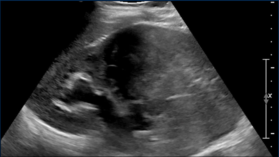
Renal cell carcinoma (RCC)
is the second most common renal tumor in children.
RCC is rare in young
children and occurs mostly in children older than 10 years.
It can also occur in
patients with von Hippel-Lindau disease and Tuberous Sclerosis Complex, or
after treatment for previous malignancies (f.e Neuroblastoma or Leukemia) Ung thư biểu mô tế bào thận (UTBMTB) là loại u thận thường gặp thứ hai ở trẻ em.
RCC hiếm gặp ở trẻ nhỏ và xảy ra chủ yếu ở trẻ em trên 10 tuổi.
Nó cũng có thể xảy ra ở những bệnh nhân mắc bệnh von Hippel-Lindau và Phức hợp xơ cứng củ, hoặc sau khi điều trị các bệnh ác tính trước đó (ví dụ U nguyên bào thần kinh hoặc Bệnh bạch cầu)
Image
A nine-year-old boy presented with severe hematuria.
On palpation a left abdominal mass was found.
On ultrasound a predominantly solid mass was seen which seemed to invade the dilated collecting system.Hình ảnh
Một cậu bé chín tuổi bị tiểu máu nặng.
Khi sờ nắn, một khối bụng trái được tìm thấy.
Trên siêu âm, một khối rắn chủ yếu được nhìn thấy dường như xâm nhập vào hệ thống thu gom giãn nở.
On imaging renal cell carcinomas can present in many different ways.
They can appear more solid or have cystic elements and may be homogeneous, or rather heterogeneous.
Hemorrhage can occur and they may contain fat.
Relative small tumor size and prominent peripheral vessels suggests a RCC.Trên hình ảnh, ung thư biểu mô tế bào thận có thể biểu hiện theo nhiều cách khác nhau.
Chúng có thể xuất hiện đặc hơn hoặc có các yếu tố dạng nang và có thể đồng nhất, hoặc không đồng nhất.
Xuất huyết có thể xảy ra và chúng có thể chứa chất béo.
Kích thước khối u tương đối nhỏ và các mạch máu ngoại vi nổi bật gợi ý RCC.
Imaging is essential for staging and a combination of ultrasound, CT and MRI is used. Chẩn đoán hình ảnh là cần thiết để phân giai đoạn và kết hợp siêu âm, CT và MRI được sử dụng.
video
MRI
nicely demonstrates the mass in the left kidney. Note the large collateral
veins.
Because
of heavy hematuria a left nephrectomy was performed.
Pathology showed a RCC which
was completely resected.video
MRI cho thấy rõ khối u ở thận trái, lưu ý các tĩnh mạch bàng hệ lớn.
Tiểu máu nhiều nên phải cắt thận trái.
Kết quả giải phẫu bệnh cho thấy một khối u đã được cắt bỏ hoàn toàn.
Lymphoma
There are two main types of lymphoma: Hodgkin lymphoma and non-Hodgkin lymphoma. Có hai loại u lympho chính: u lympho Hodgkin và u lympho không Hodgkin.
Hodgkin
lymphoma often manifests with cervical lymph node enlargement, and mediastinal
masses.
It is very rarely confined to the abdomen. U lympho Hodgkin thường biểu hiện với phì đại hạch bạch huyết cổ tử cung và khối u trung thất.
Nó rất hiếm khi giới hạn ở bụng.
Non-Hodgkin lymphoma can be
located solely in the abdomen and generally originates from a bowel loop.
Non-Hodgkin
lymphoma presents more frequently with extra-nodal disease than Hodgkin
lymphoma.
Kidney involvement in lymphoma is rarely seen, but imaging features
to look for are multiple focal lesions that are homogeneous and hypoechoic on ultrasound and show strong diffusion restriction on MRI. 5,6U lympho không Hodgkin có thể chỉ nằm ở bụng và thường bắt nguồn từ một quai ruột.
U lympho không Hodgkin thường gặp hơn với bệnh ngoài hạch so với u lympho Hodgkin.
Tổn thương thận trong u lympho hiếm gặp, nhưng các đặc điểm hình ảnh cần tìm kiếm là nhiều tổn thương khu trú đồng nhất và giảm âm trên siêu âm và cho thấy hạn chế khuếch tán mạnh trên MRI. 5,6
video
Ultrasound
of a three-year-old boy. Diffuse infiltration of both kidneys was seen.
Furthermore, multiple hypoechoic masses were detected
in pancreas, liver and bowel.
The combination of bowel
involvement and high LDH in de serum was highly suggestive for Burkitt
lymphoma. This diagnosis was confirmed by bone marrow biopsy and flow
cytometry. Video
Siêu âm của bé trai 3 tuổi cho thấy thâm nhiễm lan tỏa cả hai thận và nhiều khối giảm âm ở tụy, gan và ruột. Tổn thương ruột kết hợp với LDH huyết thanh tăng gợi mạnh u lympho Burkitt. Sinh thiết tủy xương và xét nghiệm tế bào dòng chảy xác nhận chẩn đoán.
LeukemiaBệnh bạch cầu
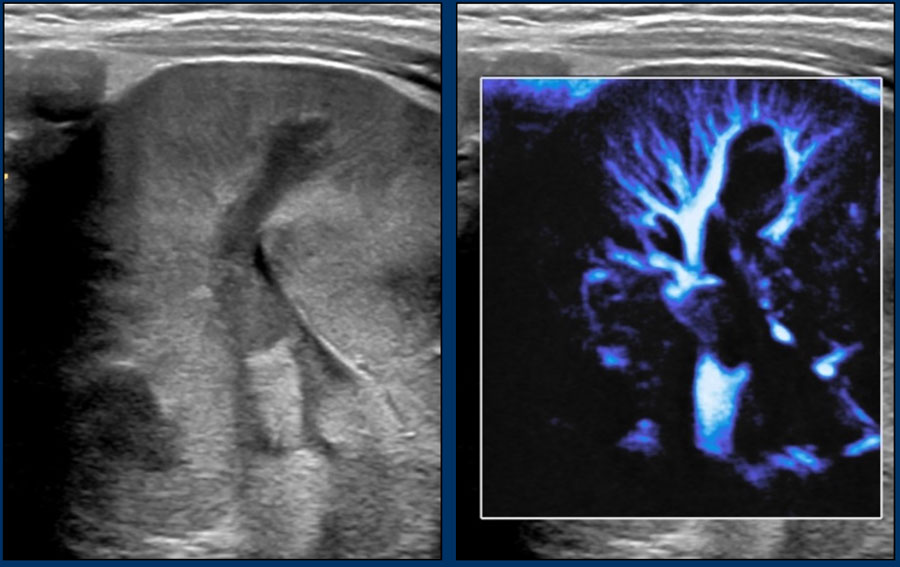
Leukemia is the most common malignancy in children.
It can affect all solid abdominal organs.
Organs can be diffusely infiltrated
or have a more nodular pattern. Bệnh bạch cầu là bệnh lý ác tính thường gặp nhất ở trẻ em.
Nó có thể ảnh hưởng đến tất cả các cơ quan đặc trong ổ bụng.
Các cơ quan có thể bị thâm nhiễm lan toả hoặc có dạng nốt hơn.
The kidneys are affected in almost half of the
patients with acute lymphoblastic leukemia.
It can be uni- or bilateral, and
there can be focal lesions or diffuse infiltration with enlargement of the
kidneys.Thận bị ảnh hưởng ở gần một nửa số bệnh nhân mắc bệnh bạch cầu cấp dòng lympho.
Nó có thể là đơn độc hoặc hai bên, và có thể có tổn thương khu trú hoặc thâm nhiễm lan toả với thận to.
The latter has a rather typical appearance with a striated pattern around the calices, similar to renal involvement of malignant lymphoma (figure).Loại thứ hai có hình dạng khá điển hình với một mô hình có vân xung quanh các khay, tương tự như sự liên quan đến thận của u lympho ác tính (hình).
Image
A
four-month-old boy presented with fever and anemia with thrombopenia. An
ultrasound examination shows enlarged kidneys with multiple hypo-echoic
lesions and linear abnormalities in both kidneys.
Retroperitoneal
lymphadenopathy was seen as well.
A hematological malignancy,
in particular leukemia was suspected due to the combination of imaging findings
and laboratory findings. Hình ảnh
Một bé trai bốn tháng tuổi bị sốt và thiếu máu kèm theo giảm tiểu cầu. Khám siêu âm cho thấy thận to với nhiều tổn thương giảm âm và bất thường tuyến tính ở cả hai thận.
bệnh hạch sau phúc mạc cũng được quan sát thấy.
Một bệnh lý ác tính về huyết học, đặc biệt là bệnh bạch cầu được nghi ngờ do sự kết hợp của các kết quả chẩn đoán hình ảnh và các kết quả xét nghiệm.
Continue with the video...Tiếp tục với video...
Distinguishing between malignant lymphoma and leukemia on imaging is difficult, when a lot of extramedullary disease is seen.
Phân biệt lymphoma ác tính và bạch cầu trên hình ảnh là khó khăn, khi có nhiều bệnh ngoài tuỷ được nhìn thấy.
Often the final diagnosis is thus based on clinical presentation, laboratory findings (anemia and thrombocytopenia) and flow cytometry. Thông thường chẩn đoán cuối cùng dựa trên biểu hiện lâm sàng, các dấu hiệu trong phòng thí nghiệm (thiếu máu và giảm tiểu cầu) và đếm tế bào dòng chảy.
For this case the final diagnosis was leukemia. Đối với trường hợp này, chẩn đoán cuối cùng là bệnh bạch cầu.
NeuroblastomaU nguyên bào thần kinh
The main differential diagnosis of nephroblastoma is neuroblastoma.Chẩn đoán phân biệt chính của u nguyên bào thận là u nguyên bào thần kinh.
Typical
features that indicate neuroblastoma are calcifications - causing a specific
ultrasound pattern, shown in the video – and vessel encasement,
without invasion.
This vessel encasement causes the aorta and caval vein to
be lifted anteriorly. Các đặc điểm điển hình cho thấy u nguyên bào thần kinh là vôi hóa - gây ra một mô hình siêu âm cụ thể, được hiển thị trong video – và bao bọc mạch, không xâm lấn.
Sự bao bọc mạch máu này làm cho động mạch chủ và tĩnh mạch chủ được nâng lên phía trước.
On ultrasound the tumor is generally inhomogeneous and echogenic, with bright calcifications.Trên siêu âm, khối u thường không đồng nhất và có tiếng vang, với vôi hóa sáng.
Children
with neuroblastoma are generally very ill, while children with a Wilms tumor are not.
The Vanillyl Mandelic Acid (VMA) and HomoVanillic Acid
(HVA) levels in the urine are in most cases elevated.
In our hospital these levels are checked in every child that presents with a tumor in the abdomen.Trẻ em bị u nguyên bào thần kinh thường rất ốm yếu, trong khi trẻ em bị khối u Wilms thì không.
Nồng độ Vanillyl Mandelic Acid (VMA) và HomoVanillic Acid trong nước tiểu trong hầu hết các trường hợp đều tăng cao.
Trong bệnh viện chúng tôi, các mức độ này được kiểm tra ở tất cả trẻ em có khối u trong bụng.
video
Three-years-old
girl referred from a peripheral hospital with a diagnosis of a tumor of the
left kidney. Ultrasound shows a mass originating from the left kidney. The mass
is very inhomogeneous with calcifications, and much more compatible with a
neuroblastoma than a nephroblastoma. Video
Bé gái 3 tuổi được chuyển từ bệnh viện tuyến dưới với chẩn đoán u thận trái. Siêu âm cho thấy khối xuất phát từ vùng thận trái. Khối rất không đồng nhất và có vôi hóa, gợi u nguyên bào thần kinh hơn nhiều so với u nguyên bào thận.
ReferencesTài liệu tham khảo
-
Renal tumors of Childhood: Radiologic-Pathologic Correlation Part 1. The 1st Decade
U thận ở trẻ em: Mối tương quan X quang – Giải phẫu bệnh Phần 1. Thập kỷ 1
Chung EM, Graeber AR, Conran RM.
RadioGraphics Vol. 36, No. 2
-
Renal tumors of childhood: Radiologic-Pathologic Correlation Part 2. The 2st Decade
U thận ở trẻ em: Mối tương quan X quang – Giải phẫu bệnh Phần 2. Thập kỷ 2
Chung EM, Lattin GE, Fagen KE, Kim AM, Pavio MA, Fehringer AF, Conran RM.
-
American Cancer Society, Wilms Tumor Stages.
Hiệp hội Ung thư Hoa Kỳ, Giai đoạn khối u Wilms.
Accessed on 27-09-2021 Truy cập ngày 27-09-2021
-
Renal Tumors of Childhood—A Histopathologic Pattern-Based Diagnostic Approach.
Các khối u thận ở trẻ em - Phương pháp tiếp cận chẩn đoán dựa trên mô bệnh học.
Ooms AHAG, Vujanic GM, D’Hooghe E, Collini P, L’Herminé-Coulomb A, Vokuhl C, Graf N, van den Heuvel-Eibrink MM, de Krijger RR.
Cancers 2020; 12(3): 729
Ooms AHAG, Vujanic GM, D ’ Hooghe E, Collini P, L ’ Herminé-Coulomb A, Vokuhl C, Graf N, van den Heuvel-Eibrink MM, de Krijger RR. -
Imaging features of extranodal involvement in paediatric Hodgkin Lymphoma
Đặc điểm hình ảnh học u lympho Hodgkin ở trẻ em
Spijkers S, Littooij AS, Humphries PD et al. .
Pediatr Radiol 2018; 49:266-246
Spijkers S, Littooij AS, Humphreys PD và cộng sự. -
Non-Hodgkin lymphoma in children and adolescents: progress through effective collaboration, current knowledge, and challenges ahead.
U lympho không Hodgkin ở trẻ em và thanh thiếu niên: tiến bộ thông qua sự hợp tác hiệu quả, kiến thức hiện tại và những thách thức phía trước.
Rosolen A, Perkins SL, Pinkerton CR.
Clin Oncol 2015;33:2112-2118
-
A review of neuroblastoma image-defined risk factors on magnetic resonance imaging.
Nhận xét các yếu tố nguy cơ hình ảnh u nguyên bào thần kinh trên cộng hưởng từ.
Chen AM, Trout AT, Towbin AJ.
Pediatr Radiol 2018; 48:1337-1347
-
Updates in diagnosis, management, and treatment of neuroblastoma.
Cập nhật chẩn đoán, xử trí và điều trị u nguyên bào thần kinh.
Swift CC, Eklund MJ, Kraveka JM, Alazraki AL.
Radiographics 2018; 38:566-580
-
Pediatric Renal Masses: Wilms Tumor and Beyond
Khối u thận ở trẻ em: Khối u Wilms và xa hơn nữa
by Lisa H. Lowe, Bernardo H. Isuani, Richard M. Heller, Sharon M. Stein, Joyce E. Johnson, Oscar M. Navarro, Marta Hernanz-Schulman.
RadioGraphics Vol. 20, No. 6
của Lisa H. Lowe, Bernardo H. Isuani, Richard M. Heller, Sharon M. Stein, Joyce E. Johnson, Oscar M. Navarro, Marta Hernanz-Schulman. - Wilms Tumor and Its Management in a Surgical Aspect Wilms Tumor và quản lý của nó trong một khía cạnh phẫu thuật