
Neonatal Brain USSiêu âm não trẻ sơ sinh
Department of Radiology and Neonatology of the Wilhelmina Children's Hospital and the University Medical Centre of Utrecht, the NetherlandsKhoa Chẩn đoán hình ảnh và Sơ sinh Bệnh viện Nhi Wilhelmina và Trung tâm Y tế Đại học Utrecht, Hà Lan
Ngày đăng / cập nhật: 2006-04-01
Bản gốc trên radiologyassistant.nl ↗ · tải về lúc 2026-11-19 20:19 · bản dịch máy có chuẩn hóa thuật ngữ chẩn đoán hình ảnh
Cranial sonography (US) is the most widely used neuroimaging procedure in premature infants.
US helps in assessing the neurologic status of the child, since clinical examination and symptoms are often nonspecific.
It gives information about immediate and long term prognosis.Siêu âm sọ (siêu âm) là phương pháp chẩn đoán hình ảnh thần kinh được sử dụng rộng rãi nhất ở trẻ sơ sinh non tháng.
siêu âm giúp đánh giá tình trạng thần kinh của trẻ, vì khám lâm sàng và các triệu chứng thường không đặc hiệu.
Nó cung cấp thông tin về tiên lượng trước mắt và lâu dài.
by Erik Beek and Floris Groenendaalbởi Erik Beek và Floris Groenendaal
IntroductionMở đầu
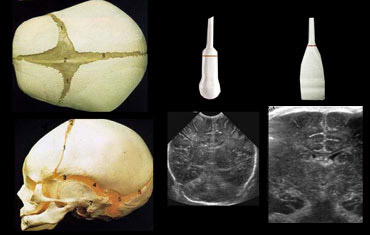 Use both the sector and linear transducer and examine the greater fontanel and if necessary also the lesser and sphenoidal fontanel.Sử dụng cả đầu dò cung từ và đầu dò tuyến tính và kiểm tra thóp lớn hơn và nếu cần thiết cũng là thóp nhỏ hơn và thóp hình bướm.
Ultrasound is a fast and bedside examination which makes it ideal for premature infants.
Try to get all the information you can.
Do not limit yourself to only one transducer or only one acustic window (figure).
Generally the large fontanel is used as acoustic window.
The small fontanel however is a good window to the occipital lobes.
This can be usefull in patients with borderline hyperechogenicity in these areas.Siêu âm là một cuộc kiểm tra nhanh chóng và bên giường, rất lý tưởng cho trẻ sinh non.
Cố gắng lấy tất cả thông tin bạn có thể.
Không giới hạn bản thân chỉ trong một bộ chuyển đổi hoặc chỉ một cửa sổ acustic (hình).
Nói chung, phông chữ lớn được sử dụng làm cửa sổ âm thanh.
Tuy nhiên, thóp nhỏ là một cửa sổ tốt cho các thùy chẩm.
Điều này có thể hữu ích ở những bệnh nhân bị tăng sản ở biên giới ở những khu vực này.
Disadvantages of US are: Nhược điểm của siêu âm là:
- Limited overview in posterior fossa and convexity of the brainTổng quan hạn chế về hố sau và lồi não
- Absence of US-signs in ischemia in full-terms in first 24 hoursSự vắng mặt của các dấu hiệu siêu âm trong thiếu máu cục bộ trong toàn bộ thời gian trong 24 giờ đầu
- Difficulty in detecting migration disorders, cortical dysplasiaKhó khăn trong phát hiện rối loạn di cư, loạn sản vỏ não
Peri Ventricular Leukomalacia (PVL)Bệnh bạch cầu ngoại tâm thu (PVL)
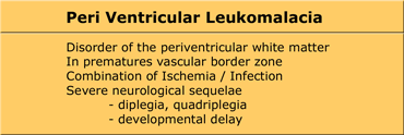
PVL is also known as Hypoxic-Ischemic Encephalopathy (HIE) of the preterm.
It is a white matter disease that affects the periventricular zones.
In prematures this white matter zone is a watershed zone between deep and superficial vessels.
Until recently ischemia was thought to be the single cause of PVL, but probably other causes (infection, vasculitis) play an additional role.
PVL presents as areas of increased periventricular echogenicity.
Normally the echogenicity of the periventricular white matter should be less than the echogenicity of the choroid plexus.
PVL còn được gọi là bệnh não thiếu máu thiếu oxy (HIE) của trẻ sinh non.
Đây là bệnh chất trắng ảnh hưởng đến vùng quanh não thất.
Ở trẻ non tháng, vùng chất trắng này là vùng đầu nguồn giữa các mạch máu sâu và nông.
Cho đến gần đây thiếu máu cục bộ được cho là nguyên nhân duy nhất của PVL, nhưng có thể các nguyên nhân khác (nhiễm trùng, viêm mạch) đóng một vai trò bổ sung.
PVL biểu hiện như các vùng tăng cường độ âm tính quanh não thất.
Thông thường, độ vang của chất trắng quanh não thất phải nhỏ hơn độ vang của đám rối mạch mạc.
PVL occurs most commonly in premature infants born at less than 33 weeks gestation (38% PVL) and less than 1500 g birth weight (45% PVL).
Detection of PVL is important because a significant percentage of surviving premature infants with PVL develop cerebral palsy, intellectual impairment or visual disturbances.
More than 50% of infants with PVL or grade III hemmorrhage develop cerebral palsy.PVL xảy ra phổ biến nhất ở trẻ sinh non dưới 33 tuần (38% PVL) và dưới 1500 g cân nặng khi sinh (45% PVL).
Phát hiện PVL là rất quan trọng vì một tỷ lệ đáng kể trẻ sơ sinh non tháng sống sót với PVL phát triển bại não, suy giảm trí tuệ hoặc rối loạn thị giác.
Hơn 50% trẻ sơ sinh bị PVL hoặc xuất huyết độ III bị bại não.
Grading PVLPhân loại PVL
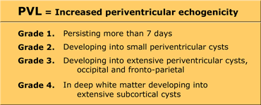
PVL is graded according to the signs as listed in the Table on the left. PVL được phân độ theo các dấu hiệu như được liệt kê trong Bảng bên trái.
Regular sonographic examination is mandatory as cysts in PVL can develop as long as 4 weeks after birth (especially in prematures
Cranial ultrasonographic findings may be normal in patients who go on to develop clinical and delayed imaging findings of PVL.
A good protocol is US-examination at least once a week until discharge
?nd at the age of 40 weeks.
Khám siêu âm thường xuyên là bắt buộc vì các nang trong PVL có thể phát triển đến 4 tuần sau khi sinh (đặc biệt là ở trẻ sinh non Các dấu hiệu siêu âm sọ não có thể bình thường ở những bệnh nhân tiếp tục phát hiện các dấu hiệu lâm sàng và hình ảnh chậm trễ của PVL.
Một phác đồ tốt là khám siêu âm ít nhất một lần một tuần cho đến khi xuất viện ở tuổi 40 tuần.
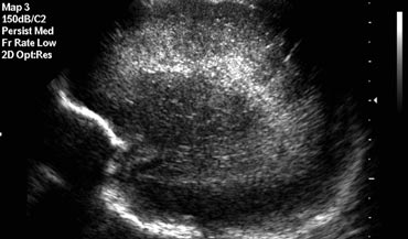 Sagittal image of a child with PVL grade 1Hình ảnh dọc của một trẻ bị PVL độ 1
PVL grade 1
PVL is diagnosed as grade 1 if there are areas of increased periventricular echogenicity without any cyst formation persisting for more than 7 days.
Increased periventricular echogenicity is however a nonspecific finding that must be differentiated from the normal periventricular halo or normal hyperechoic 'blush' posterosuperior to the ventricular trigones.
Suspect PVL if the echogenicity is asymmetric, coarse, globular or more hyperechoic than the choroid plexus.
The abnormal periventricular echotexture of PVL usually disappears at 2-3 weeks.
PVL can be differentiated from hemorrhages because PVL lacks mass effect.PVL độ 1
PVL được chẩn đoán là độ 1 nếu có các vùng tăng âm tính quanh não thất mà không có sự hình thành nang tồn tại hơn 7 ngày.
Tuy nhiên, tăng cường độ âm tính của vùng quanh thất là một dấu hiệu không đặc hiệu, phải được phân biệt với quầng quanh thất bình thường hoặc tăng âm bình thường 'màu xanh' sau tâm thất.
Nghi ngờ PVL nếu độ vang không đối xứng, thô, hình cầu hoặc tăng âm nhiều hơn đám rối mạch mạc.
Sợi vải ngoại vi quanh não thất bất thường của PVL thường biến mất sau 2-3 tuần.
PVL có thể được phân biệt với xuất huyết vì PVL thiếu hiệu ứng khối.
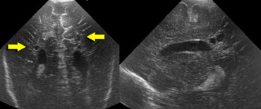 Transverse and sagittal image of a child with PVL grade 2.Hình ảnh ngang và dọc của một trẻ có PVL độ 2.
PVL grade 2
The images on the left demonstrate a PVL grade 2 with small periventricular cysts.
The echogenicity has resolved at the time of cyst formation.
2% of the preterm neonates born before 32 weeks develop cystic PVL.
The severity of PVL is related to the size and distribution of these cysts.
Cystic PVL has been identified on cranial ultrasounds on the first day of life, indicating that the adverse event was at least 2 weeks prenatal rather than perinatal or postnatal.PVL độ 2
Hình ảnh bên trái cho thấy một PVL độ 2 với các nang nhỏ quanh não thất.
Tính hồi âm đã được giải quyết tại thời điểm hình thành nang.
2% trẻ sơ sinh non tháng sinh trước 32 tuần phát triển nang PVL.
Mức độ nặng của nang lông liên quan đến kích thước và sự phân bố của nang lông.
U nang tuyến yên đã được xác định trên siêu âm sọ não vào ngày đầu tiên của cuộc đời, cho thấy tác dụng phụ ít nhất là 2 tuần trước khi sinh hơn là chu sinh hoặc sau sinh.
US is highly reliable in the detection of cystic WM injury (PVL grade II or more), but has significant limitations in the demonstration of noncystic WM injury (PVL grade I).
This deficiency of neonatal cranial US is important, because noncystic WM injury is considerably more common than cystic WM injury.Siêu âm có độ tin cậy cao trong việc phát hiện tổn thương WM dạng nang (PVL độ II trở lên), nhưng có những hạn chế đáng kể trong việc chứng minh tổn thương wm không phải dạng nang ( PVL độ I).
Sự thiếu hụt siêu âm trong sọ sơ sinh rất quan trọng, bởi vì tổn thương WM không nang thường phổ biến hơn đáng kể so với tổn thương wm nang.
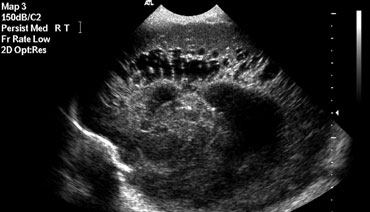 Sagittal image demonstarting extensive PVL grade 3Hình ảnh dọc bắt đầu thấy PVL độ 3 rộng
PVL grade 3
PVL is diagnosed as grade 3 if there are areas of increased periventricular echogenicity, that develop into extensive periventricular cysts in the occipital and fronto-parietal region.PVL độ 3
PVL được chẩn đoán là độ 3 nếu có các vùng tăng âm tính quanh não thất, phát triển thành các nang quanh não thất rộng ở vùng chẩm và vùng trán-phổi.
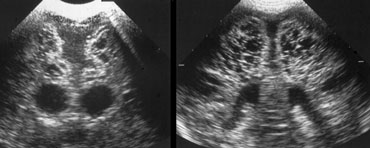 Coronal and transverse images demonstrating PVL grade 4Hình ảnh vành và ngang thể hiện PVL độ 4
PVL grade 4
PVL is diagnosed as grade 4 if there are areas of increased periventricular echogenicity in the deep white matter developing into extensive subcortical cysts.
PVL grade 4 is seen mostly in fullterm neonates as opposed to PVL grade 1-3, which is a disease of the preterm neonate.
Flaring persisting beyond the first week of life is by definition PVL garde 1.
PVL độ 4
PVL được chẩn đoán là độ 4 nếu có các vùng tăng âm quanh não thất ở sâu chất trắng phát triển thành các nang dưới vỏ rộng.
PVL độ 4 gặp chủ yếu ở trẻ sơ sinh đủ tháng so với PVL độ 1-3, là bệnh lý của trẻ sơ sinh non tháng. Phát ban kéo dài sau tuần đầu tiên của cuộc đời được định nghĩa là PVL garde 1.
FlaringBùng phát
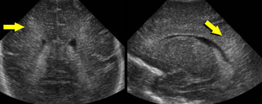 Transverse and sagittal images demonstrating flaring in a premature infant.Biểu hiện lâm sàng của trẻ sơ sinh non tháng là nổi mẩn ngứa và ảnh mặt phẳng đứng dọc.
The term flaring is used to describe the slightly echogenic periventricular zones, that are seen in many premature infants in the first week of life.
During this first week it is not sure if this is a normal variant or a sign of PVL grade 1.
Flaring persisting beyond the first week of life is by definition PVL grade 1.
Thuật ngữ bùng phát được sử dụng để mô tả các vùng quanh não thất có tiếng vang nhẹ, được thấy ở nhiều trẻ sinh non trong tuần đầu tiên của cuộc đời.
Trong tuần đầu tiên này, không chắc chắn đây là một biến thể bình thường hay là dấu hiệu của PVL độ 1.
Sự bùng phát kéo dài sau tuần đầu tiên của cuộc đời theo định nghĩa là PVL độ 1.
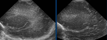 LEFT: Initial examination shows flaring.RIGHT: Follow up one week later shows normal periventricular white matter.Bên trái: Kiểm tra ban đầu cho thấy bùng phát. Phải: Theo dõi một tuần sau đó cho thấy chất trắng quanh não thất bình thường.
Follow up is needed to differentiate flaring from PVL grade I.
The case on the left shows a premature infant with flaring.
At follow up no cyst formation was found and after the first week a normal periventricular white matter was seen.
Cần theo dõi để phân biệt mức độ bùng phát với PVL độ I.
Trường hợp bên trái cho thấy một trẻ sơ sinh non tháng có bùng phát.
Theo dõi không thấy nang và sau tuần đầu tiên, chất trắng quanh não thất bình thường.
Germinal Matrix HemorrhageXuất huyết ma trận mầm bệnh
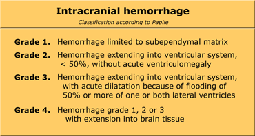
Germinal matrix hemorrhage (GMH) is also known as periventricular hemorrhage or preterm caudothalamic hemorrhage.
These germinal matrix hemorrhages occur in the highly vascular but also stress sensitive germinal matrix, which is located in the caudothalamic groove. This is the subependymal region between the caudate nucleus and thalamus.
Xuất huyết ma trận mầm bệnh (GMH) còn được gọi là xuất huyết quanh não thất hoặc xuất huyết vùng dưới đồi non tháng.
Những xuất huyết chất nền mầm này xảy ra ở ma trận mầm có mạch máu cao nhưng cũng nhạy cảm với áp lực, nằm trong rãnh vùng dưới đồi Đây là vùng dưới dưới mỏm yên giữa nhân đuôi và đồi thị.
- The germinal matrix is only transiently present as a region of thin-walled vessels, migrating neuronal components and vessel precursorsMa trận mầm chỉ xuất hiện thóang qua dưới dạng một vùng mạch máu có thành mỏng, di chuyển các thành phần thần kinh và tiền thân mạch máu
- It has matured by 34 weeks gestation, such that hemorrhage becomes very unlikely after this age.Nó đã trưởng thành ở tuần thứ 34 của thai kỳ, do đó xuất huyết trở nên rất khó xảy ra sau tuổi này.
- Most GMHs occur in the first week of lifeHầu hết các GMH xảy ra trong tuần đầu tiên của cuộc đời
- Most common in infants Phổ biến nhất ở trẻ nhũ nhi
- These hemorrhages start in the caudothalamic groove and may extend into the lateral ventricle and periventricular brain parenchyma.Những xuất huyết này bắt đầu ở rãnh giả đồi và có thể lan rộng vào não thất bên và nhu mô não quanh não thất.
Most infants are asymptomatic or demonstrate subtle signs that are easily overlooked.
These hemorhages are subsequently found on surveillance sonography.Hầu hết trẻ sơ sinh không có triệu chứng hoặc biểu hiện các dấu hiệu tinh tế dễ bị bỏ qua.
Những xuất huyết này sau đó được tìm thấy trên siêu âm giám sát.
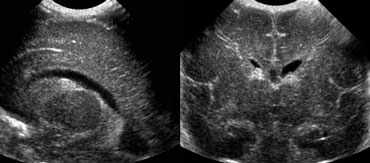 Grade 1 intracranial hemorrhageSagittal and coronal US of subependymal hemorrhage located in the groove between the thalamus and the nucleus caudatus.Xuất huyết nội sọ độ 1 Xuất huyết dưới mỏm yên vị, dọc theo trục siêu âm nằm ở rãnh giữa đồi thị và nhân mạch máu.
Grade 1 intracranial hemorrhage
On the left an intracranial hemorrhage confined to the caudothalamic groove.
It is staged as grade 1 hemorrhage.
Xuất huyết nội sọ độ 1
Ở bên trái, xuất huyết nội sọ giới hạn ở rãnh vùng dưới đồi.
Nó được phân giai đoạn như xuất huyết độ 1.
In the acute phase these bleedings are hyperechoic, changing to iso- and hypo-echoic with time. Trong giai đoạn cấp, các trường hợp này là tăng âm, thay đổi sang đẳng âm và giảm âm theo thời gian.
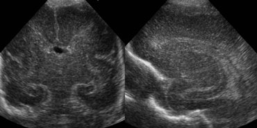 Sagittal and coronal US of a grade 2 hemorrhageSiêu âm đoạn dọc và đoạn vành của xuất huyết độ 2
Grade 2 intracranial hemorrhage
On the left a grade 2 intracranial hemorrhage.
On the coronal image only the cavum septi pellucidi is seen.
Both lateral ventricles are filled with blood, but there is no ventricular dilatation.
Xuất huyết nội sọ độ 2
Bên trái xuất huyết nội sọ độ 2.
Trên hình ảnh vành chỉ thấy cavum septi pellucidi.
Cả hai tâm thất bên đều chứa đầy máu, nhưng không có giãn não thất.
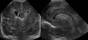
On the left the same patient after 3 days.
The ventricles are dilated and clot formation is seen.
Secondary hydrocephalus occuring several days after a grade 2 bleed should not be mislabeled as grade 3 hemorrhage.
Bên trái cùng một bệnh nhân sau 3 ngày.
Các tâm thất giãn và hình thành cục máu đông.
Tràn dịch não thứ phát xảy ra vài ngày sau khi chảy máu độ 2 không nên nhầm lẫn với xuất huyết độ 3.
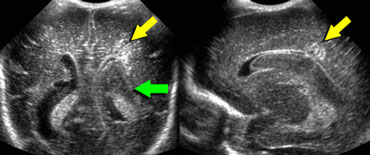 LEFT: Coronal image, green arrow indicating grade 3 hemorrhageRIGHT: Sagittal image, yellow arrow indicating venous infarction.TRÁI: Hình ảnh vành, mũi tên màu xanh lá cây cho thấy xuất huyết độ 3 RIGHT: Hình ảnh dọc, mũi tên vàng cho thấy nhồi máu tĩnh mạch.
Grade 3 intracranial hemorrhage
On the left a grade 3 intracranial hemorrhage filling the left lateral ventricle.
Also note the wedge shaped hyperechoic area on the laterosuperior side of the ventricle.
This represents a small venous infarction.
Xuất huyết nội sọ độ 3
Ở bên trái xuất huyết nội sọ độ 3 lấp đầy tâm thất trái.
Cũng lưu ý vùng tăng âm hình nêm ở phía sau tâm thất.
Biểu đồ này đại diện cho một tĩnh mạch nhỏ nhồi máu.
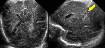 Yellow arrow indicating cyst formation within venous infarction.Mũi tên màu vàng cho thấy sự hình thành nang trong tĩnh mạch nhồi máu.
Same patient as above.
Two weeks later the venous infarction has developed into a hypoechoic area with cyst formation.Cùng một bệnh nhân như trên.
Hai tuần sau, nhồi máu tĩnh mạch đã phát triển thành một vùng giảm âm với sự hình thành nang.
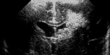 Intracranial hemorrhage grade 4Xuất huyết nội sọ độ 4
Grade 4 intracranial hemorrhage
Originally these grade 4 hemorrhages were thought to result from subependymal bleeding into the adjacent brain.
Today however most regard these grade 4 hemorrhages to be venous hemorrhagic infartions, which are the result of compression of the outflow of the veins by the subependymal hemorrhage. Xuất huyết nội sọ độ 4
Ban đầu, những xuất huyết độ 4 này được cho là do chảy máu dưới mỏm yên vào não lân cận.
Tuy nhiên, ngày nay hầu hết các trường hợp xuất huyết độ 4 này là nhồi máu tĩnh mạch, là kết quả của sự chèn ép dòng chảy của tĩnh mạch do xuất huyết dưới mỏm yên.
On the left a grade 4 hemorrhage.
There is a large subependymal bleeding but also a large area with increased echogenicity in the brain parenchyma lateral to the ventricle.
This is probably the result of a venous infarct.Bên trái xuất huyết độ 4.
Có chảy máu dưới màng cứng lớn nhưng cũng có một vùng rộng lớn với tính tăng âm trong nhu mô não bên tâm thất.
Đây có thể là kết quả của nhồi máu tĩnh mạch.
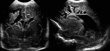
These venous infarctions resolve with cyst formation.
These cysts can merge with the lateral ventricle, finally resulting into a porencephalic cyst.
On the left a different patient with a grade 4 hemorrhage at a later stage with extensive cyst formation.
Grade 1 and 2 bleeds generally have a good prognosis.
Grade 3 and 4 bleeds have variable long-term deficits, but outcome in grade 3 hemorrhages is usually good when no parenchymal injury has occurred. Những nhồi máu tĩnh mạch này giải quyết bằng sự hình thành nang.
Những nang này có thể hợp nhất với tâm thất bên, cuối cùng tạo thành nang não.
Bên trái là một bệnh nhân khác với xuất huyết độ 4 ở giai đoạn muộn với sự hình thành nang rộng.
Chảy máu độ 1 và 2 thường có tiên lượng tốt.
Chảy máu độ 3 và 4 có những thiếu sót lâu dài khác nhau, nhưng kết quả trong xuất huyết độ 3 thường tốt khi không có tổn thương nhu mô.
Hydrocephalus is a common complication and many infants require ventriculoperitoneal shunting.
The mechanisms by which hydrocephalus develop include:Tràn dịch não thất là một biến chứng thường gặp và nhiều trẻ sơ sinh cần được dẫn lưu não thất - ổ bụng.
Các cơ chế mà não úng thủy phát triển bao gồm:
- Communicating hydrocephalus: decreased absorption of cerebral spinal fluid (CSF) secondary to obstruction of arachnoid villi by blood and debris or the development of arachnoiditisNão úng thủy thông thương: giảm hấp thu dịch não tuỷ (CSF) thứ phát do tắc nghẽn nhung mao màng nhện do máu và mảnh vụn hoặc sự phát triển của viêm màng nhệN
- Obstructive hydrocephalus: obstruction to CSF circulation.Não úng thủy do tắc nghẽn: tắc nghẽn lưu thông dịch não tuỷ.
Normal VariantsBiến thể bình thường
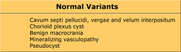
Common variants are listed in the Table on the left.Các biến thể thường gặp được liệt kê trong Bảng bên trái.
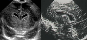 LEFT: Coronal image. Cavum septi pellucidi is seen in between the lateral ventricles.RIGHT: Midsagittal image demonstrating a cavum vergae.Bên trái: Hình ảnh Coronal. Cavum septi pellucidi được nhìn thấy ở giữa các tâm thất bên. phải: Hình ảnh midsagittal thể hiện một cavum vergae.
Cavum septi pellucidi, cavum vergae and cavum of the velum interpositumCavum septi pellucidi, cavum vergae và cavum của velum interpositum
Well known variants are the cavum of the septum pellucidum and the cavum vergae.
The more premature the baby, the more frequently these cavities are present.
They can persist until adulthood.
Các biến thể được biết đến nhiều là cavum của vách ngăn pellucidum và cavum vergae.
Trẻ càng non tháng, các lỗ sâu răng này càng xuất hiện thường xuyên.
Chúng có thể tồn tại cho đến khi trưởng thành.
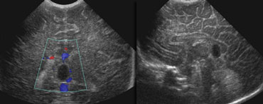
A less frequently seen variant is the cavum of the velum interpositum.
This presents as a cyst-like structure in the region of the tectum.
It's shape is compared to a helmet.
It can easily be confused with a subarachnoid cyst or a cyst of the pineal gland.Một biến thể ít gặp hơn là cavum của velum interpositum.
Biểu hiện này là một cấu trúc giống như u nang ở vùng trực tràng.
Hình dạng của nó được so sánh với mũ bảo hiểm.
Nó có thể dễ dàng bị nhầm lẫn với một nang dưới nhện hoặc một nang của tuyến tùng.
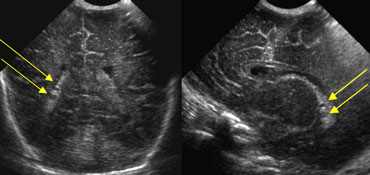
Choroid plexus cystNang đám rối mạch mạc
In postnatal US these cysts of the chorio?d plexus are often incidental findings without clinical consequences.
Choroid plexus cysts (CPC) are however of importance for obstetricians.
At prenatal US these cysts can be predictive of trisomy 18.
About half of babies with Trisomy 18 show a CPC on ultrasound, but nearly all of these babies will also have other abnormalities on the ultrasound, especially in the heart, hand, and feet.
Trong siêu âm sau sinh, các nang này của đám rối thần kinh đệm thường là những phát hiện ngẫu nhiên mà không có hậu quả lâm sàng.
Tuy nhiên, nang đám rối mạch mạc (CPC) có tầm quan trọng đối với các bác sĩ sản khoa.
Ở siêu âm trước sinh, các nang này có thể dự đoán trisomy 18.
Khoảng một nửa số trẻ có Trisomy 18 cho thấy CPC trên siêu âm, nhưng gần như tất cả những trẻ này cũng sẽ có những bất thường khác trên siêu âm.
An exeption must be made for cysts that arise close to the foramen of Monro.
Although these cysts often disappear spontaneously, follow up US is necessary to ensure disappearance.
Some may produce symptoms of raised intracranial pressure due to obstruction to the cerebrospinal fluid (CSF) flow.Cần phải lấy nang ra nếu nang phát sinh gần lỗ Monro.
Mặc dù những nang này thường tự biến mất, nhưng theo dõi siêu âm là cần thiết để đảm bảo sự biến mất.
Một số có thể tạo ra các triệu chứng của áp lực nội sọ tăng lên do tắc nghẽn dòng chảy dịch não tủy (CSF).
Benign macrocraniaĐau xương sọ lành tính
Benign macrocrania is also known as extraventricular obstructive hydrocephalus.
This is seen in children between 6 months and 2 years.
The head circumference is above the 97th percentile.
After the age of 2 years the head size normalizes.
Often the mother or father of the child had large heads at that age.
The cause is not known. Most state that it is a normal condition, although some state that these children have a slight developmental delay.Đau xương sọ lớn lành tính còn được gọi là não úng thủy do tắc nghẽn ngoài thất.
Điều này được thấy ở trẻ em từ 6 tháng đến 2 tuổi.
Vòng đầu trên bách phân vị thứ 97.
Sau 2 tuổi, kích thước đầu trở lại bình thường.
Thông thường mẹ hoặc cha của đứa trẻ có đầu to ở tuổi đó.
Nguyên nhân không rõ Hầu hết cho rằng đó là một tình trạng bình thường, mặc dù một số cho rằng những đứa trẻ này có chậm phát triển nhẹ.
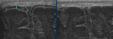 LEFT: Normal subarachnoid space is RIGHT: avoid pressure on the fontanel.Bên trái: Không gian dưới nhện bình thường là phải: tránh áp lực lên thóp.
When children with a large head are presented for US, examine the superficial subarachnoid space and the ventricles.
The normal subarachnoid space measures less then 3 mm. The ventricles are often slightly enlarged.Khi trẻ có đầu to được trình bày cho siêu âm, hãy kiểm tra khoang dưới nhện nông và tâm thất.
Không gian dưới nhện bình thường nhỏ hơn 3 mm. Thuỷ tinh thể thường hơi to.
These prominent subarachnoid space and ventricular system in these children should not be interpreted as cerebral atrophia, as in atrophia there is a small head circumpherence.Những khoảng trống dưới nhện và hệ thống não thất nổi bật ở những trẻ này không nên được hiểu là teo não, vì trong teo có một vòng đầu nhỏ.
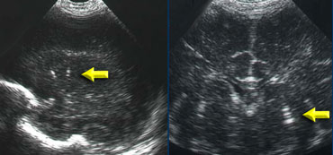
Mineralizing vasculopathyBệnh mạch máu khóang hóa
Mineralizing vasculopathy can be seen in the thalamostriatal and lenticulostriatal arteries and is caused by calcification of the arterial wall.
A wide range of perinatal, acquired, and nonspecific clinical conditions may result in this sonographic finding.
In the Wilhelmina Children's Hospital these children used to be tested for TORCH-infection, but currently the only test that is done is a urine-test for CMV.
Bệnh mạch máu khóang hóa có thể được nhìn thấy trong các động mạch đồi thị và động mạch thấu kính và được gây ra bởi vôi hóa của thành động mạch.
Một loạt các tình trạng lâm sàng chu sinh, mắc phải và không đặc hiệu có thể dẫn đến phát hiện siêu âm này.
Tại Bệnh viện Nhi Wilhelmina, những trẻ này đã từng được xét nghiệm để phát hiện nhiễm TORCH, nhưng hiện tại xét nghiệm duy nhất được thực hiện là xét nghiệm nước tiểu để phát hiện CMV.
Germinolytic cysts
Are located at the caudothalamic groove.
They are tear shaped.
There are no signs of intracerebral hemorrhage and these children have no neurological sequelae.
The etiology is not known.Nang vi khuẩn
Nằm ở rãnh vùng dưới đồi.
Chúng có hình giọt nước mắt.
Không có dấu hiệu xuất huyết trong nhu mô não và những trẻ này không có di chứng thần kinh.
Nguyên nhân không được biết.
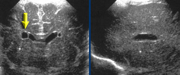
Pseudocyst
These are also called coarctation of the lateral ventricle.
They are often bilaterally and have no neurological sequelaeGiả nang
Đây còn được gọi là hẹp eo thất ngoài.
Thường là hai bên và không có di chứng thần kinh.
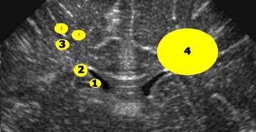 1+2 = germinolytic cysts and pseudocysts, 3 = cystic periventricular leukomalacia, 4 = cysts as a result of a venous infarct1+2 = nang và giả nang, 3 = nhuyễn chất trắng quanh não thất thể nang, 4 = nang do nhồi máu tĩnh mạch
CystsU nang
If cysts are seen around the lateral ventricles, it is important to determine their position in regard to the upper part of the lateral ventricle (figure). Nếu các nang được nhìn thấy xung quanh các tâm thất bên, điều quan trọng là xác định vị trí của chúng liên quan đến phần trên của tâm thất bên (hình).
- 1+2 = Germinolytic cysts and Pseudocysts are below or at the level of the upper part of the lateral ventricle.1 + 2 = Nang mầm và nang giả nằm dưới hoặc ở phần trên của tâm thất bên.
- 3 = Cystic periventricular leukomalacia is mostly above this level3 = nhuyễn chất trắng quanh não thất thể nang phần lớn ở trên mức này
- 4 = Cysts as a result of a venous infarct is large and can be either above, at or below this level 4 = Nang là kết quả của nhồi máu tĩnh mạch lớn và có thể ở trên, ở hoặc dưới mức này
Ventricular measurementĐo điện thế thất
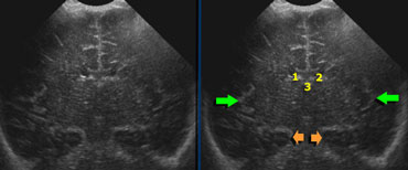
Measurement of the ventricular system should be done in an easy reproducible sonographic plane.
Use a coronal section through the lateral ventricles slightly posterior to the foramen of Monro.
You will see 3 echogenic dots representing the choroid plexus in the lateral ventricles and in the roof of the third ventricle.
Furthermore you should see a symmetrical image of the Sylvian fissure on both sides and the hippocampus (green and orange arrows).Đo lường hệ thống thất phải được thực hiện trên một mặt phẳng siêu âm dễ tái tạo.
Sử dụng mặt cắt đứng ngang qua tâm thất bên hơi sau lỗ Monro.
Bạn sẽ thấy 3 chấm echogenic đại diện cho đám rối mạch mạc ở tâm thất bên và ở tâm thất thứ ba.
Hơn nữa, bạn sẽ thấy hình ảnh đối xứng của rãnh Sylvian ở cả hai bên và hồi hải mã (các mũi tên màu xanh lá cây và màu cam).
Levene indexChỉ số Levene
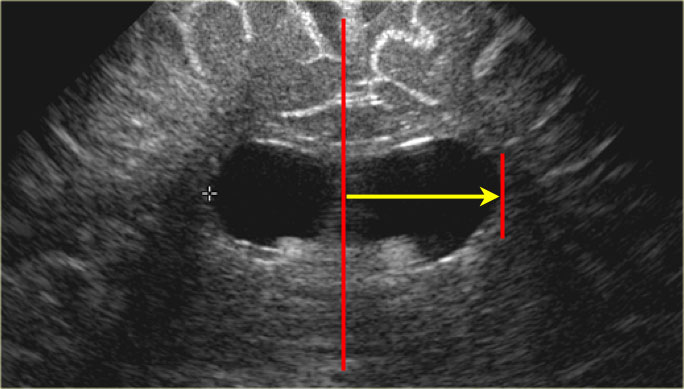
Up to 40 weeks of gestational age the Levene-index should be used and after 40 weeks the ventricular index.
The Levene index is the absolute distance between the falx and the lateral wall of the anterior horn in the coronal plane at the level of the third ventricle.
This is performed for the left and right side.
These measurements can be compared to the reference curve and are quite usefull for further follow-up.Tuổi thai đến 40 tuần chỉ số Levene nên được sử dụng và sau 40 tuần chỉ sô thất.
Chỉ số Levene là khoảng cách tuyệt đối giữa mỏm móc và thành bên sừng trước trong mặt phẳng đứng ngang ở mức thất ba.
Điều này được thực hiện cho bên trái và bên phải.
Các phép đo này có thể được so sánh với đường cong tham chiếu và khá hữu ích để theo dõi thêm.
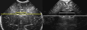 LEFT: Standard measurement of the ventricular index.RIGHT: There is ballooning of the ventricles and the index measurement underestimates the severity of the ventricular widening.Bên trái: Đo tiêu chuẩn của chỉ số tâm thất. Phải: có sự phồng lên của tâm thất và phép đo chỉ số đánh giá thấp mức độ nghiêm trọng của việc mở rộng tâm thất.
Ventricular indexChỉ số thất
After 40 weeks the ventricular index or frontal horn ratio should be used, i.e. the ratio of the distance between the lateral sides of the ventricles and the biparietal diameter.
When using this ratio you have to realise, that when the ventricular system widens, the frontal horns tend to enlarge in the craniocaudal direction more than in the left to right dimension.
Sau 40 tuần, chỉ số tâm thất hoặc tỷ lệ sừng trán nên được sử dụng, tức là tỷ lệ khoảng cách giữa các bên của tâm thất và đường kính hai cạnh.
Khi sử dụng tỷ lệ này, bạn phải nhận ra rằng khi hệ thống não thất mở rộng, các sừng trán có xu hướng mở rộng theo hướng sọ não nhiều hơn theo chiều từ trái sang phải.
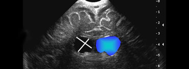
 Measurement of the falx to the most lateral point of the lateral ventricle.Đo lường điểm đỉnh của não thất ngoài.
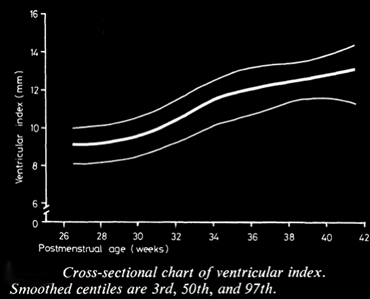
Real-time ultrasound was used to make exact measurements from the lateral wall of the body of the lateral ventricle to the falx (the ventricular index) in 273 infants of varying gestational ages (5).
The measurement performed in an axial plane through the temporoparietal bone correlated closely with an actual measurement made in coronal plane in 50 infants.
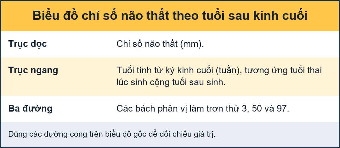
A cross-sectional centile chart was drawn up of the normal range for this measurement from 27 to 42 weeks' postmenstrual age.
A further chart showing the rate of change of the ventricular index allowed growth of the ventricles to be assessed in a longitudinal manner.
Use of these charts permits early detection of hydrocephalus or dilated ventricles secondary to cerebral atrophy.Siêu âm thời gian thực được sử dụng để đo chính xác từ thành bên của cơ thể của tâm thất bên đến falx (chỉ số tâm thất) ở 273 trẻ sơ sinh ở các độ tuổi thai khác nhau (5).
Phép đo được thực hiện trên mặt phẳng ngang qua xương thái dương hàm tương quan chặt chẽ với phép đo thực tế được thực hiện trong mặt phẳng đứng ngang ở 50 trẻ sơ sinh.
Biểu đồ centile cắt ngang được vẽ trong phạm vi bình thường cho phép đo này từ 27 đến 42 tuần tuổi sau kinh nguyệt.
Biểu đồ tiếp theo cho thấy tốc độ thay đổi của chỉ số tâm thất cho phép đánh giá sự tăng trưởng của tâm thất theo chiều dọc.
Sử dụng các biểu đồ này cho phép phát hiện sớm não úng thủy hoặc giãn não thất thứ phát do teo não.
A more realiable indicator of widening of the ventricular system would be an area- or volume-measurement.
This however is more time consuming.
So although ventricular index has shortcommings it is still the most commonly used.
In general practise, studying the images by eye is reliable, provided, that standard planes are used.Một chỉ số có thể thực hiện được hơn về sự giãn rộng của hệ thống thất phải là đo diện tích hoặc thể tích.
Tuy nhiên, điều này tốn nhiều thời gian hơn.
Vì vậy, mặc dù chỉ số thất có những thiếu sót, nó vẫn được sử dụng phổ biến nhất.
Trong thực hành chung, nghiên cứu hình ảnh bằng mắt là đáng tin cậy, với điều kiện là các mặt phẳng tiêu chuẩn được sử dụng.
ReferencesTài liệu tham khảo
-
An Atlas of Neonatal Brain Sonography
by Paul Govaert, Gent University Hospital and Linda S. de Vries, Wilhelmina Kinderziekenhuis.
with contributions of Frank van Bel, Erik Beek, Dirk Voet, An Bael, Linde Goossens
Siêu âm não sơ sinh -
Parenchymal Brain Injury in the Preterm Infant: Comparison of Cranial Ultrasound, MRI and Neurodevelopmental Outcom
Tổn thương não nhu mô ở trẻ sơ sinh non tháng: So sánh siêu âm sọ, MRI và kết quả phát triển thần kinh
Ariadne M. Roelants-van Rijn, Floris Groenendaal, F. J. A. Beek, Paula Eken, Ingrid C. van Haastert, Linda S. de Vries.
-
Periventricular Leukomalacia
Nhuyễn chất trắng quanh não thất
in eMedicine, Author: Terence Zach, MD, in eMedicine, Tác giả: Terence Zach, MD,
-
Periventricular Hemorrhage-Intraventricular Hemorrhage
Xuất huyết não thất-xuất huyết não thất
in eMedicine, Author: David J Annibale, MD Tác giả: David J. Annibale, MD
-
Levene MI., Measurement of the growth of the lateral ventricles in preterm infants with real-time ultrasound.
Arch Dis Child. 1981 Dec;56(12):900-4.
Levene MI., Đo sự phát triển của tâm thất bên ở trẻ sinh non bằng siêu âm thời gian thực.