
Ultrasound of the Neonatal spineSiêu âm cột sống trẻ sơ sinh
Department of Radiology of the Wilhelmina Childrens Hospital, University Medical Center Utrecht and the Alrijne hospital in Leiderdorp, the NetherlandsKhoa Chẩn đoán hình ảnh của Bệnh viện Nhi Wilhelmina, Trung tâm Y tế Đại học Utrecht và Bệnh viện Alrijne ở Leiderdorp, Hà Lan
Ngày đăng / cập nhật: October 29, 201929 tháng 10 năm 2019
Bản gốc trên radiologyassistant.nl ↗ · tải về lúc 2026-11-19 20:19 · bản dịch máy có chuẩn hóa thuật ngữ chẩn đoán hình ảnh
Ultrasound is the preferred modality in neonates with suspected occult spinal dysraphism (OSD).
OSD implies the presence of one or more spinal cord anomalies, which can cause tethering of the spinal cord and possible neurological and bladder or bowel function deficits. Siêu âm là phương pháp được ưu tiên ở trẻ sơ sinh nghi ngờ có rối loạn vận động cột sống (OSD).
OSD ngụ ý sự hiện diện của một hoặc nhiều bất thường tủy sống, có thể gây ra sự ràng buộc của tủy sống và có thể là thiếu hụt chức năng thần kinh và bàng quang hoặc ruột.
Ultrasound is easy to perform, since the posterior arch of the vertebra is not yet ossified, providing a perfect acoustic window.
Especially the lumbosacral part of the spinal canal with the conus medullaris and the cauda equina can be beautifully depicted with a high resolution linear array probe.Siêu âm rất dễ thực hiện, vì cung sau của đốt sống chưa được cốt hóa, cung cấp một cửa sổ âm thanh hoàn hảo.
Đặc biệt là phần thắt lưng cùng của ống sống với nón hành não và đuôi ngựa có thể được mô tả đẹp mắt bằng đầu dò mảng tuyến tính có độ phân giải cao.
Classification of Spinal dysraphismPhân loại loạn vận động cột sống
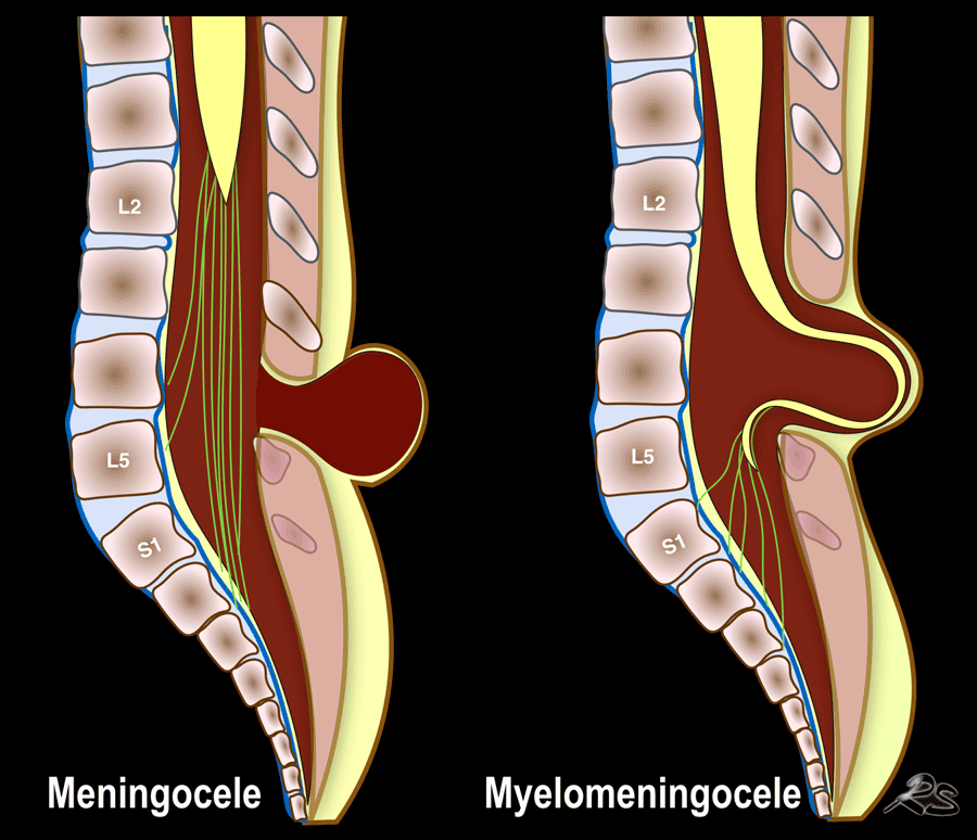

Spina bifida apertaTật nứt đốt sống hở
Spinal dysraphism or spina bifida is a congenital anomaly resulting in a defective closure of the neural arch.
It is classified into open (spina bifida aperta) and closed dysraphism (spina bifida occulta). Rối loạn vận động cột sống hoặc nứt đốt sống là một dị tật bẩm sinh dẫn đến khiếm khuyết đóng vòm thần kinh.
Nó được phân loại thành tật nứt xương quay mở (nứt xương nứt mở) và tật nứn kín (nứn xương nứn hở).
Open dysraphism presents with a swelling over the back which is noticed at birth and may contain meninges and CSF, called meningocele or contain parts of the spinal cord or nerves, called myelomeningocele.
Chứng loạn dưỡng mở biểu hiện bằng một vết sưng trên lưng được nhận thấy khi sinh và có thể chứa màng não và dịch não tuỷ, được gọi là u màng não hoặc chứa các phần của tủy sống hoặc dây thần kinh, được gọi lầ u màng não tuỷ.
Ultrasound should not be used to image open spinal dysraphism at the lesion itself.
It does not add much and can lead to infection.
Ultrasound can be used to examine more cranial parts of the vertebral column, searching for additional anomalies and is useful to measure the size of the ventricles of the brain after closure of the myelomeningocele.Không nên sử dụng siêu âm để hình ảnh rối loạn vận động cột sống hở tại chính tổn thương.
Nó không bổ sung nhiều và có thể dẫn đến nhiễm trùng.
Siêu âm có thể được sử dụng để kiểm tra các phần khác của cột sống, tìm kiếm các dị tật bổ sung và rất hữu ích để đo kích thước của não thất sau khi đóng túi thóat vị màng tuỷ.
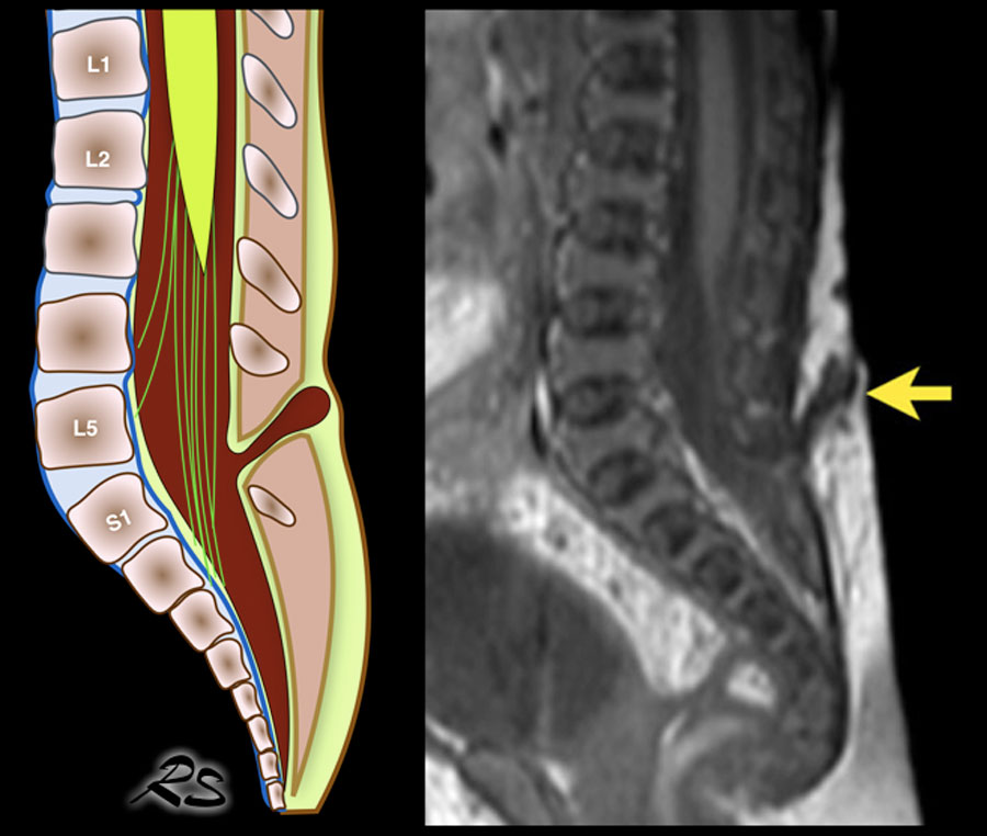
Spina bifida occultaNứt đốt sống
In closed or occult spinal dysraphism, also called spina bifida occulta, there is an intact covering of the skin.Trong loạn dưỡng cột sống kín hoặc ẩn, còn được gọi là tật nứt đốt sống, có một lớp da còn nguyên vẹn.
The anomaly is suspected when there are skin changes like hair tufts, hemangiomas, pigmented spots, cutaneous dimples or a subcutaneous mass.Nghi ngờ dị tật khi có những thay đổi trên da như búi tóc, u mạch, đốm sắc tố, lúm đồng tiền trên da hoặc khối dưới da.
Another reason to perform ultrasound is when there is a congenital anomaly that is associated with OSD like anal atresia.Một lý do khác để thực hiện siêu âm là khi có một dị tật bẩm sinh có liên quan đến OSD như teo hậu môn.
The term OSD implies the presence of one or more spinal cord anomalies, which can cause tethering of the spinal cord and possible neurological and bladder/bowel function deficits.
The terms thickened or fatty filum terminale, spinal lipomas, split cord malformations, dermoid cyst, and syringohydromyelia are all different forms of OSD. Thuật ngữ OSD ngụ ý sự hiện diện của một hoặc nhiều bất thường tủy sống, có thể gây ra sự ràng buộc của tủy sống và có thể là thiếu hụt chức năng thần kinh và bàng quang / ruột.
Các thuật ngữ filum terminale dày hoặc béo, u mỡ tuỷ sống, dị dạng tuỷ, nang bì và syringohydromyelia là các dạng OSD khác nhau.
Normal anatomyGiải phẫu bình thường
 Click to enlargeNhấp để phóng to
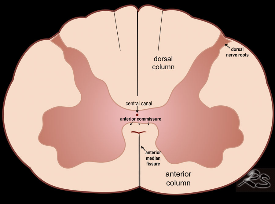
The spinal cord is depicted as a very hypoechoic structure with a central echogenicity.
This central echogenicity is supposed to represent the interface between the anterior commissure and the median anterior fissure, and not the central canal.Tủy sống được mô tả là một cấu trúc giảm âm rất có tính tạo tiếng vang trung tâm.
Tính năng tạo tiếng vang trung tâm này được cho là đại diện cho giao diện giữa mép trước và rãnh trước giữa, chứ không phải ống trung tâm.
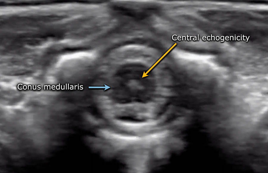

Axial image of the spinal cord with central echogenicity.Hình ảnh trục tủy sống với độ vang trung tâm.
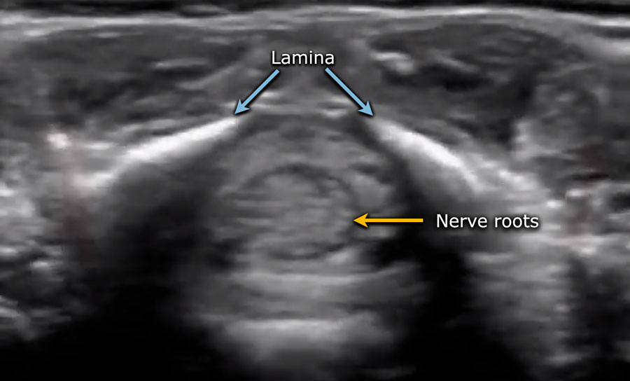

Axial image of the cauda equina.Hình ảnh trục của đuôi ngựa.
The lower end of the cord is thickened, which is the lumbar intumescence.
Đầu dưới của dây rốn dày lên, đó là sự phát triển của thắt lưng.
The cord tapers in a sharp cone (blue arrow).Dây thần kinh hình nón sắc nhọn (mũi tên màu xanh).
The cauda equina is seen as a bunch of moving strands.
If the baby is lying in the decubitus position, the strands will gravitate to the dependent posterior side.
If the baby is examined in the prone position with a pillow under the abdomen, the stands will move ventrally.Ngón đuôi ngựa được xem như một bó các sợi di chuyển.
Nếu em bé nằm ở tư thế nằm, các sợi sẽ bị hút về phía sau phụ thuộc.
Nếu trẻ được khám ở tư thế nằm sấp với một chiếc gối dưới bụng, thì tư thế đứng sẽ di chuyển theo tâm thất.
The filum terminale can be seen as a thin echogenic thread.
The dural sac ends at approximately S2.
More distally fatty tissue is present.Phần tận cùng của sợi chỉ có thể được xem như một sợi chỉ mỏng.
Túi màng cứng kết thúc ở khoảng S 2.
Có nhiều mô mỡ ở đầu xa.
Video of the normal sagittal anatomy.Video giải phẫu bình thường của dọc sống.
Always obtain axial views.
The movement of the nerve roots is better seen in the transverse plane compared to the sagittal plane and it is easier to assess intraspinal pathology like a thickened filum.Luôn luôn có được góc nhìn trục.
Sự di chuyển của rễ thần kinh được nhìn thấy rõ hơn trên mặt phẳng ngang so với mặt phẳng đứng dọc và dễ dàng đánh giá bệnh lý trong tuỷ hơn như là một sợi dây dày lên.
Film in right decubitus position.
The nerve roots are clustered in the dependent side, but move freely.Phim ở tư thế nằm nghiêng phải.
Các rễ thần kinh được nhóm lại ở phía phụ thuộc, nhưng di chuyển tự do.
When the nerve roots do not move freely, it can be a sign of OSD.Khi rễ thần kinh không di chuyển tự do, đó có thể là dấu hiệu của OSD.
Position of the conus medullarisVị trí của nón hành não
The normal position of the conus is at L!.
It should not be below L2.Vị trí bình thường của nón là L!.
Nó không nên ở dưới L 2.
The best way to determine the position of the conus medullaris is by identifying the lumbosacral junction at the lordotic angle between the lumbar and sacral vertebrae (arrow).
It can be helpful to flex and extend the pelvis to see the point of motion of the sacrum.Cách tốt nhất để xác định vị trí của nón hành não là xác định điểm nối thắt lưng cùng ở góc ưỡn giữa đốt sống thắt ngực và đốt sống cùng (mũi tên).
Có thể hữu ích khi gập và duỗi xương chậu để xem điểm chuyển động của xương cùng.
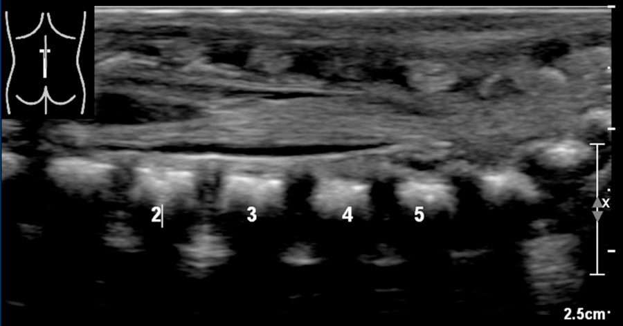
In this newborn the lumbosacral junction is less clearly seen because there is no acute angle.
The numbers that we've put in, might be wrong.Ở trẻ sơ sinh này, chỗ nối thắt lưng cùng ít được nhìn thấy rõ hơn vì không có góc nhọn.
Những con số mà chúng ta đã đưa vào, có thể sai.
If one is uncertain, make a panoramic or dual image of the lumbosacral vertebral column and compare the vertebral count from below upwards with a lateral plain film.Nếu không chắc chắn, hãy chụp toàn cảnh hoặc hình ảnh kép của cột sống thắt lưng cùng và so sánh số lượng đốt sống từ dưới lên trên với phim thường quy bên.
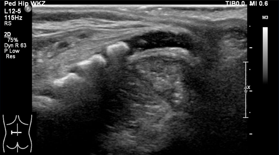 Sagittal view of a normal "kyphotic" coccyx in a 2-day-old girlHình ảnh dọc của xương cụt "siêu gù" bình thường ở một bé gái 2 ngày tuổi
The coccyx, if not yet ossified, is composed of hypoechoic cartilage.
It usually has a kyphotic shape.
On transverse views it should not be confused with a fluid collection or an abscess.Sụn sườn, nếu chưa được cốt hóa, bao gồm sụn giảm âm.
Nó thường có hình dạng gù.
Trên phim chụp cắt ngang, không nên nhầm lẫn với tụ dịch hoặc áp xe.
Normal variantsBiến thể bình thường
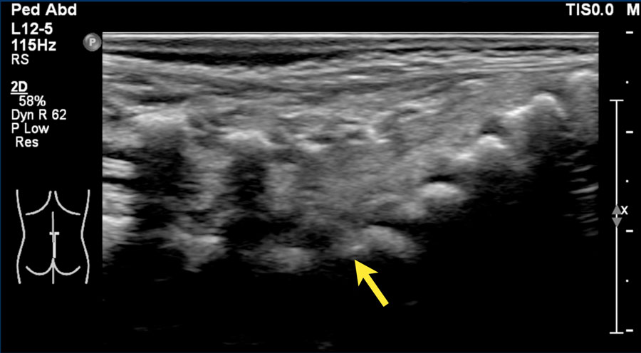
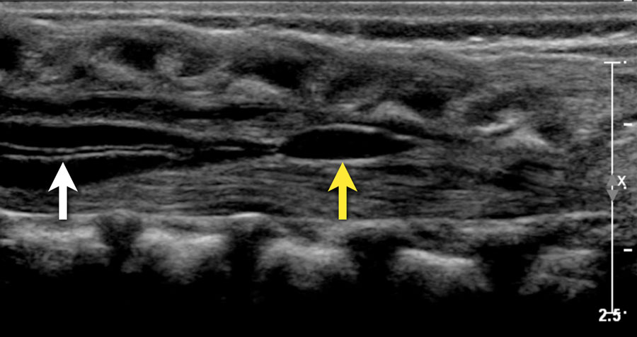 Two-week-old girl with a sacral simple. There is a slight hydromyelia (white arrow) and a cyst in the filum terminale (yellow arrow), both are normal variants.Cô bé hai tuần tuổi với một chiếc sacral đơn giản. Có một loại hydromyelia nhẹ (mũi tên màu trắng) và một u nang trong filum terminale (mũi tên màu vàng), cả hai đều là các biến thể bình thường.
Central canal
In this image the central canal is visible as a thin anechoic line in the spinal cord (white arrow).
Although this is sometimes associated with pathology it is frequently seen as a normal finding.Kênh trung tâm
Trong hình ảnh này, ống trung tâm có thể nhìn thấy dưới dạng một đường trống âm mỏng trong tủy sống (mũi tên trắng).
Mặc dù điều này đôi khi liên quan đến bệnh lý, nhưng nó thường được coi là một phát hiện bình thường.
Ventriculus terminalis
A small cyst is seen in the proximal filum terminale.
This is called a ventriculus terminalis (or fifth ventricle).
Sometimes it is seen in the conus medullaris.
It is formed during embryogenesis and usually regresses completely during early childhood.
If it stays persistent, it typically measures less than 2 cm in craniocaudal dimension and 2 mm in transverse dimension and is detected as an asymptomatic and incidental finding in adults.Lỗ não thất tận
Một nang nhỏ được nhìn thấy ở đầu gần của đầu dây chằng.
Đây được gọi là tâm thất tận (hay tâm thất thứ năm).
Đôi khi nó được nhìn thấy trong nón hành não.
Nó được hình thành trong quá trình tạo phôi và thường thóai triển hoàn toàn trong thời thơ ấu.
Nếu nó vẫn tồn tại, nó thường có kích thước nhỏ hơn 2 cm ở chiều ngang và 2 mm ở chiều ngang, và được phát hiện là không có triệu chứng và phát hiện tình cờ ở người lớn.
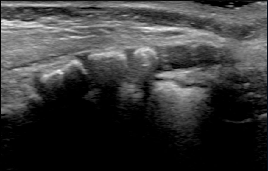
Here a sagittal image of a three-months-old girl who was imaged because of a skin discoloration of the lower back.
The spinal anatomy was normal and there was no sign of OSD.Đây là hình ảnh dọc của một bé gái ba tháng tuổi được chụp vì sự đổi màu da ở phần lưng dưới.
Giải phẫu cột sống bình thường và không có dấu hiệu của OSD.
There is a straight coccyx, which is a normal variant.
Usually the coccyx has a anteriorly bent tip, but sometimes it is straight or even dorsally bent, which is also a normal variant.Có một xương cụt thẳng, là một biến thể bình thường.
Thông thường xương cụt có đầu cong về phía trước, nhưng đôi khi nó thẳng hoặc thậm chí cong về mặt lưng, cũng là một biến thể bình thường.
PathologyBệnh lý
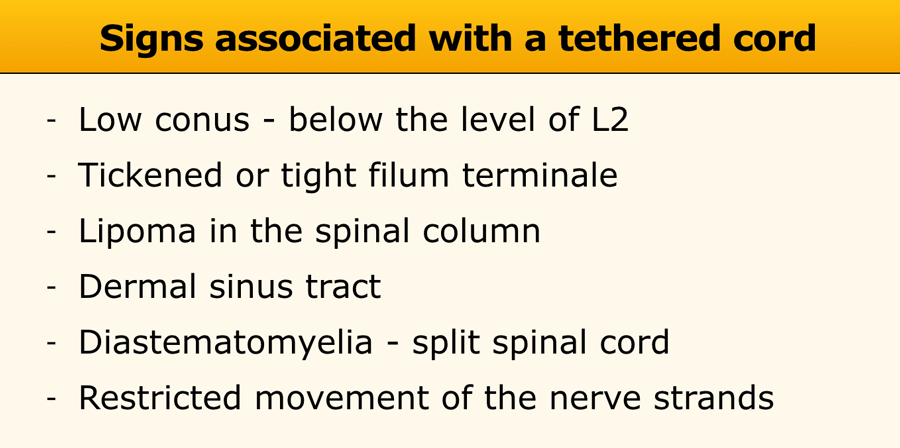
Tethered cordDây buộc
In many cases occult spinal dysraphism may not cause any symptoms.
However in some cases there may develop neurological problems due to tethering of the cord.Trong nhiều trường hợp, rối loạn vận động cột sống có thể không gây ra bất kỳ triệu chứng nào.
Tuy nhiên, trong một số trường hợp, có thể phát sinh các vấn đề thần kinh do dây rốn bị buộc chặt.
A tethered cord is a pathologic fixation of the spinal cord in an abnormal caudal location, so that the cord suffers mechanical stretching, distortion and ischemia with growth and development.Dây rốn bị buộc là sự cố định bệnh lý của tủy sống ở vị trí đuôi bất thường, do đó dây rốn bị kéo căng cơ học, biến dạng và thiếu máu cục bộ tăng trưởng và phát triển.
In these cases ultrasound is well suited to image the contents of the spinal canal and to look for findings that are associated with a tethered cord (Table).Trong những trường hợp này, siêu âm rất phù hợp để hình ảnh hóa các thành phần của ống sống và tìm kiếm các dấu hiệu có liên quan đến dây rốn (Bảng).
Low conus medullarisNón tuỷ thấp
Newborn girl with a cloacal malformation.
The conus medullaris is at L5.
No lipoma visible.
Findings were confirmed at MR imaging which was acquired at the age of 9 months.
Continue with the MR.Trẻ sơ sinh có dị dạng lỗ huyệt.
Tụy hình nón nằm ở vị trí L 5.
Không có u mỡ.
Các dấu hiệu được xác nhận trên hình ảnh cộng hưởng từ khi trẻ được 9 tháng tuổi.
Tiếp tục với chụp cộng hưởng từ.
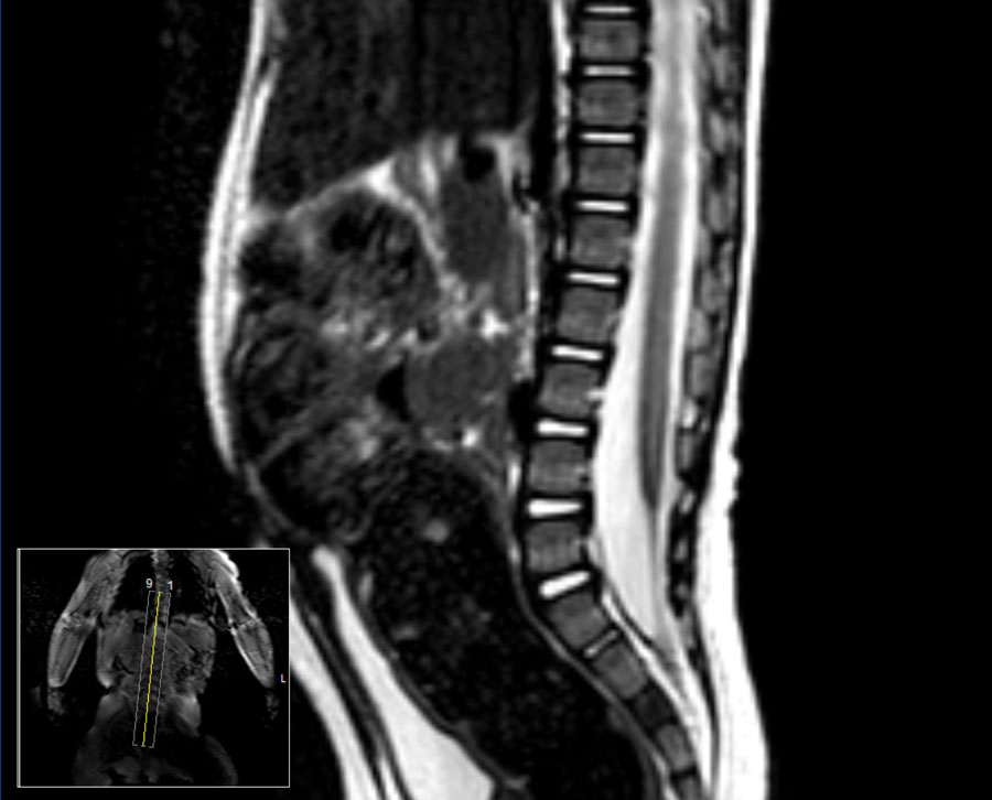
MR image at the age of 9 months.
Hình ảnh cộng hưởng từ lúc 9 tháng tuổi.
The conus medullaris is now seen at L4.Hình nón hành não hiện được nhìn thấy ở L 4.
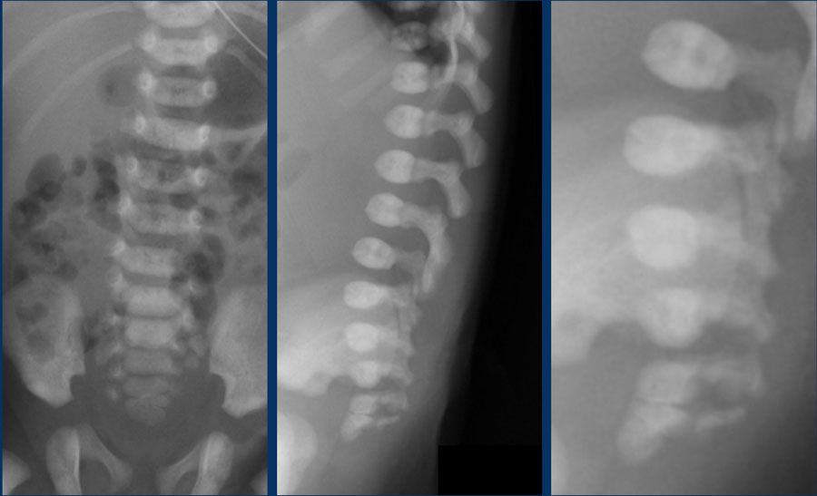
Thickened filum terminaleFilum dày
Study the image.
What are the findings?Nghiên cứu hình ảnh.
Những phát hiện này là gì?
Findings:
The plain film shows a fusion of S4 and S5.Kết quả:
phim thường quy cho thấy sự hợp nhất của S4 và S 5.
This was a newborn boy with an anorectal malformation.
An ultrasound was performed to look for signs of occult spinal dysraphism.Đây là một trẻ sơ sinh bị dị dạng hậu môn trực tràng.
Siêu âm được thực hiện để tìm các dấu hiệu của rối loạn vận động cột sống.
This is the transverse videoĐây là video ngang
There is a thickened filum with a low ending conus medullaris at L3-L4 and a mild hydromyelia.Có một lớp màng mỏng dày lên với một đầu thấp của nón hành não ở L 3 - L 4 và một chứng tràn dịch màng phổi nhẹ.
Continue with the sagittal video.Tiếp tục với video dọc.
On the sagittal video the low ending conus medullaris is seen at L3-L4.Trên đoạn phim dọc, hình nón hành não có đầu thấp được nhìn thấy ở L 3 - L 4.
Continue with the MRI.Tiếp tục với MRI.
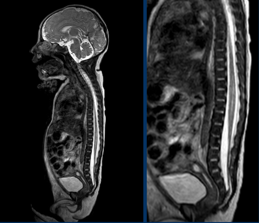
The MRI also depicts:MRI cũng mô tả:
- Hydromyelia
- Low ending conus medullaris.
Conus tuỷ sống tận cùng thấp. - Thickened filum terminale
Filum dày
The upper limit of normal for the width of the filum terminale is 2 mm.
If it is thickened it often shows fatty infiltration with hyperechoic tissue.
Giới hạn trên của giá trị bình thường đối với chiều rộng của đầu tận cùng là 2 mm.
Nếu nó dày lên, nó thường cho thấy sự thâm nhiễm chất béo với mô tăng âm.
In healthy newborns, the tip of the conus medullaris is located between L1 and L2.
The tip should not be positioned below L2-3.Ở trẻ sơ sinh khoẻ mạnh, đầu của nón hành não nằm giữa L1 và L 2.
Đầu không nên được đặt dưới L 2-3.
Hydromyelia
A hydromyelia is the abnormal widening of the central canal by cerebrospinal fluid. This condition may be either focal or diffuse, extending through the entire length of the spinal cord.
Tràn dịch màng phổi là sự giãn nở bất thường của ống trung tâm bởi dịch não tủy, tình trạng này có thể là khu trú hoặc lan toả, kéo dài qua toàn bộ chiều dài của tủy sống.
It can be associated with several congenital abnormalities including diastematomyelia, Arnold‐Chiari malformation, myelomeningocele and lipomeningocele.Nó có thể liên quan đến một số bất thường bẩm sinh bao gồm diastematomyelia, dị dạng Arnold-Chiari, thóat vị màng tuỷ và thóat vị màng não.
If there is a fluid collection in the cord outside the central canal it is called syringomyelia.
Because it is often not possible to separate these two entities on imaging, it is better to use the term syringohydromyelia.
Transverse images will show the transition of the normal cord into the cord which surrounds a CSF collectionNếu có một lượng dịch trong dây rốn bên ngoài ống trung tâm, nó được gọi là syringomyelia.
Vì thường không thể tách biệt hai thực thể này trên hình ảnh, nên tốt hơn là sử dụng thuật ngữ syringohydromyelia.
Hình ảnh ngang sẽ cho thấy sự chuyển tiếp của dây bình thường vào dây bao quanh bộ sưu tập CSF
Spinal lipomaU mỡ tuỷ sống
A spinal lipoma is an encapsulated deposit of fat, neural tissue, meninges or fibrous tissue which extends from the posterior subcutaneous tissue through a midline defect of the fascia, muscle or bone to communicate with the spinal canal or meninges.U mỡ tuỷ sống là một lắng đọng chất béo, mô thần kinh, màng não hoặc mô xơ kéo dài từ mô dưới da phía sau thông qua một khiếm khuyết đường giữa của cân, cơ hoặc xương để liên lạc với ống sống hoặc màng não.
A spinal lipoma is seen as an echogenic mass, mostly in the lower spinal canal.
The conus can be too low and buried in the lipoma.U mỡ tuỷ sống được xem như một khối echogenic, chủ yếu ở ống sống thấp hơn.
Vòng nón có thể quá thấp và vùi trong u mỡ.
Here a sagittal image of a newborn boy with anal atresia.
The conus is blunted and ends at L2-L3.
There is an echogenic mass which is likely a lipoma.
The filum is thickened.Đây là hình ảnh dọc của một bé trai sơ sinh bị teo hậu môn.
Vòng nón bị cùn và kết thúc ở L 2 - L 3.
Có một khối u tạo tiếng vang có thể là một u mỡ.
Lớp màng bẩn dày lên.
Small lipomas are often led alone. Bigger lipomas in symptomatic patients are removed.U mỡ nhỏ thường được điều trị đơn độc.U mỡ lớn hơn ở bệnh nhân có triệu chứng được loại bỏ.
On a transverse video the ecogenic mass just below the cord terminus is seen.Trên một video ngang, khối sinh thái ngay dưới đầu dây rốn được nhìn thấy.
Dorsal dermal sinusXoang bì mặt sau
This is a connection between the skin and the dural sac, sometimes into the spinal cord.
It presents with a dimple, discoloration of the skin or hairy patches.Đây là một kết nối giữa da và túi màng cứng, đôi khi vào tủy sống.
Nó biểu hiện với một lúm đồng tiền, sự đổi màu của da hoặc các mảng lông.
The conus medullaris can be too low.Tụy conus có thể quá thấp.
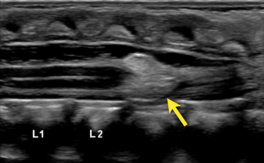
Three-day-old girl with a red stain on the lower back, a tuft of hair and a dimple.Cô bé ba ngày tuổi với một vết đỏ ở lưng dưới, một búi tóc và một má lúm đồng tiền.
US shows a tract from the skin towards the dural sac at the S1-S2 level, compatible with a dorsal dermal sinus .
The conus medullaris is at a normal level and there is no other intraspinal pathology present.Siêu âm cho thấy một đường từ da đến túi màng cứng ở mức S 1 - S 2, tương thích với xoang da lưng.
Hình nón hành não ở mức bình thường và không có bệnh lý nội tuỷ khác.
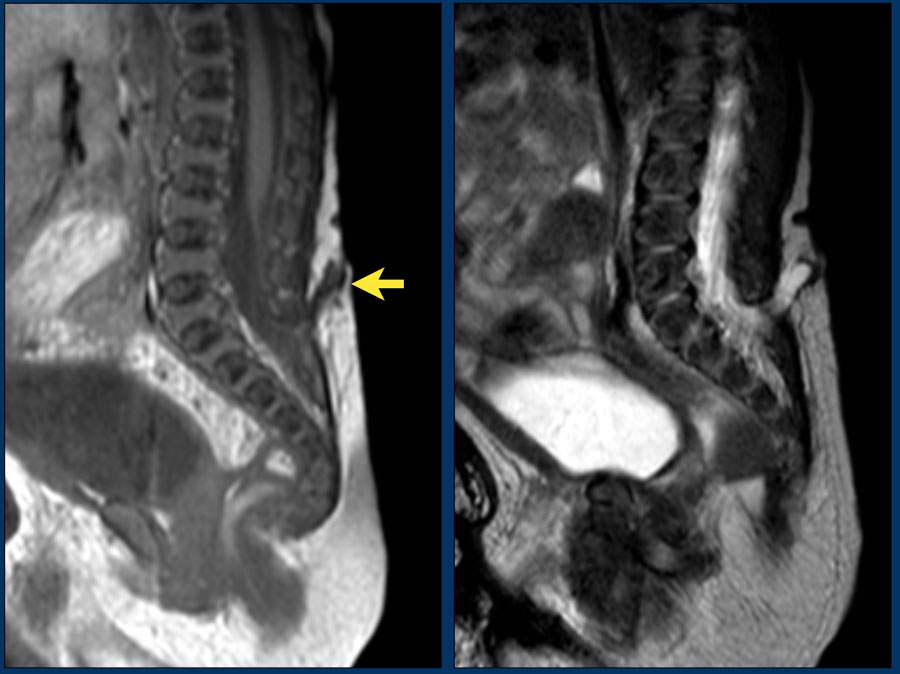
A T1-weigthed image shows the sinus tract from the skin towards the S1-S2 level. Hình ảnh T1 cho thấy đường xoang từ da về phía S 1 - S 2.
The conus medullaris is too low at L3-L4.Hành não hình nón quá thấp ở vị trí L 3 - L 4.
Dermal sinus tracts are especially important lesions to recognize because they can extend to the spinal canal and represent an increased risk of infection such as meningitis and spinal cord abscess.Các xoang da là những tổn thương đặc biệt quan trọng cần nhận biết vì chúng có thể lan rộng đến ống sống và làm tăng nguy cơ nhiễm trùng như viêm màng não và áp xe tủy sống.
DiastematomyeliaTrang chủ
Diastematomyelia, also named split cord malformation, is a longitudinal split of the spinal cord.
At the point of division, there may be an osseous, fibrous or cartilaginous dividing septum.
Diastematomyelia is commonly associated with a vertebral column abnormality and a tethered spinal cord.Diastematomyelia, còn được gọi là dị dạng dây rốn, là một sự phân chia theo chiều dọc của tủy sống.
Tại thời điểm phân chia, có thể có một vách ngăn phân chia xương, xơ hoặc sụn.
Bệnh lý hẹp thân đốt sống thường liên quan đến bất thường cột sống và tủy sống có dây buộc.
The ultrasound diagnosis is usually straightforward.
Chẩn đoán siêu âm thường đơn giản.
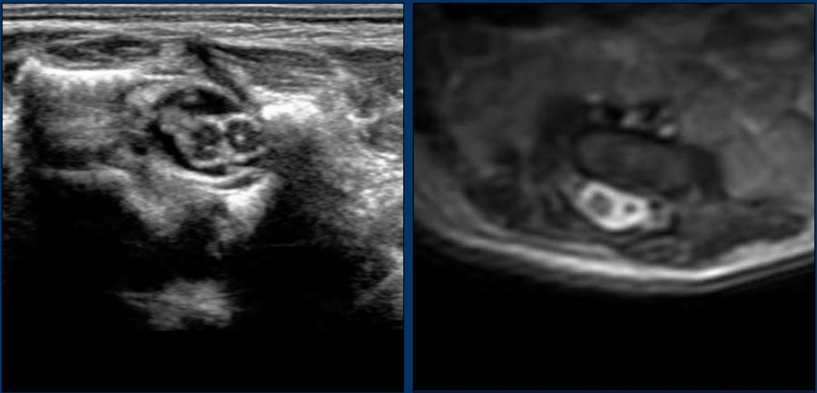
Here images of a newborn, who had several antenatal anomalies.Dưới đây là hình ảnh của một trẻ sơ sinh, người có một số dị tật trước sinh.
There was a lateral rocker bottom feet deformity, lumbar kyphoscoliosis and swelling on the lower back.Có biến dạng bàn chân đáy bên, gù cột sống thắt lưng và sưng nề vùng lưng dưới.
The spinal US and MR show a longitudinal split of the spinal cord at the thoracolumbar level. Siêu âm và MR cho thấy sự phân chia theo chiều dọc của tủy sống ở mức độ cột sống thắt lưng.
The bifid cord is asymmetric in volume.Dây nhị đầu không đối xứng về thể tích.
Additional plain films of the lumbar spine and pelvis showed thoracic and lumbar hemivertebrae.Phim phẳng bổ sung của cột sống thắt lưng và khung chậu cho thấy các nửa cột sống ngực và thắt lưng.
There was also a dislocated left hip.Ngoài ra còn có trật khớp háng trái.
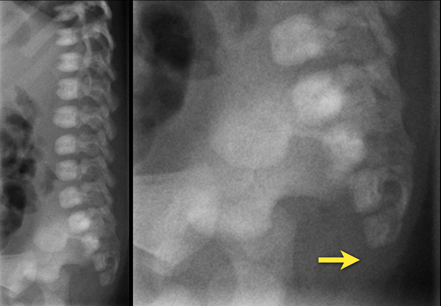 Newborn girl with an anorectal malformation. The distal sacrum below S4 is absent.Trẻ sơ sinh bị dị dạng hậu môn trực tràng. Không có xương cùng dưới S 4.
Blunt cord terminusĐầu cùn dây rốn
This image is of a newborn girl with an anorectal malformation.
The distal sacrum below S4 is absent (arrow).
Since an anorectal malformation is frequently associated with spinal pathology, an ultrasound was performed.Hình ảnh này là của một bé gái sơ sinh bị dị tật hậu môn trực tràng.
Không có xương cùng dưới S4 (mũi tên).
Vì dị dạng hậu môn trực tràng thường liên quan đến bệnh lý cột sống, nên siêu âm đã được thực hiện.
If a plain film of the vertebral column demonstrates an anomaly of the sacrum, there is a 50% change of an intraspinal anomaly.Nếu phim thường quy của cột sống cho thấy một dị tật của xương cùng, có sự thay đổi 50% của dị tật trong tuỷ.
In most cases there is a low ending tethered cord.Trong hầu hết các trường hợp có một dây buộc thấp.
In some cases however the cord ends at the normal position, but is deformed.Tuy nhiên, trong một số trường hợp, dây rốn kết thúc ở vị trí bình thường, nhưng bị biến dạng.
This is a so-called blunt cord terminus.
There is generally a wedge-shaped ending in which the dorsal side reaches further caudally than the ventral side. Đây được gọi là đầu cuối cùn.
Nói chung có một đầu hình nêm trong đó mặt lưng tiếp cận xa hơn so với mặt bụng.
Continue with the ultrasound.Tiếp tục siêu âm.
Ultrasound of the spine shows a blunt cord terminus at T12-L1.Siêu âm cột sống cho thấy một đầu dây rốn ở T 12 - L 1.
A blunt cord terminus is a sign of caudal regression syndrome characterized by abnormal development of the lower end of the spine.Đầu dây rốn bị đứt là dấu hiệu của hội chứng hồi quy đuôi đặc trưng bởi sự phát triển bất thường của đầu dưới của cột sống.
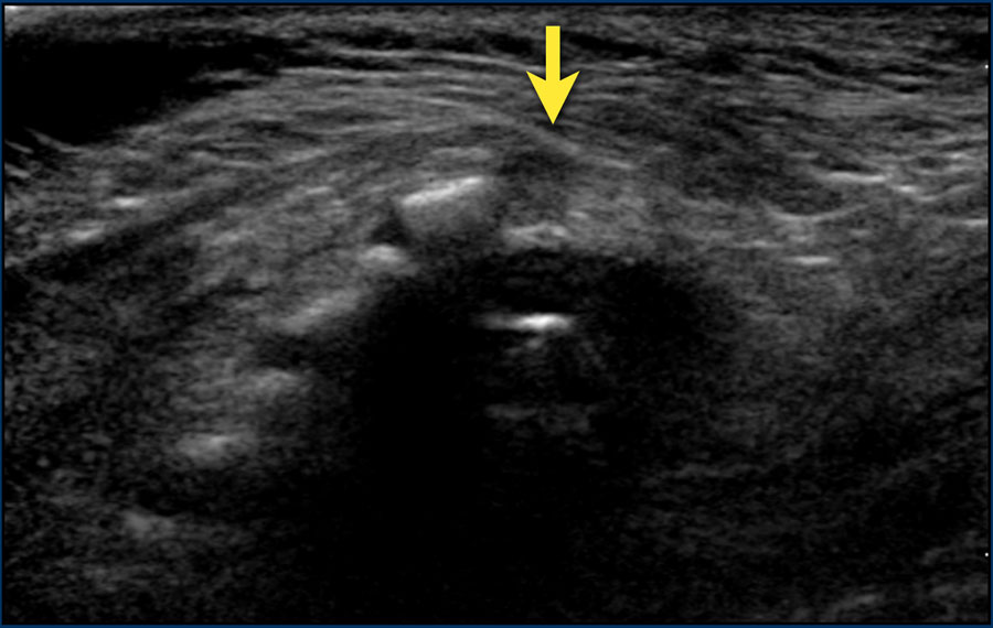
On ultrasound the absent coccyx is also visible.Trên siêu âm, cũng có thể nhìn thấy xương cụt không có.
Continue with the MRI.Tiếp tục với MRI.
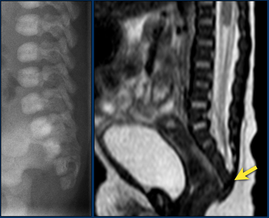
Sagittal T2-weigthed image showing the cord terminus at Th12.Hình ảnh T2 xoắn dọc cho thấy đầu tận của dây rốn ở vị trí Th 12.
ReferencesTài liệu tham khảo
-
US of the Spinal Cord in Newborns: Spectrum of Normal Findings, Variants, Congenital Anomalies, and Acquired Diseases
Siêu âm của tủy sống ở trẻ sơ sinh: Phổ các phát hiện bình thường, biến thể, dị tật bẩm sinh và bệnh mắc phải
by Karin M. Unsinn, MD et al
RadioGraphics 2000; 20:923–938
bởi Karin M. Unsinn, MD và cộng sự - Use of magnetic resonance imaging to detect occult spinal dysraphism in infants Ứng dụng cộng hưởng từ trong chẩn đoán loạn dưỡng cột sống ở trẻ nhũ nhi