
Perianal FistulasRò quanh hậu môn
Radiology Department of the Medical Centre Alkmaar and the Rijnland hospital, Leiderdorp, the Netherlands.Khoa X quang của Trung tâm Y tế Alkmaar và bệnh viện Rijnland, Leiderdorp, Hà Lan.
Ngày đăng / cập nhật: 2009-01-21
Bản gốc trên radiologyassistant.nl ↗ · tải về lúc 2026-11-19 20:19 · bản dịch máy có chuẩn hóa thuật ngữ chẩn đoán hình ảnh
Perianal fistula is a common disorder that often recurs because of infection that was missed at surgery.
Preoperative MR can help to prevent recurrence.
In this review we will address the anatomy, pathogenesis, classification and scanning protocol of perianal fistulas.
Rò hậu môn là một rối loạn thường gặp, thường tái phát do nhiễm trùng đã bỏ sót khi phẫu thuật.
Mắt cộng hưởng từ trước mổ có thể giúp ngăn ngừa tái phát.
Trong bài tổng quan này, chúng tôi sẽ đề cập đến giải phẫu, cơ chế bệnh sinh, phân loại và quy trình chụp rò hậu môn.
by Susanne Tonino and Robin Smithuisbởi Susanne Tonino và Robin Smithuis
AnatomyGiải phẫu
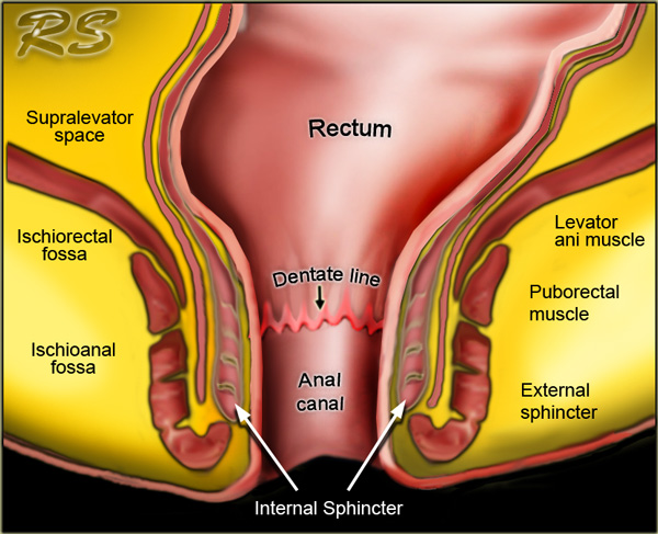
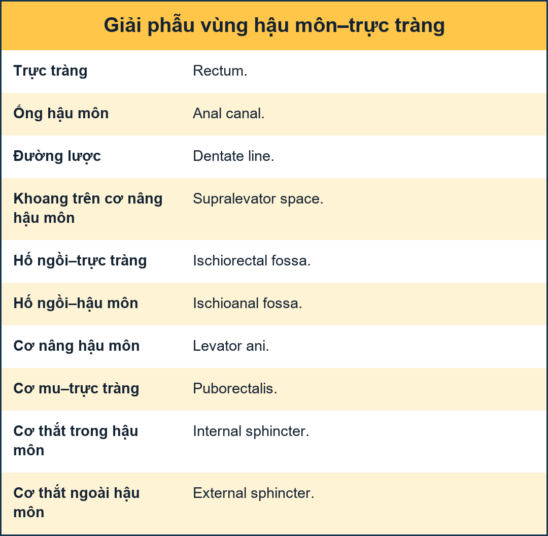
The anatomical anal canal extends from the perineal skin to the linea dentata.
Surgically, the anal canal extends from the perineal skin to the anorectal ring.
This is the circular upper border of the puborectal muscle which is digitally palpable upon rectal examination.
The anorectal ring lies approximately 1-1,5 cm above the linea dentata.
The total length of the surgical anal canal is about 4-5 cm.Ống hậu môn giải phẫu kéo dài từ da tầng sinh môn đến đường răng hàm.
Về mặt phẫu thuật, ống hậu môn kéo dài từ da tầng sinh môn đến vòng hậu môn trực tràng.
Đây là bờ trên hình tròn của cơ bắp, có thể sờ thấy bằng ngón tay khi khám trực tràng.
Vòng hậu môn trực tràng nằm khoảng 1-1,5 cm trên đường răng hàm.
Tổng chiều dài ống hậu môn phẫu thuật khoảng 4-5 cm.
The anal sphincter is comprised of three layers:Cơ thắt hậu môn bao gồm ba lớp:
- Internal sphincter: continuance of the circular smooth muscle of the rectum, involuntary and contracted during rest, relaxes at defecation.Cơ thắt trong: cơ trơn trực tràng liên tục co lại khi nghỉ ngơi, giãn ra khi đại tiện.
- Intersphincteric space.Không gian gian cơ thắt.
-
External sphincter: voluntary striated muscle, divided in three layers that function as one unit.
These three layers are continuous cranially with the puborectal muscle and levator ani (figure).Cơ vòng ngoài: cơ vân tự nguyện, chia làm ba lớp có chức năng như một đơn vị.
Ba lớp này liên tục với cơ trán và cơ nâng hậu môn (hình).
 Puborectal muscle forming a 'sling'Cơ trán tạo thành một 'cái dây'
The puborectal muscle has its origin on both sides of the pubic symphysis, forming a 'sling' around the anorectum.
Cơ mu có nguồn gốc ở cả hai bên của xương mu, tạo thành một 'cái dây' quanh hậu môn trực tràng.
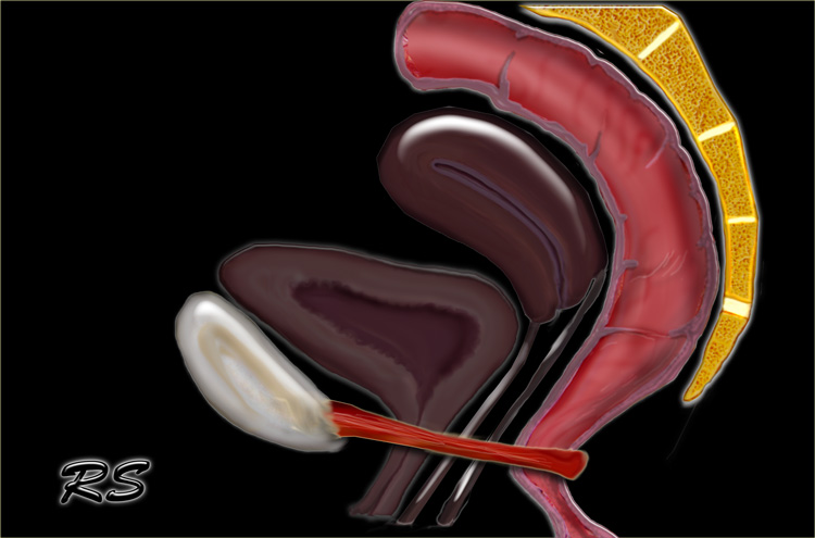
The puborectal muscle is contracted at rest and accounts for the 80? angulation of the anorectal junction.
It relaxes during defecation.Cơ lưng-trực tràng co khi nghỉ và chiếm 80? góc của chỗ nối hậu môn trực tràng.
Nó thư giãn trong quá trình đại tiện.
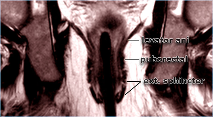
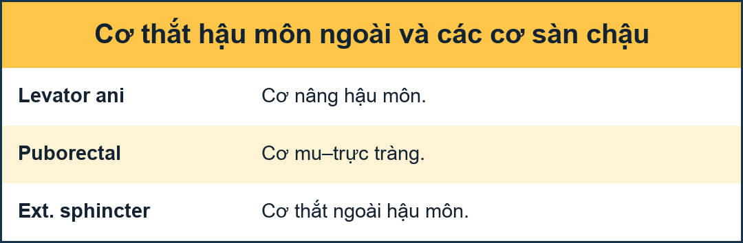 Coronal T2W-imageHình ảnh T2W vành
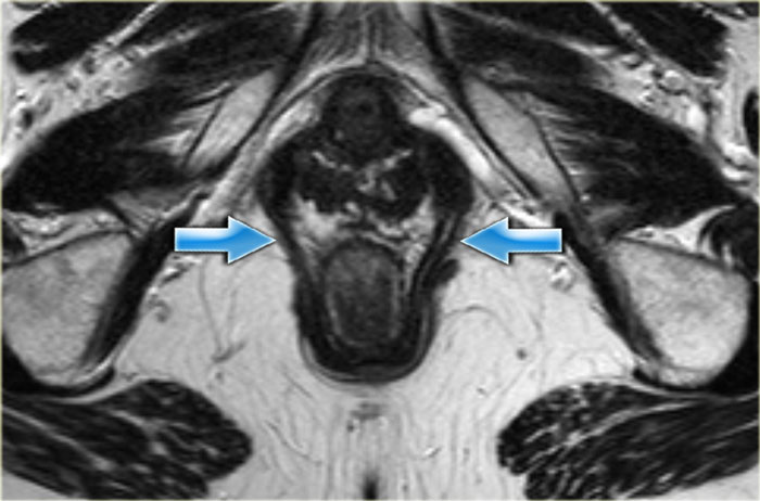
On axial and coronal MR-images the different layers of the anal sphincter and the surrounding structures can be displayed perfectly. Trên hình ảnh MR-hình ảnh trục và vành, các lớp khác nhau của cơ thắt hậu môn và các cấu trúc xung quanh có thể được hiển thị hoàn hảo.
Perianal fistulaRò hậu môn
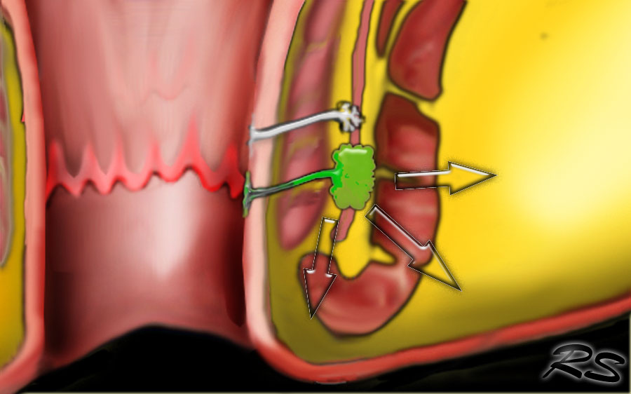
A perianal fistula is an abnormal connection between the epithilialised surface of the anal canal and the skin.
The causes of perianal fistulas: Rò hậu môn là một kết nối bất thường giữa bề mặt thượng bì của ống hậu môn và da.
Nguyên nhân của rò hậu môn:
-
PrimaryTiểu học
- Obstruction of anal gland which leads to stasis and infection with absces and fistula formation (most common cause).Tắc nghẽn tuyến hậu môn dẫn đến ứ đọng và nhiễm trùng với các ổ áp xe và hình thành lỗ rò (nguyên nhân phổ biến nhất).
- SecondaryTrung học
- Iatrogenic (hemorrhoideal surgery)Điều trị (phẫu thuật trĩ lý tưởng)
- Inflammatory bowel diseases (Crohn's disease more common than colitis ulcerosa) Bệnh viêm ruột (bệnh Crohn thường gặp hơn viêm đại tràng thể loét)
- Infections (viral, fungal or TB) Nhiễm trùng (vi rút, nấm hoặc lao)
- Malignancy Đau đớn

ClassificationPhân loại
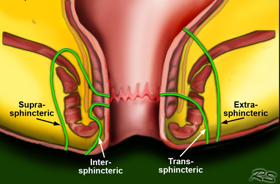
The most widely used classification is the Parks Classification which distinguishes four kinds of fistula: intersphincteric, transsphincteric, suprasphincteric and extrasphincteric. Phân loại được sử dụng rộng rãi nhất là Phân loại Parks phân biệt bốn loại rò: giữa cơ thắt, qua cơ thắT, trên cơ thắ T và ngoài cơ thắ.
The most common fistulas are the intersphincteric and the transsphincteric.
The extrasphincteric fistula is uncommon and only seen in patients who had multiple operations.
In these cases the connection with the original fistula tract to the bowel is lost.
Các lỗ rò phổ biến nhất là lỗ rò giữa cơ thắt và lỗ rò xuyên cơ thắT.
Rò ngoài cơ thắt không phổ biến và chỉ gặp ở những bệnh nhân đã phẫu thuật nhiều lần.
Trong những trường hợp này, đường nối với đường rò ban đầu với ruột bị mất.
A superficial fistula is a fistula that has no relation to the sphincter or the perianal glands and is not part of the Parks classification.
These are more often due to Crohns disease or anorectal procedures such as haemorrhoidectomy or sphincterotomy.Rò nông là một lỗ rò không liên quan đến cơ thắt hoặc các tuyến quanh hậu môn và không thuộc phân loại Parks.
Những trường hợp này thường do bệnh Crohns hoặc các thủ thuật hậu môn trực tràng như cắt trĩ hoặc mở cơ thắt.
MR Protocol and ReportingGiao thức và báo cáo MR
Protocol
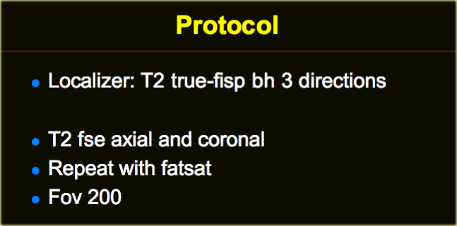
A localizer in three directions is needed in order to align the T2 sequences axial and coronal to the anal canal.
Any localizer that properly displays the anal canal can be used.
We use a TRUE FISP, which is the name that Siemens uses for a steady-state precession gradient-echo sequence (GE: FIESTA, Philips: balanced FFE).
Giao thức
Cần có một máy nội soi theo ba hướng để sắp xếp các chuỗi T2 theo trục và vành vào ống hậu môn.
Bất kỳ máy nội soi nào hiển thị đúng ống hậu môn đều có thể được sử dụng.
Chúng tôi sử dụng một TRUE FISP, đó là tên mà Siemens sử dụng cho một trình tự gradient-echo tiến động trạng thái ổn định (GE: FIESTA, Philips: cân bằng FFE).
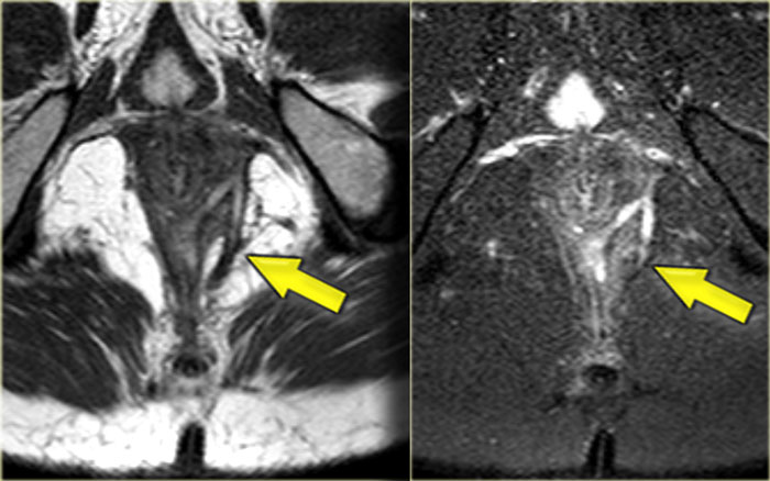 Perianal fistula: axial T2WI without fatsat (left) and with fatsat (right)Rò quanh hậu môn: T2WI trục không có fatsat (trái) và có fatsat(phải)
T2W images without fatsat better display the anatomy, while the fatsat images better depict the fistulas.Hình ảnh T2W không có fatsat thể hiện tốt hơn giải phẫu, trong khi hình ảnh fatsat thể hiết tốt hơn các lỗ rò.
Reporting
When you describe a fistula, it is important to mention the following characteristics: Báo cáo
Khi bạn mô tả một lỗ rò, điều quan trọng là phải đề cập đến các đặc điểm sau:
- Position of the mucosal opening on axial images (using the anal clock). Vị trí lỗ niêm mạc trên ảnh mặt phẳng ngang (sử dụng đồng hồ hậu môn).
- Distance of the mucosal defect to the perianal skin on coronal images. Khoảng cách của khuyết hổng niêm mạc đến da quanh hậu môn trên ảnh mặt phẳng đứng ngang.
- Secondary fistulas or abscesses. Rò hoặc áp xe thứ phát.
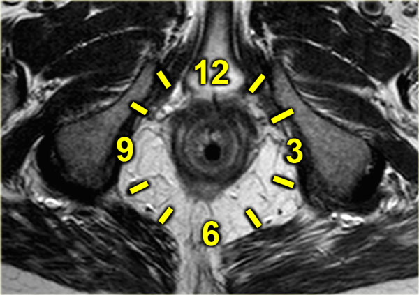
The drawing on the left illustrates the anal clock, which is the surgeon's view of the perianal region when the patient is in the supine lithotomy position (2).
This scheme corresponds to the orientation of axial MR images of the perianal region.Bản vẽ bên trái minh hoạ đồng hồ hậu môn, đó là góc nhìn của bác sĩ phẫu thuật về vùng quanh hậu môn khi bệnh nhân ở tư thế cắt sỏi nằm ngửa (2).
Sơ đồ này tương ứng với hướng của hình ảnh MR trục của vùng quanh hậu môn.
Examples of Perirectal FistulasVí dụ về rò quanh trực tràng
Intersphincteric fistulaRò gian cơ thắt
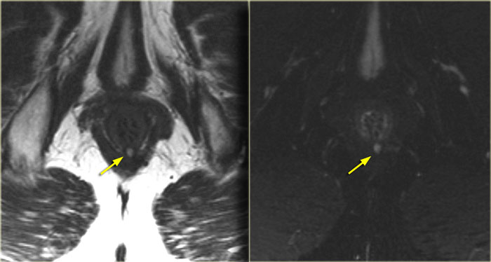
On the left axial T2W images with and without fat saturation.
An intersphincteric fistula is located at 6 o'clock.
Continue with coronal images.Trên hình ảnh T2W trục trái có và không có độ bão hoà mỡ.
Một lỗ rò giữa cơ thắt nằm ở vị trí 6 giờ.
Tiếp tục với ảnh mặt phẳng đứng ngang.
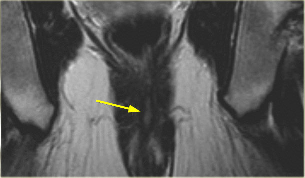
On the coronal image the fistula runs caudally towards the skin.
There is no connection with the external sphincter.
Trên hình ảnh vành, lỗ rò chạy về phía da.
Không có mối liên quan với cơ thắt ngoài.
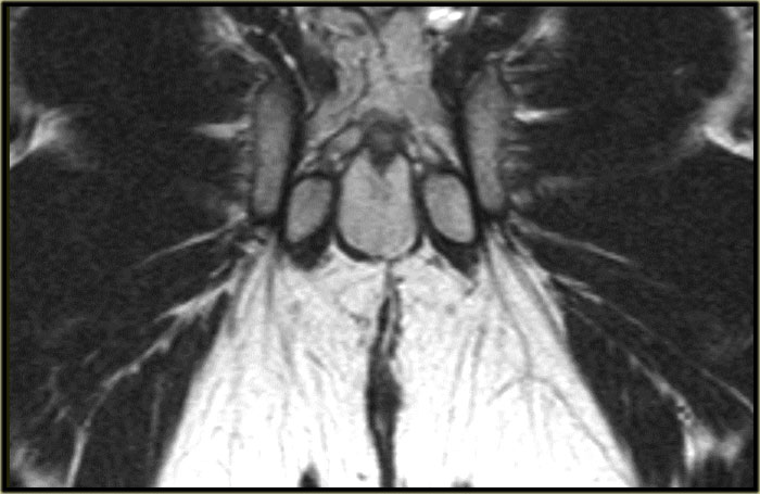
On the left coronal images of another patient with an intersphincteric fistula.
Use the arrows to scroll through the images.Trên ảnh mặt phẳng đứng ngang bên trái của một bệnh nhân khác có rò gian cơ thắt.
Sử dụng các mũi tên để cuộn qua các hình ảnh.
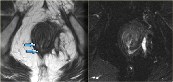 Transsphincteric fistula with sphincter defect at 6 o'clockRò xuyên cơ thắt với khuyết cơ thắt tại vị trí 6 giờ
Transsphincteric fistulaRò xuyên cơ thắt
On the left an axial T2WI and T2WI + fatsat of a transsphincteric fistula.
The defect through the internal and external sphincter at 6 o'clock is clearly visible and more apparent on the fat sat images.Bên trái là T2WI và T2WI + fatsat của lỗ rò qua cơ thắt.
Khiếm khuyết qua nội soi và cơ thắt ngoài ở vị trí 6 giờ có thể nhìn thấy rõ hơn và rõ ràng hơn trên hình ảnh sat.
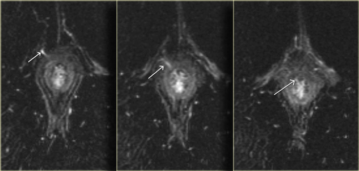 Transsphincteric fistula with sphincter defect at 11 o'clockRò xuyên cơ thắt với khuyết cơ thắt tại vị trí 11 giờ
On the left axial T2W-fatsat images of a transsphincteric fistula with the mucosal opening at 11 o'clock.Trên hình ảnh T2W-fatsat dọc trục trái của lỗ rò xuyên cơ thắt với lỗ mở niêm mạc lúc 11 giờ.
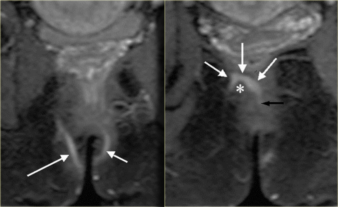
On the left an example of a suprasphincteric fistula.
There are two tracts in the ischioanal region.
The right sided tract runs over the puborectal muscle (asterisk) and the mucosal opening lies at the
level of the linea dentata (black arrow).Bên trái là một ví dụ về lỗ rò trên cơ thắt.
Có hai vùng ở vùng cơ cẳng chân.
Đường bên phải chạy qua cơ bắp (asterisk) và lỗ mở niêm mạc nằm ngang mức của đường răng (mũi tên đen).
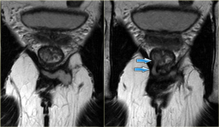
Extrasphincteric fistulaRò ngoài cơ thắt
On the left coronal T2W-images of a small abscess in the left ischioanal fossa, the fistula runs through the levator ani.
It is therefore above the sphincter complex and extrasphincteric.Trên hình ảnh T2W của một áp xe nhỏ ở hố cùng bên trái, đường rò chạy qua cơ nâng hậu môn.
Do đó, nó nằm trên phức hợp cơ thắt và ngoài cơ thắt.
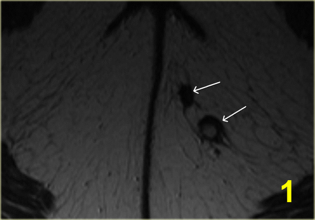
Complex fistulaRò phức tạp
On the left an example of a complex fistula.
Two tracts in the left buttock form a single tract (no. 1-2).
This fistula breaks through the external sphincter (no. 4).
In the intersphincteric space it divides again into two tracts (no. 5).
One ends blindly in the intersphincteric space (no. 6).
The other breaks through the internal sphincter with the mucosal defect at 1 o'clock.
Bên trái là một ví dụ về một lỗ rò phức tạp.
Hai đường ở mông trái tạo thành một đường duy nhất (số 1-2).
Rò này xuyên qua cơ thắt ngoài (số 4).
Trong khoang gian cơ thắt, nó lại chia thành hai vùng (số 5).
Một đầu bịt kín trong khoang giữa cơ thắt (số 6).
Một đường khác xuyên qua cơ thắt trong với khuyết niêm mạc ở góc 1 giờ.
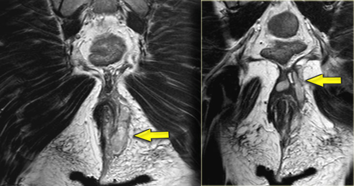
Crohn's diseaseBệnh Crohn
On the left a patient with a perianal fistula who has Crohn's disease.
Continue with the coronal images.Bên trái là một bệnh nhân bị rò hậu môn mắc bệnh Crohn.
Tiếp tục với ảnh mặt phẳng đứng ngang.
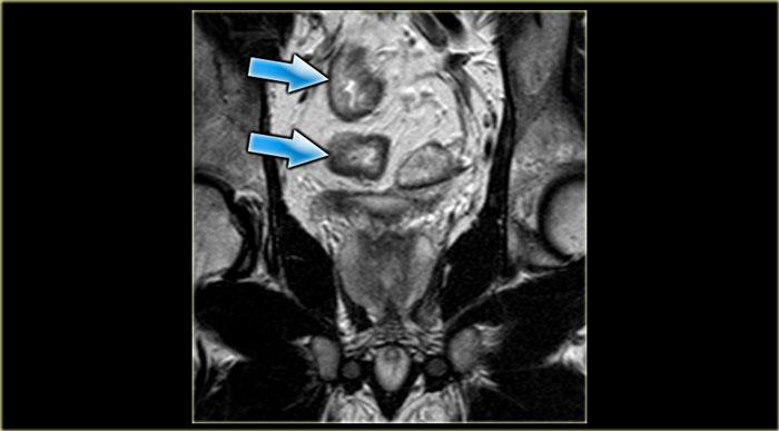
On the coronal images the thickening of the bowel wall is demonstrated.Trên ảnh mặt phẳng đứng ngang, thành ruột dày lên.
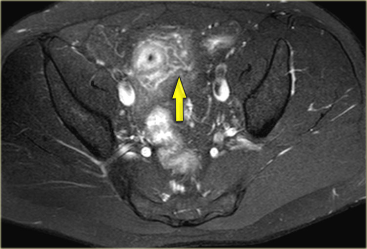
Axial fatsat images depict the transmural inflammation with infiltration of the mesenteric fat.Hình ảnh fatsat trục mô tả tình trạng viêm xuyên thành mạc treo kèm theo thâm nhiễm mỡ mạc treo.
TreatmentĐiều trị
Treatment is focussed on the elimination of the primary and secondary tracts, prevention of
recurrence and to retain continence.
The treatment given depends on the anatomy of the fistula, if it is a simple fistula with a
low mucosal defect is can be probed in the OR to identify the mucosal defect at the
linea dentata, then the tract can be opened.
This is only possible if the external sphincter is not involved.
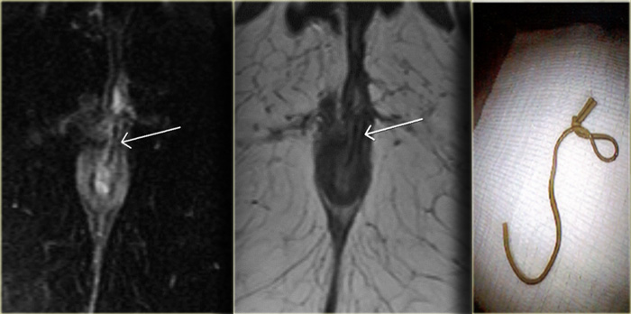
Seton fistulotomy is a technique where a rubber ligature or vessel loop is pulled through the
fistula, it then is tightened every 2 weeks or so in order to obtain pressure necrosis so that
the Seton is slowly pulled through the muscle.
This has the advantage that the muscle is slowly cut and fibroses at the same time in order
to cause as little damage as possible to the sphincter complex.
If there is an extrasphincteric fistula, the lower part is opened.
The mucosal defect, which is by definition in the rectum, is then surgically closed.
Điều trị tập trung vào việc loại bỏ các đường tiểu học và đường tiểu học thứ phát, ngăn ngừa tái phát và duy trì sự tự chủ.
Việc điều trị tuỳ thuộc vào giải phẫu của lỗ rò, nếu là lỗ rò đơn giản có khuyết hổng niêm mạc thấp có thể thăm dò trong OĐ để xác định khuyết hổng ở đường răng sữa, sau đó có thể mở đường rò.
Điều này chỉ có thể xảy ra nếu cơ thắt ngoài không liên quan.
Phẫu thuật mở thông rò Seton là kỹ thuật trong đó một dây chằng cao su hoặc vòng mạch được kéo qua lỗ rò, sau đó được thắt chặt khoảng 2 tuần một lần để có được sự hoại tử áp lực để Seton được kéo chậm qua cơ.
Điều này có ưu điểm là cơ được cắt chậm và xơ hóa cùng một lúc để gây ra ít tổn thương nhất có thể cho phức hợp cơ thắt.
Nếu có một lỗ rò ngoài cơ thắt, phần dưới được mở ra.
Khiếm khuyết niêm mạc, theo định nghĩa là ở trực tràng, sau đó được phẫu thuật đóng lại.
This patient was already known to have an intersfincteric fistula, the mucosal defect is at 1 o'clock.
In the tract there is a linear structure with a low signal intensity.
This is the Seton which was inserted to treat the fistula.Bệnh nhân này đã được biết là có rò gian cơ thắt, lỗ khuyết niêm mạc là ở vị trí 1 giờ.
Trong đường gân có cấu trúc tuyến tính với cường độ tín hiệu thấp.
Đây là Seton được đưa vào để điều trị rò.
Differential diagnosisChẩn đoán phân biệt
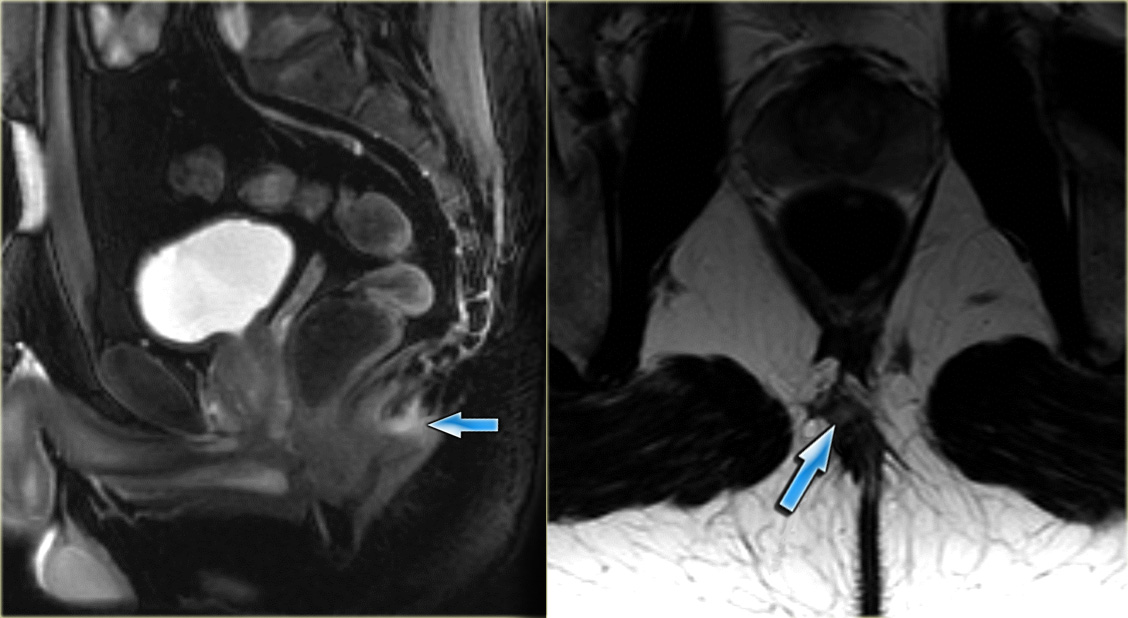 Sinus pilonidalisXoang pilonidalis
Sinus pilonidalis
On the left an example of a sinus pilonidalis.
There is a small abscess just above the nates.
There is no relation with the sphincter complex.
Xoang pilonidalis
Bên trái là một ví dụ về xoang pilonidalis.
Có một áp xe nhỏ ngay trên nates.
Không có mối liên quan với phức hợp cơ thắt.
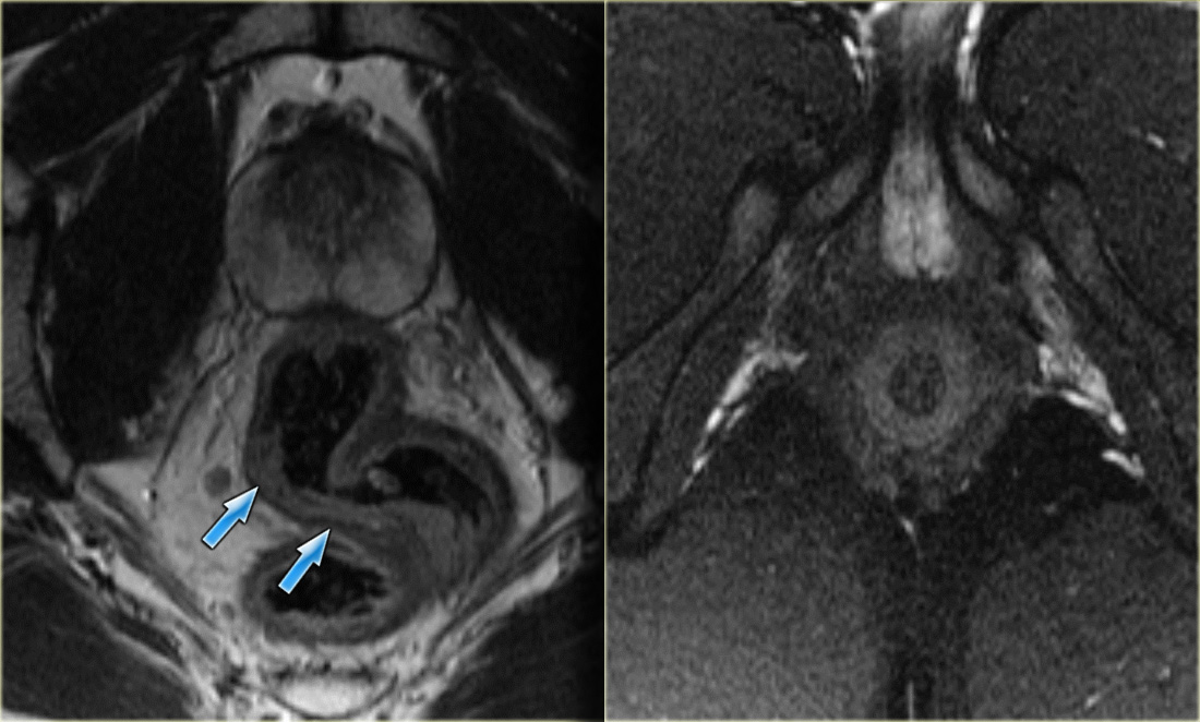
Proctitis
On the left images of a patient who presented with anal complaints.
No fistula was seen.
There is, however, a diffuse thickening of the rectal mucosa due to a proctitis.Viêm trực tràng
Trên hình ảnh bên trái của một bệnh nhân có khiếu nại về hậu môn.
Không có rò.
Tuy nhiên, có sự dày lên lan toả của niêm mạc trực tràng do viêm trực tràng.
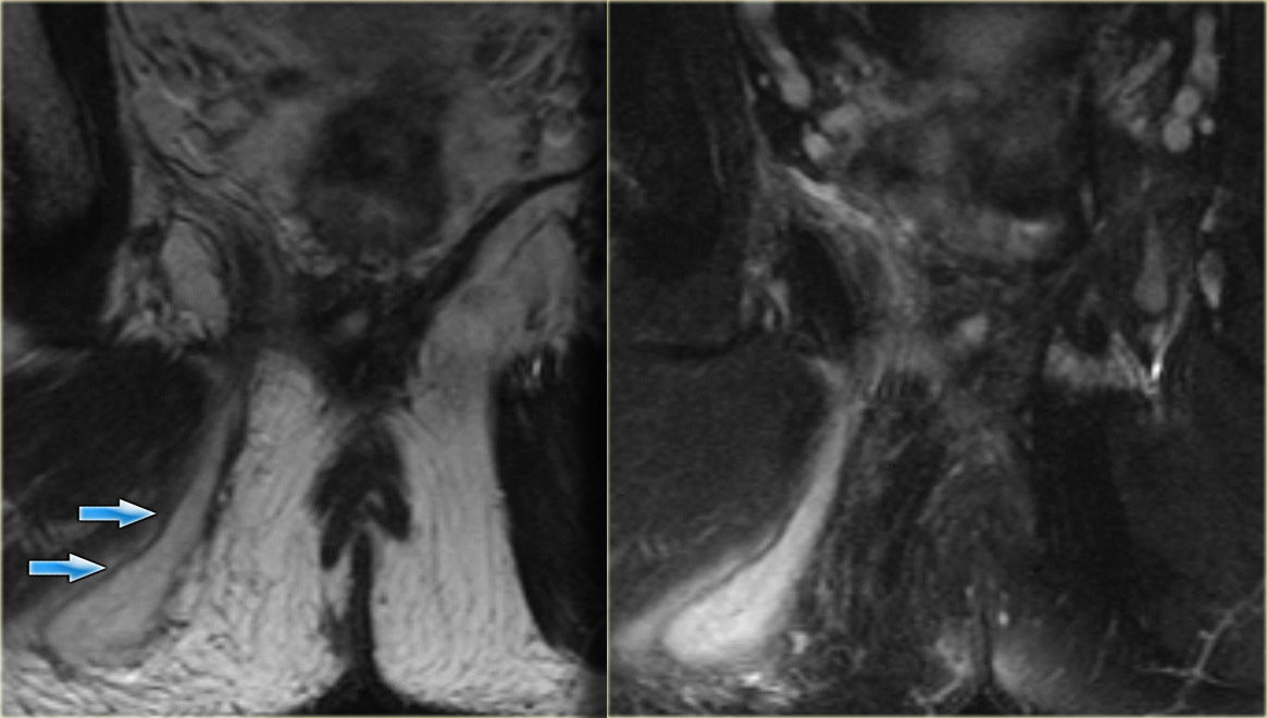 Perianal abscessÁp xe quanh hậu môn
Abscess in the Ischioanal space
An abscess in the ischioanal space with no connection to the sphincter complexÁp xe trong khoang Ischioanal
Áp xe trong khoang cùng vai không có liên quan đến phức hợp cơ thắt
ReferencesTài liệu tham khảo
-
Imaging of Fistula in Ano
Chẩn đoán hình ảnh rò ở hậu môn nhân tạo
Halligan, Stoker et al.
Radiology 2006;239:18-33
Halligan, Stoker và cộng sự. -
MR Imaging Classification of Perianal Fistulas and Its Implications for Patient Management.
Chụp cộng hưởng từ phân loại rò quanh hậu môn và ảnh hưởng của nó đối với quản lý bệnh nhân.
by John Morris
Radiographics. 2000;20:623-635
bởi John Morris -
MRI of Perianal Crohn's Disease
MRI của bệnh Crohn quanh hậu môn
by Karin Horsthuis and Jaap Stoker Tác giả: Karin Horsthuis và Jaap Stoker