
Aortic Aneurysm Rupture.Vỡ phình động mạch chủ.
CT signs of pending aortic aneurysm ruptureDấu hiệu chụp cắt lớp vi tính của phình động mạch chủ đang chờ vỡ
Mallinckrodt Institute of Radiology of the Washington University School of Medicine, St. Louis, MissouriMallinckrodt Viện X-quang của Trường Y thuộc Đại học Washington, St. Louis, Missouri
Ngày đăng / cập nhật: 2006-11-01
Bản gốc trên radiologyassistant.nl ↗ · tải về lúc 2026-11-19 20:19 · bản dịch máy có chuẩn hóa thuật ngữ chẩn đoán hình ảnh
This article is based on a presentation given by Jay Heiken and adapted for the Radiology Assistant by Robin Smithuis.
Jay Heiken is professor of radiology with special interest in abdominal imaging and co-author of the well known book 'Computed Body Tomography With Mri Correlation'.
The classical findings in aortic aneurysm rupture are well known.
In this article we will present the more subtle findings of contained leak and pending rupture of aortic aneurysm.Bài viết này dựa trên một bài thuyết trình của Jay Heiken và được Robin Smithuis chuyển thể cho Trợ lý X quang.
Jay Heiken là giáo sư về X quang có mối quan tâm đặc biệt đến hình ảnh bụng và là đồng tác giả của cuốn sách nổi tiếng 'Chụp cắt lớp cơ thể được tính toán với sự tương quan Mri'.
Các dấu hiệu kinh điển trong vỡ phình động mạch chủ đã được biết đến.
Trong bài báo này chúng tôi sẽ trình bày những dấu hiệu tinh tế hơn của rò có trong túi phình và vỡ trong thời gian chờ của phình động mạch chủ.
Primary signs of Aortic Aneurysm ruptureDấu hiệu chính của vỡ phình động mạch chủ
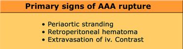
Aortic aneurysm rupture is the most important diagnosis you want to be able to exclude in patients with acute abdominal pain especially when they present with back or flank pain.
The primary signs of AAA rupture are periaortic stranding, retroperitoneal hematoma and extravasation of iv. contrast.
Phình động mạch chủ là chẩn đoán quan trọng nhất mà bạn muốn loại trừ ở những bệnh nhân đau bụng cấp tính đặc biệt khi họ có biểu hiện đau lưng hoặc đau thắt lưng.
Các dấu hiệu chính của vỡ ĐMC là kẹt động mạch chủ, tụ máu sau phúc mạc và thóat mạch máu.
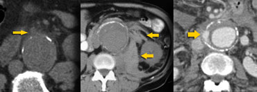 LEFT: Subtle periaortic stranding, MIDDLE: Hemorrhage into posterior pararenal and perirenal compartment, RIGHT: Extravasation of iv. contrastBên trái: Sợi quanh động mạch vành tinh vi, giữa: xuất huyết vào khoang sau cầu sau và khoang thận, bên phải: thóat mạch IV. độ tương phản
On the left we see three patients with aortic aneurysm rupture.
In the image on the far left we only see a little bit of peripheral soft tissue density adjacent to the aneurysm and indeed this is a sign that this patient is at risk for frank rupture.
The other two cases show retroperitoneal hematoma and contrast leakage outside the aorta, which makes it easier to diagnose.Ở bên trái, chúng tôi thấy ba bệnh nhân bị vỡ động mạch chủ động mạch chủ.
Trong hình ảnh bên trái bên trái, chúng ta chỉ thấy một chút mật độ mô mềm ngoại vi tiếp giáp với chứng phình động mạch và thực sự đây là dấu hiệu cho thấy bệnh nhân này có nguy cơ bị vỡ Frank.
Hai trường hợp còn lại cho thấy tụ máu sau phúc mạc và rò tương phản bên ngoài động mạch chủ, giúp chẩn đoán dễ dàng hơn.
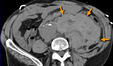
On the left a classical case in a patient with an aneurysm of the abdominal aorta and a large hyperdense retroperitoneal hematoma due to rupture.
The majority of these cases show posterior periaortic hemorrhage and in cases of massive hemorrhage, the posterior pararenal and perirenal compartments are the most frequently involved sites.
Bên trái một trường hợp kinh điển ở bệnh nhân có phình động mạch chủ bụng và máu tụ tăng tỷ trọng lớn sau phúc mạc do vỡ.
Phần lớn các trường hợp này xuất huyết quanh động mạch chủ sau và trong trường hợp xuất huyết lớn, khoang sau thượng thận và khoang quanh thận là những vị trí thường gặp nhất.
Signs of Pending Aneurysm RuptureDấu hiệu của vỡ phình động mạch đang chờ
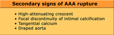
The CT features of contained leak or pending rupture of an aortic aneurysm may be subtle and easily overlooked.
We have to look for the high-attenuating crescent sign, focal discontinuity of intimal calcification or tangential calcium or the draped aorta sign (table on the left).
Các đặc điểm trên CT của túi phình động mạch chủ bị rò hoặc đang chờ vỡ có thể khó thấy và dễ bị bỏ qua.
Chúng ta phải tìm dấu hiệu lưỡi liềm đậm, dấu hiệu đứt đoạn khu trú của vôi hóa nội mạc hoặc dấu hiệu tiếp tuyến canxi hoặc dấu hiệu động mạch chủ bị che phủ (bảng bên trái).
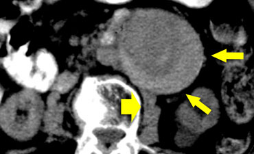 High-attenuating crescent sign in a patient with subtle evidence of leak adjacent to the right psoas muscle (broad arrow).Dấu hiệu lưỡi liềm giảm nhẹ ở một bệnh nhân có bằng chứng tinh tế về rò rỉ tiếp giáp với cơ psoas bên phải (mũi tên rộng).
High-attenuating crescentLiềm giảm âm cao
The high attenuating crescent represents an acute hematoma within either the mural thrombus or the aneurysmal wall.
This sign is strongly associated with AAA rupture.
Sensitivity of the high-attenuating crescent sign as an indication of complicated aneurysm is 77%; specificity, 93%; and positive predictive value of 53%.
So even if there are no primary signs of rupture, we need to indicate to the referring physician, that this patient is at very high risk for aneurysm rupture within the next few days.
Hình liềm đậm độ cao thể hiện một khối máu tụ cấp tính trong huyết khối thành hoặc thành phình mạch.
Dấu hiệu này có liên quan chặt chẽ với vỡ mạch máu não.
độ nhạy của dấu hiệu liềm đậm là dấu hiệu của phình mạch phức tạp là 77%; độ đặc hiệu, 93%; và giá trị dự báo dương tính là 53%.
Vì vậy, ngay cả khi không có dấu hiệu vỡ nguyên phát, chúng ta cần chỉ định cho bác sĩ giới thiệu, rằng bệnh nhân này có nguy cơ vỡ phình mạch rất cao trong vài ngày tới.
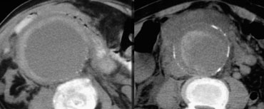 High-attenuating crescent signDấu hiệu lưỡi liềm giảm nhẹ
On the left two more cases of the high-attenuating crescent sign.
In the case on the right we can also identify a retroperitoneal hematoma, so in this case there is a frank AAA rupture.
Bên trái thêm hai trường hợp của dấu hiệu lưỡi liềm cao.
Trong trường hợp bên phải, chúng tôi cũng có thể xác định được tụ máu sau phúc mạc, vì vậy trong trường hợp này có sự vỡ AAA thẳng thắn.
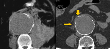
Focal discontinuity of intimal calcificationGián đoạn khu trú của vôi hóa nội sinh
Another sign of impending rupture or contained leakage is focal discontinuity of intimal calcification.
In most of these cases we can also identify the tangential calcium sign.
In these cases it looks as if the calcium is pointing out away from the expected circumference of the aneurysm.
Một dấu hiệu khác của vỡ hoặc rò có chứa là sự gián đoạn khu trú của vôi hóa nội mạc.
Trong hầu hết các trường hợp này, chúng ta cũng có thể xác định dấu hiệu canxi tiếp tuyến.
Trong những trường hợp này, có vẻ như canxi đang chỉ ra khỏi chu vi dự kiến của phình mạch.
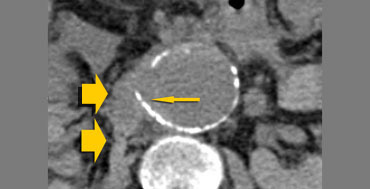 Tangential calcium sign (small arrow) and hemorrhage (broad arrow)Dấu hiệu canxi tiếp tuyến (mũi tên nhỏ) và xuất huyết (mũI tên rộng)
Tangential calcium signDấu hiệu canxi tiếp tuyến
On the left we see another example of the tangential calcium sign.
The intimal calcification points away from the aneurysm and there is retroperitoneal leakage.Ở bên trái, chúng ta thấy một ví dụ khác về dấu hiệu canxi tiếp tuyến.
vôi hóa nội mạc hướng ra xa túi phình và có rò sau phúc mạc.
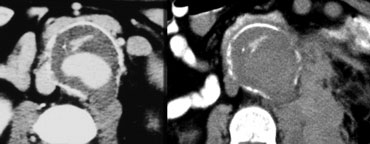 LEFT: draped aorta sign.RIGHT: two weeks later there is a ruptureTRÁI: dấu hiệu động mạch chủ bị che lấp, bên phải: hai tuần sau có vỡ
Draped AortaĐộng mạch chủ bị kéo
On the left a patient who presented with backpain.
The image on the far left shows a bulge of the aorta.
This either represents a focal weakening of the aortic wall or a localized leak.
This patient unfortunately was reported as having no leak and discharged from the emergency departement.
Two weeks later there was a frank rupture.Bên trái là một bệnh nhân có biểu hiện đau lưng.
Hình ảnh ở xa bên trái cho thấy một chỗ phình của động mạch chủ.
Điều này hoặc là biểu hiện của sự suy yếu cục bộ của thành động mạch chủ hoặc rò rỉ cục bộ.
Bệnh nhân này không may được báo cáo là không bị rò rỉ và được xuất viện khỏi khoa cấp cứu.
Hai tuần sau đó có một vết rách rõ.
A positive aortic drape sign is considered to be present when the following features are seen: Dấu hiệu treo động mạch chủ dương tính được coi là có mặt khi thấy các đặc điểm sau:
- area in which the posterior aortic wall is unidentifiable as a distinct line.vùng mà thành sau động mạch chủ không thể xác định được là một đường phân biệt.
- the posterior aorta follows the contour of the spine on one or both sides. động mạch chủ sau đi theo đường viền của cột sống ở một hoặc cả hai bên.
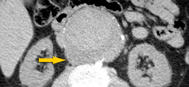 Draped aorta sign. Patient died 3 hours later.Dấu hiệu động mạch chủ rút, bệnh nhân tử vong sau 3 giờ.
On the left another patient who presented with backpain.
There was no evidence of aneurysm leakage, but we see a draped aorta.
The posterior contour of the aorta follows the contour of the spine as if the aorta is draped over the vertebral body.
There is no imaging follow up in this patient, but three hours after this image was taken, this patient exsanguinated from a ruptured AAA.
The leakage was probably where the bulge was (arrow).Bên trái một bệnh nhân khác có biểu hiện đau lưng, không có bằng chứng rò rỉ phình mạch, nhưng chúng tôi thấy động mạch chủ bị che phủ.
Đường viền sau của động mạch chủ đi theo đường viền của cột sống như thể động mạch chủ được phủ lên thân đốt sống.
Không có theo dõi hình ảnh ở bệnh nhân này, nhưng ba giờ sau khi hình ảnh này được chụp, bệnh nhân này đã hết sẩn sau khi vỡ AAA.
Vị trí rò có thể là vị trí phình (mũi tên).
ReferencesTài liệu tham khảo
-
High-attenuating crescent in abdominal aortic aneurysm wall at CT: a sign of acute or impending rupture
Hình liềm giảm âm cao trên phim CT thành phình động mạch chủ bụng: dấu hiệu vỡ cấp hoặc sắp vỡ
Radiology 1994; 192:359-362
WB Mehard, JP Heiken and GA Sicard
Mallinckrodt Institute of Radiology, Washington University School of Medicine, St Louis, MO 63110.
X quang 1994; 192:359-362 -
Abdominal aortic aneurysm morphology: CT features in patients with ruptured and nonruptured aneurysms
Hình thái phình động mạch chủ bụng: Đặc điểm hình ảnh CT ở bệnh nhân phình động vật có túi vỡ và không vỡ
CL Siegel, RH Cohan, M Korobkin, MB Alpern, DL Courneya and RA Leder
Department of Radiology, University of Michigan Medical Center, Ann Arbor
AJR 1994;163:1123-1129
CL Siegel, RH Cohan, M Korobkin, MB Alpern, DL Courneya và RA Leder -
Draped aorta: CT sign of contained leak of aortic aneurysms
Dấu hiệu động mạch chủ bị kéo ra: Dấu vết CT của túi phình động mạch chủ rò rỉ
KE Halliday and A al-Kutoubi
Department of Radiology, St Mary's Hospital, London, England.
Radiology 1996; 199:41-43
KE Halliday và A. al-Kutoubi