
HRCT - Common diagnosesHRCT - Chẩn đoán thường gặp
Radiology Department of the Rijnland Hospital, Leiderdorp and the Academical Medical Centre, Amsterdam, the NetherlandsKhoa X quang của Bệnh viện Rijnland, Leiderdorp và Trung tâm Y khoa Học thuật, Amsterdam, Hà Lan
Ngày đăng / cập nhật: 2007-12-20 / update 2022-03-1520-12-2007 / cập nhật 2022-03-15
Bản gốc trên radiologyassistant.nl ↗ · tải về lúc 2026-11-19 20:19 · bản dịch máy có chuẩn hóa thuật ngữ chẩn đoán hình ảnh
In this review we present the key findings in the most common interstitial lung diseases.
There are numerous interstitial lung diseases, but in clinical practice only about ten diseases account for approximately 90% of cases.
Knowledge of both radiological and clinical appearances of these more common interstitial lung diseases is therefore important for recognizing them in daily practice and including them in the differential diagnosis.
Some less common interstitial lung diseases will also be presented because their HRCT presentation may be very typical, allowing for a 'spot diagnosis' in selected cases.
In 'HRCT - basic interpretation' the terminology is introduced and a practical approach is given for the interpretation of HRCT examinations.
Trong bài tổng quan này, chúng tôi trình bày những phát hiện chính trong các bệnh phổi kẽ phổ biến nhất.
Có rất nhiều bệnh phổi kẽ, nhưng trong thực hành lâm sàng chỉ có khoảng mười bệnh chiếm khoảng 90% các trường hợp. Do đó, kiến thức về cả sự xuất hiện của cả phóng xạ và lâm sàng của những bệnh phổi kẽ phổ biến hơn này rất quan trọng để nhận ra chúng trong thực hành hàng ngày và bao gồm chúng trong chẩn đoán phân biệt.
Một số bệnh phổi kẽ ít phổ biến hơn cũng sẽ được trình bày vì biểu hiện HRCT của chúng có thể rất điển hình, cho phép 'chẩn đoán tại chỗ' trong một số trường hợp được chọn.
Trong 'HRCT - Diễn giải cơ bản', thuật ngữ được giới thiệu và cách tiếp cận thực tế được đưa ra để giải thích các kỳ thi HRCT.
IntroductionMở đầu
More than 100 entities manifest as diffuse lung disease.
Fortunately only about 10 of these account for about 90% of all diffuse lung diseases, that are assessed by open lung biopsy.
Knowing the common and also uncommon HRCT-presentations of these frequently encountered diffuse lung diseases is extremely important.
Hơn 100 thực thể biểu hiện như bệnh phổi lan toả.
May mắn thay, chỉ có khoảng 10 trong số này chiếm khoảng 90% tổng số các bệnh phổi lan toả, được đánh giá bằng sinh thiết phổi mở.
Biết được các biểu hiện HRCT phổ biến và cũng không phổ biến của những bệnh phổi lan toả thường gặp này là vô cùng quan trọng.
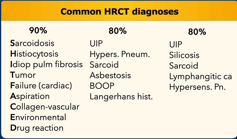
On the left you find three different lists of diagnoses.
Accounting for 80 - 90% of all diagnoses according to various literature references.
In some of them the old names are used and in some the newer ones.
The 'mnemonic' for the first list is 'SHIT FACED' (alternative shaded fit).Ở bên trái, bạn tìm thấy ba danh sách chẩn đoán khác nhau.
Chiếm 80-90% các chẩn đoán theo các tài liệu tham khảo.
Trong một số trường hợp, tên cũ được sử dụng và trong một số trường họp mới hơn.
'Mnemonic' cho danh sách đầu tiên là 'SHIT FACED' (phù hợp với bóng thay thế).
SarcoidosisBệnh sarcoid

Sarcoidosis is a systemic disorder of unknown origin.
It is characterized by non-caseating granulomas in multiple organs, that may resolve spontaneously or progress to fibrosis.
Pulmonary manifestations are present in 90% of patients.
Systemic symptoms such as fatigue, night sweats and weight loss are common.
Loefgren's syndrome, an acute presentation of sarcoidosis, consists of arthritis, erythema nodosum, bilateral hilar adenopathy and occurs in 9-34% of patients.
Erythema nodosum is seen predominantly in women and arthritis is more common in men.Sarcoidosis là một bệnh lý hệ thống không rõ nguyên nhân.
Nó được đặc trưng bởi u hạt không vỏ ở nhiều cơ quan, có thể tự giải quyết hoặc tiến triển thành xơ hóa.
Biểu hiện phổi xuất hiện ở 90% bệnh nhân.
Triệu chứng toàn thân như mệt mỏi, đổ mồ hôi ban đêm và giảm cân là phổ biến.
Hội chứng Loefgren, một biểu hiện cấp tính của bệnh sarcoid, bao gồm viêm khớp, hồng ban nút, hạch rốn gan hai bên và xảy ra ở 9-34% bệnh nhân.
Hồng ban nút gặp chủ yếu ở nữ giới và viêm khớp gặp nhiều hơn ở nam giới.
Two third of patients have a remission within ten years.
One third have continuing disease leading to clinically significant organ impairment.
Less than 5% of patients die from sarcoidosis usually as a result of pulmonary fibrosis.Hai phần ba số bệnh nhân thuyên giảm trong vòng mười năm.
Một phần ba bệnh nhân có bệnh lý kéo dài dẫn đến suy giảm chức năng các cơ quan trên lâm sàng.
Ít hơn 5% bệnh nhân tử vong do bệnh sarcoid thường là do xơ phổi.
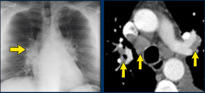 Sarcoidosis stage I: left and right hilar and paratracheal adenopathy (1-2-3 sign)Sarcoidosis giai đoạn I: hạch rốn phổi trái và phải và hạch cạnh khí quản (1-2-3 dấu hiệu)
Stages
Chest films in sarcoidosis have been classified into four stages:Giai đoạn
Phim chụp ngực trong bệnh sarcoid được phân thành bốn giai đoạn:
- Bilateral hilar lymphadenopathy Răng giả hai bên bệnh hạch
- Bilateral hilar lymphadenopath + pulmonary disease Bệnh hạch rốn gan hai bên + bệnh phổi
- Only pulmonary disease Chỉ bệnh phổi
- Irreversible fibrosisXơ hóa không hồi phục
These stages do not indicate disease chronicity or correlate with changes in pulmonary function.Những giai đoạn này không cho thấy bệnh mạn tính hoặc tương quan với những thay đổi về chức năng phổi.
Images
A patient with stage I disease.
There is hilar and paratracheal adenopathy and no sign of pulmonary involvement.Hình ảnh
Bệnh nhân mắc bệnh giai đoạn I.
Có hạch rốn phổi và hạch cạnh khí quản và không có dấu hiệu tổn thương phổi.
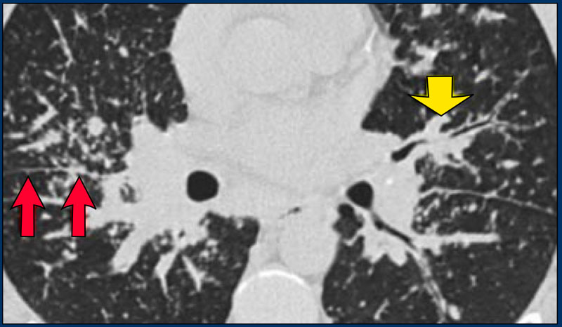 Sarcoidosis: typical presentation with nodules along the bronchovascular bundle and fthe issuresNotice the partially calcified node in the left hilum.Sarcoidosis: biểu hiện điển hình với các nốt dọc theo bó phế quản và các nốt sần Lưu ý nút vôi hóa một phần ở rốn trái.
HRCT findings in Sarcoidosis
Đặc điểm HRCT trong bệnh sarcoid
- Common findings:Các dấu hiệu thường gặp:
- Small nodules in a perilymphatic distribution (i.e. along subpleural surface and fissures, along interlobular septa and the peribronchovascular bundle).Các nốt nhỏ trong phân bố quanh bạch mạch (tức là dọc theo bề mặt dưới màng phổi và các vết nứt, dọc theo vách liên tiểu thùy và bó quanh mạch máu).
- Upper and middle zone predominance.Vùng trên và vùng giữa chiếm ưu thế.
- Lymphadenopathy in left hilus, right hilus and paratracheal (1-2-3 sign). Often with calcifications.Bệnh hạch ở rốn trái, rốn phải và cạnh khí quản (1-2-3 dấu). Thường có vôi hóa.
- Uncommon findings:Những phát hiện không phổ biến:
- Conglomerate masses in a perihilar location.Khối u kết tụ ở vị trí quanh rốn.
- Larger nodules (> 1cm in diameter, in Grouped nodules or coalescent nodlues surrounded by multiple satellite nodules (Galaxy sign) Các nốt lớn hơn (đường kính > 1 cm, trong các nốt nhóm hoặc nốt kết tụ được bao quanh bởi nhiều nốt vệ tinh (dấu hiệu Galaxy)
- Nodules so small and dense that they appear as ground glass or even as consolidations (alveolar sarcoidosis) Các nốt nhỏ và dày đặc đến mức chúng xuất hiện dưới dạng kính mờ hoặc thậm chí là các nốt đông đặc (sarcoidosis phế nang)
Image
A typical presentation of sarcoidosis with hilar lymphadenopathy and small nodules along bronchovascular bundles (yellow arrow) and along fissures (red arrows).Hình ảnh
Biểu hiện điển hình của bệnh sarcoid với hạch rốn phổi và các nốt nhỏ dọc theo các bó mạch phế quản (mũi tên màu vàng) và dọc theo vết nứt (mũa tên đỏ).
Image
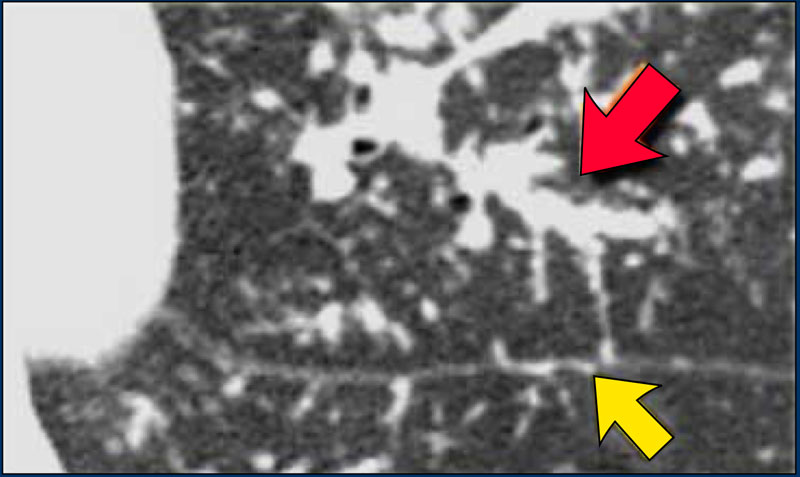
A detailed view with the typical HRCT-presentation with nodules along bronchovascular bundle (red arrow) and fissures (yellow arrow).
This is the typical perilymphatic distribution of the nodules.Hình ảnh
Hình ảnh chi tiết với HRCT điển hình với các nốt dọc theo bó mạch phế quản (mũi tên đỏ) và các vết nứt (mũa tên vàng).
Đây là sự phân bố quanh bạch mạch điển hình của các nốt.
 Sarcoidosis: typical presentationSarcoidosis: biểu hiện điển hình
The HRCT appearance of pulmonary sarcoidosis varies greatly and is known to mimic many other diffuse infiltrative lung diseases.
Approximately 60 to 70% of patients with sarcoidosis have characteristic radiologic findings.
In 25 to 30% of cases the radiologic findings are atypical.
In 5 to 10% of patients the chest radiograph is normal.
Sự xuất hiện HRCT của bệnh sarcoid phổi rất khác nhau và được biết là giống với nhiều bệnh phổi thâm nhiễm lan toả khác.
Khoảng 60 đến 70% bệnh nhân mắc bệnh sarcoid có các dấu hiệu đặc trưng trên X quang.
Trong 25 đến 30% trường hợp, các dấu hiệu X quang không điển hình.
Trong 5 đến 10% bệnh nhân, phim X-quang ngực là bình thường.
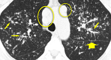
On the left another typical presentation of sarcoidosis with mediastinal lymphadenopathy and small nodules in a perilymphatic distribution along bronchovascular bundles and along fissures (yellow arrows).
Always look for small nodules along the fissures, because this is a very specific and typical sign of sarcoidosis.Ở bên trái, một biểu hiện điển hình khác của bệnh sarcoid với bệnh hạch trung thất và các nốt nhỏ trong phân bố quanh bạch mạch dọc theo các bó phế quản và dọc theo vết nứt (các mũi tên màu vàng).
Luôn tìm các nốt nhỏ dọc theo các vết nứt, bởi vì đây là một dấu hiệu rất đặc hiệu và điển hình của bệnh sarcoid.
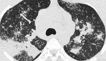 Sarcoidosis with conglomerate masses of fibrous tissueSarcoidosis với khối kết tụ của mô xơ
Fibrosis in Sarcoidosis.
Progressive fibrosis in sarcoidosis may lead to peribronchovascular (perihilar) conglomerate masses of fibrous tissue.
The typical location is posteriorly in the upper lobes, leading to volume loss of the upper lobes with displacement of the interlobar fissure.
Xơ hóa trong bệnh Sarcoid.
Xơ hóa tiến triển trong bệnh sarcoid có thể dẫn đến các khối kết tụ quanh cuống mạch (perihilar) của mô xơ.
Vị trí điển hình là ở phía sau thuỳ trên, dẫn đến mất thể tích thuỳ trên với sự dịch chuyển của rãnh liên thuỳ.
Other diseases that commonly result in this appearance are:Các bệnh khác thường dẫn đến sự xuất hiện này là:
- SilicosisBệnh bụi phổi silic
- TuberculosisBệnh lao
- TalcosisBệnh nấm men
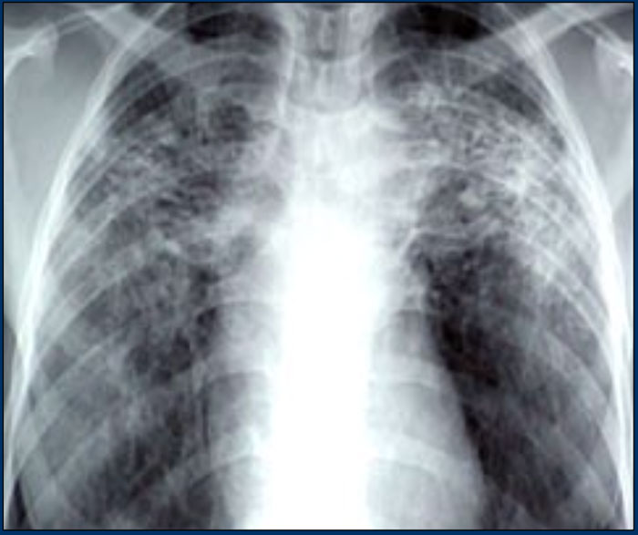 Sarcoidosis with fibrosis in the upper lobes. Typical chest film.Sarcoidosis với xơ hóa ở thuỳ trên. Điển hình phim ngực.
Here a typical chest film of long standing sarcoidosis (stage IV) with fibrosis in the upper zones and volume loss of the upper lobes resulting in hilar elevation.
Fibrosis results in obliteration of pulmonary vessels, which can lead to pulmonary hypertension.
Ở đây là một phim ngực điển hình của bệnh sarcoid kéo dài (giai đoạn IV) với xơ hóa ở các vùng trên và mất thể tích của các thuỳ trên dẫn đến tăng độ cao của rốn gan.
Xơ hóa dẫn đến sự tắc nghẽn các mạch máu phổi, có thể dẫn đến tăng áp phổi.
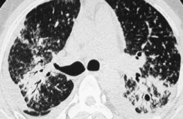 Sarcoidosis with fibrosis in the upper lobes. Typical HRCT findings.Sarcoidosis với xơ hóa ở thuỳ trên. HRCT điển hình.
Here another case of stage IV sarcoidosis.
Notice the distribution of the conglomerate masses of fibrosis in the posterior part of the lungs.
In addition there are multiple small well-defined nodules.
Some of these nodules have the typical subpleural distribution.Đây là một trường hợp khác của bệnh sarcoid giai đoạn IV.
Lưu ý sự phân bố của các khối kết tụ của xơ hóa ở phần sau của phổi.
Ngoài ra còn có nhiều nốt nhỏ được xác định rõ.
Một số nốt này có phân bố dưới màng phổi điển hình.
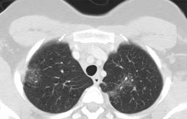
Alveolar Sarcoidosis.
Sarcoidosis phế nang.
This is a case of alveolar sarcoidosis.
Scroll through the images.Đây là một trường hợp bệnh lý sarcoid phế nang.
Cuộn qua các hình ảnh.
The appearance resembles a ground glass attenuation, but with a closer look you may appreciate that the increased attenuation is the result of many tiny grouped nodules.
Also notice the hilar lymphadenopathy.Hình dạng giống như sự suy giảm kính mờ, nhưng với một cái nhìn gần hơn, bạn có thể đánh giá cao rằng sự suy giảm tăng lên là kết quả của nhiều nốt nhỏ được nhóm lại.
Cũng lưu ý đến rốn gan bệnh hạch.
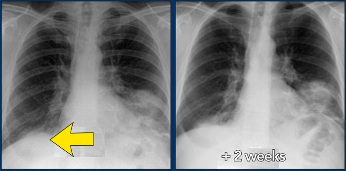
 47-year old female patient treated for possible infectionBệnh nhân nữ 47 tuổi, điều trị nhiễm trùng
Alveolar Sarcoidosis (2)
On the left a 47-year old female patient with a dry cough, slightly breathless and a normal blood analysis.
A chest film was taken and she was treated with antibiotics.
A follow up film was made, because she did not improve.Bệnh sarcoid phế nang (2)
Bên trái là một bệnh nhân nữ 47 tuổi bị ho khan, hơi khó thở và xét nghiệm máu bình thường.
Bệnh nhân được điều trị bằng kháng sinh với phim ngực.
Một bộ phim tiếp theo đã được thực hiện, bởi vì cô ấy không cải thiện.
The first chest film shows bilateral consolidations in the lower lobes (arrow), initially interpreted as infection.
After two weeks of treatment with antibiotics, there is no improvement.
The differential diagnosis now includes tumor (bronchoalveolar carcinoma or lymphoma), eosinophilic pneumonia , organizing pneumonia, Wegener's disease or an uncommon presentation of sarcoidosis.
Now continue with the HRCT.Phim ngực đầu tiên cho thấy sự hợp nhất hai bên ở các thùy dưới (mũi tên), ban đầu được hiểu là nhiễm trùng.
Sau hai tuần điều trị bằng kháng sinh, không có cải thiện.
chẩn đoán phân biệt hiện bao gồm khối u (ung thư biểu mô phế quản hoặc ung thư hạch), viêm phổi bạch cầu ái toan, tổ chức viêm phổi, bệnh Wegener hoặc biểu hiện bệnh sarcoidosis không phổ biến.
Bây giờ tiếp tục với HRCT.
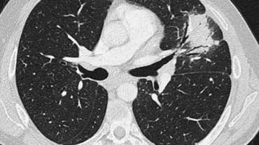
Scroll through the images on the left .
There are multiple areas of consolidation.
Ancillary findings are hilar and mediastinal lymphadenopathy.
Cuộn qua các hình ảnh bên trái.
Có nhiều vùng của đông đặc.
Các dấu hiệu phụ là bệnh hạch rốn phổi và trung thất.
The differental diagnosis of the CT-images is basically the same as of the chest film.
Histology revealed alveolar sarcoid.
There is only one clue to the diagnosis and that is the presence of small nodules that can be identified in image 3, but these are difficult to see.
This case nicely demonstrates that sarcoidosis truely is 'the great mimicker'.
Sarcoidosis should be therefore in our differential diagnostic list!.
Chẩn đoán hình ảnh CT khác nhau về cơ bản giống như phim ngực.
Mô bệnh học cho thấy u hạt dạng nang.
Chỉ có một manh mối để chẩn đoán và đó là sự hiện diện của các nốt nhỏ có thể được xác định trong hình 3, nhưng những nốt này rất khó nhìn thấy.
Trường hợp này chứng minh một cách độc đáo rằng bệnh sarcoid thực sự là 'kẻ bắt chước tuyệt vời'.
Sarcoidosis nên được đưa vào danh sách chẩn đoán phân biệt của chúng tôi!.
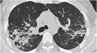
On the left a case of fibrosing sarcoidosis, showing fibrosis, traction bronchiectases and crowding of the involved bronchi, predominantly in the perihilar region and upper lobes.
Nodular abnormalities are absent, but the appearance and the location of the fibrosis are very suggestive of the diagnosis of sarcoidosis.Ở bên trái một trường hợp xơ hóa sarcoidosis, cho thấy xơ hóa, giãn phế quản kéo và đông tụ phế quản liên quan, chủ yếu ở vùng quanh rốn phổi và thuỳ trên.
Không có bất thường dạng nốt, nhưng sự xuất hiện và vị trí của xơ hóa rất gợi ý chẩn đoán bệnh sarcoid.
Differential diagnosis of Sarcoidosis. Chẩn đoán phân biệt của bệnh Sarcoid.
- Lymphadenopathy:Bệnh hạch:
- Primary TB: asymmetrical adenopathyLao nguyên phát: hạch không đối xứng
- HistoplasmosisNhiễm nấm Histoplasma
- Lymphoma
- Small cell lung cancer with nodal metastasesUng thư phổi tế bào nhỏ di căn hạch
- Nodular pattern:Mẫu nốt:
- Silicosis / Pneumoconiosis: predominantly centrilobular and subpleural nodules.Bệnh bụi phổi silic / bụi phổi con: chủ yếu là các nốt trung tâm tiểu thùy và dưới màng phổi.
- Miliary TB: random nodules.TB từng phần: các nốt ngẫu nhiên.
- Fibrotic pattern:Mô hình xơ hóa:
- Usual Interstitial Pneumonia (UIP): basal and peripheral fibrosis, honeycombing.Viêm phổi kẽ thông thường (UIP) (UIP): xơ hóa nền và xơ hóa ngoại vi, tổ ong.
- Chronic Hypersensitivity Pneumonitis: mid zone fibrosis with mosaic pattern.Viêm phổi quá mẫn mạn tính: xơ hóa vùng giữa với hình thái khảm.
- Tuberculosis (more unilateral). Bệnh lao (một bên).
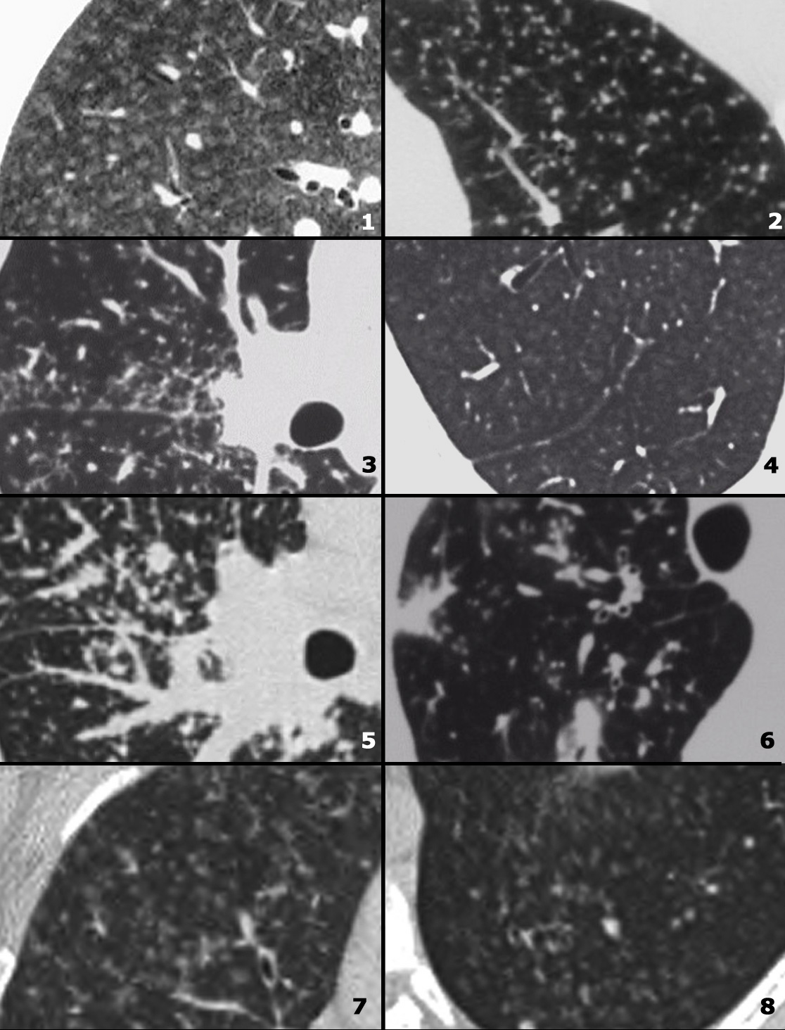
On the left some diseases with a nodular pattern.Bên trái một số bệnh có dạng nốt.
- Hypersensitivity pneumonitis: ill defined centrilobular nodules. Viêm phổi quá mẫn: nốt trung tâm tiểu thùy không rõ.
- Miliary TB: random nodules of the same size. Miliary TB: các nốt ngẫu nhiên có cùng kích thước.
- Sarcoidosis: nodules with perilymphatic distribution, along fissures, adenopathy. Sarcoid: nốt có phân bố quanh bạch mạch, dọc theo các khe nứt, hạch.
- Hypersensitivity pneumonitis: centrilobular nodules, notice sparing of the subpleural area. Viêm phổi quá mẫn: Nốt trung tâm tiểu thùy, lưu ý đến diện tích dưới màng phổi.
- Sarcoidosis: nodules with perilymphatic distribution, along fissures, adenopathy. Sarcoid: nốt có phân bố quanh bạch mạch, dọc theo các khe nứt, hạch.
- TB: Tree-in-bud appearance in a patient with active TB. TB: nốt mầm cây (tree-in-bud) xuất hiện ở một bệnh nhân lao hoạt động.
- Langerhans cell histiocytosis: early nodular stage before the typical cysts appear. Bệnh mô bào Langerhans: giai đoạn nốt sớm trước khi xuất hiện nang điển hình.
- Respiratory bronchiolitis: ill defined centrilobular nodules of ground-glass opacity.Viêm tiểu phế quản hô hấp: Nốt trung tâm tiểu thùy không rõ của đám mờ kính mờ.
Silicosis / Coal worker pneumoconiosisBệnh bụi phổi silic / Bệnh bụi phổI công nhân than
Silicosis and Coal worker pneumoconiosis (CWP) are pathologically distinct entities with differing histology, resulting from the inhalation of different inorganic dusts.
The radiographic and HRCT appearances of these diseases, however, may not be distinguishable from each other and may be similar to sarcoidosis.
It is important to realize that these diseases are rare compared to sarcoidosis.
Silicosis and CWP occur in a specific patient group (construction workers, mining workers, workers exposed to sandblasting, glass blowing and pottery).
Bệnh bụi phổi silic và bệnh bụi phổi công nhân than (CWP) là những thực thể bệnh lý khác biệt với mô học khác nhau, do hít phải các loại bụi vô cơ khác nhau.
Tuy nhiên, hình ảnh X quang và HRCT của các bệnh này có thể không phân biệt được với nhau và có thể tương tự như bệnh sarcoid.
Điều quan trọng là phải nhận ra rằng những bệnh này hiếm gặp so với bệnh sarcoid.
Bệnh bụi phổi silic và CWP xảy ra trong một nhóm bệnh nhân cụ thể (công nhân xây dựng, công nhân khai thác mỏ, công nhân tiếp xúc với phun cát, thổi thủy tinh và làm gốm).
HRCT findings in Silicosis/CWP
Đặc điểm HRCT ở bệnh nhân suy tuyến yên
- Small well-defined nodules of 2 to 5mm in diameter in both lungs.Các nốt nhỏ được xác định rõ đường kính từ 2 đến 5 mm ở cả hai phổi.
- Upper lobe predominanceThuỳ trên chiếm ưu thế
- Nodules may be calcifiedNốt có thể bị vôi hóa
- Centrilobular and subpleural distribution Phân bố trung tâm tiểu thùy và dưới màng phổi
- Sometimes random distributionĐôi khi phân bố ngẫu nhiên
- Irregular conglomerate masses, known as progressive massive fibrosisKhối u kết tụ không đều, được gọi là xơ hóa khối tiến triển
- Masses may cavitate due to ischemic necrosis.Các khối có thể bị xâm lấn do hoại tử do thiếu máu cục bộ.
- Often hilar and mediastinal lymphnodes.Thường là hạch rốn phổi và trung thất.
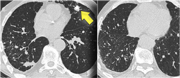
On the left a case of silicosis showing nodules of varying sizes with a random and subpleural distribution. One nodule contains calcification (arrow). Note the absence of a lymphatic distribution pattern (peribronchovascular and along fissures), which would be suggestive of sarcoidosis.Bên trái là một trường hợp bệnh bụi phổi silic cho thấy các nốt có kích thước khác nhau với sự phân bố ngẫu nhiên và dưới màng phổi. Một nốt chứa vôi hóa (mũi tên). Lưu ý rằng không có một mô hình phân bố bạch huyết (peribronchovascular và dọc theo các vết nứt), điều này sẽ gợi ý bệnh sarcoidosis.
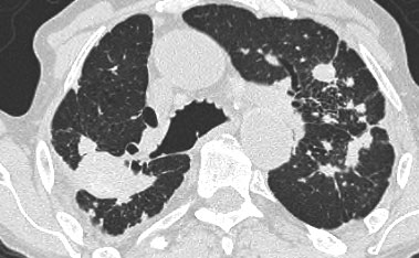 Same patient with silicosis as previous images showing a conglomerate mass in a perihilar location in the right upper lobe. The left lobe shows multiple nodules of varying size.Cùng một bệnh nhân bị bệnh bụi phổi silic như các hình ảnh trước đây cho thấy một khối kết tụ ở vị trí quanh rốn ở thuỳ trên bên phải Thuỳ trái cho thấy nhiều nốt có kích thước khác nhau.
Differential diagnosis of Silicosis / Pneumoconiosis. Chẩn đoán phân biệt của bệnh bụi phổi silic / viêm phổi liên cầu.
- Sarcoidosis : can be difficult to distinguish (look for distribution of nodules).Sarcoidosis: có thể khó phân biệt (tìm kiếm sự phân bố của các nốt).
- Infection: miliairy TB, fungus.Nhiễm trùng: lao, nấm.
- Hematogenous metastases: silicotic nodules in subpleural and peribronchiolar location up to the level of the secundary pulmonary lobule, may have a seemingly random distribution and simulate metastases and miliary infections.Di căn tạo máu: các nốt silic ở dưới màng phổi và vị trí quanh phế quản đến mức thuỳ phổi, có thể có sự phân bố dường như ngẫu nhiên và mô phỏng di căn và nhiễm trùng kê.
- Langerhans cell histiocytosis: can be difficult to distinguish from silicosis in the early stage, when LCH is solely characterized by the presence of small nodules. Look for nodules with cavitation. Bệnh mô bào Langerhans: có thể khó phân biệt với bệnh bụi phổi silic trong giai đoạn đầu, khi LCH chỉ được đặc trưng bởi sự hiện diện của các nốt nhỏ. Tìm các nốt có cavitation.
Lymphangitic CarcinomatosisUng thư biểu mô bạch huyết

Lymphangitic Carcinomatosis results from hematogenous spread to the lung, with subsequent invasion of interstitium and lymphatics.
The presenting symptoms are dyspnea and cough and can predate the radiographic abnormalities.
In many cases however the patients are asymptomatic.
Lymphangitic Carcinomatosis is seen in carcinoma of the lung, breast, stomach, pancreas, prostate, cervix, thyroid and metastatic adenocarcinoma from an unknown primary. Ung thư biểu mô bạch huyết là kết quả của sự lan truyền theo đường máu đến phổi, với sự xâm lấn tiếp theo của mô kẽ và bạch huyết.
Các triệu chứng hiện tại là khó thở và ho và có thể xuất hiện trước các bất thường trên X-quang.
Tuy nhiên, trong nhiều trường hợp, bệnh nhân không có triệu chứng.
Ung thư biểu mô bạch huyết được thấy trong ung thư biểu mô phổi, vú, dạ dày, tuyến tụy, tuyến tiền liệt, cổ tử cung, tuyến giáp và di căn ung thư biểu mô tuyến từ một nguyên phát không rõ.
HRCT findings in Lymphangitic Carcinomatosis
Đặc điểm HRCT trong ung thư biểu mô tuyến bạch huyết
- Interlobular septal thickening, thickening of fissures and thickening of the peribronchovascular interstitium (bronchial cuffing).Dày vách liên tiểu thùy, dày lên các vết nứt và dày lên các khoảng kẽ quanh cuống mạch (cuffing phế quản).
- Depending on filling with fluid or with tumor cells, septal thickening is irregular or smooth.Tuỳ thuộc vào việc đổ đầy dịch hoặc với tế bào khối u, dày vách không đều hoặc nhẵn.
- Focal or unilateral abnormalities in 50% of patients.Bất thường khu trú hoặc một bên ở 50% bệnh nhân.
- Hilar lymphadenopathy in 50% of patients.Hilar bệnh hạch ở 50% bệnh nhân.
- Pleural effusion due to pleuritic carcinomatosis ( > 50% of patients). Tràn dịch màng phổi do ung thư biểu mô màng phổi (> 50% bệnh nhân).
Image
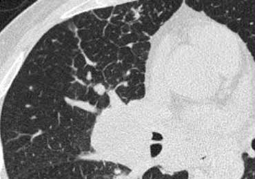
A patient with Lymphangitic Carcinomatosis.
Notice the focal distribution.
This finding is helpful in distinguishing Lymphangitic Carcinomatosis from other causes of interlobular septal thickening like pulmonary edema or sarcoid.
There is also lymphadenopathy.Hình ảnh
Một bệnh nhân bị ung thư biểu mô bạch huyết.
Chú ý sự phân bố khu trú.
Phát hiện này rất hữu ích trong việc phân biệt ung thư biểu mô bạch huyết với các nguyên nhân khác của dày vách liên tiểu thùy như phù phổi hoặc sarcoid.
Ngoài ra còn có bệnh hạch.
Image
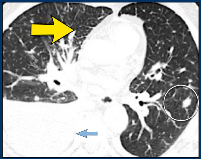
Another patient with Lymphangitic Carcinomatosis with interlobular septal thickening (yellow arrow).
Additional pleural fluid (blue arrow) and a lung metastasis (circle).Hình ảnh
Một bệnh nhân khác bị ung thư biểu mô bạch huyết với dày vách liên tiểu thùy (mũi tên màu vàng).
Dịch màng phổi (mũi tên màu xanh) và di căn phổi (vòng tròn).
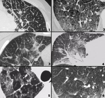
Examples of Lymphangitic carcinomatosis and differential diagnosis
Ví dụ về ung thư biểu mô bạch huyết và chẩn đoán phân biệt
- Lymphangitic carcinomatosis: irregular septal thickening, usually focal or unilateral, in 50% adenopathy, known carcinoma. Ung thư biểu mô bạch huyết: dày vách không đều, thường là khu trú hoặc một bên, trong 50% bệnh lý hạch, ung thư biểu mô đã biết.
- Cardiogenic pulmonary edema: bilateral abnormalities, filling of alveoli, enlarged heart, rapid response to diuretics, ground-glass opacity due to filling of alveoli with fluid, gravitational distribution of the alveolar fluid. Phù phổi do tim: bất thường hai bên, lấp đầy phế nang, tim to, đáp ứng nhanh với thuốc lợi tiểu, đám mờ kính mờ do lấp kín phế nang bằng dịch, phân bố hấp dẫn dịch phế nang.
- Lymphangitic carcinomatosis. Ung thư biểu mô bạch huyết.
- Lymphangitic carcinomatosis with hilar adenopathy and thickening of the central bronchovascular interstitium. Ung thư biểu mô bạch huyết với hạch rốn phổi và dày mô kẽ phế quản trung tâm.
- Alveolar proteinosis: sharply demarcated secondary lobeles with ground glass attenuation as opposed to secondary lobules with normal aeration, superimposed inter and intralobular septal thickening (crazy paving). Protein phế nang: các thuỳ thứ phát có ranh giới rõ với sự suy giảm kính mờ so với các thuỳ có thông khí bình thường, chồng chất dày vách (lát đá điên (crazy paving)) giữa và trong thuỳ.
- Cardiogenic pulmonary edema.Phù phổi do tim.
Cardiogenic pulmonary edemaPhù phổi do tim

Patients with pulmonary edema are not imaged with HRCT as their diagnosis is usually based on a combination of clinical and chest radiographic findings.
However sometimes the diagnosis is not that straightforward and knowledge of the HRCT appearance of pulmonary edema can be helpful in avoiding misdiagnosis.Bệnh nhân bị phù phổi không được chụp HRCT vì chẩn đoán của họ thường dựa trên sự kết hợp của các dấu hiệu lâm sàng và X quang ngực.
Tuy nhiên, đôi khi chẩn đoán không đơn giản và kiến thức về HRCT phù phổi có thể hữu ích trong việc tránh chẩn định sai.
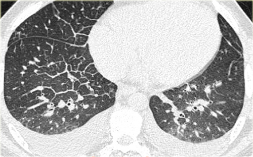 Cardiogenic pulmonary edemaPhù phổi do tim
HRCT findings in cardiogenic pulmonary edema
Đặc điểm hình ảnh HRCT trong phù phổi do tim
- Bilateral septal thickening and ground-glass opacity.Dày vách và đám mờ kính mờ hai bên.
- Perihilar and gravitational distribution predominatly in the dependent lung.Phân bố quanh rốn phổi và hấp dẫn chủ yếu ở phổi phụ thuộc.
- Cardiomegaly and pleural fluid. Tim to và dịch màng phổi.
On the left typical features of cardiogenic pulmonary edema
There is smooth septal thickening and some ground glass opacity in the dependent part of the lungs.
In addition there is bilateral pleural fluid.
In a patient with a known malignancy lymphangitic carcinomatosis would be high in the differential diagnostic list.Đặc điểm bên trái của phù phổi do tim
Có dày vách trơn và một số đám mờ kính mờ ở phần phụ thuộc của phổi.
Ngoài ra còn có dịch màng phổi hai bên.
Ở bệnh nhân có carcinôm tế bào lymphô ác tính đã biết sẽ có tỷ lệ cao trong danh sách chẩn đoán phân biệt.
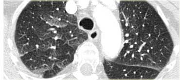 Cardiogenic pulmonary edemaPhù phổi do tim
Differential diagnosis of cardiogenic pulmonary edema. Chẩn đoán phân biệt của phù phổi do tim.
- Lymphangitic carcinomatosisUng thư biểu mô bạch huyết
- Interstitial pneumonia (viral, mycoplasma)Viêm phổi kẽ (virus, mycoplasma)
- ARDSARDSComment
- Pulmonary hemorrhage Xuất huyết phổi
On the left another example of cardiogenic pulmonary edema.
This patient had a CT to rule out pulmonary emboli.
There is smooth septal thickening and ground glass opacity in a more patchy distribution.
Note: edema can have a very unusual appearance and be distributed very patchy: some areas are filled with fluid as opposed to other areas in immediate vicinity which appear normal.
Bên trái là một ví dụ khác về phù phổi do tim.
Bệnh nhân này được chụp CT để loại trừ thuyên tắc phổi.
Có dày vách và đám mờ kính mờ mịn trong một phân bố chắp vá hơn.
Lưu ý: phù có thể có hình dạng rất bất thường và phân bố rất rải rác: một số khu vực chứa đầy dịch trái ngược với các khu vực khác trong vùng lân cận có vẻ bình thường.
Hypersensitivity PneumonitisViêm phổi quá mẫn

Hypersensitivity pneumonitis (HP) is also known as extrinsic allergic alveolitis (EAA).
HP is an allergic lung disease caused by the inhalation of a variety of antigens (farmer's lung, bird fancier's lung, 'hot tub' lung, humidifier lung).
The radiographic and pathologic abnormalities in patients can be classified into acute, subacute, and chronic stages.
Mostly HRCT is performed in the subacute stage of HP, weeks to months following the first exposure to the antigen or in the chronic phase.Viêm phổi quá mẫn (HP) còn được gọi là viêm phế nang dị ứng ngoại sinh (EAA).
HP là bệnh phổi dị ứng do hít phải nhiều loại kháng nguyên (phổi người nông dân, phổi chim, phổi bồn tắm nóng, phổi máy tạo ẩm).
Các bất thường về X quang và bệnh lý ở bệnh nhân có thể được phân loại thành các giai đoạn cấp tính, bán cấp và mạn tính.
Hầu hết HRCT được thực hiện trong giai đoạn bán cấp của HP, vài tuần đến vài tháng sau khi tiếp xúc với kháng nguyên đầu tiên hoặc trong giai đoạn mạn tính.
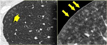 Subacute hypersensitivity pneumonitis with ill-defined centrilobular nodulesNốt viêm phổi quá mẫn bán cấp với nốt bờ không rõ trung tâm tiểu thùy
Subacute hypersensitivity pneumonitis
The key findings in the subacute hypersensitivity pneumonitis are:Viêm phổi quá mẫn bán cấp
Các dấu hiệu chính trong bán cấp viêm phổi quá mẫn là:
- ill-defined centrilobular nodules of ground-glass opacity (80% of cases) orNốt bờ không rõ trung tâm tiểu thùy của đám mờ kính mờ (80% trường hợp) hoặc
- mosaic pattern of a combination of patchy ground-glass opacity due to lung infiltration and patchy lucency due to bronchiolitis with air trappingmô hình khảm của sự kết hợp đám mờ kính mờ loang lổ do thâm nhiễm phổi và loang lơ do viêm tiểu phế quản với bẫy khí
Here two examples of subacute hypersensitivity pneumonitis.
There are ill-defined centrilobular nodules of ground-glass opacity.Dưới đây là hai ví dụ về bán cấp viêm phổi quá mẫn.
Có các nốt bờ không rõ trung tâm tiểu thùy của đám mờ kính mờ.
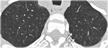
Here another case of subacute hypersensitivity pneumonitis.
There is subtle opacity in the centre of the secondary lobules (arrows) with sparing of the subpleural region.Đây là một trường hợp bán cấp viêm phổi quá mẫn.
Có sự mờ đục tinh tế ở trung tâm của thuỳ thứ cấp (các mũi tên) với sự tiết kiệm của vùng dưới màng phổi.
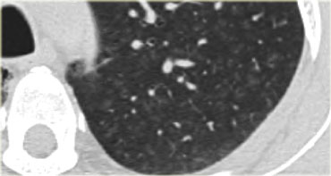
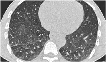
This HRCT-image also demonstrates subtle centrilobular opacity in a patient with subacute HP.
Notice how ill-defined these centrilobular nodules are.Hình ảnh HRCT này cũng cho thấy độ mờ đục trung tâm tiểu thùy tinh tế ở một bệnh nhân HP bán cấp.
Lưu ý bờ không rõ như thế nào các nốt trung tâm tiểu thùy này.
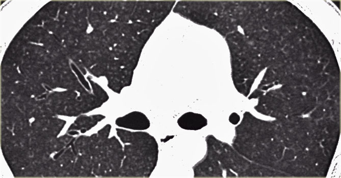
Sometimes the centrilobular opacities are more nodular in appearance as in this case.Đôi khi các nốt mờ trung tâm tiểu thùy có hình dạng nốt hơn như trong trường hợp này.
 Subacute hypersensitivity pneumonitis with mosaic patternViêm phổi quá mẫn bán cấp với mô hình khảm
Here another case of hypersensitivity pneumonitis.
There is a mosaic pattern.
Some secundary lobules demonstrate ground-glass opacity due to lung infiltration, while others are more lucent due to bronchiolitis with air trapping.Đây là một trường hợp khác của viêm phổi quá mẫn.
Có một mô hình khảm.
Một số tiểu thuỳ thể mở biểu hiện đám mờ kính mờ do thâm nhiễm phổi, trong khi một số khác thì sáng hơn do viêm tiểu phế quản với bẫy khí.
 LEFT: HRCT at presentation. RIGHT: HRCT ten days laterBên trái: HRCT khi trình bày. Phải: HRCT mười ngày sau
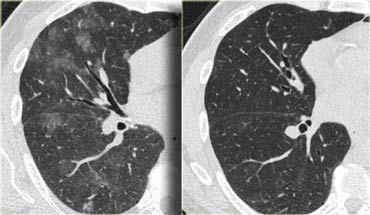
This patient presented with acute dyspnoe and a normal chest film (not shown).
The HRCT at presentation (left) shows lobular areas of ground glass attenuation.
A control HRCT ten days later (right) demonstrated, that the findings had resolved without any treatment.
The findings were thought to be due to hypersensitivity pneumonitis.Bệnh nhân này có triệu chứng khó thở cấp và phim ngực bình thường (không có trên phim).
HRCT ở hình ảnh (trái) cho thấy các vùng thuỳ của sự suy giảm kính mờ.
Một HRCT đối chứng mười ngày sau đó (phải) chứng minh, rằng những phát hiện đã được giải quyết mà không cần điều trị.
Các kết quả được cho là do viêm phổi quá mẫn.
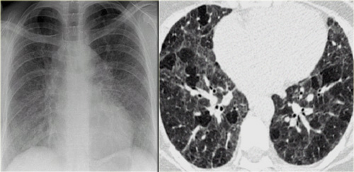 Chronic hypersensitivity pneumonitis: Nonspecific chest film, typical HRCT-findings.Viêm phổi quá mẫn mạn tính: phim ngực không đặc hiệu, HRCT-tìm thấy điển hình.
Chronic hypersensitivity pneumonitis
The key findings in chronic hypersensitivity pneumonitis are:Viêm phổi quá mẫn mạn tính
Các dấu hiệu chính trong viêm phổi quá mẫn mạn tính là:
- Mosaic pattern with areas of ground-glass atenuation and areas of low attenuation. Mô hình khảm với các vùng suy giảm kính mờ và các vùng suy hao thấp.
- Fibrosis and parenchymal distortion in a mid zone distribution.Xơ hóa và biến dạng nhu mô ở vùng giữa.
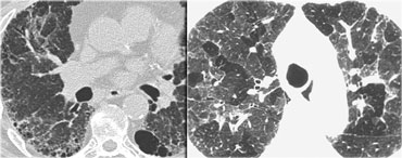
On the left a patient with chronic hypersensitivity pneumonitis.
The HRCT shows a mosaic pattern with hyperaerated secondary nodules and secondry nodules of increased attenuation.
Additionally there is septal and intralobular reticular thickening, indicating already existing irreversible fibrosis.Bên trái là bệnh nhân có viêm phổi quá mẫn mạn tính.
HRCT cho thấy một mô hình khảm với các nốt thứ phát tăng khí hóa và các nốt tăng độ đậm độ.
Ngoài ra còn có sự dày lên của vách ngăn và mạng lưới trong thuỳ, cho thấy tình trạng xơ hóa không hồi phục đã tồn tại.
 UIP with honeycombing (left) and chronic HP (right)UIP với tổ ong (trái) và HP mạn tính (phải)
Differential diagnosis of Hypersensitivity Pneumonitis. Chẩn đoán phân biệt của viêm phổi quá mẫn.
-
Subacute stage:Giai đoạn bán cấp:
- RB-ILD: seen in smokers, upper lobe predilection, usually associated with centrilobular emphysema.RB-ILD: gặp ở người hút thuốc lá, khuynh hướng thuỳ trên, thường liên quan đến trung tâm tiểu thùy khí phế thũng.
- Alveolar proteinosis.Protein phế nang.
-
Chronic stage:Giai đoạn mạn tính:
-
UIP: may show very similar HRCT findings.
UIP has a strong lower zone distribution.
In chronic HP fibrotic changes are typically seen throughout the whole lung parenchyma from the periphery towards the centrum. UIP: có thể cho thấy những kết quả HRCT rất giống nhau.
UIP có phân bố vùng dưới mạnh.
Trong HP mạn tính, những thay đổi xơ hóa thường thấy trên toàn bộ nhu mô phổi từ ngoại vi về phía trung tâm.
-
UIP: may show very similar HRCT findings.
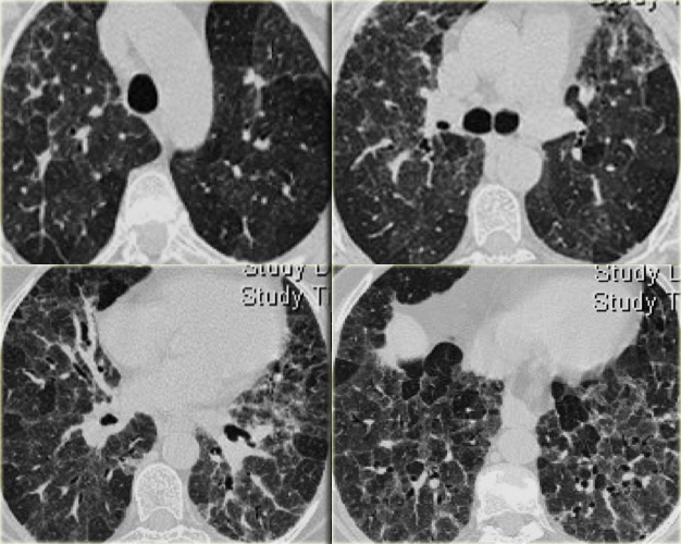 Chronic hypersensitivity pneumonitis.Viêm phổi quá mẫn mạn tính.
Chronic hypersensitivity pneumonitis (2)
The case on the left shows an inspiratory and expiratory scan: the mosaic pattern with areas of ground-glass attenuation and areas of low attenuation, that become more evident on the expiratory scan, indicating air trapping.
Signs of fibrosis such as distorted vessels and bronchi as well as septal thickening are more pronounced in the mid and lower lung zones, but not limited to the subpleural area.Viêm phổi quá mẫn mạn tính (2)
Trường hợp bên trái cho thấy hình ảnh chụp XQ và XQ: mô hình khảm với các vùng suy giảm kính mờ và các vùng suy yếu thấp, trở nên rõ ràng hơn trên XQ, cho thấy bẫy khí.
Các dấu hiệu xơ hóa như mạch máu và phế quản bị biến dạng cũng như dày vách rõ rệt hơn ở vùng giữa và dưới phổi, nhưng không giới hạn ở vùng dưới màng phổi.
The images on the left suggest the diagnosis hypersensitivity pneumonitis.
Based on the imaging findigs alone, alveolar proteinosis and other diseases with a mozaic pattern should be included in the differential diagnosis.Hình ảnh bên trái gợi ý chẩn đoán viêm phổi quá mẫn.
Dựa trên các phát hiện hình ảnh đơn thuần, protein phế nang và các bệnh khác có dạng mozaic nên được đưa vào chẩn đoán phân biệt.
TuberculosisBệnh lao
Primary TB:
Initial infection with consolidation, adenopathy and pleural effusion.
Secondary TB :
Post-primary or reactivation TB.
This is the reactivation of the original infection.
Usually located in the apical segments of upper lobes with cavitation
Endobronchial spread: May occur in both primary and secondary TB, when the infection is not contained.
Hematogenous spread (miliary TB): May occur in both primary and secondary TB, when the infection is not contained.Bệnh lao nguyên phát: Nhiễm trùng ban đầu với đông đặc, hạch và tràn dịch màng phổi.
Lao thứ phát: Lao sau nguyên phát hoặc tái hoạt.
Đây là sự tái hoạt của nhiễm trùng ban đầu.
Thường nằm ở các đoạn đỉnh của thuỳ trên có tạo hang
Sự lây lan nội phế quản: Có thể xảy ra ở cả lao nguyên phát và thứ phát, khi nhiễm trùng không được kiểm soát.
Sự lây lan theo đường máu (bệnh lao quen thuộc): Có thể xảy ra ở cả bệnh lao nguyên phát và thứ phát, khi nhiễm trùng không được kiểm soát.
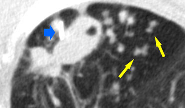 TB: cavitating lesion and endobronchial spreadLao: tổn thương dạng hang và lan rộng nội phế quản
HRCT findings in TB Đặc điểm HRCT ở bệnh nhân lao
-
Primary TB: Lao nguyên phát:
- Consolidation anywhere, lymphadenopathy and pleural effusion. Usually regresses to calcified lung nodule and calcified ipsilateral lymph node.Đông đặc bất cứ đâu, bệnh hạch và tràn dịch màng phổi. Thường thóai triển thành nốt vôi hóa phổi và vôi hóa cùng bên hạch bạch huyết.
- Secondary TB: Lao thứ phát:
- Consolidation in apical segments of upper lobes or superior segments of lower lobes. Đông đặc ở các đoạn đỉnh thuỳ trên hoặc các đoạn trên thuỳ dưới.
- CavitationTạo hang
- Endobronchial spread: Sự lan rộng nội phế quản:
- Tree in bud appearanceCây mọc chồi
- Miliary TB: Lao thể nhẹ:
- 2-3 mm nodules with random disrtibution.Nốt 2-3 mm với kích thước ngẫu nhiên.
On the left a patient with TB. There is a cavitating lesion and typical tree-in-bud appearance. The blue arrow indicates the biopsy needle.Bên trái là bệnh nhân lao, có tổn thương dạng hang và hình dạng nốt mầm cây (tree-in-bud) điển hình, mũi tên màu xanh dương chỉ kim sinh thiết.
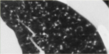 Miliary TBTB Miliary
Miliary TB
This represents a hematogenous dissemination of infection and may occur in association with either primary or postprimary disease.
It is characterized by uniform small nodules with a random distribution.
TB Miliary
Điều này thể hiện sự lan truyền nhiễm trùng theo đường máu và có thể xảy ra liên quan đến bệnh nguyên phát hoặc sau nguyên phát.
Nó được đặc trưng bởi các nốt nhỏ đồng nhất với sự phân bố ngẫu nhiên.
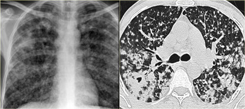 TB with cavitationLao có xâm lấn
Cavitation in TB
The chest film on the left shows diffuse areas with nodular air space opacifications.
The HRCT demonstrates multiple nodules in peribronchial distribution, partially confluent, and a cavitation in the right lung, strongly suggestive for tuberculosis.
Other diseases in the differential are Wegener granulomatosis or malignancy (both show no tree-in-bud).Cavitation trong lao
phim ngực bên trái cho thấy các vùng lan toả với các nốt mờ trong không gian khí.
HRCT cho thấy nhiều nốt trong phân bố quanh phế quản, hợp nhất một phần, và một lỗ thông ở phổi phải, gợi ý mạnh mẽ về bệnh lao.
Các bệnh khác trong phân biệt là U hạt Wegener hoặc ác tính (cả hai đều không có nốt mầm cây (tree-in-bud)).
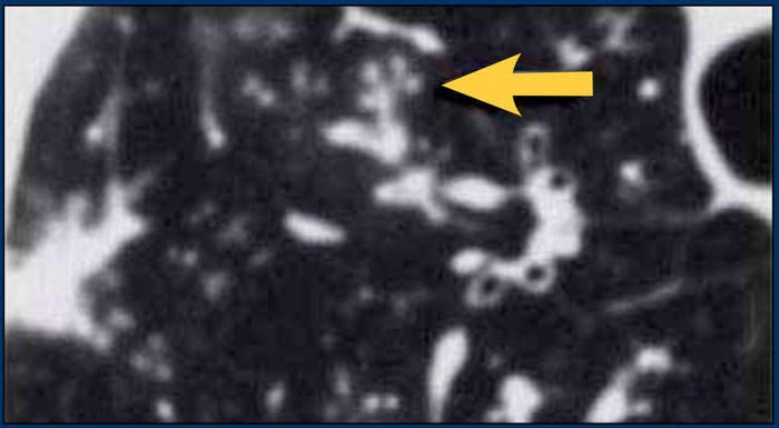 Endobronchial spread of TB with tree-in-budSự lan rộng nội phế quản của lao với nốt mầm cây (tree-in-bud)
Endobronchial spread of TB
This can occur with primary or postprimary infection.
In most subjects, the primary infection is localized and clinically inapparent.
However, in 5 to 10% of patients with primary TB, the infection is poorly contained and dissemination occurs.
This is termed progressive primary tuberculosis.
Extensive cavitation of the tuberculous pneumonia lead to endobronchial spread of the infection.
Rupture of necrotic lymph nodes into the bronchi can also result in endobronchial dissemination.
Sự lan rộng của lao trong nội phế quản
Điều này có thể xảy ra với nhiễm trùng nguyên phát hoặc sau nguyên phát.
Ở hầu hết các đối tượng, nhiễm trùng nguyên phát là khu trú và lâm sàng không rõ ràng.
Tuy nhiên, trong 5 đến 10% số bệnh nhân lao nguyên phát, nhiễm trùng ít được kiểm soát và lan truyền.
Đây được gọi là lao tiên phát tiến triển.
Sự xâm lấn của viêm phổi do lao lan rộng dẫn đến sự lây lan của nhiễm trùng vào nội phế quản.
Vỡ hoại tử hạch bạch huyết vào phế quản cũng có thể dẫn đến sự lan toả nội phế quản.
Tree-in-bud appearance is typical for active endobronchial spread of infection.
It occurs in acute tuberculosis but also in any other bacterial infection. Sự xuất hiện của nốt mầm cây (tree-in-bud) là điển hình cho sự lây lan nội phế quản đang hoạt động của nhiễm trùng.
Nó xảy ra trong bệnh lao cấp tính nhưng cũng có trong bất kỳ nhiễm trùng do vi khuẩn nào khác.
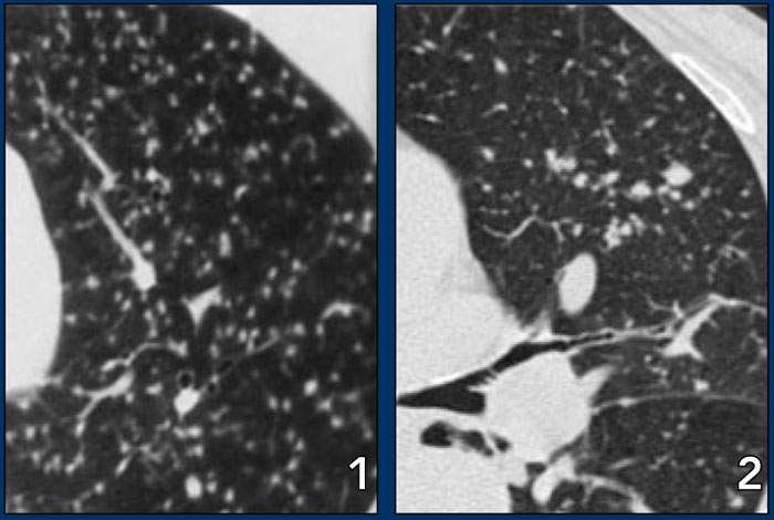 LEFT: miliary TB. RIGHT: metastasesTRÁI: lao kê, PHẢI: di căn
Differential diagnosis of TB.
Chẩn đoán phân biệt của bệnh lao.
- Primary TB: Acute bacterial pneumoniaLao nguyên phát: Viêm phổi cấp do vi khuẩn
- Secondary TB: Sarcoidosis, Silicosis, PneumoconiosisLao thứ phát: Sarcoidosis, Silicosis, Pneumoconiosis
- Endobronchial spread of TB: Bronchopneumonia, Hypersensitivity pneumonitisSự lan rộng nội phế quản của lao: Viêm phế quản phổi, viêm phổi quá mẫn
- Miliary TB: metastases of medullary thyroid ca, chorionca and melanoma. Lao thể tuỷ: di căn của u tuỷ, u hắc tố và u tuyến giáp thể tuỷ.
In both miliary TB and metastases the nodules have a random distribution.
In miliary TB the nodules are more uniform in size.Trong cả lao kê và di căn, các nốt có phân bố ngẫu nhiên.
Trong lao kê, các nốt sần có kích thước đồng đều hơn.
Images
Hình minh họa
- TB - uniform small nodules in random distributionTB - nốt nhỏ đồng nhất phân bố ngẫu nhiên
- Metastases - nodules of different sizes in random distribution.Di căn - các nốt có kích thước khác nhau phân bố ngẫu nhiên.
Chronic eosinophilic pneumoniaViêm phổi tăng bạch cầu ái toan mạn tính

Chronic eosinophilic pneumonia is an idiopathic condition characterized by filling of the alveoli with eosinophils.
It is associated with an increased number of eosinophils in the peripheral blood and patients present with fever, cough, weight loss, malaise, and shortness of breath.
The symptoms are often severe and last three months or more.
Patients respond promptly to treatment with steroids.Viêm phổi tăng bạch cầu ái toan mạn tính là một tình trạng vô căn đặc trưng bởi sự lấp đầy các phế nang với bạch cầu áI toan.
Nó có liên quan đến sự gia tăng số lượng bạch cầu ái toan trong máu ngoại vi và bệnh nhân có sốt, ho, sụt cân, khó chịu và khó thở.
Các triệu chứng thường nặng và kéo dài từ ba tháng trở lên.
Bệnh nhân đáp ứng nhanh với điều trị bằng steroid.
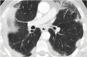 Chronic eosinophilic pneumoniaViêm phổi tăng bạch cầu ái toan mạn tính
HRCT findings in Chronic eosinophilic pneumonia Đặc điểm hình ảnh HRCT trong viêm phổi tăng bạch cầu ái toan mạn tính
- Peripheral consolidations with upper lobe predominance (photo negative of pulmonary edema).Củng cố ngoại vi với ưu thế thuỳ trên (ảnh âm tính của phù phổi).
On the left a contrast enhanced CT in a patient with chronic eosinophilic pneumonia.
Notice peripheral distribution of the consolidations.Bên trái là hình ảnh chụp CT tăng cường thuốc cản quang ở bệnh nhân viêm phổi tăng bạch cầu ái toan mạn tính.
Lưu ý sự phân bố ngoại vi của các khối đông đặc.
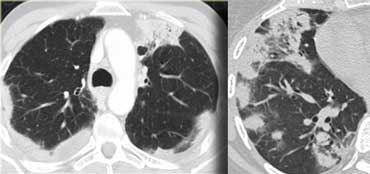 Chronic eosinophilic pneumonia (left) versus Organizing pneumonia (right)Viêm phổi tăng bạch cầu ái toan mạn tính (trái) so với viêm phổi tổ chức hóa (phải)
Differential diagnosis of Chronic eosinophilic pneumonia Chẩn đoán phân biệt của viêm phổi tăng bạch cầu ái toan mạn tính
- Organizing pneumonia (COP)Viêm phổi tổ chức hóa (COP)
- Loffler syndrome (eosinophilia and vanishing peripheral consolidations)Hội chứng Loffler (bạch cầu ái toan và biến mất sự đông đặc ngoại vi)
- Churg-Strauss syndrome (also serum eosinophilia, asthma, systemic vasculitis affecting multiple organs: renal insufficiency, arthralgia and myocarditis and pericarditis)Hội chứng Churg-Strauss (cũng tăng bạch cầu ái toan huyết thanh, hen suyễn, viêm mạch hệ thống ảnh hưởng đến nhiều cơ quan: suy thận, đau khớp và viêm cơ tim và viêm màng ngoài tim)
- Pulmonary infarctsNhồi máu phổi
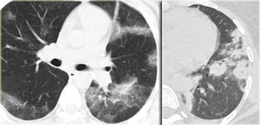
The images on the left show the similarities between chronic eosinophilic pneumonia and organizing pneumonia.
Differentiation has to be made on the basis of clinical and laboratory findings.Hình ảnh bên trái cho thấy sự tương đồng giữa viêm phổi tăng bạch cầu ái toan mạn tính và viêm phổi tổ chức hóa.
Phân biệt phải được thực hiện trên cơ sở các kết quả lâm sàng và xét nghiệm.
Pneumocystis carinii pneumoniaViêm phổi do Pneumocystis carinii
Pneumocystis carinii pneumonia (PCP) or pneumocystis jiroveci as it is currently named, is an opportumistic infection in immunocompromised patients.
PCP used to affect most HIV-infected patients at some point during the course of their disease, but with the new anti-viral drugs it has become less common.
Nowadays PCP is seen more in immunosuppressed patients, i.e. transplant recipients and patients on chemotherapy.Viêm phổi do Pneumocystis carinii (PCP) hoặc pneumocystis jiroveci như tên gọi hiện tại, là một bệnh nhiễm trùng cơ hội ở những bệnh nhân suy giảm miễn dịch.
PCP từng ảnh hưởng đến hầu hết các bệnh nhân nhiễm HIV tại một số thời điểm trong quá trình bệnh của họ, nhưng với các loại thuốc chống vi rút mới, nó đã trở nên ít phổ biến hơn.
Ngày nay, PCP được thấy nhiều hơn ở những bệnh nhân bị ức chế miễn dịch, tức là những người nhận ghép tạng và những bệnh nhân đang hóa trị.
HRCT findings in PCP Đặc điểm HRCT trong PCP
- Perihilar or diffuse ground-glass opacification. Đục quanh rốn hoặc đục lan toả kính mờ.
- Sometimes thickened septal lines in association with areas of ground-glass Đôi khi các vách ngăn dày lên liên quan đến các vùng của kính mờ
- Later cysts (or pneumatoceles) in 10-35% of patients, typically involving upper lobes Nang sau này (hoặc tràn khí màng phổi) ở 10-35% bệnh nhân, thường liên quan đến thuỳ trên.
- Cysts may have bizarre shapes and thick walls U nang có thể có hình dạng kỳ lạ và thành dày
- Following therapy these lesions eventually regress, resulting either in complete disappearance, or residual nodules or scars Sau khi điều trị, những tổn thương này cuối cùng sẽ thóai triển, dẫn đến sự biến mất hoàn toàn, hoặc các nốt hoặc sẹo còn sót lại.
- Pneumothorax in 35% of patients with cystsTràn khí màng phổi ở 35% bệnh nhân có nang
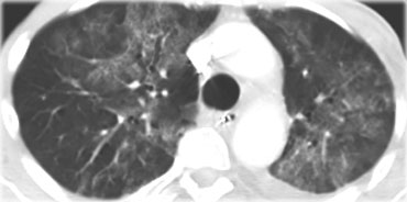 PCP with diffuse ground-glass opacificationPCP với đục kính mờ khuếch tán
On the left an immunocompromised patient with PCP.
The CT findings are diffuse ground-glass opacification.
The findings are not specific for PCP, but in this clinical setting PCP is the most likely diagnosis.Bên trái là một bệnh nhân suy giảm miễn dịch với PCP.
Hình ảnh trên CT là hình ảnh kính mờ mờ lan toả.
Các dấu hiệu không đặc hiệu cho PCP, nhưng trong bối cảnh lâm sàng này PCP là chẩn đoán có khả năng nhất.
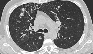
On the left another patient with PCP.
Scroll through the images.Bên trái một bệnh nhân khác có PCP.
Cuộn qua các hình ảnh.
ARDSARDSComment
Acute respiratory distress syndrome (ARDS) is a sudden, life-threatening lung failure requiring mechanical ventilation.
ARDS represents the result of increased permeability often in combination with injury to the respiratory epithelium.
A variety of underlying conditions, from infections to major trauma, can cause ARDS.
Primary pulmonary risk factors include aspiration, pneumonia, toxic inhalation and pulmonary contusion.
Extrapulmonary risk factors are sepsis, pancreatitis, multiple blood transfusions, trauma and the use of drugs such as heroin.
Hội chứng suy hô hấp cấp (ARDS) là một tình trạng suy phổi đột ngột, đe doạ tính mạng cần phải thở máy.
ARDS là kết quả của sự gia tăng tính thấm thường kết hợp với tổn thương biểu mô hô hấp.
Một loạt các bệnh lý nền, từ nhiễm trùng đến chấn thương nặng, có thể gây ARDS.
Các yếu tố nguy cơ phổi chính bao gồm hít phải, viêm phổi, hít phải chất độc và đụng dập phổi.
Các yếu tố nguy cơ ngoài phổi là nhiễm trùng huyết, viêm tụy, truyền máu nhiều lần, chấn thương và sử dụng ma tuý như heroin.
Mild forms of ARDS may resolve completely, while severe forms result in irreverible fibrosis.
Why some people develop ARDS and others do not is unknown.
Các dạng ARDS nhẹ có thể khỏi hoàn toàn, trong khi các dạng nặng dẫn đến xơ hóa không hồi phục.
Tại sao một số người mắc ARDS và những người khác thì không thì vẫn chưa rõ.
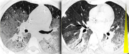 Extra-pulmonary ARDS with gravity dependent gradientARDS ngoài phổi với gradient phụ thuộc trọng lực
Extra-pulmonary ARDS
On the left a patient who was involved in a traffic accident and within hours developed ARDS.
The dominant pattern is ground glass opacity.
In the dependent parts of the lung there is also some consolidation, so there is a gradient from front to back.
An important finding in extra-pulmonary ARDS is the symmetry of the abnormalities.
ARDS ngoài phổi
Bên trái là một bệnh nhân bị tai nạn giao thông và trong vòng vài giờ đã tiến triển ARDS.
Mô hình trội là đám mờ kính mờ.
Ở các phần phụ thuộc của phổi cũng có một số đông đặc, do đó có một gradient từ trước ra sau.
Một dấu hiệu quan trọng trong ARDS ngoài phổi là sự đối xứng của các bất thường.
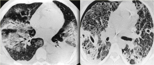 Pulmonary ARDS with asymmetric patchy distribution of consolidations.ARDS phổi với sự phân bố không đối xứng của các khối đông đặc.
Pulmonary ARDS
On the left a patient who developed ARDS as a result of pneumonia (i.e. pulmonary ARDS).
Note the patchy distribution of lung disease and the almost complete distorsion more basal.
Patient is ventilated with PEEP (positive end expiratory pressure ) leading to a barotrauma of the lung parenchyma: there are multiple subpleural cysts and a bilateral pneumothorax.
ARDS phổi
Ở bên trái, một bệnh nhân bị ARDS do viêm phổi (tức là ARDS phổi).
Lưu ý sự phân bố loang lổ của bệnh phổi và sự biến dạng gần như hoàn toàn cơ bản hơn.
Bệnh nhân được thông khí với PEEP (áp lực cuối thở ra dương tính) dẫn đến chấn thương khí áp của nhu mô phổi: có nhiều nang dưới màng phổi và tràn khí màng phổi hai bên.
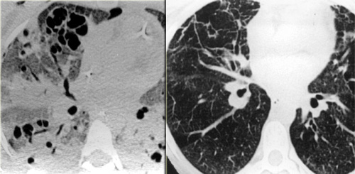 LEFT: ARDS. RIGHT: Fibrosis in the anterior parts as a result of damage related to barotraumas and PEEP ventilationTRÁI: CÁNH TAY. PHẢI: Xơ hóa phần trước do tổn thương liên quan đến chấn thương áp lực và thông khí PEEP
Consolidations have a protecting effect on the lung parenchyma under PEEP ventilation, while the ventrally located areas of more normal lung are most prone to the effects of barotrauma.
As a result we find cystic destruction ventrally and residual fibrosis mostly in the ventral lung areas. Sự đông đặc có tác dụng bảo vệ nhu mô phổi dưới thông khí PEEP, trong khi các vùng nằm ở tâm thất của phổi bình thường hơn dễ bị ảnh hưởng nhất bởi chấn thương khí áp.
Kết quả chúng tôi thấy có tổn thương nang thất và xơ hóa còn sót lại chủ yếu ở vùng phổi bụng.
Idiopathic interstitial pneumoniasViêm phổi kẽ tự phát
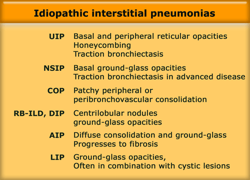
The idiopathic interstitial pneumonias (IIPs) comprise a heterogenous group of disorders.
They represent fundamental responses of the lung to injury and do not represent 'diseases' per se.
Idiopathic indicates unknown cause and interstitial pneumonia refers to involvement of the lung parenchyma by varying combinations of fibrosis and inflammation.
IIPs include seven entities listed in the table on the left in order of relative frequency.
Viêm phổi kẽ tự phát (VKDT) bao gồm một nhóm rối loạn không đồng nhất.
Chúng đại diện cho các phản ứng cơ bản của phổi đối với tổn thương và không đại diện cho 'bệnh'.
Viêm phổi kẽ tự phát không rõ nguyên nhân liên quan đến sự tham gia của nhu mô phổi bằng sự kết hợp khác nhau của xơ hóa và viêm.
Các IIP bao gồm bảy thực thể được liệt kê trong bảng bên trái theo thứ tự tần suất tương đối.
These diseases have specific patterns of morphologic findings on HRCT and histology.
Before we call these findings idiopathic or cryptogenic, we should realise, that these patterns are also common findings in collagen vascular diseases (e.g., sclerodermia, rheumatoid arthritis) and drug-related lung diseases.
For instance in patients with rheumatoid arthritis findings of NSIP, UIP, OP and LIP have been reported.
Các bệnh này có các đặc điểm hình thái đặc trưng trên HRCT và mô học.
Trước khi chúng ta gọi những phát hiện này là vô căn hoặc do nguyên nhân, chúng ta nên nhận ra rằng những mô hình này cũng là những phát hiện phổ biến trong các bệnh mạch máu collagen (ví dụ, xơ cứng bì, viêm khớp dạng thấp) và các bệnh phổi liên quan đến thuốc.
Ví dụ ở những bệnh nhân viêm khớp dạng thấp, các dấu hiệu của NSIP, UIP, OP và LIP đã được báo cáo.
UIP
Usual Interstitial Pneumonitis (UIP) is a histologic diagnosis.
UIP has distinctive HRCT findings and is usually shown at lungbiopsy, when honeycombing is visible.
If the UIP pattern is of unknown cause (i.e. idiopathic), the disease is called Idiopathic pulmonary fibrosis (IPF).
IPF accounts for more than 60% of the cases of UIP. Viêm phổi kẽ thường gặp (UIP) là một chẩn đoán mô học.
UIP có các dấu hiệu HRCT đặc biệt và thường được hiển thị khi sinh thiết phổi, khi có thể nhìn thấy tổ ong. Nếu mô hình UIP không rõ nguyên nhân (tức là vô căn), bệnh được gọi là Vô căn xơ phổi (IPF).
IPF chiếm hơn 60% các trường hợp UIP.
In the presence of a surgical biopsy showing a UIP pattern the diagnosis of IPF requires exclusion of other known causes of UIP including drug toxicities, environmental exposures (asbest), and collagen vascular diseases like RA, SLE, polyarteritis nodosa and sclerodermia.
A long list of drugs have been implicated, but this pattern is most commonly the result of cytotoxic chemotherapeutic agents such as bleomycin, busulfan, vincristine, methotrexate, adriamycin, and carmustine (BCNU).Khi có sinh thiết phẫu thuật cho thấy một mẫu UIP, chẩn đoán IPF đòi hỏi phải loại trừ các nguyên nhân khác đã biết của UIP bao gồm độc tính của thuốc, phơi nhiễm môi trường (tốt nhất) và các bệnh mạch máu collagen như RA, SLE, viêm nút đa động mạch và xơ cứng bì.
Một danh sách dài các loại thuốc đã được liên quan, nhưng mô hình này thường là kết quả của các tác nhân hóa trị liệu gây độc tế bào như bleomycin, busulfan, vincristine, methotrexate, adriamycin và carmustine (BCNU).
The differentiation between NSIP and UIP has tremendous prognostic implication for the patient.
UIP is more progressive and more than 50% of patients with UIP die within 3 years. Sự khác biệt giữa NSIP và UIP có ý nghĩa tiên lượng rất lớn đối với bệnh nhân.
UIP tiến triển nhiều hơn và hơn 50% bệnh nhân bị UIP chết trong vòng 3 năm.
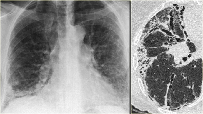 Chest film in a patient with UIP demonstrating the reticular pattern in basal and subpleural distribution due to honeycombing.Phim ngực ở bệnh nhân UIP thể hiện dạng lưới trong phân bố đáy và dưới màng phổi do tổ ong.
On the left a chest film of a patient with UIP due to IPF.
The findings on the chest film comprise volume loss and fibrotic changes in the basal lung area.
The radiographic appearance of honeycombing comprises reticular densities caused by the thick walls of the cysts.
Whenever you see a chest film with long standing reticulation with a lower lobe and peripheral preference also think 'UIP'.Bên trái là phim ngực của một bệnh nhân UIP do IPF.
Các dấu hiệu trên phim ngực bao gồm mất thể tích và thay đổi xơ hóa ở vùng đáy phổi.
Biểu hiện trên X-quang của tổ ong bao gồm mật độ dạng lưới do thành dày của nang gây ra.
Bất cứ khi nào bạn thấy một phim ngực với hình lưới dài với thuỳ dưới và ưu tiên ngoại vi cũng nghĩ rằng 'UIP'.
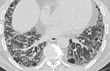 Typical UIPUIP điển hình
HRCT findings in UIP Kết quả HRCT trong UIP
- Honeycombing consisting of multilayered thick-walled cysts. Tổ ong gồm các nang vách dày đa lớp.
- Architectural distortion with traction bronchiectasis due to fibrosis. Biến dạng cấu trúc với giãn phế quản co kéo do xơ hóa.
- Predominance in basal and subpleural region. Đa số ở vùng đáy và dưới màng phổi.
- Mild mediastinal lymphadenopathyTrung thất nhẹ bệnh hạch
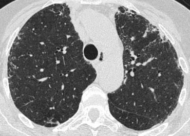
Differential diagnosis of UIP. Chẩn đoán phân biệt của UIP.
- Chronic Hypersensitivity PneumonitisViêm phổi quá mẫn mạn tính
- End stage SarcoidosisGiai đoạn cuối Sarcoidosis
- NSIP
Chronic HP may be indistinguishable.
It is suspected if there is a mosaic pattern with sparing of the lung bases or when there are centrilobular nodules.
Sarcoidosis is a more likely diagnosis if the fibrosis is located in the posterior parts of the upper lobes or in the perihilar area and if there are also nodules in a perilymphatic distribution or if there is extensive mediastinal lymphadenopathy.HP mạn tính có thể không thể phân biệt được.
Nghi ngờ nếu có một mẫu khảm với sự tiết kiệm của hai đáy phổi hoặc khi có nốt trung tâm tiểu thùy.
Sarcoidosis là một chẩn đoán có nhiều khả năng hơn nếu xơ hóa nằm ở phần sau của thuỳ trên hoặc ở vùng quanh rốn và nếu cũng có các nốt trong phân bố quanh bạch mạch hoặc nếu có bệnh hạch trung thất rộng.
The presence of pleural plaques helps for the differentiation between IPF and asbestosis.Sự hiện diện của mảng màng phổi giúp phân biệt giữa IPF và bệnh bụi phổi amiăng.
On the left a patient with UIP.
Notice the honeycombing and the preference of the subpleural and basal lung areas.
Bên trái là một bệnh nhân có UIP.
Lưu ý tổ ong và sự ưu tiên của dưới màng phổi và các vùng phổi đáy.
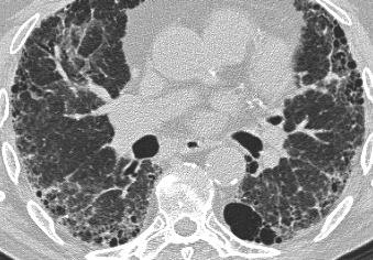 UIP: Typical caseUIP: Ca lâm sàng điển hình
It is usually easy to recognize the pattern of UIP on HRCT.Thường dễ dàng nhận biết được dạng UIP trên HRCT.
NSIP
Nonspecific interstitial pneumonia (NSIP) is by some considered as a specific entity, with specific histologic characteristics, but by others as a 'wastebasket' diagnosis, representing cases of idiopathic interstitial pneumonia that cannot be classified as UIP, DIP, or OP.
NSIP is histologically characterized by a homogeneous, uniform pattern of cellular interstitial inflammation associated with variable degrees of fibrosis.
In contrast, UIP is associated with extensive fibrosis which is temporally inhomogeneous (i.e. various lesions are of different ages).
Viêm phổi kẽ không đặc hiệu (NSIP) (NSIP) được một số người coi là một thực thể cụ thể, với các đặc điểm mô học cụ thể, nhưng những người khác coi là chẩn đoán 'chứa rác', đại diện cho các trường hợp viêm phổi kẽ tự phát không thể phân loại là UIP, DIP hoặc OP.
NSIP được đặc trưng về mặt mô học bởi một mô hình đồng nhất, đồng nhất của viêm mô kẽ tế bào liên quan đến các mức độ xơ hóa khác nhau.
Ngược lại, UIP có liên quan đến xơ hóa lan rộng, không đồng nhất về mặt thời gian (tức là các tổn thương khác nhau ở các độ tuổi khác nhau).
NSIP is a very inhomogeneous group.
NSIP ranges from type I which is a cellular pattern seen as ground glass opacity on HRCT to type IV with a fibrotic pattern, which may be indistinguishable from UIP. NSIP là một nhóm rất không đồng nhất.
NSIP dao động từ loại I là mô hình tế bào được xem là đám mờ kính mờ trên HRCT đến loại IV có mô hình xơ hóa, có thể không thể phân biệt được với UIP.
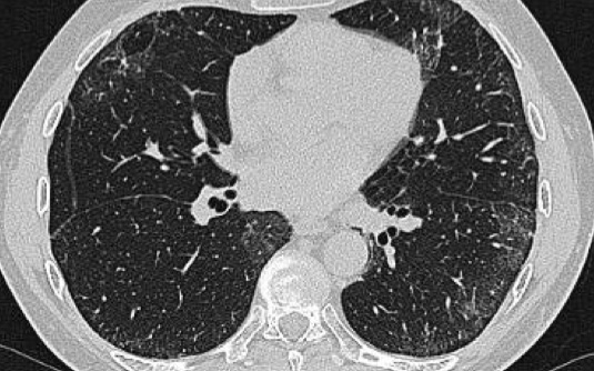
On the left a patient with a NSIP.
This patient had a rash and muscle weakness.
Scroll through the images.Bên trái là một bệnh nhân có NSIP.
Bệnh nhân này có biểu hiện phát ban và yếu cơ.
Cuộn qua các hình ảnh.
The predominant finding is ground glass opacity (GGO).
There is very subtle traction bronchiectasis, indicating that the GGO is the result of fibrosis and therefore irreversible.
It is important to note that we do not see the classic distribution of UIP, from which NSIP has to be differentiated.
The history of this patient is suggestive for the diagnosis dermatomyositis.
NSIP is by far the most common interstitial lung disease in patients with connective tissue disease.Dấu hiệu chủ yếu là đám mờ kính mờ (GGO).
Có giãn phế quản co kéo rất tinh tế, chỉ ra rằng GGO là kết quả của sự xơ hóa và do đó không thể đảo ngược.
Điều quan trọng cần lưu ý là chúng ta không thấy sự phân bố cổ điển của UIP, từ đó NSIP phải được phân biệt.
Tiền sử bệnh nhân này gợi ý chẩn đoán viêm da cơ.
NSIP là bệnh phổi kẽ phổ biến nhất ở bệnh nhân mắc bệnh mô liên kết.
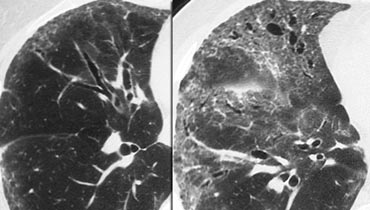
NSIP (2)
NSIP is not a diagnosis on it's own.
It is a pattern of lung damage.
For the pathologist the key feature is the uniformity of the abnormality within the lung.
The role of the radiologist is more to 'exclude UIP pattern' rather than to make the diagnosis of NSIP.
The diagnosis of NSIP requires histological proof.
In all patients with a NSIP pattern, the clinician should be advised to look for connective tissue diseases, hypersensitivity pneumonitis or drugs .CK-C (2)
NSIP không phải là một chẩn đoán riêng.
Đây là một dạng tổn thương phổi.
Đối với nhà giải phẫu bệnh, đặc điểm chính là sự đồng nhất của các bất thường trong phổi.
Vai trò của bác sĩ chẩn đoán hình ảnh là loại trừ UIP hơn là chẩn định NSIP.
Chẩn đoán NSIP cần phải có bằng chứng mô học.
Ở tất cả các bệnh nhân có mô hình NSIP, bác sĩ lâm sàng nên được khuyên nên tìm kiếm các bệnh mô liên kết, viêm phổi quá mẫn hoặc thuốc.
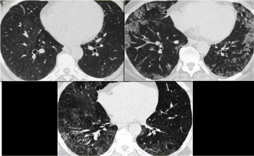
On the left two cases of NSIP.
Note the varying combination of GGO and fibrosis (traction bronchiectasis), but the lack of honeycombing.
Bên trái hai trường hợp NSIP.
Lưu ý sự kết hợp khác nhau của GGO và xơ hóa (giãn phế quản co kéo), nhưng thiếu tổ ong.
NSIP (3)
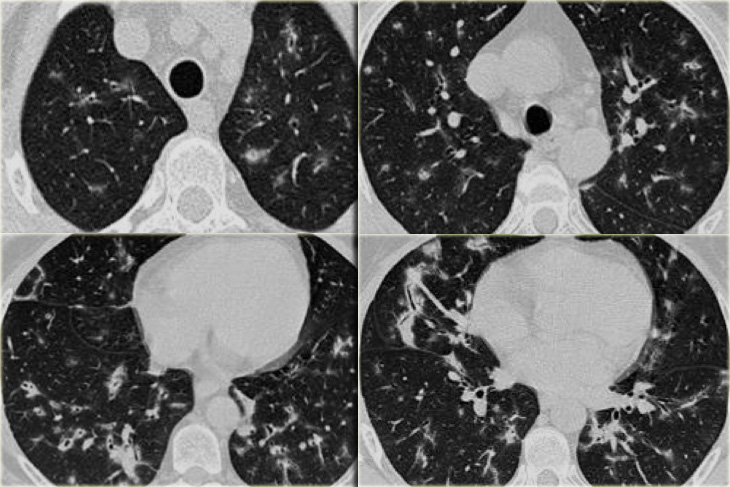
NSIP is the prevalent lung pattern in systemic sclerosis and polymyosisits/dermatomyositis (more than 90%), but also may occur in RA, SLE, Sj?gren's and MCTD.
In the images on your left you can appreciate again the spectrum of findings seen in NSIP.
All three patients were suffering from connective tissue disease, all cases were biopsy proven.
The first (top left) shows a very subtle GGO.
Note the difference in the density of the air within the bronchus and surrounding lungparenchyma (dark bronchus sign).
The second (top right) is a more obvious example of GGO with a superimposed fine reticular densities as a result of thickening of the intralobular septa.
The last image also shows GGO with a fine reticular pattern.
Notice the lack of honeycombing in all three cases, excluding UIP as diagnosis.
NSIP (3)
NSIP là mô hình phổi phổ biến trong bệnh xơ cứng hệ thống và viêm da toàn thân / viêm da (hơn 90%), nhưng cũng có thể xảy ra ở RA, SLE, SJ? Gren và MCTD.
Trong các hình ảnh bên trái của bạn, bạn có thể đánh giá lại phổ phát hiện được thấy trong NSIP.
Cả ba bệnh nhân đều bị bệnh mô liên kết, tất cả các trường hợp đều được chứng minh sinh thiết.
Đầu tiên (trên cùng bên trái) cho thấy một GGO rất tinh tế.
Lưu ý sự khác biệt về mật độ không khí trong phế quản và nhu mô phổi xung quanh (dấu hiệu phế quản tối).
Thứ hai (trên cùng bên phải) là một ví dụ rõ ràng hơn về GGO với mật độ lưới nhỏ chồng lên nhau do sự dày lên của vách ngăn trong.
Hình ảnh cuối cùng cũng cho thấy GGO với một mẫu lưới nhỏ.
Lưu ý việc thiếu tổ ong trong cả ba trường hợp, không bao gồm UIP là chẩn đoán.
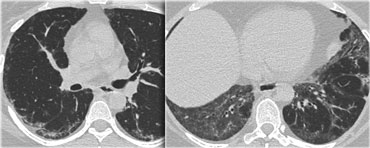
NSIP (4)
The HRCT of this patient with scleroderma and NSIP shows a fine subpleural reticular pattern in the upper lobes and more extensive abnormalities in the lower lung zones.
There are also areas of ground-glass and traction bronchiectases, but honeycombing is typically lacking.
Note also the mildly dilated esophagus, which is consistent with scleroderma.Máy cắt cỏ (4)
Trên phim HRCT của bệnh nhân xơ cứng bì và NSIP cho thấy hình ảnh dạng lưới dưới màng phổi nhỏ ở thuỳ trên và các bất thường lan toả hơn ở vùng phổi dưới.
Ngoài ra còn có các khu vực của kính mờ và giãn phế quản kéo, nhưng tổ ong thường thiếu.
Cũng lưu ý thực quản giãn nhẹ, phù hợp với xơ cứng bì.

COP
Cryptogenic organizing pneumonia (COP) used to be described as bronchiolitis obliterans with organizing pneumonia (BOOP) in an earlier version of the classification of idiopathic interstitial pneumonias.
It is a inflammatory process in which the healing process is characterized by organization of the exudate rather than by resorption ('unresolved pneumonia').
Organizing pneumonia is mostly idiopathic and then called cryptogenic, but is also seen in patients with pulmonary infection, drug reactions, collagen vascular disease, Wegener's granulomatosis and after toxic-fume inhalation.
OP presents with a several-month history of nonproductive cough, low-grade fever, malaise and shortness of breath.
There is a good response to corticosteroid therapy and a good prognosis. Viêm phổi tổ chức hóa do Cryptogenic (COP) từng được mô tả là viêm tiểu phế quản tắc nghẽn với viêm phổi tổ hợp (BOOP) trong một phiên bản trước đó của phân loại viêm phổi kẽ tự phát.
Đây là một quá trình viêm trong đó quá trình lành thương được đặc trưng bởi sự tổ chức của dịch tiết chứ không phải do sự tái hấp thu ('viêm phổi không được giải quyết').
Viêm phổi tổ chức hóa hầu hết là tự phát và sau đó được gọi là cryptogenic, nhưng cũng thấy ở những bệnh nhân bị nhiễm trùng phổi, phản ứng thuốc, bệnh mạch máu collagen, u hạt Wegener và sau khi hít phải khói độc.
Bệnh nhân có tiền sử ho khan, sốt nhẹ, khó chịu và khó thở trong vài tháng.
Đáp ứng tốt với điều trị corticosteroid và tiên lượng tốt.
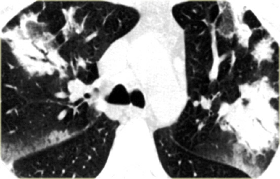
OP is again a great mimicker and can show a broad variety of HRCT findings, which makes it a frequent differential diagnosis and actually represents a diagnosis of exclusion.
Frequently biopsy is needed for final proof.OP một lần nữa là một sự bắt chước tuyệt vời và có thể cho thấy một loạt các phát hiện HRCT, mà làm cho nó thường xuyên chẩn đoán phân biệt và thực sự đại diện cho một chẩn đoán loại trừ.
Sinh thiết thường xuyên là cần thiết để chứng minh cuối cùng.
HRCT findings in OP Đặc điểm HRCT trong phẫu thuật nội soi
- Bilateral peripheral consolidations, sharply demarcated.Củng cố ngoại vi hai bên, ranh giới rõ.
- Consolidations may be migratory.Sự hợp nhất có thể là di cư.
- Lesions may show pleural tags or spiculae and give the impression of volume loss and slight retraction of the surrounding parenchyma (DD bronchogenic carcinoma).Các tổn thương có thể cho thấy các thẻ hoặc gai màng phổi và cho thấy sự mất thể tích và sự co lại nhẹ của nhu mô xung quanh (ung thư biểu mô phế quản DD).
- Bronchial wall thickening and dilatation are seen in most patients and are usually restricted to areas of consolidation or ground glass opacifications. Dày thành phế quản và giãn mạch được thấy ở hầu hết các bệnh nhân và thường bị hạn chế ở các vùng đục đông đặc hoặc kính mờ.
- Additional findings are pleural thickening, small pleural effusions and parenchymal bands. Các dấu hiệu khác là dày màng phổi, tràn dịch màng phổi nhỏ và các dải nhu mô.
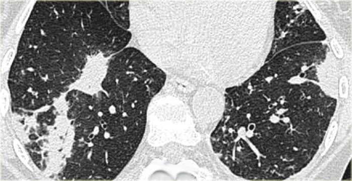
On the left a patient with the typical bilateral peripheral consolidations of OP.
After exclusion of other diseases such as lymphoma, infection, bronchoalveolar carcinoma, the diagnosis of cryptogenic organizing pneumonia was made.Bên trái là một bệnh nhân với hai bên điển hình là củng cố của OP.
Sau khi loại trừ các bệnh lý khác như u lympho, nhiễm trùng, ung thư biểu mô phế quản phế nang, chẩn đoán viêm phổi tổ chức hóa được thực hiện.
 OP in a patient with collagen vascular diseaseNhân một trường hợp phẫu thuật nội soi điều trị bệnh mạch máu collagen
On the left a patient who complained of arthritic pain.
There are multiple small bilateral peripheral consolidations.
The findings in this patient are not as specific as in the former case, but this was also organizing pneumonia, but now related to collagen vascular disease.Bên trái là một bệnh nhân than phiền đau khớp.
Có nhiều khối đông đặc nhỏ ở ngoại vi hai bên.
Kết quả trên bệnh nhân này không đặc hiệu như trường hợp trước, nhưng đây cũng là tổ chức viêm phổi, nhưng hiện nay liên quan đến bệnh mạch máu collagen.
 OP in rheumatoid arthritisPhẫu thuật nội soi trong viêm khớp dạng thấp
On the left a patient with rheumatoid arthritis and bilateral peripheral consolidations as a result of organizing pneumonia.
Patients with OP associated with collagen vascular diseases respond less well to therapy with steroids.
Bên trái là một bệnh nhân bị viêm khớp dạng thấp và đông đặc hai bên do viêm phổi tổ chức hóa.
Bệnh nhân có OP liên quan đến bệnh mạch máu collagen đáp ứng kém hơn với điều trị bằng steroid.
 Chronic eosinophilic pneumonia (left) versus Organizing pneumonia (right)Viêm phổi tăng bạch cầu ái toan mạn tính (trái) so với viêm phổi tổ chức hóa (phải)
Differential diagnosis of Organizing Pneumonia. Chẩn đoán phân biệt của viêm phổi tổ chức hóa.
- Chronic eosinophilic pneumonia Viêm phổi tăng bạch cầu ái toan mạn tính
- Bronchoalveolar cell carcinoma Ung thư biểu mô tế bào phế quản phế nang
- Aspiration pneumonia Viêm phổi hít
- Pulmonary infarction Phổi nhồi máu
- Lymphoma
The images on the left show the similarities between chronic eosinophilic pneumonia and organizing pneumonia.
Differentiation has to be made on the basis of clinical and laboratory findings.Hình ảnh bên trái cho thấy sự tương đồng giữa viêm phổi tăng bạch cầu ái toan mạn tính và viêm phổi tổ chức hóa.
Phân biệt phải được thực hiện trên cơ sở các kết quả lâm sàng và xét nghiệm.

RB-ILD and DIPRB-ILD và DIP
Respiratory bronchiolitis (RB), respiratory bronchiolitis-associated interstitial lung disease (RB-ILD), and desquamative interstitial pneumonia (DIP) represent different degrees of severity of small airway and parenchymal reaction to cigarette smoke (8).
Viêm tiểu phế quản đường hô hấp (RB), viêm tiểu phế nang liên quan đến viêm phế quản bệnh phổi kẽ (RB-ILD) và viêm phổi kẽ bong vảy (DIP) biểu hiện các mức độ nghiêm trọng khác nhau của phản ứng đường thở nhỏ và phản ứng nhu mô với khói thuốc lá (8).
All smokers have various degrees of respiratory bronchiolitis, but it is usually asymptomatic.
However 5-10% of smokers have a clinically significant lung disease in association with RB, presenting with symptoms, lung function tests and auscultatory findings at clinical examination.
The term RB-ILD was proposed to describe the bronchocentric (or centrilobular) lung disease in these patients and the term DIP was used to describe the more diffuse disorder.
Radiologically however these diseases cannot be clearly separated because of the overlap of CT findings.
Tất cả những người hút thuốc đều có các mức độ viêm tiểu phế quản hô hấp khác nhau, nhưng thường không có triệu chứng.
Tuy nhiên 5-10% người hút thuốc lá có bệnh phổi liên quan đến RB, biểu hiện lâm sàng với các triệu chứng, xét nghiệm chức năng phổi và các dấu hiệu nghe được khi khám lâm sàng.
Thuật ngữ RB-ILD được đề xuất để mô tả bệnh phổi trung tâm phế quản (hoặc trung tâm tiểu thùy) ở những bệnh nhân này và thuật ngữ DIP được sử dụng để mô tả rối loạn lan toả hơn.
Tuy nhiên, về mặt X quang, các bệnh này không thể phân tách rõ ràng vì có sự chồng chéo của các dấu hiệu trên CT.
HRCT findings in RB-ILD Đặc điểm HRCT ở bệnh nhân RB-ILD
- Centrilobular nodules of ground glass opacity with upper lobe predominance Nốt trung tâm tiểu thùy của đám mờ kính mờ có ưu thế ở thuỳ trên
- Bronchial wall thickening Dày thành phế quản
- Secondary lobuli with decreased attenuation (air trapping) Lobuli thứ phát giảm độ suy giảm (bẫy khí)
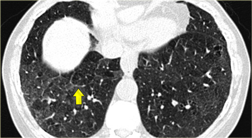
On the left a patient with RB-ILD.
The dominant feature is ground glass opacification and there are some thickened interlobular septa (arrow).
Usually these patients will also have smoking induced centrilobular emphysema and there is some evidence that respiratory bronchiolitis is the precursor of emphysema.
Bên trái là một bệnh nhân có RB-ILD.
Đặc điểm nổi bật là kính mờ mờ và có một số vách liên thuỳ dày (mũi tên).
Thông thường những bệnh nhân này cũng sẽ có trung tâm tiểu thùy khí phế thũng do hút thuốc và có một số bằng chứng cho thấy viêm tiểu phế quản hô hấp là tiền thân của khí phế thũng.
RB-ILD (2)
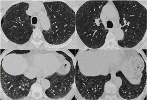
On the left a smoker with RB-ILD with subtle HRCT-findings.
The dominant pattern is ground glass opacification.
Additional findings in this patient are paraseptal emphysema in the upper lobes and some subtle septal thickening in the basal parts.RB-ILD (2)
Bên trái là một người hút thuốc lá có RB-ILD với các phát hiện HRCT tinh tế.
Mô hình ưu thế là kính mờ.
Các dấu hiệu bổ sung ở bệnh nhân này là khí phế thũng ở thuỳ trên và một số dày vách tinh tế ở phần đáy.
Based on these non-specific CT findings there is a broad differential diagnosis and additional clinical information is mandatory for the interpretion of the HRCT.
Since this patient is a smoker we first think RB-ILD.
In a immunocompromised patient PCP would be on top of the list.
If this patient was coughing up blood, this probably would be pulmonary hemorrhage (although we would expect more pulmonary densities in these patients).
If this patient was a bird-fancier we would first think hypersensitivity pneumonitis, but mostly these patients do not smoke.Dựa trên những phát hiện CT không đặc hiệu này, có một chẩn đoán phân biệt rộng và thông tin lâm sàng bổ sung là bắt buộc để diễn giải HRCT.
Vì bệnh nhân này là người hút thuốc nên chúng tôi nghĩ đến RB-ILD trước tiên.
Ở bệnh nhân suy giảm miễn dịch, PCP sẽ đứng đầu danh sách.
Nếu bệnh nhân này ho ra máu, có thể là xuất huyết phổi (mặc dù chúng tôi mong đợi mật độ phổi nhiều hơn ở những bệnh nhân này).
Nếu bệnh nhân này là người nuôi chim thì đầu tiên chúng ta sẽ nghĩ đến viêm phổi quá mẫn, nhưng hầu hết những bệnh nhân này không hút thuốc.
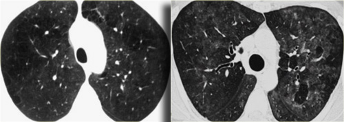 LEFT: RB-ILD in smoker. RIGHT: Hypersensitivity pneumonitis in non-smokerTrái: RB-Id trong Smoker. Phải: viêm phổi quá mẫn trong không hút thuốc
On the left two different patients with similarl HRCT findings.
The left one is a smoker with RB-ILD and the patient on the right has hypersensitivity pneumonits.
Note the difference in severity of ground glass opacities and the well defined areas of airtrapping in HP.
Bên trái hai bệnh nhân khác nhau có kết quả HRCT tương tự nhau.
Bên trái là người hút thuốc lá có RB-ILD và bên phải là người có phế nang quá mẫn, lưu ý sự khác biệt về mức độ nặng của đục kính mờ và các vùng kẹt khí được xác định rõ trong HP.
- If a patient is a smoker, think RB-ILD and look for additional smoking related features.Nếu bệnh nhân là người hút thuốc, hãy nghĩ đến RB-ILD và tìm kiếm các đặc điểm liên quan đến hút thuốc bổ sung.
- If a patient is a non-smoker, think HP, and look at the expiratory CT scans.Nếu bệnh nhân không hút thuốc, hãy nghĩ đến HP và nhìn vào phim chụp CT khi thở ra.
Somehow smoking seems to protect against HP.
This is not a 100% specific criterium but is quite helpful for differential diagnosis. Bằng cách nào đó, hút thuốc lá dường như bảo vệ chống lại HP.
Đây không phải là tiêu chuẩn cụ thể 100% nhưng khá hữu ích cho chẩn đoán phân biệt.
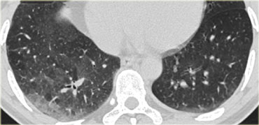
RB-ILD (3)
On the left a patient with DIP.
The HRCT shows diffuse areas of ground-glass density in the lower lobes and some mosaic pattern as the sole abnormality.
Reticular abnormalities and signs of fibrosis are typically absent.
These abnormalities are usually reversible and will disappear upon cessation of smoking.
RB-ILD (3)
Bên trái là một bệnh nhân bị DIP.
HRCT cho thấy các vùng khuếch tán của kính mờ ở thuỳ dưới và một số mẫu khảm như bất thường duy nhất.
Các bất thường về võng mạc và dấu hiệu xơ hóa thường không có.
Những bất thường này thường có thể đảo ngược và sẽ biến mất khi ngừng hút thuốc.
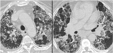 AIPAIPName
Acute interstitial pneumonia (AIP, earlier named Hamman Rich Pneumonitis) is a rare idiopathic lung disease characterized by diffuse alveolar damage with subsequent fibrosis.
It has a fatal outcome in many cases.
The histologic pattern aswell as the HRCT findings in AIP are indistinguishable from acute respiratory distress syndrome (ARDS).Viêm phổi kẽ cấp tính (AIP, trước đây có tên là viêm phổi kẹt Hamman) là một bệnh phổi vô căn hiếm gặp, đặc trưng bởi tổn thương phế nang lan toả với xơ hóa sau đó.
Kết cục tử vong trong nhiều trường hợp.
Mô hình mô học cũng như các dấu hiệu HRCT trong AIP không thể phân biệt được với hội chứng suy hô hấp cấp tính (ARDS).
The HRCT characteristics are diffuse or patchy consolidation, often with a crazy paving appearance like in the case on the left.
There are areas of consolidation and extensive areas of ground-glass density with a crazy-paving appearance.
These abnormalities developed in several days and this rapid progression of disease combined with these imaging findings are very suggestive of the diagnosis AIP.
Các đặc điểm HRCT là đông đặc lan toả hoặc loang lổ, thường có hình dạng lát đá điên (crazy paving) giống như trường hợp bên trái.
Có các vùng đông đặc và các vùng rộng lớn của kính mờ với mật độ lát đá điên (crazy paving).
Những bất thường này phát triển trong vài ngày và sự tiến triển nhanh chóng của bệnh kết hợp với những phát hiện hình ảnh này rất gợi ý chẩn đoán AIP.
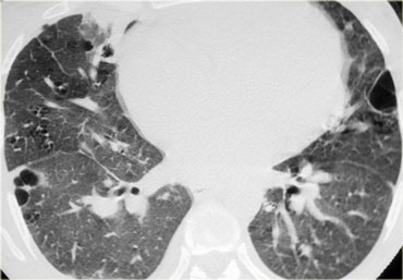
LIPLIPLanguage
Lymphocytic interstitial pneumonitis or LIP is uncommon, being seen mainly in patients with autoimmune disease, particularly Sj?gren's syndrome, and in patients with AIDS.
Symptoms are nonspecific and often those of the patient's underlying disease
Viêm phổi kẽ bạch cầu lympho hoặc LIP là không phổ biến, được thấy chủ yếu ở những bệnh nhân mắc bệnh tự miễn, đặc biệt là hội chứng Sj?gren và ở bệnh nhân AIDS.
Triệu chứng không đặc hiệu và thường là triệu chứng của bệnh nền của bệnh nhân
HRCT findings are usually nonspecific.
On the left a patient with Sjogren's syndrome with LIP.HRCT thường không đặc hiệu.
Bên trái là một bệnh nhân có hội chứng Sjogren với LIP.
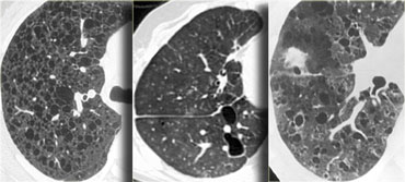 Lymphangiomyomatosis, LIP and Langerhans cell histiocytosisU cơ bạch huyết, LIP và bệnh mô bào Langerhans
On the left three different patients with lung cysts.
From left to right: Lymphangiomyomatosis, LIP and Langerhans cell histiocytosis.Bên trái là ba bệnh nhân khác nhau có nang phổi.
Từ trái sang phải: U cơ bạch huyết, LIP và mô bào Langerhans.
Drug-induced lung diseaseBệnh phổi do thuốc
Drug-induced lung disease is a major source of iatrogenic lung injury.
The major diagnostic problem is, that it may present with a large variety of radiologic patterns.
It may present as organizing pneumonia, eosinophilic pneumonia, fibrosis, hypersensitivity pneumonitis or even as ARDS.
The diagnosis of drug-induced pulmonary disease is usually one of exclusion.
Bệnh phổi do thuốc là một nguyên nhân chính gây tổn thương phổi do điều trị.
Vấn đề chẩn đoán chính là nó có thể xuất hiện với nhiều kiểu hình X quang khác nhau.
Nó có thể biểu hiện như viêm phổi tổ chức hóa, viêm phổi tăng bạch cầu ái toan, xơ hóa, viêm phổi quá mẫn hoặc thậm chí là ARDS.
Chẩn đoán bệnh phổi do thuốc thường là loại trừ.
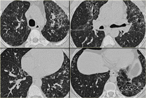 Drug-induced interstitial lung fibrosisXơ hóa phổi kẽ do thuốc
On the left a patient who is treated with cytotoxic drugs for a hematologic malignancy.
Bên trái là một bệnh nhân đang điều trị bằng thuốc gây độc tế bào cho một khối u ác tính về huyết học.
The radiographic findings are areas of ground glass opacity, some traction bronchiectasis and subtle honeycombing in the left lower lobe.
This could be the result of an idiopathic form of fibrosis like idiopathic pulmonary fibrosis and non-specific interstitial pneumonitis or fibrosis in chronic hypersensitivity pneumonitis and longstanding sarcoid.
However it is not one of the typical forms of fibrosis, that we commonly encounter in patients with a UIP pattern or NSIP pattern seen in collagenvascular diseases.
When there is fibrosis, that does not fit into any of the common diseases with fibrosis always consider drug-related lung disease in the differential.
Các dấu hiệu trên X quang là các vùng đám mờ kính mờ, một số giãn phế quản co kéo và tổ ong tinh tế ở thuỳ dưới bên trái.
Đây có thể là kết quả của một dạng xơ hóa vô căn như xơ phổi vô căn và viêm phổi kẽ không đặc hiệu hoặc xơ hóa ở viêm phổi quá mẫn mạn tính và sarcoid lâu năm.
Tuy nhiên, đây không phải là một trong những dạng xơ hóa điển hình mà chúng ta thường gặp ở những bệnh nhân có dạng UIP hoặc NSIP được thấy trong các bệnh mạch máu collagen.
Khi có xơ hóa, không phù hợp với bất kỳ bệnh phổ biến nào có xơ hóa luôn xem xét bệnh phổi liên quan đến thuốc trong phân biệt.
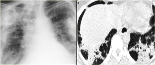 Drug-induced organizing pneumoniaViêm phổi tổ chức do thuốc
Drug-induced organizing pneumonia is commonly caused by Bleomycin and Cyclophosphamide and other drugs like Methotrexate, Amiodarone, Nitrofurantoin and Penicillamine (9).
The HRCT findings are the same as in cryptogenic organizing pneumonia.Viêm phổi tổ chức do thuốc thường do Bleomycin và Cyclophosphamide và các loại thuốc khác như Methotrexate, Amiodarone, Nitrofurantoin và Penicillamine (9) gây ra.
Các kết quả HRCT cũng giống như trong viêm phổi tổ chức hóa do cryptogenic.
Drug-induced non-specific interstitial pneumonita (NSIP) occurs most commonly as a manifestation of carmustine toxicity or of toxicity from noncytotoxic drugs such as amiodarone.
The radiologic findings are the same as in other forms of NSIP.Viêm phổi kẽ không đặc hiệu do thuốc (NSIP) xảy ra phổ biến nhất là biểu hiện của độc tính carmustine hoặc độc tính từ các thuốc không gây độc tế bào như amiodarone.
Các dấu hiệu X quang cũng giống như trong các dạng NSIP khác.
Uncommon interstitial lung diseasesBệnh phổi kẽ không phổ biến

LymphangiomyomatosisBệnh u cơ bạch huyết
- Rare disease, that occurs only in premenopausal women Bệnh hiếm gặp, chỉ xảy ra ở phụ nữ tiền mãn kinh
- Characterized by progressive proliferation of atypical muscle cells along the bronchioles leading to air trapping and the development of thin-walled cysts, that replace normal lung parenchyma.Đặc trưng bởi sự tăng sinh tiến triển của các tế bào cơ không điển hình dọc theo tiểu phế quản dẫn đến bẫy khí và sự phát triển của các nang có thành mỏng, thay thế nhu mô phổi bình thường.
- Identical pulmonary changes seen in 1% of patients with tuberous sclerosis (predominant involvement of young men). Những thay đổi phổi giống hệt nhau được thấy ở 1% bệnh nhân bị xơ cứng củ (chủ yếu là nam giới trẻ tuổi).
Clinical findings: Các dấu hiệu lâm sàng:
- Majority of patients present with dyspnea. Đa số bệnh nhân có biểu hiện khó thở.
- Chylous pleural effusions (40%), Pneumothorax (40%), hemoptysis (40%). Tràn dịch màng phổi tỳ đè (40%), tràn khí màng phổi (40%), ho ra máu (40%).
- Patients die within 10 years of the onset of symptoms. Bệnh nhân tử vong trong vòng 10 năm kể từ khi khởi phát triệu chứng.
- Pregnancy may exacerbate disease.Mang thai có thể làm trầm trọng thêm bệnh.
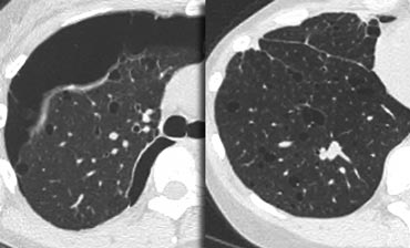
Key findings in Lymphangiomyomatosis:Những đặc điểm chính trong bệnh u cơ bạch huyết:
- Numerous thin-walled cysts, surrounded by normal parenchyma. Nhiều nang có thành mỏng, bao quanh bởi nhu mô bình thường.
- Cysts range from 2mm to 5cm in diameter, are round in shape and more or less uniform. U nang có đường kính từ 2 mm đến 5 cm, hình tròn và ít nhiều đồng nhất.
- Cysts are distributed diffusely throughout the lungs and upper and lower lobes are involved to a similar degree. U nang phân bố lan toả khắp phổi và thuỳ trên và dưới cũng có liên quan đến mức độ tương tự.
- Wall thickness of the cysts ranges from barely perceptible to 4 mm. Độ dày thành của u nang dao động từ chỉ nhận biết được đến 4 mm.
- Mediastinal or hilar adenopathy and pleural effusions (40%). Dị dạng hạch trung thất hoặc rốn phổi và tràn dịch màng phổi (40%).
- Recurrent pneumothorax (40%).Tái phát tràn khí màng phổi (40%).
On the left a typical case of LAM with multiple evenly spread thin walled cysts complicated by a pneumothorax.
Bên trái là một trường hợp LAM điển hình với nhiều nang mỏng lan toả đều, biến chứng là một tràn khí màng phổi.
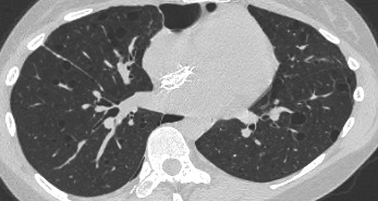 .
Differential diagnosis of Lymphangiomyomatosis: Chẩn đoán phân biệt của bệnh u cơ bạch huyết:
- Langerhans cell histiocytosis: > 90% are smokers, cysts have irregular shapes and the basal costophrenic angles are spared. Bệnh mô bào Langerhans: > 90% là người hút thuốc, nang có hình dạng không đều và các góc sườn-cổ không bị ảnh hưởng.
- Centrilobular emphysema: characterized by airspaces that have no perceptible wall, centrilobular artery seen as dot in the centre.Trung tâm tiểu thùy khí phế thũng: đặc trưng bởi các khoảng không khí không có thành cảm nhận được, động mạch trung tâm tiểu thùy được nhìn thấy như chấm ở trung tâm.
- Lymphoid interstitial pneumonitis: seen in patients with HIV and Sj?gren syndrome.Viêm phổi kẽ do lympho: gặp ở bệnh nhân HIV và hội chứng Sj?gren.
On the left another typical case of LAM.Bên trái là một trường hợp điển hình khác của LAM.

Langerhans cell histiocytosisBệnh mô bào Langerhans
Langerhans cell histiocytosis is also known as pulmonary histiocytosis X or eosinophilic granuloma.
LCH is probably an allergic reaction to cigarette smoke since more than 90% of patients are active smokers.
In the early nodular stage it is characterized by a centrilobular granulomatous reaction by Langerhans histiocytes.
In the cystic stage bronchiolar obliteration causes alveolar wall fibrosis and cyst formation.
Bệnh mô bào Langerhans còn được gọi là bệnh mô bào phổi X hoặc u hạt bạch cầu ái toan.
LCH có thể là một phản ứng dị ứng với khói thuốc lá vì hơn 90% bệnh nhân là người hút thuốc lá.
Trong giai đoạn đầu của nốt, nó được đặc trưng bởi phản ứng u hạt trung tâm tiểu thùy bởi các mô bào Langerhans.
Trong giai đoạn nang, sự tắc nghẽn phế quản gây ra xơ hóa thành phế nang và hình thành nang.
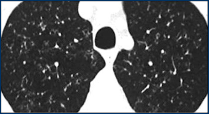 Early stage Langerhans cell histiocytosis with small nodulesGiai đoạn sớm của bệnh Langerhans với các nốt nhỏ
HRCT findings in Langerhans cell histiocytosis: Đặc điểm HRCT trong bệnh mô bào Langerhans:
- Early stage:Giai đoạn sớm:
- Small irregular or stellate nodules in centrilobular location.Các nốt nhỏ không đều hoặc hình sao ở vị trí trung tâm tiểu thùy.
- Late stage (more commonly seen) Giai đoạn muộn (thường gặp hơn)
- Cystic airspaces Cysts have bizarre shapes, they may coalesce and than become larger.Nang khí Nang có hình dạng kỳ lạ, chúng có thể kết hợp lại và trở nên lớn hơn.
- Upper and mid lobe predominance.Thuỳ trên và giữa chiếm ưu thế.
- Recurrent pneumothorax.Tràn khí màng phổi tái phát.
On the left early stage Langerhans cell histiocytosis with small nodules.
There are no cysts visible.
Ở giai đoạn sớm bên trái, mô bào Langerhans có các nốt nhỏ.
Không có nang có thể nhìn thấy.
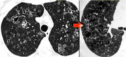 Late stage Langerhans' cell histiocytosis. Cysts progress to typical bizarre shaped cysts.Giai đoạn cuối Bệnh mô bào Langerhans Nang tiến triển thành nang hình dạng kỳ lạ điển hình.
In a later stage the nodules start to cavitate and become cysts.
These cysts start as round structures but finally coalesce to become the typical bizarre shaped cysts of LCH.
In patients with LCH 95% have a smoking history.Ở giai đoạn sau, các nốt bắt đầu hình thành và trở thành nang.
Những nang này bắt đầu như các cấu trúc tròn nhưng cuối cùng kết hợp lại để trở thành các nang có hình dạng kỳ lạ điển hình của LCH.
Ở những bệnh nhân có LCH 95% có tiền sử hút thuốc.
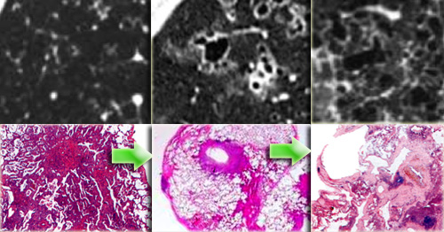 Specimen of Langerhans cell histiocytosis in three different stagesMẫu mô bệnh học của bệnh mô bào Langerhans ở ba giai đoạn khác nhau
On the left radiological pathological correlation of Langerhans cell histiocytosis in respectively nodular stage and early and late cystic stage.Về tương quan giải phẫu bệnh trên X quang bên trái của bệnh mô bào Langerhans ở giai đoạn nốt và giai đoạn nang sớm và muộn.
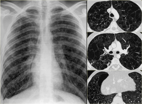 Langerhans' cell histiocytosisBệnh mô bào Langerhans
On the left a chest film of a 19 year old patient with Langerhans cell histiocytosis.
The dominant findig on the chest film is a reticular patern and that's about as far as you can go.
There is also hyperinflation.
No way you would have recognized that this pattern was caused by multiple cysts.
This is late stage Langerhans cell histiocytosis.Bên trái là phim ngực của một bệnh nhân 19 tuổi mắc bệnh mô bào Langerhans.
Phát hiện nổi bật trên phim ngực là một patern dạng lưới và đó là khoảng cách xa nhất mà bạn có thể đi.
Ngoài ra còn có tình trạng tăng áp lực.
Không có cách nào bạn có thể nhận ra rằng mô hình này là do nhiều nang.
Đây là bệnh mô bào Langerhans giai đoạn muộn.
The most challenging differential diagnosis in this patient is centrilobular emphysema.
Emphysema however is defined as airspaces without definable walls.
Usually we can identify the central dot sign.
The upper lobe predominance is not helpfull in the differential as we can appreciate this in many inhalational diseases and also in emphysema.
Chẩn đoán phân biệt khó nhất ở bệnh nhân này là trung tâm tiểu thùy khí phế thũng.
Tuy nhiên, khí phế thũng được định nghĩa là các vùng không khí không có các bức tường xác định được.
Thông thường chúng ta có thể xác định dấu chấm trung tâm.
Thuỳ trên chiếm ưu thế không có ích trong chẩn đoán phân biệt vì chúng ta có thể đánh giá được điều này trong nhiều bệnh lý hô hấp và cả trong khí phế thũng.
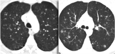 Langerhans cell histiosytosis: early phase and late phaseBệnh mô học tế bào Langerhans: giai đoạn sớm và giai đoạn muộn
On the left another case of Langerhans' cell histiocytosis.
It started as small noduli, which progressed over time to cavitating nodules.
In the end this will progress to bizarre shaped cysts, that replace normal lung tissue.Bên trái là một trường hợp mô bào Langerhans khác.
Nó bắt đầu như những nốt nhỏ, tiến triển theo thời gian để tạo thành các nốt sần.
Cuối cùng, điều này sẽ tiến triển thành các nang có hình dạng kỳ lạ, thay thế mô phổi bình thường.
Video
Images of a young male smoker with Langerhans cell histiocytosis.
Notice progression on second scan 7 years later.Video
Hình ảnh một người đàn ông trẻ hút thuốc lá mắc bệnh mô bào Langerhans.
Lưu ý sự tiến triển trên lần chụp thứ hai 7 năm sau.
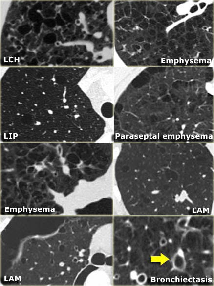
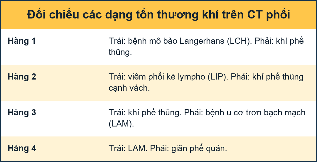
Differential diagnosis of Langerhans cell histiocytosis. Chẩn đoán phân biệt của bệnh mô bào Langerhans.
-
Nodular LCH:Nốt LCH:
- Sarcoidosis: perilymphatic distribution. Sarcoidosis: Phân bố quanh bạch mạch.
- Metastases: random distribution.Di căn: phân bố ngẫu nhiên.
- Cystic LCH:U nang LCH:
- LAM: round cysts, evenly distribution in women in the child-bearing ageLAM: nang tròn, phân bố đều ở phụ nữ trong độ tuổi sinh đẻ
- Cystic bronchiectasis: 'signet ring sign'.Cystic giãn phế quản: là dấu hiệu nhẫn siget.
- Centrilobular emphysema: no walls, central dot.Trung tâm tiểu thùy khí phế thũng: không có vách, điểm trung tâm.
- LIPLIPLanguage
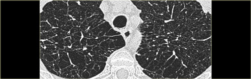 Emphysema mimicking Langerhans cell histiocytosisKhí phế thũng bắt chước bệnh mô bào Langerhans
Emphysema, when it is severe, can mimick Langerhans cell histiosytosis.
When it extends beyond the centrilobular area to the edge of the secondary lobule, it may look as if it is cystic with walls.
In patients with LCH, the pathologist may find LCH, but also areas of emphysema, respiratory bronchiolitis and even fibrosis.
So these smoking-related diseases do not represent discrete entities.
Khí phế thũng, khi nó nặng, có thể bắt chước bệnh mô bào Langerhans.
Khi nó kéo dài ra ngoài khu vực trung tâm tiểu thùy đến rìa của thuỳ thứ cấp, nó có thể trông như thể nó là nang có thành.
Ở những bệnh nhân bị LCH, nhà nghiên cứu bệnh học có thể tìm thấy LCH, nhưng cũng có các khu vực của khí phế thũng, viêm tiểu phế quản hô hấp và thậm chí là xơ hóa.
Vì vậy, các bệnh liên quan đến hút thuốc này không đại diện cho các thực thể riêng biệt.
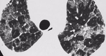 Alveolar proteinosis with crazy paving patternProtein phế nang với mô hình lát đá điên (crazy paving)
Alveolar proteinosisProtein phế nang
Alveolar proteinosis is a rare disease characterized by filling of the alveolar spaces with PAS positive material due to an abnormality in surfactant metabolism.
The diagnosis is based on the suggestive HRCT pattern (crazy paving) and the characteristic features of BAL fluid (Broncho Alveolar Lavage)
Bệnh protein phế nang là một bệnh hiếm gặp, đặc trưng bởi sự lấp đầy các khoang phế nang bằng vật liệu PAS dương tính do bất thường trong chuyển hóa chất hoạt động bề mặt.
Chẩn đoán dựa vào mô hình HRCT gợi ý (lát đá điên (crazy paving)) và các đặc điểm dịch BAL (Broncho Alveolar Lavage)
- Usually between 30 and 50 years old. Thường từ 30 đến 50 tuổi.
- Nonproductive cough, fever, and mild dyspnea. Ho khan, sốt, và khó thở nhẹ.
- Prognosis has improved since the advent of treatment using bronchoalveolar lavage.Tiên lượng đã được cải thiện kể từ khi điều trị bằng phương pháp rửa phế quản phế nang.
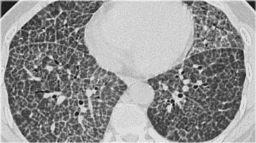
Key findings in alveolar proteinosis Những đặc điểm chính trong bệnh lý protein phế nang
- Crazy paving pattern: reticular pattern superimposed on ground glass opacification.Mô hình lát đá điên (crazy paving): mô hình dạng lưới chồng lên kính mờ.
- Opacifications range from ground glass to consolidation.Các vùng đục dao động từ kính mờ đến đông đặc.
On the left a typical case of alveolar proteinosis with extensive thickening of interlobular and intra-lobular septa.
This is caused by the fact that the proteinacious material, which is removed from the alveolar space by macrophages is transported to the interstitium and thus leads to thickening of septa.Bên trái là một trường hợp điển hình của bệnh protein phế nang với sự dày lên của vách liên thuỳ và vách trong thuỳ.
Điều này là do thực tế là các chất đạm, được các đại thực bào loại bỏ khỏi phế nang, được vận chuyển đến mô kẽ và do đó dẫn đến sự dày lên của vách ngăn.
The crazy paving pattern is a rather non-specific finding.
Many other diseases may present with this finding and are listed in the differential diagnosis.
Mô hình lát đá điên (crazy paving) là một phát hiện khá không đặc hiệu.
Nhiều bệnh khác có thể xuất hiện với dấu hiệu này và được liệt kê trong chẩn đoán phân biệt.
Differential diagnosis of alveolar proteinosis Chẩn đoán phân biệt của bệnh protein phế nang
- Non cardiogenic edema: ARDS, Acute Interstitial Pneumonia.Phù không do tim: ARDS, viêm phổi kẽ cấp.
-
Pneumonia:Viêm phổi:
- Infection (PCP and CMV).Nhiễm trùng (PCP và CMV).
- OP (organizing pneumonia).OP (viêm phổi tổ chức).
- Chronic eosinophilic pneumonia.Viêm phổi tăng bạch cầu ái toan mạn tính.
- Hemorrhage.Xuất huyết.
- Bronchoalveolar ca.Nắp phế quản.
Special Thanks
The authors like to thank Dr. Sujal Desai of the King's College Hospital in London for his inspiring lectures.
Some of the images used in this overview were provide by him.
We would also like to thank Dr. Richard Webb who produced such a fabulous educational CD (1).
Đặc biệt cảm ơn
Các tác giả muốn cảm ơn Tiến sĩ Sujal Desai của Bệnh viện King 's College ở London vì những bài giảng đầy cảm hứng của ông.
Một số hình ảnh được sử dụng trong tổng quan này đã được ông cung cấp.
Chúng tôi cũng muốn cảm ơn Tiến sĩ Richard Webb, người đã tạo ra một CD giáo dục tuyệt vời như vậy (1).
ReferencesTài liệu tham khảo
-
by Schaefer-Prokop, C, Prokop M, Fleischmann D, Herold CJ.
European Radiology 2001;11: 373-392
Schaefer-Prokop, C, Prokop M, Fleischmann D, Herold CJ. -
High Resolution Lung CT, UCSF Interactive Radiology Series on CD-ROM
Phim chụp cắt lớp vi tính phổi độ phân giải cao, UCSF Interactive Radiology Series trên CD-ROM
This browser-based learning file is based on Dr. Webb's HRCT text.
It offers a wide variety of cases dealing with common HRCT patterns of disease, diffuse lung diseases and their significance, and clinical characteristics.
It is one of the best educational CD's ever made.
Tập tin học tập dựa trên trình duyệt này dựa trên văn bản HRCT của Tiến sĩ Webb. -
by Hansell DM.
Radiol Clin North Am 2001:39: 1115-35
bởi Hansell DM. -
by Webb, Mueller and Naidich. Webb, Mueller và Naidich.
-
by Zampatori M, Sverzellati N, Poletti V et al.
Semin Ultrasound CT MR
2005;20(3): 176-85
Zampatori M, Sverzellati N, Poletti V và cộng sự. -
American Thoracic Society/European Respiratory Society International Multidisciplinary Consensus Classification of the Idiopathic Interstitial Pneumonias
Hiệp hội lồng ngực Hoa Kỳ / Hiệp hội hô hấp châu Âu Phân loại đồng thuận đa ngành quốc tế về viêm phổi kẽ tự phát
This Joint Statement of the American Thoracic Society (ATS), and the European Respiratory Society (ERS) was adopted by the ATS Board of Directors, June 2001 and by The ERS Executive Committee, June 2001 Tuyên bố chung này của Hiệp hội Lồng ngực Hoa Kỳ (ATS) và Hiệp hội Hô hấp Châu Âu (ERS) đã được Hội đồng Quản trị ATS, June 2001 và Ủy ban Điều hành ERS, June 2001 thông qua.
-
What Every Radiologist Should Know about Idiopathic Interstitial Pneumonias
Những gì mỗi bác sĩ chẩn đoán hình ảnh nên biết về viêm phổi kẽ tự phát
by Christina Mueller-Mang, MD, Claudia Grosse, MD, Katharina Schmid, MD, Leopold Stiebellehner, MD, and Alexander A. Bankier, MD
RadioGraphics 2007;27:595-615
Christina Mueller-Mang, MD, Claudia Grosse, MD, Katharina Schmid, MD, Leopold Stiebellehner, MD, và Alexander A. Bankier, MD -
Respiratory bronchiolitis, respiratory bronchiolitis-associated interstitial lung disease, and desquamative interstitial pneumonia: different entities or part of the spectrum of the same disease process?
Viêm tiểu phế quản hô hấp, viêm tiểu phế nang liên quan đến viêm tiểu phẳng hô hấp bệnh phổi kẽ và viêm phổi kẽ bong vảy: các thực thể khác nhau hoặc một phần của phổ của cùng một quá trình bệnh?
by LE Heyneman, S Ward, DA Lynch, M Remy-Jardin, T Johkoh and NL Muller
American Journal of Roentgenology, Vol 173, 1617-1622
bởi LE Heyneman, S. Ward, DA Lynch, M. Remy-Jardin, T. Johkoh và NL Muller -
Pulmonary Drug Toxicity: Radiologic and Pathologic Manifestations
Ngộ độc thuốc phổi: Biểu hiện X quang và bệnh lý
by Santiago E. Rossi, MD, Jeremy J. Erasmus, MD, H. Page McAdams, MD, Thomas A. Sporn, MD and Philip C. Goodman, MD.
Radiographics. 2000;20:1245-1259
bởi Santiago E. Rossi, MD, Jeremy J. Erasmus, MD, H. Page McAdams, MD, Thomas A. Sporn, MD và Philip C. Goodman, MD.