
Shoulder instability - MRIMất vững khớp vai trên MRI
Radiology department of the Rijnland hospital, Leiden and the Onze Lieve Vrouwe Gasthuis, Amsterdam, the NetherlandsKhoa Chẩn đoán hình ảnh của bệnh viện Rijnland, Leiden và Onze Lieve Vrouwe Gasthuis, Amsterdam, Hà Lan
Ngày đăng / cập nhật: 2012-05-21
Bản gốc trên radiologyassistant.nl ↗ · tải về lúc 2026-11-19 20:19 · bản dịch máy có chuẩn hóa thuật ngữ chẩn đoán hình ảnh
A Bankart lesion is an injury of the anterior glenoid labrum due to anterior shoulder dislocation.
These labral tears make the shoulder unstable and susceptible to repeated dislocations.
In this article we will focus on:Tổn thương Bankart là tổn thương của sụn viền ổ chảo trước do trật khớp vai trước.
Những vết rách này làm cho vai không ổn định và dễ bị trật khớp lặp đi lặp lại.
Trong bài viết này, chúng tôi sẽ tập trung vào:
- Shouder dislocationsTrật khớp shouder
- Bankart tears and variantsRách Bankart và các biến thể
IntroductionMở đầu
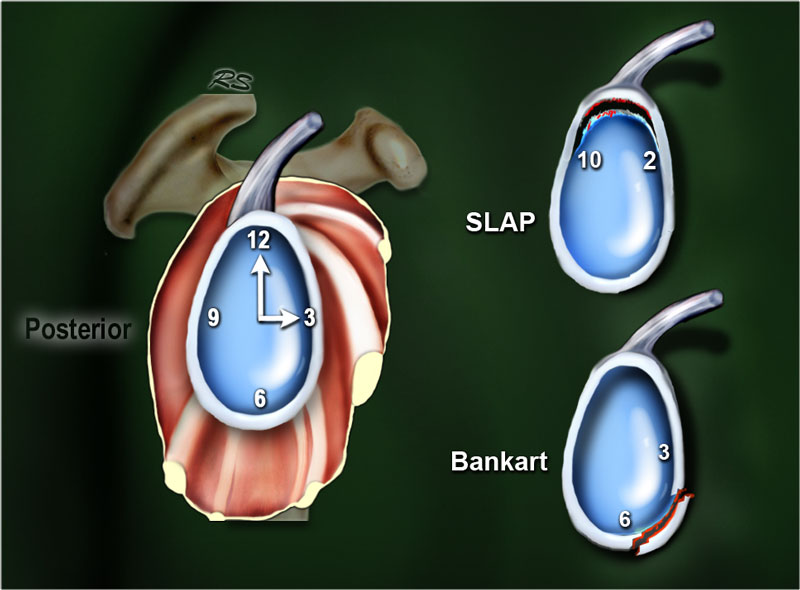
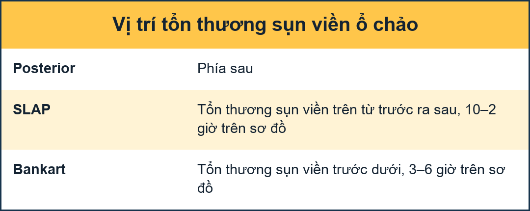 Clockwise approach. Click on image to enlargeCách tiếp cận theo chiều kim đồng hồ. Nhấp vào hình ảnh để phóng to
Clockwise approach to labral pathologyTiếp cận theo chiều kim đồng hồ trong bệnh lý sụn viền
A Clockwise approach to the labrum is the easiest way to diagnose labral tears and to differentiate them from normal labral variants.
There are two types of labral tears: SLAP tears and Bankart lesions.
SLAP is an acronym that stands for 'Superior Labral tear from Anterior to Posterior'.
SLAP tears start at the 12 o'clock position where the biceps anchor is located, which tears the labrum off the glenoid.
SLAP tears typically extend from the 10 to the 2 o'clock position, but can extend more posteriorly or anteriorly and even extend into the biceps tendon.
Bankart lesions are typically located in the 3-6 o'clock position because that's where the humeral head dislocates. Cách tiếp cận theo chiều kim đồng hồ đến xương ức là cách dễ nhất để chẩn đoán rách sụn viền và phân biệt chúng với các biến thể sụn bình thường.
Có hai loại rách sụn viền: rách SLAP và tổn thương Bankart.
SLAP là từ viết tắt của cụm từ tiếng Anh “Superior Labral tear from anterior to Posterior ” (tạm dịch: Rách sụn viền trên từ trước ra sau).
Rách SLAP bắt đầu ở vị trí 12 giờ, nơi neo cơ nhị đầu nằm, làm rách xương ức ra khỏi ổ chảo.
Rách SLAP thường kéo dài từ vị trí 10 đến 2 giờ, nhưng có thể kéo dài ra phía sau hoặc phía trước và thậm chí kéo dài vào gân cơ nhị đầu.
Các tổn thương Bankart thường nằm ở vị trí 3-6 giờ vì đó là nơi đầu xương cánh tay bị trật.
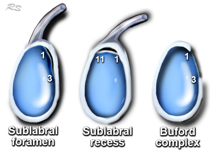
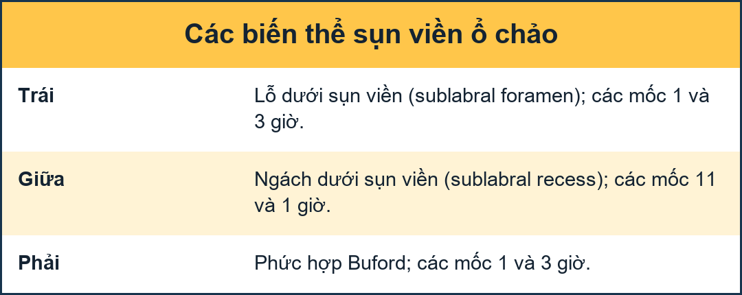
There are many labral variants that may simulate a labral tear.
They also have a typical location.
They are not in the 3-6 o'clock position, which makes it easy to differentiate them from a Bankart tear.
A Bankart tear can extend to the 1-3 o'clock position, but then there should also be a tear in the 3-6 o'clock position.
Labral variants however may mimick a SLAP tear.Có nhiều biến thể labral có thể mô phỏng một vết rách labral.
Chúng cũng có một vị trí điển hình.
Chúng không nằm ở vị trí 3-6 giờ, điều này giúp dễ dàng phân biệt chúng với rách Bankart.
Rách Bankart có thể kéo dài đến vị trí 1-3 giờ, nhưng sau đó cũng nên có một vết rách ở vị trí 3-6 giờ.
Tuy nhiên, các biến thể Labral có thể giống với rách SLAP.
DislocationTrật khớp
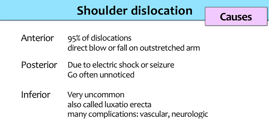
The shoulder is a very mobile and therefore unstable joint.
It is the most dislocated joint in the body.
Khớp vai là một khớp rất di động và do đó không ổn định.
Đây là khớp trật khớp nhiều nhất trong cơ thể.
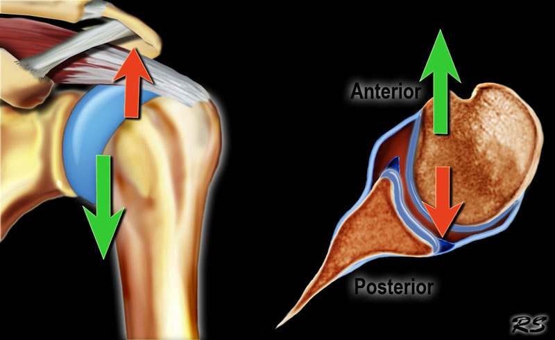
The shoulder most commonly dislocates in an anterior and inferior direction, as superior motion is restricted by the acromion, coracoid process, and rotator cuff.
Posterior motion is limited by the posterior rim of the glenoid, which is typically in an anteverted position.
Vai thường trật khớp theo hướng trước và dưới, vì chuyển động trên bị hạn chế bởi mỏm cùng vai, mỏm quạ và chóp xoay.
Chuyển động ra sau bị giới hạn bởi bờ sau của ổ chảo, thường ở vị trí lệch về phía trước.
Posterior dislocations are uncommon and seen due to seizures, electric shock or in patients with dysplasia of the glenoid.Trật khớp sau ít gặp và gặp do co giật, sốc điện hoặc ở bệnh nhân loạn sản ổ chảo.
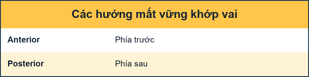
Anterior dislocationTrật khớp trước
The shoulder most commonly dislocates in an anterior and inferior direction, as superior motion is restricted by the acromion, coracoid process, and rotator cuff (Figure).
Posterior motion is limited by the posterior rim of the glenoid, which is typically in an anteverted position (red arrow).Vai thường trật khớp theo hướng trước và dưới, vì chuyển động trên bị hạn chế bởi mỏm cùng vai, mỏm quạ và chóp xoay (Hình).
Chuyển động ra sau bị giới hạn bởi vành sau của ổ chảo, thường ở vị trí lệch về phía trước (mũi tên màu đỏ).
The dislocation of the humeral head to antero-inferior causes damage to the antero-inferior rim of the glenoid in the 3 - 6 o'clock position (marked in red).
Especially in younger patients this results in a Bankart fracture or a Bankart lesion which is a tear of the anteroinferior labrum.
This results in instability and recurrent dislocations.
Due to these recurrent dislocations significant bone loss and erosion of the anterior glenoid rim may occur, which maintains the unstable situation.Trật khớp chỏm xương cánh tay ra trước-hạ gây tổn thương đến vành trước-hậu của ổ chảo ở vị trí 3-6 giờ (được đánh dấu màu đỏ).
Đặc biệt ở những bệnh nhân trẻ tuổi, điều này dẫn đến gãy xương Bankart hoặc tổn thương Bankart là rách xương ức trước dưới.
Điều này dẫn đến mất vững và trật khớp tái phát.
Do những trật khớp tái phát này có thể gây mất xương đáng kể và xói mòn bờ trước của ổ chảo, điều này duy trì tình trạng không ổn định.
Scroll through images.Cuộn qua các hình ảnh.
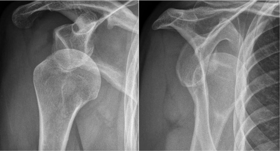
The images show a subtle Bankart fracture (arrows).Hình ảnh cho thấy một gãy Bankart tinh tế (các mũi tên).
Scroll through images.Cuộn qua các hình ảnh.
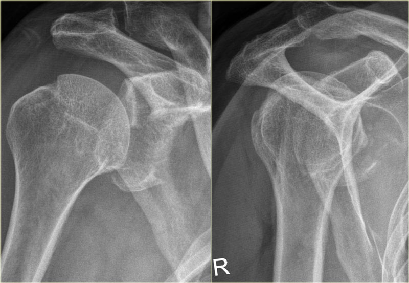
Bankart fractureGãy Bankart
This is a post-reduction view.
Notice the very large fracture of the glenoid rim with displacement.Đây là quan điểm sau nắn chỉnh.
Chú ý đến vết gãy rất lớn của vành ổ chảo với sự dịch chuyển.
Scroll through images.Cuộn qua các hình ảnh.
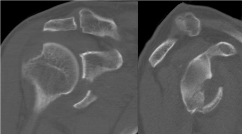
On the coronal-oblique and sagittal reconstruction the displaced fragment of the glenoid rim is seen in the 3-6 o'clock position.
On the coronal image a large Hill-Sachs defect is seen.
This is a bone defect as result of the impaction of the glenoid rim on the humeral head.Trên tái tạo đoạn vành tai-xoắn và dọc, mảnh vỡ di lệch của vành ổ chảo được nhìn thấy ở vị trí 3-6 giờ.
Trên hình ảnh vành, có thể thấy một lỗ hổng Hill-Sachs lớn.
Đây là một khiếm khuyết xương do sự tác động của vành ổ chảo lên đầu xương cánh tay.
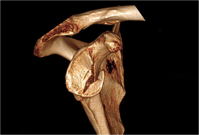
3D-reconstruction of a large bony Bankart in the 2 - 6 o'clock position.Tái tạo 3D một xương lớn Bankart ở vị trí 2 - 6 giờ.
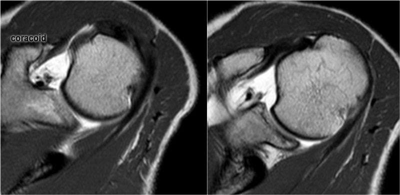
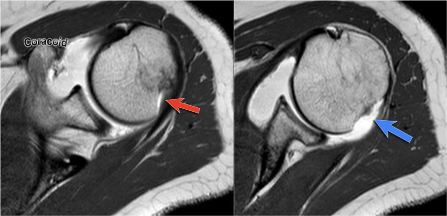 LEFT: Hill-Sachs at level of coracoid. RIGHT: Normal groove seen at level below coracoid.Trái: Hill-Sachs ở mức coracoid. Phải: Rãnh bình thường được nhìn thấy ở mức dưới coracoid.
Hill-Sachs
On MR a Hill-Sachs defect is seen at or above the level of the coracoid process.Trên cộng hưởng từ, có thể thấy một lỗ Hill-Sachs ở hoặc trên mức của mỏm quạ.
Hill-Sachs is a posterolateral depression of the humeral head.
It is above or at the level of the coracoid in the first 18 mm of the proximal humeral head.
It is seen in 75-100% of patients with anterior instability.
The physiologic groove in the humerus or cysts and erosions at the attachment site of the infraspinatus tendon can simulate a Hill-Sachs, but usually this is not a diagnostic problem (figure).Hill-Sachs là một chỗ lõm sau bên của đầu xương cánh tay.
Nó nằm trên hoặc ngang mức mỏm quạ trong 18 mm đầu gần xương cánh tay đầu tiên.
Nó được thấy ở 75-100% bệnh nhân có mất vững trước.
Rãnh sinh lý ở xương cánh tay hoặc nang và chỗ bám của gân cơ dưới gai có thể mô phỏng Hill-Sachs, nhưng thường thì đây không phải là vấn đề chẩn đoán.
Posterior dislocationTrật khớp sau
Posterior dislocations are uncommon and easily missed, because there is less displacement compared to the anterior dislocation.
In cases of glenoid dysplasia, the posterior glenoid rim is often flattened, reducing bony stability against posterior translation of the humeral head.
This anatomical variation is more frequently associated with posterior instability and, consequently, posterior dislocations.Trật khớp ra sau ít gặp và dễ bị bỏ sót, vì có ít di lệch so với trật khớp ra trước.
Trong các trường hợp loạn sản ổ chảo, vành ổ chải sau thường bị dẹt, làm giảm sự ổn định của xương đối với sự dịch chuyển sau của đầu xương cánh tay.
Sự biến đổi giải phẫu này thường liên quan đến sự mất vững sau và do đó, trật khớp sau.
Images
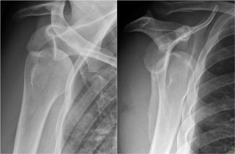
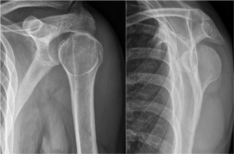
On the AP-view the head looks strange due to the internal rotation.
On the transscapular-Y view the humeral head is displaced posteriorly.
Sometimes the displacement is difficult to appreciate, especially when the transscapular-Y view is slightly rotated.
An axillary view can be helpful, but when in doubt CT is necessary.Hình ảnh
Trên chế độ xem AP, đầu trông lạ do xoay trong.
Trên phim X-quang xương vai, đầu xương cánh tay di lệch ra sau.
Đôi khi rất khó đánh giá sự dịch chuyển, đặc biệt là khi góc nhìn xuyên xương vai-Y hơi xoay.
Quan sát vùng nách có thể hữu ích, nhưng khi nghi ngờ CT là cần thiết.
Scroll through images.Cuộn qua các hình ảnh.
 Posterior dislocationTrật khớp sau
Images Hình minh họa
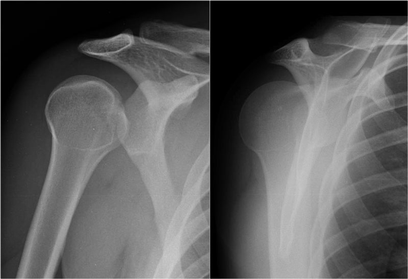
- AP-view: Distance between the humeral head and the glenoid is abnormally wide.AP-view: Khoảng cách giữa đầu xương cánh tay và ổ chảo rộng bất thường.
- Transscapular-Y view: the humeral head is displaced posteriorly. Hình ảnh xuyên vai-Y: đầu xương cánh tay di lệch ra sau.
Posterior dislocations are not as obvious on the X-rays as an anterior dislocation.
Approximately half of the posterior shoulder dislocations go undiagnosed on initial presentation, because of a low level of clinical suspicion and insufficient imaging.Trật khớp ra sau không rõ ràng trên X-quang như trật khớp ra trước.
Khoảng một nửa số trật khớp vai sau không được chẩn đoán khi có biểu hiện ban đầu, vì mức độ nghi ngờ lâm sàng thấp và chẩn đoán hình ảnh không đầy đủ.
 Posterior dislocation-fractureTrật khớp sau-gãy xương
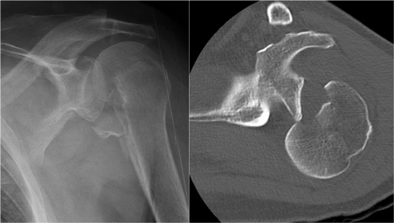
Posterior dislocations account for 2-4% of all shoulder dislocations.
Posterior dislocations are associated with epileptic seizures, high energy trauma, electroconvulsive therapy and dysplasia of the glenoid.Trật khớp vai sau chiếm 2-4% tổng số trật khớp vai.
Trật khớp sau có liên quan đến động kinh, chấn thương năng lượng cao, liệu pháp sốc điện và loạn sản ổ chảo.
On the images a posterior dislocation is seen with a fracture.Trên hình ảnh, trật khớp sau được nhìn thấy với một vết gãy.
The MR-images are of a patient who had undergone both an anterior aswell as a posterior dislocation.
This resulted in both a Hill-Sachs impression fracture on the posterior aspect of the humeral head (blue arrow) and an impression fracture on the anterior aspect as a result of posterior dislocation (red arrow).Hình ảnh cộng hưởng từ là một bệnh nhân đã trải qua cả trật khớp trước và trật khớp sau.
Điều này dẫn đến cả gãy xương Hill-Sachs ở mặt sau của đầu xương cánh tay (mũi tên màu xanh) và gãy xương ở mặt trước do trật khớp sau (mũI tên màu đỏ).
Scroll through images.Cuộn qua các hình ảnh.
 Superior dislocationTrật khớp trên
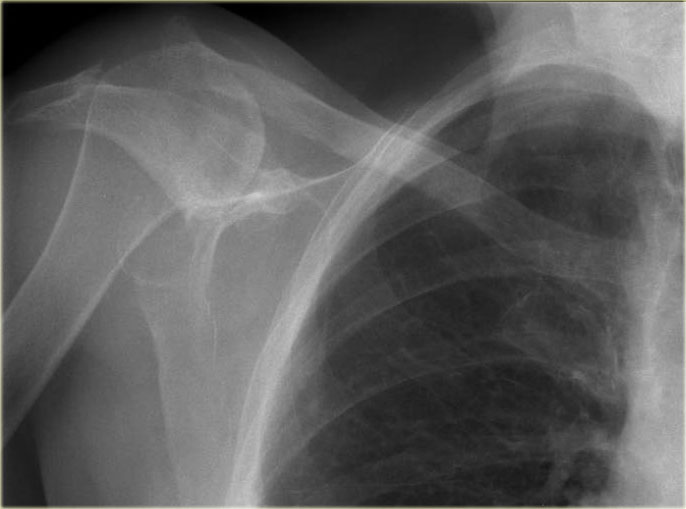
This was an incidental finding on a chest radiograph.Đây là phát hiện tình cờ trên phim X-quang ngực.
There is a superior dislocation of the humeral head, likely secondary to a chronic, massive rotator cuff tear.
This has resulted in progressive cranialization of the humeral head and acromial erosion.Có trật khớp trên của đầu xương cánh tay, có thể là thứ phát sau rách chóp xoay mạn tính, lớn.
Điều này đã dẫn đến sự sọ não tiến triển của đầu xương cánh tay và xói mòn mỏm cùng vai.
Bankart and variantsBankart và các biến thể
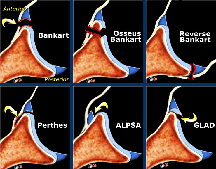
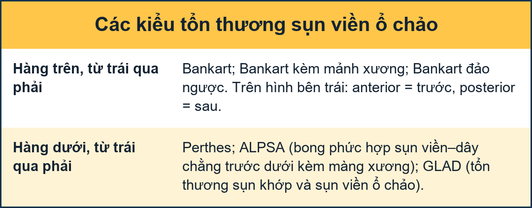 Axial images of Bankart lesions and variantsẢnh mặt phẳng ngang của tổn thương Bankart và các biến thể
Bankart-lesions and variants like Perthes and ALPSA are injuries to the anteroinferior labrum.
These injuries are always located in the 3-6 o'clock position because they are caused by an anterior-inferior dislocation.
The only exception to this rule is the reverse Bankart, which is the result of a posterior dislocation and injury to the inferoposterior labrum.
Bankart tears may extend to superior, but this is uncommon. Tổn thương Bankart và các biến thể như Perthes và ALPSA là những tổn thương ở vùng trước xương ức.
Những chấn thương này luôn nằm ở vị trí 3-6 giờ vì chúng gây ra bởi trật khớp trước-sau.
Ngoại lệ duy nhất đối với quy tắc này là Bankart đảo ngược, đó là kết quả của trật khớp sau và tổn thương xương ức sau.
Rách Bankart có thể lan lên trên, nhưng điều này không phổ biến.
- Bankart
Detachment of the anteroinferior labrum (3-6 o'clock) with complete tearing of the anterior scapular periosteum with or without an osseus fragment of the glenoid. Trang chủ
Tách xương ức trước dưới (3-6 giờ) với rách hoàn toàn màng xương mỏm móc trước có hoặc không có mảnh xương con của ổ chảo. - Reverse Bankart
Detachment of the posteroinferior labrum (6-9 o'clock) with tearing of the posterior scapular periosteum with or without an osseus fragment of the glenoid. Đảo ngược Bankart
Tháo xương ức sau dưới (6-9 giờ) với rách màng xương vai sau có hoặc không có mảnh xương con của xương ức. - Perthes
Detachment of the anteroinferior labrum (3-6 o'clock) with medially stripped but intact periosteum.Trang chủ
Tách xương ức trước dưới (3-6 giờ) với lớp màng xương bị tước bỏ ở giữa nhưng còn nguyên vẹn. - ALPSA = Anterior Labral Periosteal Sleeve Avulsion.
Medially displaced labroligamentous complex with absence of the labrum on the glenoid rim.ALPSA = Nhổ sụn viền trước sụn.
Phức hợp dạng sợi chuyển vị vào trong với sự vắng mặt của xương ức trên vành ổ chảo. - GLAD = GlenoLabral Articular Disruption.
Represents a partial tear of anteroinferior labrum with adjacent cartilage damage.GLAD = GlenoLabral Articular Disruption.
Biểu hiện rách một phần xương ức trước dưới kèm theo tổn thương sụn kế cận.
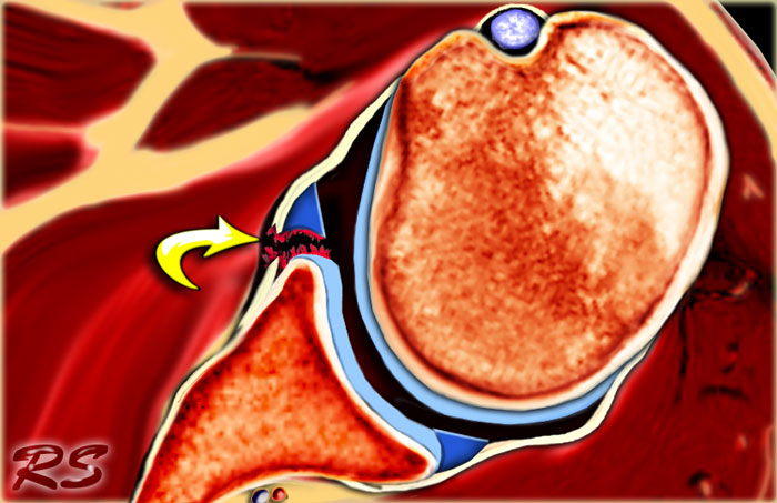 Bankart lesionTổn thương Bankart
Bankart lesions are labral tears without an osseus fragment.
MR arthrography or arthroscopy are optimal to diagnose Bankart or Bankart-like lesions.
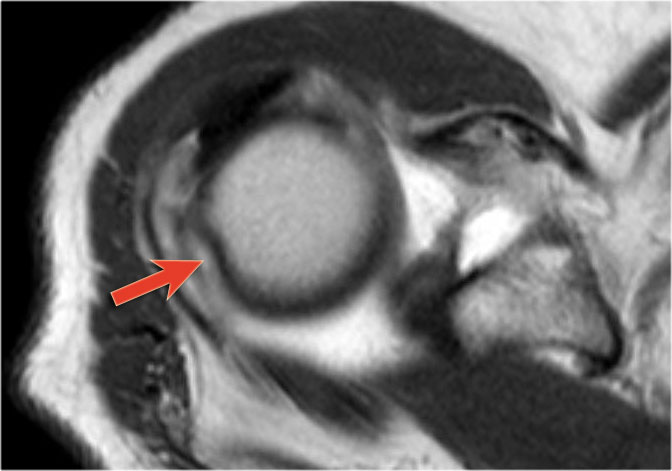
There is a detachment of the anteroinferior labrum (3-6 o'clock) with complete tearing of the anterior scapular periosteum.
The arrow points to the disrupted periosteum.Tổn thương Bankart là rách sụn viền mà không có mảnh xương con.
Chụp cộng hưởng từ hoặc nội soi khớp là phương pháp tối ưu để chẩn đoán tổn thương Bankart hoặc tổn thương giống Bankart.
Có một bong gân trước dưới (3-6 o 'clock) với rách hoàn toàn màng xương vai trước.
mũi tên chỉ ra màng xương bị đứt.
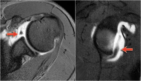
On MR-athrography the labrum is missing on the anterior glenoid and the labral fragment is displaced anteriorly (arrow). Trên MR-athrography, xương ức bị mất ở ổ chảo trước và mảnh xương ức di lệch ra trước (mũi tên).
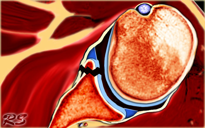 Osseus Bankart
Bankart lesions with an osseus fragment are common findings in patients with an anterior dislocation and are frequently seen on the x-rays or CT-scan.
Tổn thương Bankart với mảnh xương con là những phát hiện phổ biến ở bệnh nhân trật khớp trước và thường thấy trên phim X-quang hoặc CT-scan.
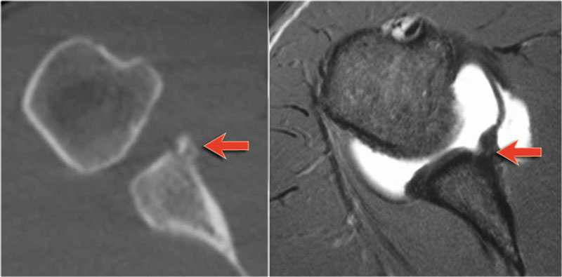 CT and MR-arthrography in a patient with an osseus Bankart (arrow)Chụp CT và MR-động kinh ở bệnh nhân có xương con Bankart (mũi tên)
On MR-arthrography it may be difficult to depict the osseus fragment.
On CT it is easy to appreciate the osseus fragment of the anterior glenoid (arrow).Trên cộng hưởng từ có thể khó mô tả mảnh xương con.
Trên CT, dễ dàng đánh giá được mảnh xương con của lườn trước (mũi tên).
Scroll through the images.
There is an osseus Bankart lesion (curved red arrow).
The tear extends to superior (black arrows).
There is also a Hill-Sachs defect (red arrow).Cuộn qua các hình ảnh.
Có tổn thương xương cong Bankart (mũi tên cong đỏ).
Vết rách lan lên trên (các mũi tên màu đen).
Ngoài ra còn có một khuyết tật Hill-Sachs (mũi tên màu đỏ).
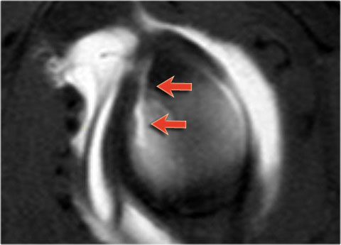 Bankart lesion with superior extentionTổn thương Bankart với phần trên giãn rộng
Sagittal MR-arthrogram demonstrates the superior extension of the Bankart tear.Chụp MR-arthrogram dọc cho thấy sự mở rộng trên của rách Bankart.
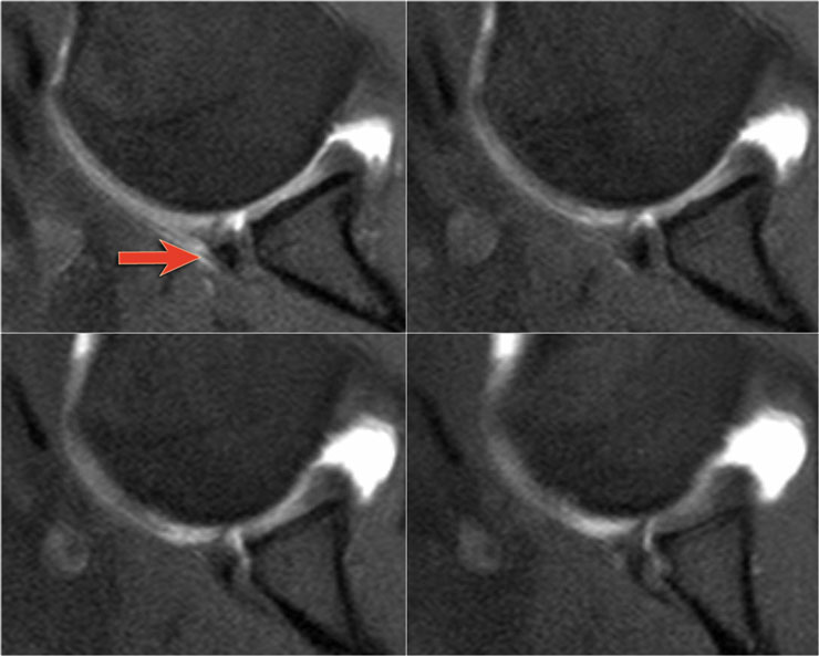 Osseus Bankart on a MR arthrogram ABER-viewOsseus Bankart trên phim cộng hưởng từ khớp ABER-view
Here another patient with an osseus Bankart seen on four consecutive images of a MR arthrogram in ABER-view.
Notice the abnormal contour of the anterior glenoid and the avulsed anterior rim (arrow)Ở đây một bệnh nhân khác có xương con Bankart được nhìn thấy trên bốn hình ảnh chụp cộng hưởng từ khớp liên tiếp trong chế độ xem ABER.
Nhận xét đường viền bất thường của ổ chảo trước và bờ trước bị bong (mũi tên)
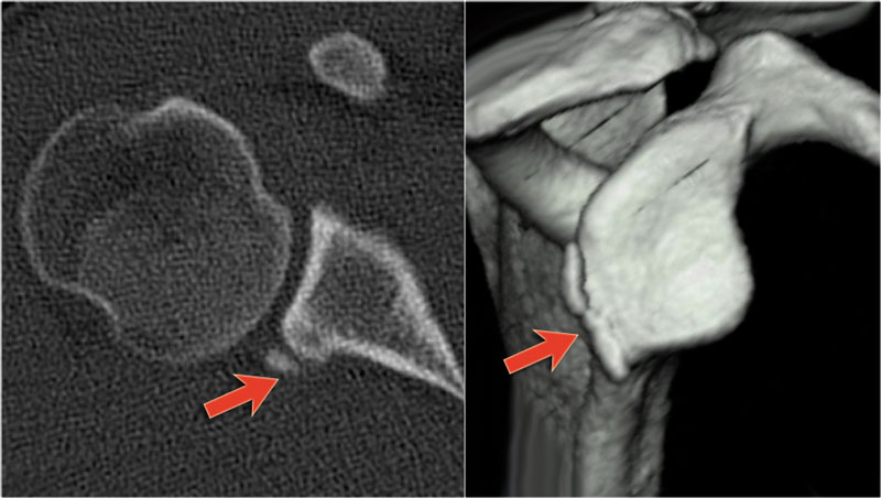
Reverse BankartĐảo ngược Bankart
CT-images in another patient show a reversed osseus Bankart in a patient with posterior dislocation.Hình ảnh CT ở một bệnh nhân khác cho thấy xương con Bankart bị đảo ngược ở một bệnh nhi trật khớp sau.
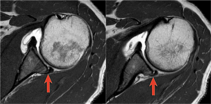
Axial MR-arthrogram of a reverse Bankart.Chụp cộng hưởng từ khớp vai của bệnh nhân Bankart.
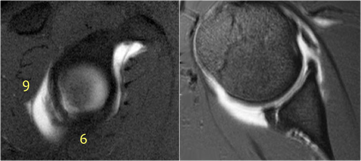 Reverse BankartĐảo ngược Bankart
Another example of a reverse Bankart.
Notice the detatched labrum at the 6-9 o'clock position on the sagittal MR-arthrogram.Một ví dụ khác về một Bankart đảo ngược.
Lưu ý xương ức bị xước ở vị trí 6-9 giờ trên MR-arthrogram đứng dọc.
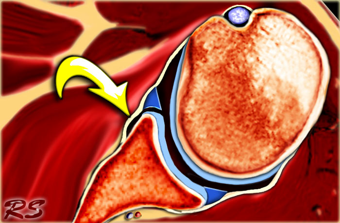 Perthes lesion: the labrum is thorn, but the periosteum is intact and only stripped (arrow)Tổn thương quanh thân xương: sụn viền là gai, nhưng màng xương còn nguyên vẹn và chỉ bị tước bỏ (mũi tên)
Perthes lesionTổn thương Perthes
A Perthes lesion is a labroligamentous avulsion like a Bankart, but with a medially stripped intact periosteum.
On images of the shoulder with the arm in a neutral position, the torn labrum may be held in its normal anatomic position by the intact scapular periosteum, which thereby prevents contrast media from entering the tear.
This means that MR-arthrography with the arm in the neutral position may fail to detect the labral tear.
In the ABER position however there is tension on the antero-inferior labrum by the stretched anterior band of the inferior glenohumeral ligament and you have more chance to detect the tear.
The arrow points to the intact periosteum.Tổn thương Perthes là một tổn thương dạng bong bóng giống như tổn thương Bankart, nhưng với màng xương còn nguyên vẹn.
Trên hình ảnh của vai với cánh tay ở vị trí trung tính, xương ức bị rách có thể được giữ ở vị trí giải phẫu bình thường bởi màng xương vai nguyên vẹn, do đó ngăn không cho chất cản quang xâm nhập vào vết rách.
Điều này có nghĩa là chụp cộng hưởng từ với cánh tay ở vị trí trung tính có thể không phát hiện được rách sụn viền.
Tuy nhiên, ở tư thế ABER, có sự căng thẳng lên xương ức trước-sau bởi dải trước của dây chằng ổ chảo dưới được kéo căng và bạn có nhiều cơ hội phát hiện ra vết rách hơn.
mũi tên chỉ vào màng xương còn nguyên vẹn.
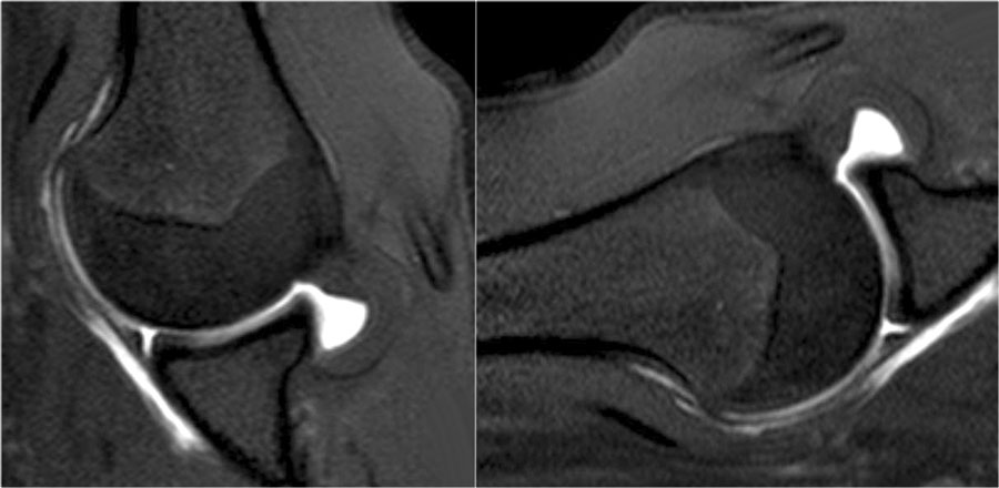 LEFT: normal ABER-view. RIGHT: Image rotated 90? anti-clockwise.Bên trái: chế độ xem ABER bình thường. Bên phải: Hình ảnh xoay 90 độ ngược chiều kim đồng hồ.
The images in ABER-position demonstrate a detached anterior labrum.
The image on the right is rotated 90? anti-clockwise.
Sometimes this makes it easier to understand the anatomy.Hình ảnh ở vị trí ABER cho thấy một xương ức trước bị bong ra.
Hình ảnh bên phải được xoay 90 độ ngược chiều kim đồng hồ.
Đôi khi điều này làm cho việc hiểu giải phẫu dễ dàng hơn.
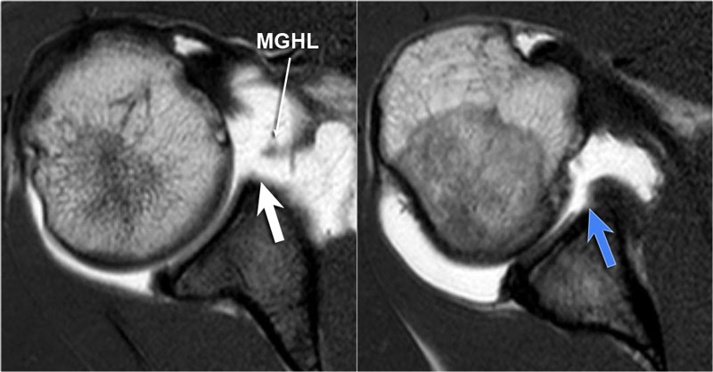

Images of a MR-arthrogram.
The image on the left shows an absent anterosuperior labrum, which is called a Buford complex.
The image on the right shows a cartilage defect in the 4 o'clock position.
It is not clear whether the labrum is normal.
Continue with the images in ABER-position.Hình ảnh chụp cộng hưởng từ khớp.
Hình ảnh bên trái cho thấy một vùng trước trên không có xương ức, được gọi là phức hợp Buford.
Hình ảnh bên phải cho thấy một khiếm khuyết sụn ở vị trí 4 giờ.
Không rõ liệu xương ức có bình thường hay không.
Tiếp tục với các hình ảnh ở vị trí ABER.
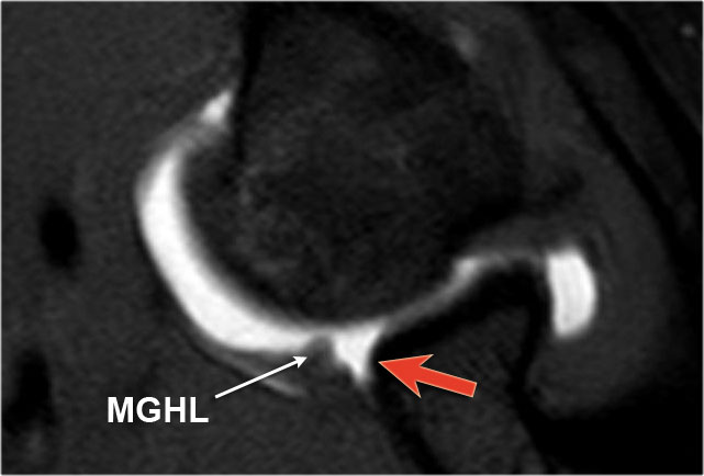
In the ABER-position it is obvious that there is a Perthes lesion (black arrow).
Due to the ABER-position the anterior band of the inferior GHL creates tension on the anteroinferior labrum and contrast fills the tear.Ở vị trí ABER, rõ ràng là có tổn thương Perthes (mũi tên đen).
Do vị trí ABER, dải trước của GHL dưới tạo ra sức căng trên xương ức trước dưới và thuốc cản quang lấp đầy vết rách.
The red arrow points to the absent labrum - Buford complex.Mũi tên màu đỏ chỉ ra phức hợp Labrum-Buford không có.
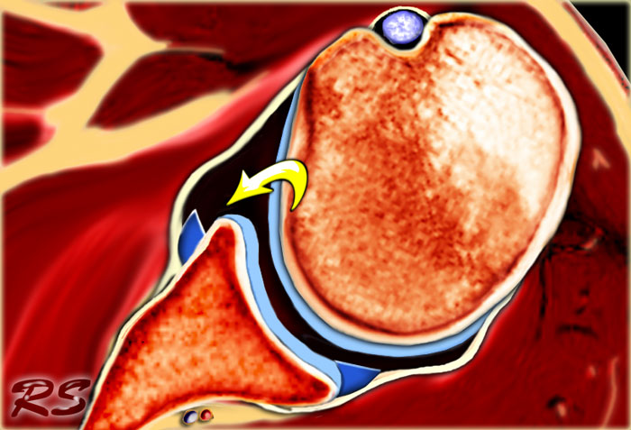 ALPSA.
ALPSAALPSAName
An ALPSA-lesion is an Anterior Labral Periosteal Sleeve Avulsion.
The anterior labrum is absent on the glenoid rim.
The arrow points to the medially displaced labroligamentous complex.Tổn thương ALPSA là một tổn thương bong vạt sụn màng xương sụn trước.
Không có xương ức trước ở bờ ổ chảo.
mũi tên chỉ ra phức hợp dạng sợi dịch chuyển vào trong.
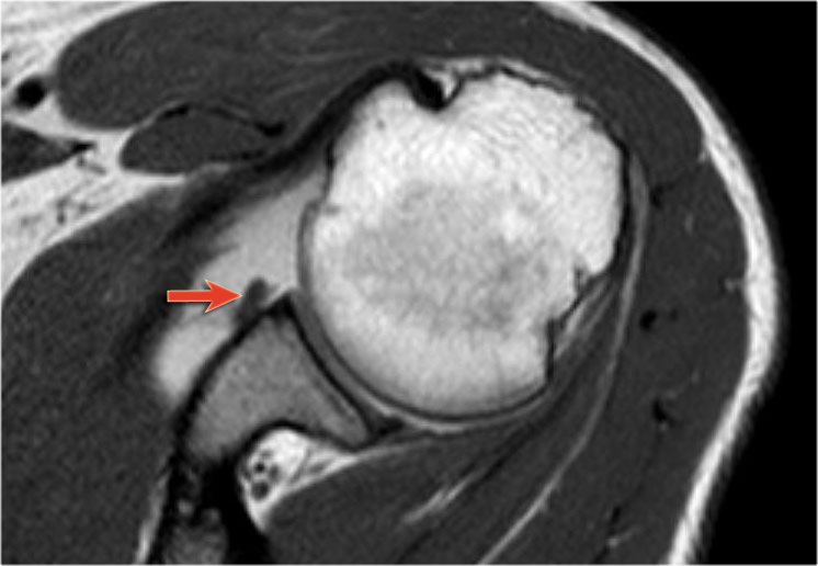
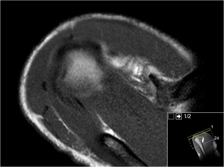
Images of a patient with an ALPSA-lesion.
Notice the medially displaced labrum.Hình ảnh một bệnh nhân có tổn thương ALPSA.
Chú ý đến xương ức di lệch về phía trong.
 ALPSA-lesionALPSA-sự tổn thương
Images of another patient with an ALPSA-lesion.
The ABER-view shows an absent antero-inferior labrum.
The coronal images shows the medially displaced labrum (red arrow).Hình ảnh của một bệnh nhân khác bị tổn thương ALPSA.
Hình ảnh ABER cho thấy không có xương ức trước-dưới.
ảnh mặt phẳng đứng ngang cho thấy xương ức di lệch về phía trong (mũi tên màu đỏ).
This is a difficult case.
First scroll through the images and try to find out what is going on.
Then continue reading.Đây là một trường hợp khó.
Đầu tiên, hãy cuộn qua các hình ảnh và cố gắng tìm hiểu những gì đang xảy ra.
Sau đó tiếp tục đọc.
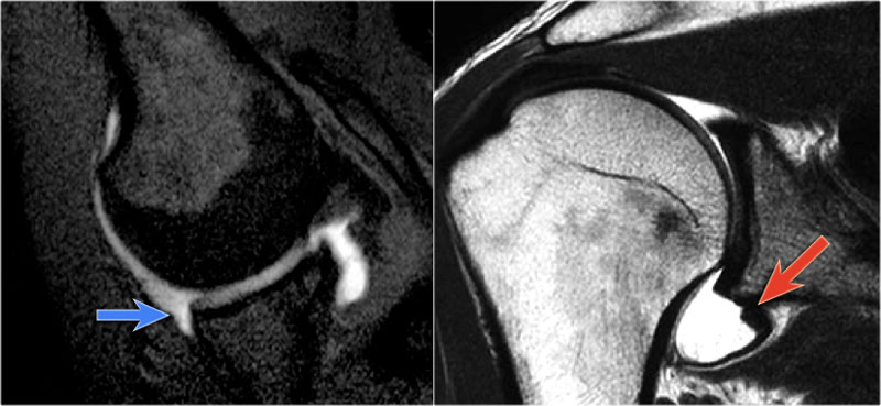
First notice the Hill-Sachs defect indicating a prior anterior dislocation (blue arrow).
Now you know that you have to look for a Bankart or variant.
Next notice the high signal at 12 o' clock (red arrows).
On coronal images you want to make sure whether this is a variant like a labral recess or labral foramen or whether this is a SLAP.
Notice how this high signal continues posteriorly, which means that it is a SLAP-lesion.
The yellow arrow points to the anterior glenoid rim.
The anterior labrum is absent at the 1-3 o 'clock position
This is a Buford complex, which is a normal variant.
The structure anterior to the glenoid is not a thorn labrum, but the middle glenohumeral ligament.
Notice extention of the SLAP-tear further to posterior (red arrow).
Finally there is a medially displaced inferoanterior labrum at the 3-6 o 'clock position, i.e. an ALPSA-lesion (black arrow).
Đầu tiên là thông báo khuyết Hill-Sachs cho thấy trật khớp trước (mũi tên màu xanh).
Bây giờ bạn biết rằng bạn phải tìm kiếm một Bankart hoặc biến thể.
Tiếp theo, nhận thấy tín hiệu cao ở 12 giờ (các mũi tên màu đỏ).
Trên ảnh mặt phẳng đứng ngang bạn muốn chắc chắn rằng đây là một biến thể như một hốc labral hay lỗ labral hay đây là một SLAP.
Lưu ý cách tín hiệu cao này tiếp tục về phía sau, điều đó có nghĩa là đó là một tổn thương SLAP.
mũi tên màu vàng chỉ vào vành trước của ổ chảo.
Không có xương ức trước ở vị trí 1-3 giờ
Đây là một phức hợp Buford, là một biến thể bình thường.
Cấu trúc trước ổ chảo không phải là gai xương cánh tay, mà là dây chằng ổ chải giữa.
Lưu ý sự giãn rộng của rách SLAP ra phía sau (mũi tên màu đỏ).
Cuối cùng là một vùng xương ức phía trong di lệch vào trong ở vị trí 3-6 giờ, tức là một tổn thương ALPSA (mũi tên đen).
 GLAD-lesionGLAD-sự tổn thương
GLAD
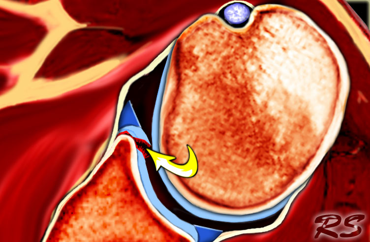
A GLAD-lesion is a GlenoLabral Articular Disruption.
It represents a patial tear of the anteroinferior labrum with adjacent cartilage damage.
The arrow points to the cartilage defect.Tổn thương GLAD là tổn thương GlenoLabral.
Nó đại diện cho một vết rách của xương ức trước dưới với tổn thương sụn liền kề.
mũi tên chỉ ra khuyết sụn.
 GLAD-lesionGLAD-sự tổn thương
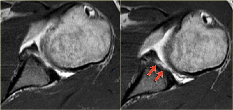
The images show a partial tear of the anteroinferior labrum with adjacent cartilage damage at the 4-6 o 'clock position (arrows).Hình ảnh cho thấy một vết rách một phần của xương ức trước dưới với tổn thương sụn liền kề ở vị trí 4 - 6 giờ (các mũi tên).
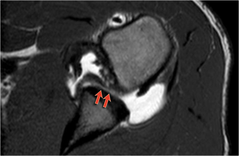
Scroll through the images.
There is a Bankart lesion with extension into the cartilage, i.e a GLAD-lesion (red arrows).Cuộn qua các hình ảnh.
Có một tổn thương Bankart lan rộng vào sụn, tức là tổn thương GLAD (các mũi tên màu đỏ).
HAGLHAGLName
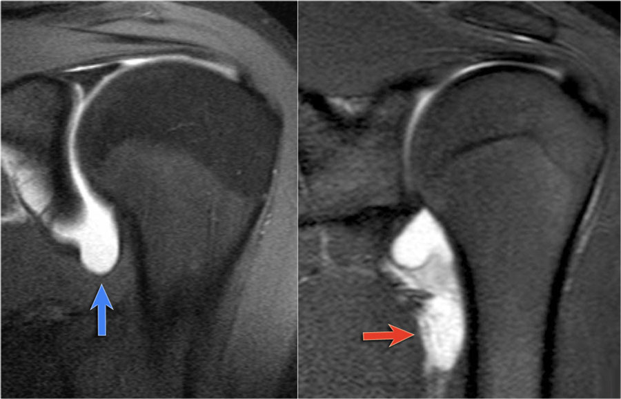 LEFT: Normal axillary recess (blue arrow). RIGHT: Abnormal axillary recess due to avulsion of the IGHL (red arrow)Trái: lõm nách bình thường (mũi tên màu xanh). Phải: hốc nách bất thường do IGHL (mũi tên màu đỏ)
HAGL is a Humeral Avulsion of the inferior Glenohumeral Ligament.
There is discontinuity of the IGHL attachment on the humerus with leakage of contrast. HAGL là một bong gân cơ cánh tay của dây chằng Glenohumeral dưới.
Có sự gián đoạn của sự gắn kết IGHL trên xương cánh tay với sự rò rỉ chất cản quang.
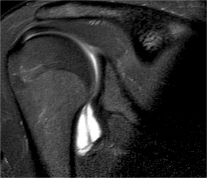
Another patient with an avulsion of the inferior glenohumeral ligament from the humeral insertion.Một bệnh nhân khác bị đứt dây chằng ổ chảo cánh tay dưới do chèn ép xương cánh tay.
ReferencesTài liệu tham khảo
-
Usefulness of the Abduction and External Rotation Views in Shoulder MR Arthrography
Tính hữu ích của chế độ xem giạng và xoay ngoài trong chụp cộng hưởng từ khớp vai
by Asgar M. Saleem, Joong K. Lee, Leon M. Novak AJR 2008; 191:1024-1030 của Asgar M. Saleem, Joong K. Lee, Leon M. Novak AJR 2008; 191: 1024-1030
-
MR Imaging and MR Arthrography of Paraglenoid Labral Cysts
Chụp cộng hưởng từ và cộng hưởng từ sỏi tuyến yên
by Glenn A. Tung et al
AJR June 2000 vol. 174 no. 6 1707-1715
Glenn A. Tung và cộng sự -
CT and MR Arthrography of the Normal and Pathologic Anterosuperior Labrum and Labral-Bicipital Complex
Chụp cắt lớp vi tính và cộng hưởng từ khớp vai bình thường và bệnh lý vùng trước trên xương chày và phức hợp xương chẩm
by Michel De Maeseneer et al
October 2000 RadioGraphics, 20, S67-S81.
bởi Michel De Maeseneer và cộng sự