
Vascular territories of the BrainCác vùng cấp máu của não
Radiology department of the Alrijne Hospital in Leiderdorp, the NetherlandsKhoa Chẩn đoán hình ảnh của Bệnh viện Alrijne ở Leiderdorp, Hà Lan
Ngày đăng / cập nhật: 2008-11-24
Bản gốc trên radiologyassistant.nl ↗ · tải về lúc 2026-11-19 20:19 · bản dịch máy có chuẩn hóa thuật ngữ chẩn đoán hình ảnh
Knowledge of the vascular territories is important, because it enables you to recognize infarctions in arterial territories, in watershed regions and also venous infarctions.
It also helps you to differentiate infarction from other pathology.Kiến thức về các vùng mạch máu là rất quan trọng, bởi vì nó cho phép bạn nhận ra nhồi máu ở các vùng động mạch, ở các vùng đầu nguồn và cả nhồi máu tĩnh mạch.
Nó cũng giúp bạn phân biệt nhồi máu với các bệnh lý khác.
Cerebral Arterial TerritoryLãnh thổ động mạch não
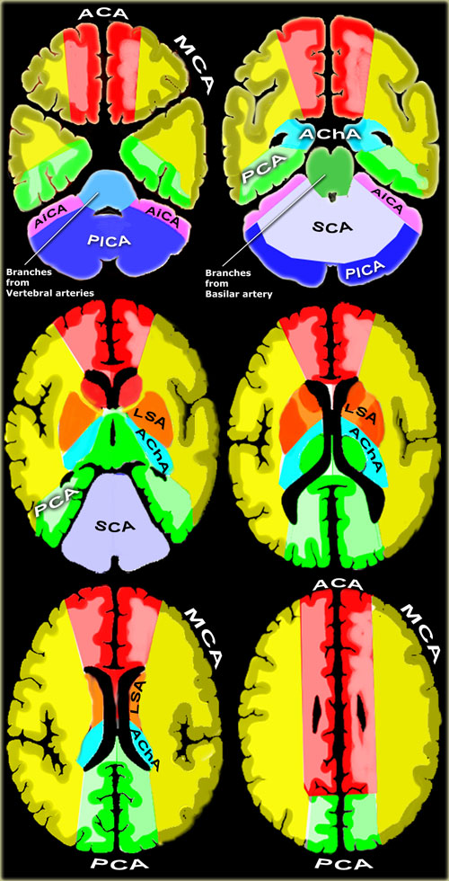
 Vascular territories of the cerebral arteries (adapted and modified with permission from M. Savoiardo (1)Lãnh thổ mạch máu của động mạch não (đã được điều chỉnh và sửa đổi với sự cho phép của M. Savoiardo (1)
- Posterior Inferior Cerebellar Artery (PICA in blue)
The PICA territory is on the inferior occipital surface of the cerebellum and is in equilibrium with the territory of the AICA in purple, which is on the lateral side (1).
The larger the PICA territory, the smaller the AICA and viceversa. Động mạch tiểu não dưới sau (PICA màu xanh)
Lãnh thổ PICA nằm trên bề mặt dưới chẩm của tiểu não và ở trạng thái cân bằng với lãnh thổ của AICA màu tím, nằm ở phía bên (1).
Lãnh thổ PICA càng lớn thì AICA càng nhỏ và ngược lại. - Superior Cerebellar Artery (SCA in grey)
The SCA territory is in the superior and tentorial surface of the cerebellum.
Động mạch tiểu não trên (SCA màu xám)
Lãnh thổ SCA nằm ở bề mặt trên và lều của tiểu não. - Branches from vertebral and basilar artery
These branches supply the medulla oblongata (in blue) and the pons (in green). Các nhánh từ đốt sống và động mạch nền
Các nhánh này cung cấp máu cho hành não (màu xanh) và các cầu não (mầu xanh lá). - Anterior Choroideal artery (AchA in blue))
The territory of the AChA is part of the hippocampus, the posterior limb of the internal capsule and extends upwards to an area lateral to the posterior part of the cella media.
Động mạch Choroideal trước (AchA màu xanh))
Lãnh thổ của AChA là một phần của hồi hải mã, chi sau của bao trong và kéo dài lên trên đến một khu vực bên cạnh phần sau của màng đệm. - Lenticulo-striate arteries
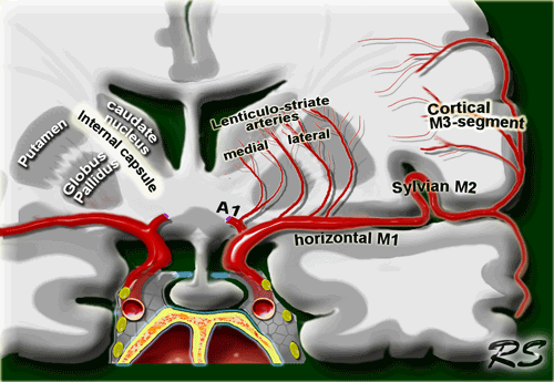
The lateral LSA' s (in orange) are deep penetrating arteries of the middle cerebral artery (MCA).
Their territory includes most of the basal ganglia.
The medial LSA' s (indicated in dark red) arise from the anterior cerebral artery (usually the A1-segment).
Heubner's artery is the largest of the medial lenticulostriate arteries and supplies the anteromedial part of the head of the caudate and anteroinferior internal capsule.
Động mạch vân Lenticulo
Động mạch não giữa ngoài (màu cam) là các động mạch xuyên sâu của động mạch não giữa (MCA).
Lãnh thổ của họ bao gồm hầu hết các hạch nền.
LSA trong (đỏ sẫm) phát sinh từ động mạch não trước (thường là đoạn A 1).
Động mạch Heubner là động mạch lớn nhất trong số các động mạch dạng thấu kính trong và cấp máu cho phần trước trong của đầu đuôi và bao trong trước dưới. - Anterior cerebral artery (ACA in red)
The ACA supplies the medial part of the frontal and the parietal lobe and the anterior portion of the corpus callosum, basal ganglia and internal capsule. Động mạch não trước (ACA màu đỏ)
ACA cung cấp phần trong của thuỳ trán và thuỳ đỉnh và phần trước của thể chai, hạch nền và bao trong. - Middle cerebral artery (MCA in yellow)
The cortical branches of the MCA supply the lateral surface of the hemisphere, except for the medial part of the frontal and the parietal lobe (anterior cerebral artery), and the inferior part of the temporal lobe (posterior cerebral artery).
The deep penetrating LSA-branches are discussed above. Động mạch não giữa (MCA màu vàng)
Các nhánh vỏ não của não tuỷ ngoài cung cấp máu cho bề mặt ngoài của bán cầu, ngoại trừ phần trong của thuỳ trán và thuỳ đỉnh (động mạch não trước) và phần dưới của thuỳ thái dương (độNG mạch não sau).
Các nhánh xuyên thấu sâu của LSA được thảo luận ở trên. - Posterior cerebral artery (PCA in green)
P1 extends from origin of the PCA to the posterior communicating artery, contributing to the circle of Willis.
Posterior thalamoperforating arteries branch off the P1 segment and supply blood to the midbrain and thalamus.
Cortical branches of the PCA supply the inferomedial part of the temporal lobe, occipital pole, visual cortex, and splenium of the corpus callosum. Động mạch não sau (PCA màu xanh lá cây)
P1 kéo dài từ nguồn gốc của PCA đến động mạch thông sau, góp phần vào vòng tròn Willis.
Các động mạch xuyên não sau chia nhánh ra khỏi đoạn P1 và cung cấp máu cho não giữa và đồi thị.
Các nhánh vỏ não của PCA cung cấp cho phần trong cùng của thuỳ thái dương, cực chẩm, vỏ não thị giác và lách của thể chai.
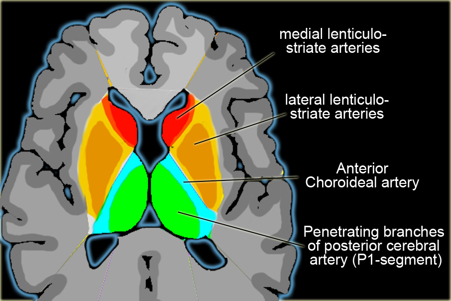

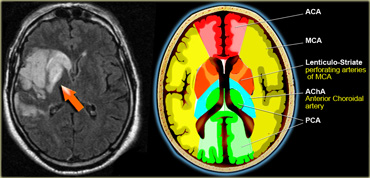
On the left a detail to illustrate the vascular supply to the basal ganglia.Bên trái là chi tiết minh hoạ cung cấp mạch máu cho hạch nền.
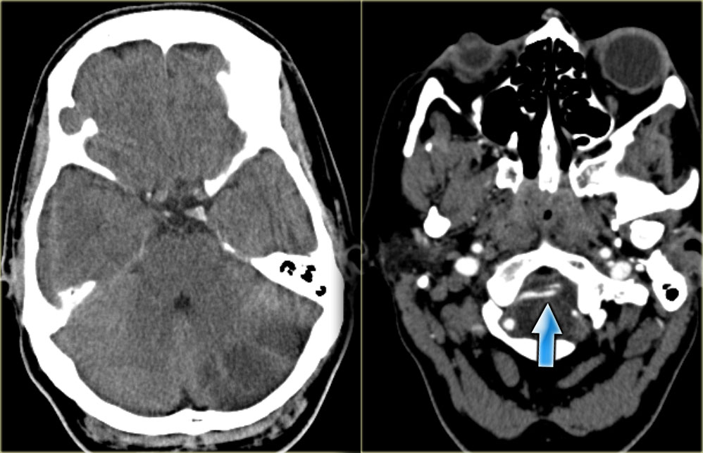
PICAPICAName
On the left CT-images of a left-sided PICA-infarction.
Notice the posterior extention.
The infarction was the result of a dissection (blue arrow).Trên CT-scan bên trái của một PICA-nhồi máu bên trái.
Chú ý đến phần sau của cơ thắt.
nhồi máu là kết quả của một lần bóc tách (mũi tên xanh).
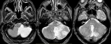
On the left MR-images of a left-sided PICA-infarction.
In unilateral infarcts there is always a sharp delineation in the midline because the superior vermian branches do not cross the midline, but have a sagittal course.
This sharp delineation may not be evident until the late phase of infarction.
In the early phase, edema may cross the midline and create diagnostic difficulties.
Trên hình ảnh MR bên trái của một PICA-nhồi máu bên trái.
Trong nhồi máu một bên, luôn có một đường nét rõ ở đường giữa vì các nhánh trên không đi qua đường giữa, nhưng có một đường dọc.
Sự phân định rõ ràng này có thể không được hiển nhiên cho đến giai đoạn cuối của nhồi máu.
Trong giai đoạn đầu, phù nề có thể vượt qua đường giữa và tạo ra những khó khăn trong chẩn đoán.
Infarctions at pontine level are usually paramedian and sharply defined because the branches of the basilar arery have a sagittal course and do not cross the midline.
Bilateral infarcts are rarely observed because these patients do not survive long enough to be studied, but sometimes small bilateral infarcts can be seen.Nhồi máu ở mức cầu não thường ở cạnh trong và được xác định rõ ràng vì các nhánh của quầng đáy có đường đi dọc và không vượt qua đường giữa.
Nhồi máu hai bên hiếm khi được quan sát thấy vì những bệnh nhân này không sống sót đủ lâu để được nghiên cứu, nhưng đôi khi có thể thấy nhồi máu nhỏ hai bên.
SCA
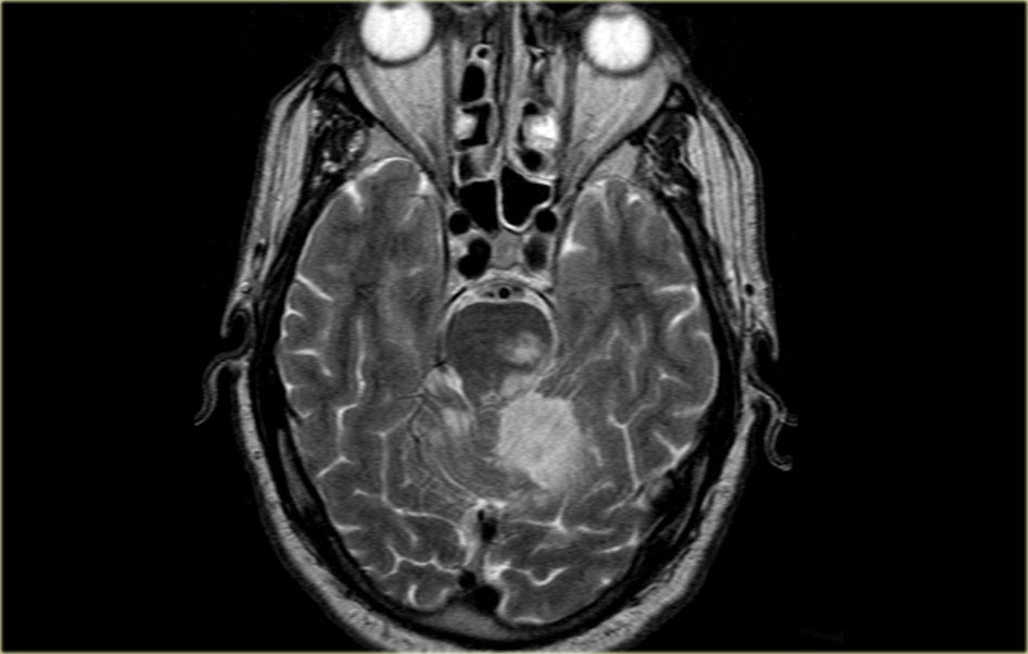
On the left MR-image of a cerebellar infarction in the region of the superior cerebellar artery and also in the brainstem in the territory of the PCA.
Notice the limitation to the midline.Trên hình ảnh MR bên trái của một tiểu não nhồi máu ở vùng động mạch tiểu não trên và cũng ở thân não trong lãnh thổ của PCA.
Lưu ý giới hạn của đường giữa.
 ACA infarctionNhồi máu ACA
ACA
Anterior cerebral artery: Động mạch não trước:
- A1 segment: from origin to anterior communicating artery and gives rise to medial lenticulostriate arteries (inferior parts of the head of the caudate and the anterior limb of the internal capsule). Đoạn A1: từ gốc đến động mạch thông trước và tạo ra các động mạch dạng thấu kính trong (phần dưới đầu đuôi và chi trước bao trong).
- A2 segment: from anterior communicating artery to bifurcation of pericallosal and callosomarginal arteries. Đoạn A2: từ động mạch thông trước đến chỗ chia đôi của động mạch quanh chóp và động mạch rìa.
- A3 segment: major branches (medial portions of frontal lobes, superior medial part of parietal lobes, anterior part of the corpus callosum). Đoạn A3: các nhánh chính (phần giữa thuỳ trán, phần giữa trên thuỳ đỉnh, phần trước thể chai).
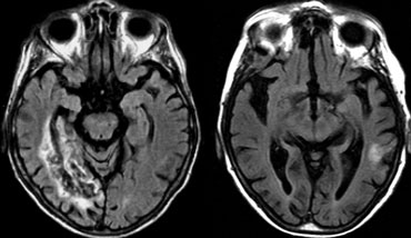 Infarction of right hippocampal region (courtesy Frederik Barkhof)Nhồi máu của vùng hải mã phải (do Frederik Barkhof cung cấp)
Anterior choroidal arteryĐộng mạch hắc mạc trước
The anterior choroidal artery originates from the internal carotid artery.
Rarely it arises from the middle cerebral artery.Động mạch mạc treo trước bắt nguồn từ động mạch cảnh trong.
Hiếm khi nó phát sinh từ động mạch não giữa.
The territory of the anterior choroidal artery encompasses part of the hippocampus, the posterior limb of the internal capsule and extends upwards to an area lateral to the posterior part of the cella media.
The whole area is rarely involved in AChA infarcts.
The posterior limb of the internal capsule also receives blood from the lateral lenticulostriate arteries.Lãnh thổ của động mạch màng đệm trước bao gồm một phần của hồi hải mã, chi sau của bao trong và kéo dài lên trên đến một khu vực bên phía sau của màng đệm.
Toàn bộ khu vực hiếm khi liên quan đến nhồi máu AChA.
Chi sau của bao trong cũng nhận máu từ các động mạch thấu kính ngoài.
On the left an uncommon infarction in the hippocampal region.
Part of the territory of the anterior choroidal artery and the PCA are involved.Bên trái một nhồi máu không phổ biến ở vùng hải mã.
Một phần của lãnh thổ của động mạch màng đệm trước và PCA có liên quan.
 Xem hình gốc (EN)
 MCA infarction. Involvement of cortical branches and deep perforating lenticulo-striate arteriesMCA nhồi máu. Sự tham gia của các nhánh vỏ não và các động mạch vân thấu kính xuyên sâu
Middle cerebral arteryĐộng mạch não giữa
The MCA has cortical branches and deep penetrating branches, which are called the lateral lenticulo-striate arteries.Động mạch não giữa có các nhánh vỏ và các nhánh xuyên sâu, được gọi là các động mạch vân thấu kính ngoài.
The territory of the lateral lenticulo-striate perforating arteries of the MCA is indicated with a different color from the rest of the territory of the MCA because it is a well-defined area supplied by penetrating branches, which may be involved or spared in infarcts separately from the main cortical territory of the MCA.
On the left a T2W-image of a patient with an infarction in the territory of the middle cerebral artery (MCA).
Notice that the lateral lenticulo-striate perforating arteries of the MCA are also involved (orange arrow).Lãnh thổ của các động mạch xuyên thấu kính-sơ đồ bên của MMCA được chỉ định với một màu khác với phần còn lại của lãnh thổ của MMCA vì nó là một khu vực được xác định rõ ràng được cung cấp bởi các nhánh xuyên thấU, có thể có liên quan hoặc không có trong các trường hợp nhồi máu tách biệt với lãnh thổ vỏ não chính của MMCA.
Bên trái là hình ảnh T2W của một bệnh nhân có nhồi máu trong vùng động mạch não giữa (MCA).
Lưu ý rằng các động mạch xuyên thấu kính bên của động mạch não giữa cũng có liên quan (mũi tên màu cam).
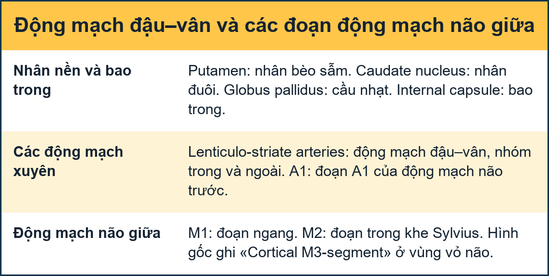
Lenticulostriate arteriesĐộng mạch bèo mắt
Medial lenticulostriate arteries
They are branches of the A1-segment of the anterior cerebral artery.
They supply the anterior inferior parts of the basal nuclei.
They also supply the anterior limb of the internal capsule together with the recurrent artery of Huebner, which also is a branch of the anterior cerebral artery.Động mạch vân thấu kính trong
Chúng là các nhánh của đoạn A1 của động mạch não trước.
Chúng cung cấp cho các phần trước dưới của nhân đáy.
Chúng cũng cung cấp máu cho chi trước của bao trong cùng với động mạch tái phát của Huebner, cũng là một nhánh của động mạch não trước.
Lateral lenticulostriate arteries
They are branches of the horizontal M1-segment of the middle cerebral artery.
They supply the superior part of the head and the body of the caudate nucleus, most of the globus pallidus and putamen.
They also supply the anterior limb of the internal capsule and parts of the posterior limb of the internal capsule, which is largely supplied by the anterior choroidal artery.Động mạch vân thấu kính ngoài
Chúng là các nhánh của đoạn M1 nằm ngang của động mạch não giữa.
Chúng cung cấp phần trên của đầu và thân của nhân đuôi, hầu hết cầu nhạt và nhân bèo sẫm.
Chúng cũng cung cấp máu cho chi trước của bao trong và các phần của chi sau của bao trong, phần lớn được cung cấp bởi động mạch hắc mạc trước.
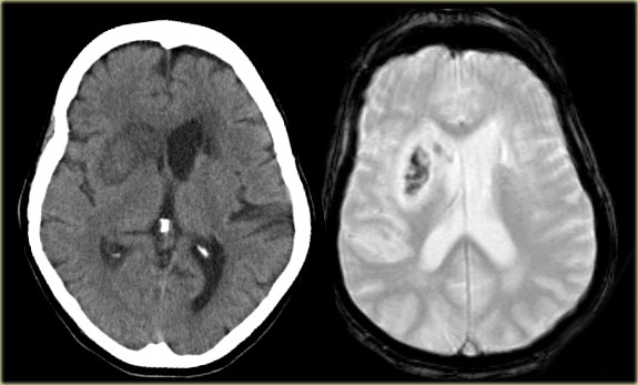 CT and T2W-gradient echo image of a hemorrhagic infarction limited to the territory of the lateral lenticulo-striate arteriesHình ảnh cắt lớp vi tính và siêu âm T2W của nhồi máu xuất huyết giới hạn trong phạm vi của các động mạch vân thấu kính bên
On the left images of a hemorrhagic infarction in the area of the deep perforating lenticulostriate branches of the MCA.Trên hình ảnh bên trái của một nhồi máu xuất huyết trong khu vực của các nhánh thấu kính xuyên sâu của MCA.
 MCA infarction with luxury perfusionMCA nhồi máu với tưới máu cao cấp
On the left enhanced CT-images of a patient with an infarction in the territory of the middle cerebral artery (MCA).
There is extensive gyral enhancement (luxury perfusion).
Sometimes this luxury perfusion may lead to confusion with tumoral enhancement.Trên hình ảnh CT tăng cường bên trái của một bệnh nhân có nhồi máu trong lãnh thổ của động mạch não giữa (MCA).
Có ngấm thuốc theo hồi não lan toả (tràn dịch xa).
Đôi khi sự tưới máu quá mức này có thể dẫn đến nhầm lẫn với sự tăng cường khối u.
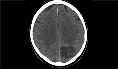 PCA infarctionNhồi máu
Posterior cerebral artery (PCA)Động mạch não sau (PCA)
Deep or proximal PCA strokes cause ischemia in the thalamus and/or midbrain, as well as in the cortex.
Superficial or distal PCA infarctions involve only cortical structures (4).Đột quỵ PCA sâu hoặc gần gây ra thiếu máu cục bộ ở đồi thị và / hoặc não giữa, cũng như ở vỏ não.
Nhồi máu PCA nông hoặc đầu xa chỉ liên quan đến cấu trúc vỏ não (4).
On the left a patient with acute vision loss in the right half of the visual field.
The CT demonstrates an infarction in the contralateral visual cortex, i.e left occipital lobe.
Bên trái là một bệnh nhân bị mất thị lực cấp tính ở nửa phải của thị trường.
CT cho thấy một nhồi máu ở vỏ não thị giác đối diện, tức là thuỳ chẩm trái.
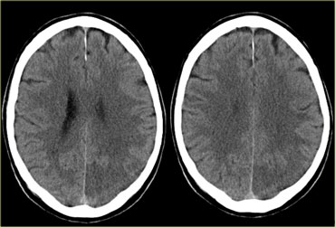 PCA infarctionNhồi máu
Only about 5% of ischemic strokes involve the PCA or its branches (3).
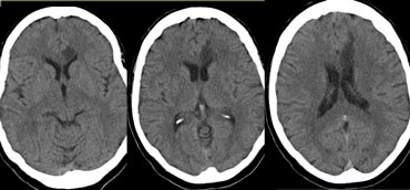
On the left CT-images of a patient with a PCA-infarction.
Notice the loss of gray/white matter differentiation in the regio of the left occipital lobe.Chỉ có khoảng 5% đột quỵ thiếu máu não cục bộ liên quan đến PCA hoặc các nhánh của nó (3).
Trên hình ảnh CT bên trái của một bệnh nhân có PCA-nhồi máu. Lưu ý sự mất biệt hóa Gray / chất trắng ở vùng vùng cùng thuỳ chẩm trái.
Variations in Arterial TerritoriesBiến thể trong lãnh thổ động mạch
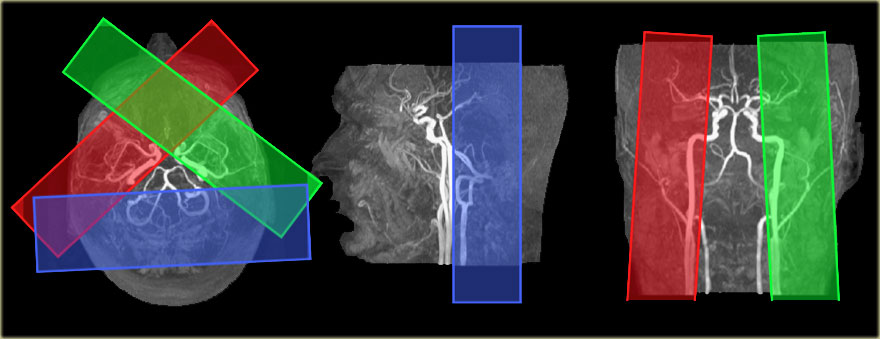 Images courtesy Jeroen Hendrikse (9)Hình ảnh do Jeroen Hendrikse (9) cung cấp
Variations in perfusion territories in the brain can be visualized with selective arterial spin-labeling (9).
The ability to visualize these perfusion territories is important in specific patient groups with cerebrovascular disease, such as acute stroke, large artery steno-occlusive disease, and arteriovenous malformation, as it provides valuable hemodynamic information.Các biến thể trong vùng tưới máu trong não có thể được quan sát bằng cách dán nhãn chọn lọc động mạch (9).
Khả năng hình dung các vùng tưới máu này rất quan trọng trong các nhóm bệnh nhân cụ thể mắc bệnh mạch máu não, chẳng hạn như đột quỵ cấp tính, bệnh tắc hẹp động mạch lớn và dị dạng động tĩnh mạch, vì nó cung cấp thông tin huyết động có giá trị.
On the left the time-of-flight MR angiography-images of brain-feeding arteries showing the planning of the selective slabs for perfusion territory imaging of the left and right internal carotid artery and the vertebrobasilar artery.Bên trái chụp cộng hưởng từ thời gian bay-hình ảnh của các động mạch nuôi não cho thấy việc lập kế hoạch của các tấm chọn lọc để chụp hình ảnh lãnh thổ tưới máu của động mạch cảnh trong bên trái và bên phải và động mạch thân đốt sống.
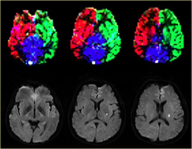 Normal perfusion territories in a patient with a lacunar infarction Images courtesy Jeroen Hendrikse (9)Vùng tưới máu bình thường ở bệnh nhân có lỗ khuyết nhồi máu Hình ảnh do Jeroen Hendrikse cung cấp (9)
On the left a patient with a lacunar infarction on the left with normal perfusion territories.Bên trái bệnh nhân có lỗ khuyết nhồi máu bên trái với vùng tưới máu bình thường.
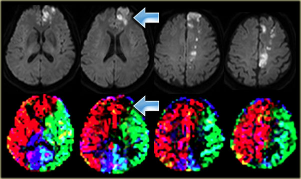 Cortical infarction in the left frontal lobe which is supplied by the right ICA Images courtesy Jeroen Hendrikse (9)Thuỳ trán trái nhồi máu ở thuỳ trán bên trái được cung cấp bởi ICA bên phải Hình ảnh do Jeroen Hendrikse cung cấp (9)
On the left a patient with a watershed infarct in the left hemisphere and also a cortical infarction in the left frontal lobe (arrow).
Notice that there is a variation in the brain perfusion since the left frontal lobe is supplied by the right internal carotid artery. Ở bên trái, một bệnh nhân bị nhồi máu đầu nguồn ở bán cầu trái và cũng có một nhồi máu vỏ não ở thuỳ trán trái (mũi tên).
Lưu ý rằng có sự thay đổi trong tưới máu não vì thuỳ trán trái được cung cấp bởi động mạch cảnh trong bên phải.
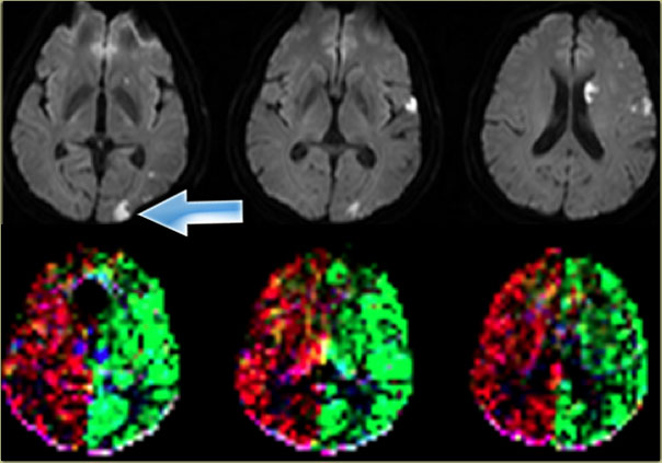 Images courtesy Jeroen Hendrikse (9)Hình ảnh do Jeroen Hendrikse (9) cung cấp
On the left another variation in the brain perfusion in a patient with multiple infarctions as demonstrated on the diffusion images.
There is a small cortical infarction in the left occipital lobe which happens to be perfused by the left internal carotid artery (arrow).
Notice that there is no contribution by the vertebrobasilar arteries.Ở bên trái, một biến thể khác trong tưới máu não ở bệnh nhân nhồi máu não nhiều lần như đã được chứng minh trên hình ảnh khuếch tán.
Có một nhồi máu nhỏ ở vỏ não ở thuỳ chẩm trái, được tưới máu bởi động mạch cảnh trong bên trái (mũi tên).
Lưu ý rằng không có sự đóng góp của các động mạch thân đốt sống.
Watershed InfarctsNhồi máu đầu nguồn
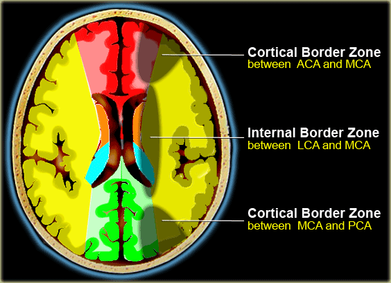
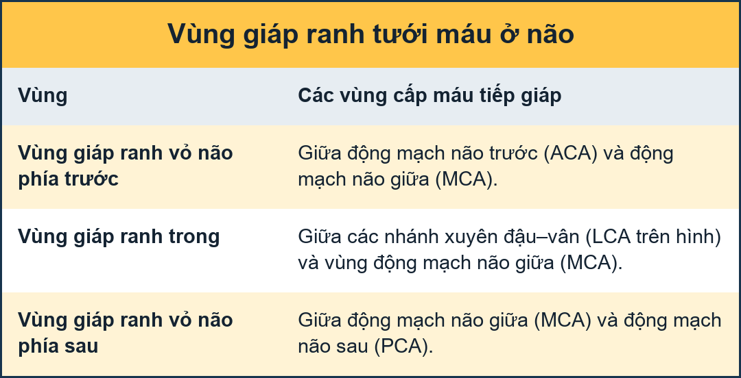
Watershed infarcts occur at the border zones between major cerebral arterial territories as a result of hypoperfusion.
There are two patterns of border zone infarcts:Nhồi máu đầu nguồn xảy ra ở các vùng biên giới giữa các vùng động mạch não chính do giảm tưới máu.
Có hai dạng nhồi máu vùng biên giới:
- Cortical border zone infarctions
Infarctions of the cortex and adjacent subcortical white matter located at the border zone of ACA/MCA and MCA/PCA Nhồi máu vùng bờ rìa
Nhồi máu vỏ não và dưới vỏ chất trắng liền kề nằm ở vùng biên giới của ACA / MCA và MCA / PCA - Internal border zone infarctions
Infarctions of the deep white matter of the centrum semiovale and corona radiata at the border zone between lenticulostriate perforators and the deep penetrating cortical branches of the MCA or at the border zone of deep white matter branches of the MCA and the ACA. Nhồi máu vùng biên giới trong
Nhồi máu chất trắng sâu của trung tâm nửa bầu dục và mạch vành tại vùng giáp ranh giữa các nhánh xuyên dạng thấu kính và các nhánh xuyên sâu vào vỏ não của MCA hoặc tại vùng giáp biên của các nhánh chất trắng sâu của MCA và ACA.
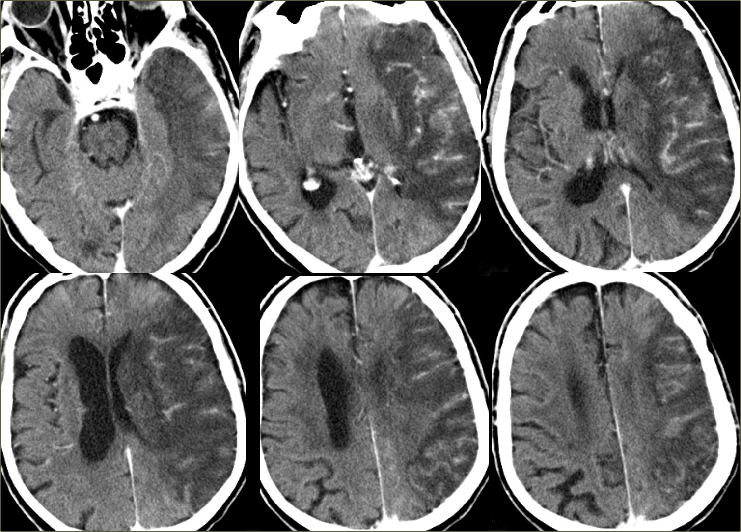
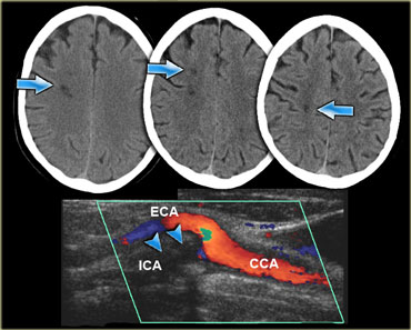 Deep watershed infarction in a patient with an occlusion of the right internal carotid arteryNghiên cứu đặc điểm dòng chảy nhồi máu ở bệnh nhân tắc động mạch cảnh trong bên phải
On the left three consecutive CT-images of a patient with an occlusion of the right internal carotid artery.
The hypoperfusion in the right hemisphere resulted in multiple internal border zone infarctions.
This pattern of deep watershed infarction is quite common and should urge you to examine the carotids.
Bên trái là ba hình ảnh CT liên tiếp của một bệnh nhân bị tắc động mạch cảnh trong bên phải.
Giảm tưới máu ở bán cầu phải dẫn đến nhiều trường hợp nhồi máu vùng biên trong.
Kiểu hình nhồi máu đầu nguồn sâu này khá phổ biến và nên khuyến khích bạn kiểm tra động mạch cảnh.
See the article on Multiple Sclerosis for the differentiation between internal border zone infarctions, lacunar infarctions and MS.Xem bài viết về Bệnh đa xơ cứng để phân biệt giữa nhồi máu vùng biên trong, nhồi máu ổ khuyết và MS.
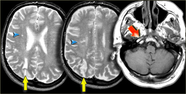 Borderzone infarcts due to occlusion of the internal carotidNhồi máu vùng biên do tắc động mạch cảnh trong
On the left images of a patient who has small infarctions in the right hemisphere in the deep borderzone (blue arrowheads) and also in the cortical borderzone between the MCA- and PCA-territory (yellow arrows).
There is abnormal signal in the right carotid (red arrow) as a result of occlusion.
In patients with abnormalities that may indicate borderzone infarcts, always study the images of the carotid artery to look for abnormal signal.Trên hình ảnh bên trái của một bệnh nhân bị nhồi máu nhỏ ở bán cầu phải ở vùng biên sâu (đầu mũi tên màu xanh) và cũng ở vùng biên vỏ não giữa MCA- và PCA-territory (mũi tên màu vàng).
Có tín hiệu bất thường ở động mạch cảnh phải (mũi tên màu đỏ) do tắc mạch.
Ở những bệnh nhân có bất thường có thể chỉ ra nhồi máu vùng biên, luôn nghiên cứu hình ảnh động mạch cảnh để tìm tín hiệu bất thường.
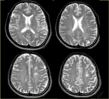
On the left another example of small infarctions in the deep borderzone and in the cortical borderzone between the MCA- and PCA-territory in the left hemisphere.
Ở bên trái, một ví dụ khác về các nhồi máu nhỏ ở vùng biên giới sâu và ở vùng biên vỏ não giữa vùng MCA và PCA ở bán cầu trái.
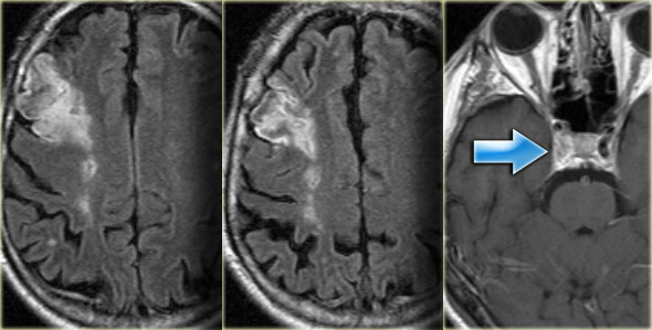
On the left an example of infarctions in the deep borderzone and in the cortical borderzone between the ACA- and MCA-territory.
The abnormal signal intensity in the right carotid is the result of an occlusion.
This combination of findings is so common, that once you know the pattern, you will see it many times.Bên trái là một ví dụ về nhồi máu ở vùng biên giới sâu và ở vùng biên giữa vỏ não giữa lãnh thổ ACA và MCA.
Bất thường cường độ tín hiệu trong động mạch cảnh phải là kết quả của tắc nghẽn.
Sự kết hợp các dấu hiệu này rất phổ biến, một khi bạn biết được mô hình, bạn sẽ thấy nó nhiều lần.
Lacunar InfarctsNhồi máu ổ khuyết
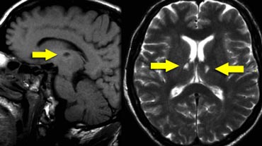
Lacunar infarcts are small infarcts in the deeper parts of the brain (basal ganglia, thalamus, white matter) and in the brain stem.
Lacunar infarcts are caused by occlusion of a single deep penetrating artery.
Lacunar infarcts account for 25% of all ischemic strokes.
Atherosclerosis is the most common cause of lacunar infarcts followed by emboli.
25% of patients with clinical and radiologically defined lacunes had a potential cardiac cause for their strokes. Nhồi máu ổ khuyết là những nhồi máu nhỏ ở các phần sâu hơn của não (hạch nền, đồi thị, chất trắng) và ở thân não.
Nhồi máu ổ khuyết là do tắc một động mạch thâm nhập sâu.
Nhồi máu ổ khuyết chiếm 25% tổng số các trường hợp nhồi máu não.
Xơ vữa động mạch là nguyên nhân phổ biến nhất của nhồi máu lỗ khuyết, tiếp theo là tắc mạch.
25% bệnh nhân có tổn thương mạch máu não trên lâm sàng và X quang có thể là nguyên nhân tim mạch gây đột quỵ.
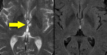
On the left a T2W- and FLAIR image of a lacunar infarct in the left thalamus.
On the FLAIR image the infarct is hardly seen.
There is only a small area of subtle hyperintensity.Bên trái là hình ảnh T2W- và FLAIR của một nhồi máu khuyết ở đồi thị trái.
Trên hình ảnh FLAIR, hầu như không thấy nhồi máu.
Chỉ có một vùng nhỏ của cường độ tinh tế.
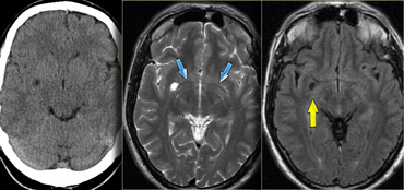
Lacunes may be confused with other empty spaces, such as enlarged perivascular Virchow-Robin spaces (VRS).
The VRS are extensions of the subarachnoid space that accompany vessels entering the brain parenchyma.
Widening of VRS often first occurs around penetrating arteries in the substantia perforata and can be seen on transverse MRI slices around the anterior commisure, even in young subjects (5).Các lỗ thông có thể bị nhầm lẫn với các khoảng trống khác, chẳng hạn như các khoảng thông Virchow-Robin mở rộng (VRS).
VRS là phần mở rộng của khoang dưới nhện đi kèm với các mạch máu đi vào nhu mô não.
Sự giãn rộng của VRS thường xảy ra đầu tiên xung quanh các động mạch xuyên thấu trong cơ quan xuyên và có thể được nhìn thấy trên các lát cắt MRI ngang xung quanh mép trước, ngay cả ở những đối tượng trẻ tuổi (5).
On the left CT- and MR-images at the level of the anterior commisure (blue arrows).
On the CT there is a hypodense area in the right hemisphere, which follows the signal intensity of CSF on T2W- and FLAIR-images, which is typical for widened VRS.Trên hình ảnh CT và MR bên trái ở mức của mép trước (các mũi tên màu xanh).
Trên CT có một vùng giảm tỷ trọng ở bán cầu phải, theo sau cường độ tín hiệu của CSF trên hình ảnh T2W và FLAIR, là điển hình cho VRS mở rộng.
PRES
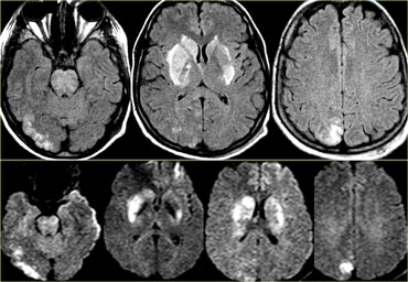 PRES (courtesy Madja Turnher)PRES (thư mời Madja Turnher)
PRES is short for Posterior Reversible Encephalopathy Syndrome.
It is also known as reversible posterior Leukoencephalopathy syndrome [RPLS].
It classically consists of potentially reversible vasogenic edema in the posterior circulation territories, but anterior circulation structures can also be involved (6).
Many causes have been described including hypertension, eclampsia and preeclampsia, immunosuppressive medications such as cyclosporine.
The mechanism is not entirely understood but is thought to be related to a hyperperfusion state, with blood-brain-barrier breakthrough, extravasation of fluid potentially containing blood or macromolecules, and resulting cortical or subcortical edema.
PRES là viết tắt của Hội chứng bệnh não đảo ngược sau.
Nó còn được gọi là hội chứng não chất trắng sau có thể hồi phục [RPLS].
Nó bao gồm phù do mạch có khả năng đảo ngược trong các vùng tuần hoàn não sau, nhưng các cấu trúc tuần hoàn não trước cũng có thể liên quan (6).
Nhiều nguyên nhân đã được mô tả bao gồm tăng huyết áp, sản giật và tiền sản giật, thuốc ức chế miễn dịch như cyclosporine.
Cơ chế này chưa được hiểu hoàn toàn nhưng được cho là có liên quan đến tình trạng tăng tưới máu, với sự đột phá về rào cản máu-não, thóat mạch của chất lỏng có khả năng chứa máu hoặc các đại phân tử, và dẫn đến phù vỏ não hoặc dưới vỏ.
The typical imaging findings of PRES are most apparent as hyperintensity on FLAIR images in the parietooccipital and posterior frontal cortical and subcortical white matter; less commonly, the brainstem, basal ganglia, and cerebellum are involved.
Các dấu hiệu hình ảnh điển hình của PRES rõ ràng nhất là cường độ trên hình ảnh FLAIR ở vỏ não vùng đỉnh chẩm và sau trán và dưới vỏ chất trắng; ít phổ biến hơn, có liên quan đến thân não, hạch nền và tiểu não.
On the left images of a patient with reversible neurological symptoms.
The abnormalities are seen both in the posterior circulation as well as in the basal ganglia.
Continue.
Trên hình ảnh bên trái của một bệnh nhân có các triệu chứng thần kinh có thể hồi phục.
Các bất thường được thấy cả ở tuần hoàn sau cũng như ở hạch nền.
Tiếp tục.
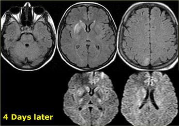

Four days later most of the abnormalities have disappeared.Bốn ngày sau, hầu hết các bất thường đã biến mất.
Cerebral Venous territoryLãnh thổ tĩnh mạch não
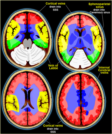
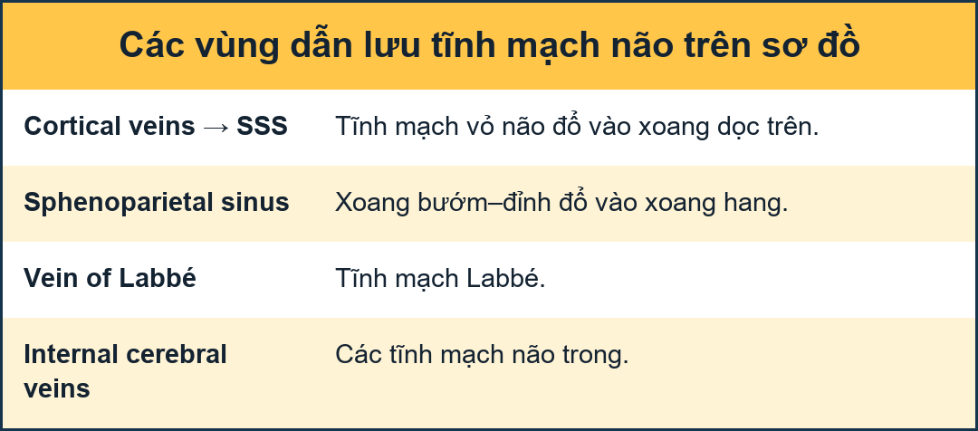
There is great variation in the territories of venous drainage.
The illustrations on the left should be regarded as a rough guide. Có sự khác biệt lớn trong các vùng dẫn lưu tĩnh mạch.
Các hình minh hoạ bên trái nên được coi là một hướng dẫn sơ bộ.
Cerebral venous thrombosisHuyết khối tĩnh mạch não
Cerebral venous thrombosis results from occlusion of a venous sinus and/or cortical vein and usually is caused by a partial thrombus or an extrinsic compression that subsequently progresses to complete occlusion (7).
Dehydration, pregnancy, a hypercoagulable state and adjacent infection (eg, mastoiditis) are predisposing factors.
Cerebral venous thrombosis is an elusive diagnosis because of its nonspecific presentation.
It often presents with hemorrhagic infarction in areas atypical for arterial vascular distribution.
Huyết khối tĩnh mạch não là kết quả của sự tắc nghẽn của xoang tĩnh mạch và / hoặc tĩnh mạch vỏ não và thường do huyết khối cục bộ hoặc chèn ép bên ngoài mà sau đó tiến triển thành tắc nghẹt hoàn toàn (7).
Mất nước, mang thai, tình trạng tăng đông và nhiễm trùng lân cận (ví dụ, viêm xương chũm) là những yếu tố ảnh hưởng.
Huyết khối tĩnh mạch não là một chẩn đoán khó nắm bắt vì biểu hiện không đặc hiệu.
Nó thường xuất hiện với nhồi máu xuất huyết ở những vùng không điển hình cho sự phân bố mạch máu động mạch.
Imaging plays a key role in the diagnosis.
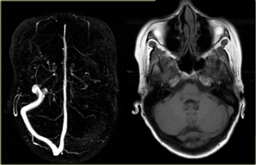
On the far left a MRA with non-visualization of the left transverse sinus.
Since the venous anatomy is variable, this can be due to absence of the transverse sinus or thrombosis.
The T1W-image on the right clearly demonstrates, that there is a transverse sinus on the left, so the MRA findings are due to thrombosis.
Continue with next images.Chẩn đoán hình ảnh đóng vai trò quan trọng trong chẩn đoán.
Ở xa bên trái, chụp cộng hưởng từ xoang ngang trái không nhìn thấy.
Vì giải phẫu tĩnh mạch rất đa dạng, điều này có thể là do không có xoang ngang hoặc huyết khối.
Hình ảnh T1W bên phải cho thấy rõ ràng, có một xoang ngang bên trái, do đó các dấu hiệu MRA là do huyết khối.
Tiếp tục với những hình ảnh tiếp theo.
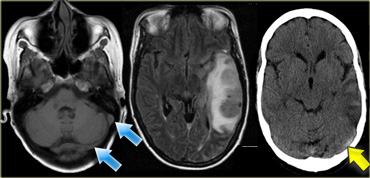
On the left the CT nicely demonstrates the dense thrombosed transverse sinus (yellow arrow).
The FLAIR image demonstrates the venous infarction in the temporal lobe.
Ở bên trái, CT cho thấy rõ hình ảnh xoang ngang bị huyết khối dày đặc (mũi tên màu vàng).
Hình ảnh FLAIR cho thấy tĩnh mạch nhồi máu ở thuỳ thái dương.
Thrombosis of deep cerebral veins
The clinical presentation of thrombosis of the deep cerebral venous system are severe dysfunction of the diencephalon, reflected by coma and disturbances of eye movements and pupillary reflexes.
Usually this results in a poor outcome.
However, partial syndromes without a decrease in the level of consciousness or brainstem signs exist, which may lead to initial misdiagnoses.
Deep cerebral venous system thrombosis is an underdiagnosed condition when symptoms are mild and should be suspected if the patient is a young woman, if the lesions are within the basal ganglia or thalamus and especially if they are bilateral.
Huyết khối tĩnh mạch não sâu
Biểu hiện lâm sàng của huyết khối hệ tĩnh mạch não sâu là rối loạn chức năng nặng của não, phản ánh bởi tình trạng hôn mê và rối loạn vận động mắt và phản xạ đồng tử.
Thông thường điều này dẫn đến kết quả xấu.
Tuy nhiên, có các hội chứng một phần mà không có sự giảm mức độ ý thức hoặc các dấu hiệu của thân não, có thể dẫn đến chẩn đoán sai ban đầu.
Huyết khối tĩnh mạch não sâu là một tình trạng chưa được chẩn đoán khi các triệu chứng nhẹ và nên được nghi ngờ nếu bệnh nhân là phụ nữ trẻ, nếu các tổn thương nằm trong hạch nền hoặc đồi thị và đặc biệt là nếu chúng ở hai bên.
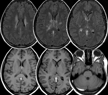
On the left images of a patient with deep cerebral vein thrombosis.
Notice the bilateral infarctions in the basal ganglia.
Continue.
Trên hình ảnh bên trái của một bệnh nhân bị huyết khối tĩnh mạch não sâu.
Lưu ý các nhồi máu hai bên ở hạch nền.
Tiếp tục.
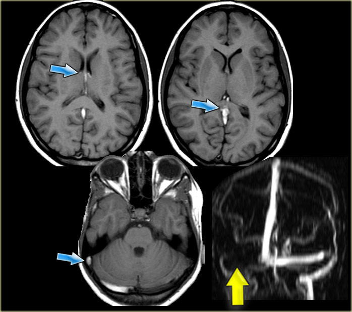 Deep cerebral vein thrombosisHuyết khối tĩnh mạch não sâu
There is absence of flow void in the internal cerebral veins, sinus rectus and right transverse sinus (blue arrows).
On the MRA the right transverse sinus is not visualized.Không có mất tín hiệu dòng chảy ở tĩnh mạch não trong, xoang thẳng và xoang ngang phải (các mũi tên xanh).
Trên MRA, không thấy xoang ngang bên phải.

Charity
Hoạt động từ thiện
All the profits of the Radiology Assistant go to Medical Action Myanmar which is run by Dr. Nini Tun and Dr. Frank Smithuis sr, who is a professor at Oxford university and happens to be the brother of Robin Smithuis.Toàn bộ lợi nhuận của Radiology Assistant được chuyển cho Medical Action Myanmar, do bác sĩ Nini Tun và bác sĩ Frank Smithuis Sr. điều hành. Frank Smithuis Sr. là giáo sư tại Đại học Oxford và là anh em ruột của Robin Smithuis.
Click here to watch the video of Medical Action Myanmar and if you like the Radiology Assistant, please support Medical Action Myanmar with a small gift.Bấm để xem video về Medical Action Myanmar. Nếu thấy Radiology Assistant hữu ích, bạn có thể đóng góp cho Medical Action Myanmar.
ReferencesTài liệu tham khảo
-
The vascular territories of the carotid and vertebrobasilar systems. Diagrams based on CT studies of infarcts.
by Savoiardo M.
Ital J Neurol Sci. 1986 Aug;7(4):405-9.
Các vùng mạch máu của hệ thống động mạch cảnh và hệ thống đốt sống.Sơ đồ dựa trên các nghiên cứu CT của nhồi máu. -
The vascular territories in the cerebellum and brainstem: CT and MR study.
Các vùng mạch máu ở tiểu não và thân não: Nghiên cứu trên CT và MR.
by Savoiardo M, Bracchi M, Passerini A, Visciani A.AJNR Am J Neuroradiol. 1987 Mar-Apr;8(2):199-209. Savoiardo M, Bracchi M, Passerini A, Visciani A. AJNR Am J Neuroradiol. Tháng 3–4/1987, tập 8, số 2, tr. 199–209.
-
Severe Hemodynamic Impairment and Border Zone-Region Infarction
Suy giảm huyết động nặng và Vùng biên giới nhồi máu
by Colin P. Derdeyn et al Radiology. 2001;220:195-201 Colin P. Derdeyn và cộng sự. X quang học. 2001;220:195-201
-
Posterior Cerebral Artery Stroke
Động mạch não sau đột quỵ
by Michael D Hill in eMedicine Michael D. Hill trên eMedicine
-
The clinical significance of widened Virchow-Robin spaces
Ý nghĩa lâm sàng của mở rộng không gian Virchow-Robin
by Frederik Barkhof
Journal of Neurology Neurosurgery and Psychiatry 2004;75:1516-1517
Tác giả: Frederik Barkhof -
Posterior Reversible Encephalopathy Syndrome: Incidence of Atypical Regions of Involvement and Imaging Findings
Hội chứng bệnh não đảo ngược sau: Tỷ lệ các vùng liên quan và các dấu hiệu hình ảnh không điển hình
by Alexander M. McKinney et al.
AJR 2007; 189:904-912
bởi Alexander M. McKinney và cộng sự. -
Brain, Venous Sinus Thrombosis
Huyết khối xoang tĩnh mạch, não
in eMedicine
by Mahesh R Patel
-
The spectrum of presentations of venous infarction caused by deep cerebral vein thrombosis
Khảo sát phổ biểu hiện nhồi máu tĩnh mạch do huyết khối tĩnh mạch não sâu
by Walter M. van den Bergh
NEUROLOGY 2005;65:192-196
Tác giả Walter M. van den Bergh -
Brain Perfusion Territory Imaging: Methods and Clinical Applications of Selective Arterial Spin-labeling MR Imaging
Hình ảnh vùng tưới máu não: Phương pháp và ứng dụng lâm sàng của hình ảnh MR đánh dấu spin động mạch chọn lọc
by Peter Jan van Laar, Jeroen van der Grond and Jeroen Hendrikse
February 2008 Radiology, 246, 354-364.
tác giả: Peter Jan van Laar, Jeroen van der Grond và Jeroen Hendrikse