
Lumbar Disc HerniationThoát vị đĩa đệm cột sống thắt lưng
and other causes of nerve compressionvà các nguyên nhân khác gây chèn ép thần kinh
Radiology department of the Rijnland hospital in Leiderdorp, the NetherlandsKhoa Chẩn đoán hình ảnh của bệnh viện Rijnland, Leiderdorp, Hà Lan
Ngày đăng / cập nhật: 2014-12-14
Bản gốc trên radiologyassistant.nl ↗ · tải về lúc 2026-11-19 20:19 · bản dịch máy có chuẩn hóa thuật ngữ chẩn đoán hình ảnh
In this article a systematic approach to patients with nerve root compression in the lumbar region is presented.Trong bài viết này, chúng tôi trình bày một cách tiếp cận có hệ thống đối với bệnh nhân bị chèn ép rễ thần kinh ở vùng thắt lưng.
We wil discuss disc herniation, facet arthrosis, synovial cysts, spondylolisthesis and epidural lipomatosis.Chúng tôi sẽ thảo luận về thóat vị đĩa đệm, khía cạnh khớp, nang hoạt dịch, trượt đốt sống và u mỡ ngoài màng cứng.
The images can be enlarged by clicking on them.Các hình ảnh có thể được phóng to bằng cách nhấp vào chúng.
Systematic approachPhương pháp tiếp cận hệ thống
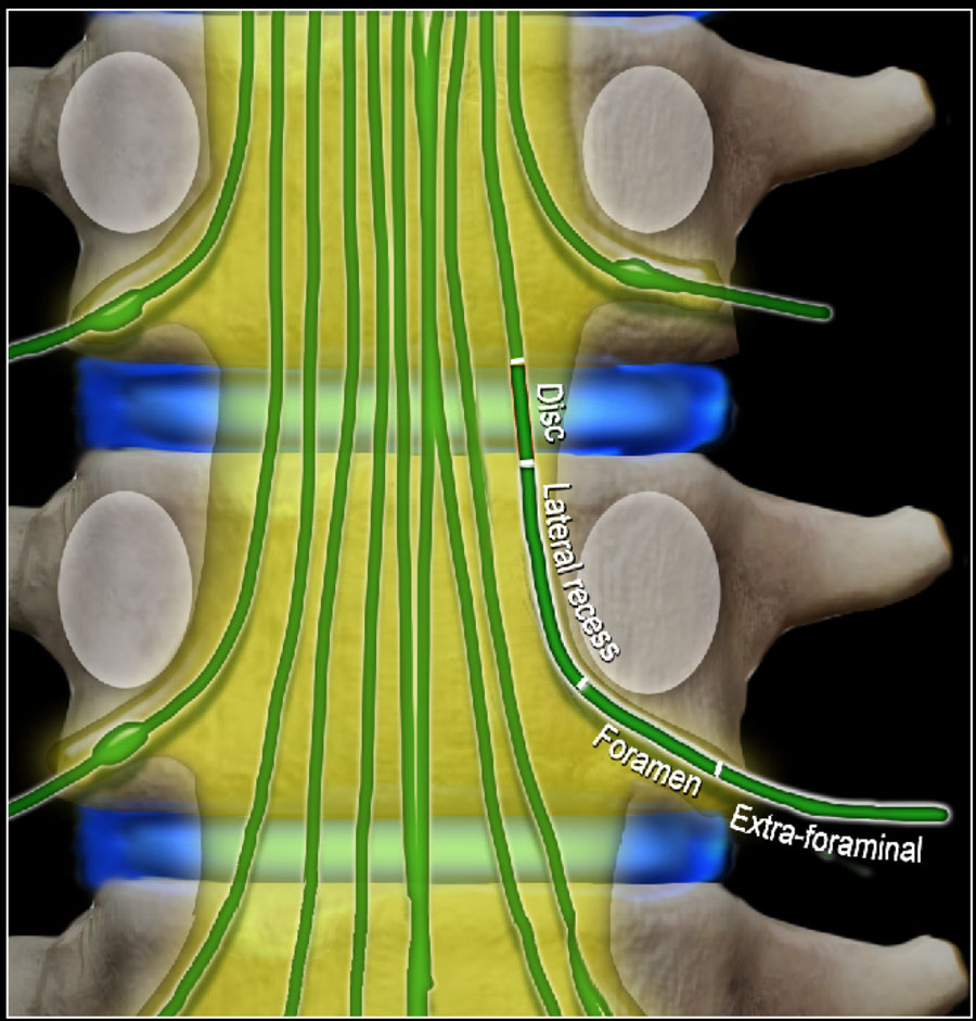
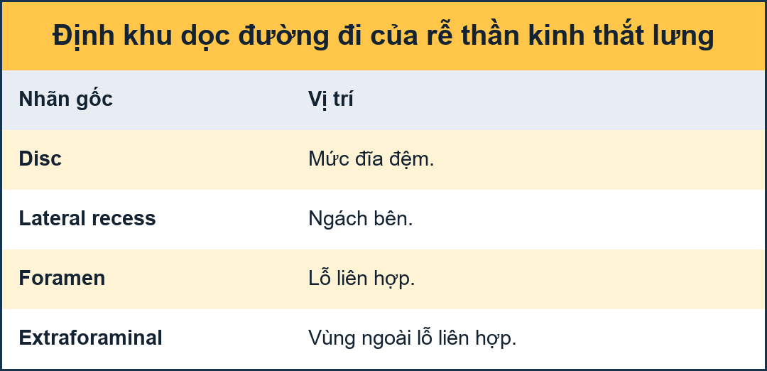
Four levels of nerve compressionBốn mức độ chèn ép thần kinh
In patients with symptoms of nerve root compression, there are four levels that need to be studied:Ở những bệnh nhân có triệu chứng rễ thần kinh, có bốn mức độ cần được nghiên cứu:
- Disc level.
This is the most common area where nerves are compressed.
Mostly by herniated discs and less frequently due to spinal stenosis.Mức độ đĩa đệm.
Đây là khu vực phổ biến nhất mà các dây thần kinh bị chèn ép.
Chủ yếu do thóat vị đĩa đệm và ít gặp hơn do hẹp ống sống. - Level of lateral recess.
This is the area below the disc where the nerve runs more laterally towards the foramen.
Narrowing of the lateral recess is caused by facet arthrosis, usually in combination with hypertrophy of the flavum ligament and bulging of the disc.Mức độ lõm ngoài.
Đây là khu vực bên dưới đĩa đệm nơi dây thần kinh chạy theo chiều ngang hơn về phía lỗ.
Hẹp ngách ngoài là do khớp mặt, thường kết hợp với phì đại dây chằng flavum và phình đĩa đệm. - Foramen.
This is the area between two pedicles, where the nerve leaves the spinal canal.
Narrowing of the foramen is seen in facet arthrosis, spondylolisthesis and foraminal disc herniation - usually a migrated disc from a lower level.Foramen.
Đây là khu vực giữa hai cuống, nơi dây thần kinh rời khỏi ống sống.
Hẹp lỗ thông được thấy trong các khía cạnh khớp, trượt đốt sống và lỗ thông thóat vị đĩa đệm - thường là đĩa đệm di chuyển từ mức thấp hơn. - Extra-foraminal.
This is the area lateral to the foramen.
Nerve compression in this area is uncommon, but is sometimes caused by a laterally herniated disc.Ngoài hậu môn.
Đây là khu vực bên cạnh lỗ.
Chèn ép thần kinh ở khu vực này không phổ biến, nhưng đôi khi do thóat vị đĩa đệm bên.
At these four levels there can be a lot of overlap of pathology.
For instance a herniated disc can cause nerve compression at the level of the disc, but also at the level of the foramen or extra-foraminal when there is migration of the disc.Ở bốn mức độ này có thể có nhiều sự chồng chéo của bệnh lý.
Ví dụ, thóat vị đĩa đệm có thể gây chèn ép thần kinh ở mức của đĩa đệm, nhưng cũng có thể ở mức của lỗ hoặc ngoài lỗ khi có sự di chuyển của đĩa đệM.
When you are looking for nerve compression, you have to study all these levels.Khi bạn tìm kiếm chèn ép thần kinh, bạn phải nghiên cứu tất cả các mức độ này.
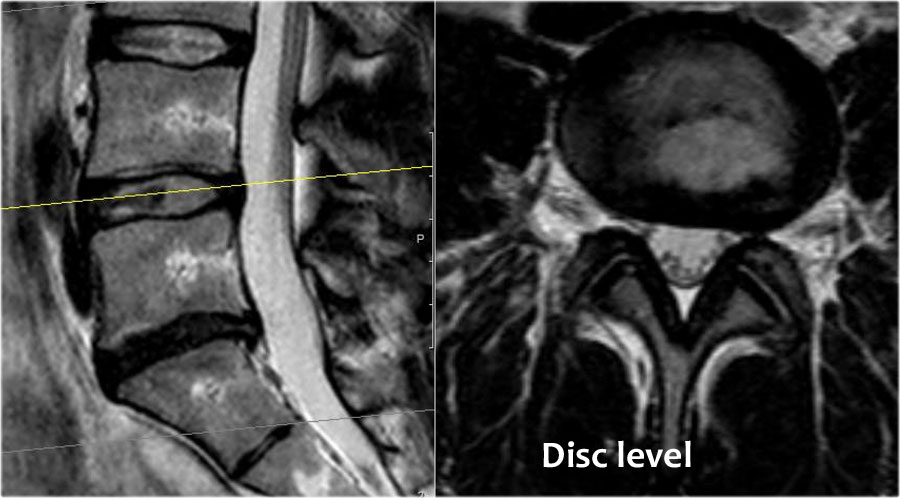
Scroll through the images to see how the nerves run at the level of the disc, lateral recess, foramen and extra-foraminal.Cuộn qua các hình ảnh để xem các dây thần kinh chạy như thế nào ở mức đĩa đệm, hốc bên, lỗ và ngoài lỗ.
At each level specific pathology can be seen, but there is a lot of overlap.
For instance a disc can herniate and cause nerve compression at the level of the disc, but can also migrate to a lower level and compress the nerve in the lateral recess or move upward and cause compression at the level of the foramen or extra-foraminal.
In patients with facet arthrosis the bony spurs can move medially and narrow the lateral recess or move upward and narrow the foramen.
When there is extreme facet arthrosis bilaterally, it can cause stenosis of the spinal canal and compress all the nerve roots at that level.Ở mỗi mức độ có thể thấy được bệnh lý cụ thể, nhưng có rất nhiều sự chồng chéo.
Ví dụ, một đĩa đệm có thể thóat vị và gây chèn ép thần kinh ở mức của đĩa đệm, nhưng cũng có thể di chuyển xuống mức thấp hơn và chèn ép dây thần kinh ở ngách ngoài hoặc di chuyển lên trên và gây chẹn ở mức của lỗ hoặc ngoài lỗ.
Ở những bệnh nhân bị viêm khớp dạng mặt, các gai xương có thể di chuyển vào trong và thu hẹp hốc bên hoặc di chuyển lên trên và thu hẹp lỗ.
Khi có khía cạnh khớp cực đoan hai bên, nó có thể gây hẹp ống sống và chèn ép tất cả các rễ thần kinh ở mức đó.
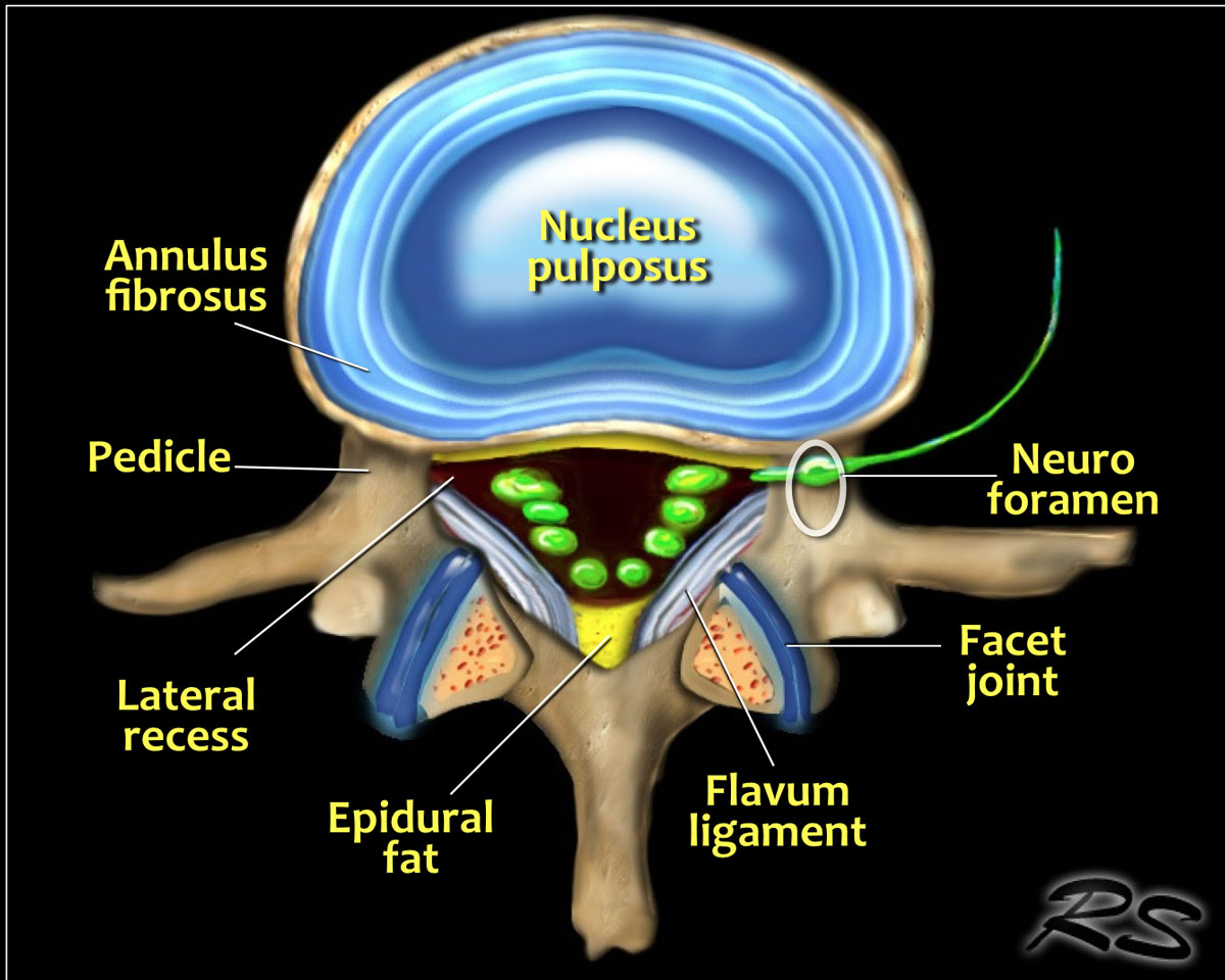

AnatomyGiải phẫu
The illustration demonstrates the structures that surround the nerves within the spinal canal.Hình minh hoạ cho thấy các cấu trúc bao quanh các dây thần kinh trong ống sống.
Flavum ligament
The flavum ligament is a strong ligament on the interior posterior side of the vertebral canal that connects the laminae of adjacent vertebrae.
As a result of aging and instability of the vertebral column due to facet arthrosis there will be more stress on the flavum ligament resulting in hypertrophy and fibrosis.Dây chằng Flavum
Dây chằng flavum là một dây chằng mạnh ở phía sau bên trong của ống sống, nối các lớp của các đốt sống liền kề.
Do sự lão hóa và mất vững cột sống do khía cạnh khớp sẽ có nhiều áp lực lên dây chằng flavum dẫn đến phì đại và xơ hóa.
Hypertrophy of the flavum ligament is usually seen in combination with facet arthrosis and both result in stenosis of the lateral recess or when it is bilateral, in spinal stenosis.Phì đại dây chằng flavum thường được thấy kết hợp với viêm khớp mặt và cả hai đều dẫn đến hẹp hốc bên hoặc khi nó ở hai bên, trong hẹp ống sống.
Epidural fat
This is the fat that surrounds the dural sac, that contains the nerves.
Abundant fat can be seen in steroid therapy, extreme obesitas and rarely idiopathic.
Abundant epidural fat can contribute to stenosis of the spinal canal.Mỡ ngoài màng cứng
Đây là mỡ bao quanh túi màng cứng, chứa các dây thần kinh.
Chất béo dồi dào có thể được thấy trong liệu pháp steroid, béo phì cực độ và hiếm khi vô căn.
Mỡ ngoài màng cứng dư thừa có thể góp phần gây hẹp ống sống.
MRI protocolQuy trình chụp MRI
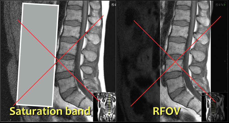
The MRI protocol for examination of the lumbar spine in patients with symptoms of nerve compression is quite simple.Quy trình chụp MRI để kiểm tra cột sống thắt lưng ở bệnh nhân có triệu chứng chèn ép thần kinh khá đơn giản.
Basically we rely on the sagittal T1W- and T2W-images and correlate the findings with the transverse T2W-images of the levels of suspected pathology.Về cơ bản chúng tôi dựa vào hình ảnh T1W và T2W dọc và tương quan các phát hiện với hình ảnh T2W ngang của các mức độ bệnh lý nghi ngờ.
Do not use a saturation band on the anterior side or a rectangular field of view (RFOV), because you also want to image the prevertebral soft tissues.Không sử dụng băng bão hoà ở phía trước hoặc trường nhìn hình chữ nhật (RFOV), bởi vì bạn cũng muốn chụp hình ảnh mô mềm trước cột sống.
Especially look for an aneurysm of the abdominal aorta, since this may also be the cause of low back pain.
Sometimes it can be difficult to clinically differentiate neurogenic claudication - which is caused by spinal stenosis - from vascular claudication - which is caused by arterial stenosis.Đặc biệt là tìm phình động mạch chủ bụng, vì đây cũng có thể là nguyên nhân gây đau lưng.
Đôi khi khó phân biệt lâm sàng đau cách hồi do nguyên nhân thần kinh - do hẹp ống sống gây ra - với đau cách hồi mạch máu - do hẹp động mạch.
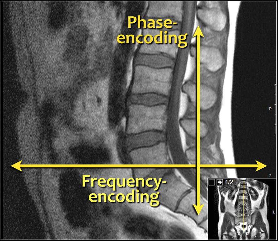

The frequency-encoding should be in the AP-direction and consequently the phase-encoding feet-head.Tần số mã hóa nên ở hướng AP và do đó là đầu chân mã hóa pha.
This has several advantages:Điều này có một số ưu điểm:
- The resolution is the highest in the frequency-encoded direction.
We want the highest resolution in the AP-direction to look for small herniations and delineation of nerve roots.Độ phân giải cao nhất theo hướng mã hóa tần số.
Chúng tôi muốn có độ phân giải cao nhất theo hướng AP để tìm các thóat vị nhỏ và phân định các rễ thần kinh. - When you have the phase encoding in the AP-direction, you get breating artifacts. That is why some use a saturation band or the RFOV.
With the frequency-encoding in the AP-direction you do not have these problems and you do not need a saturation band.Khi bạn có mã hóa pha theo hướng AP, bạn sẽ có được các tạo tác giả mạo. Đó là lý do tại sao một số người sử dụng dải bão hoà hoặc RFOV.
Với tần số mã hóa theo hướng AP, bạn không gặp phải những vấn đề này và bạn không cần dải bão hoà. - Frequency encoding in the feet-head direction can result in poor deliniation of the vertebral endplates due to chemical shift artifact when the fat in the vertebral body meets the water in the disc.
This is another reason to use a feet-head phase encoding and an AP frequency encoding.Việc mã hóa tần số theo hướng chân-đầu có thể dẫn đến sự phân bố kém của các mấu cuối đốt sống do tạo tác dịch chuyển hóa học khi mỡ trong thân đốt sống gặp nước trong đĩa đệm.
Đây là một lý do khác để sử dụng mã hóa pha đầu-chân và mã hóa tần số AP.
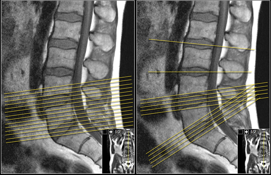
It is better to have continuous slices with the same angulation parallel to the level where nerve compression is suspected.
This has the advantage, that you can follow the involved nerve along the four levels of possible compression.Tốt hơn là có các lát cắt liên tục với cùng một góc song song với mức độ nghi ngờ chèn ép thần kinh.
Điều này có lợi thế là bạn có thể theo dõi dây thần kinh bị ảnh hưởng dọc theo bốn mức độ chèn ép có thể.
When you examine multiple levels with different angulation like in the example on the right, you will not be able to follow the whole nerve tract and you will not get the whole picture.Khi bạn kiểm tra nhiều mức độ với các góc khác nhau như trong ví dụ bên phải, bạn sẽ không thể theo dõi toàn bộ dây thần kinh và bạn sẽ không có được toàn bộ bức tranh.
InterpretationGiải thích
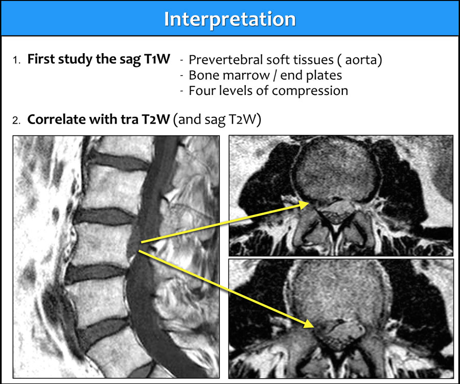

The sagittal T1W-images give you the most diagnostic information.
Before you start looking for any hernias, first take a good look at the prevertebral tissues and bone marrow.Hình ảnh T1W dọc cung cấp cho bạn thông tin chẩn đoán nhiều nhất.
Trước khi bạn bắt đầu tìm kiếm bất kỳ thóat vị nào, trước tiên hãy nhìn kỹ các mô trước cột sống và tủy xương.
Once you have detected any abnormality, correlate these findings with the T2W-images (figure).Khi bạn đã phát hiện bất kỳ bất thường nào, hãy tương quan những phát hiện này với hình ảnh T2W (hình).
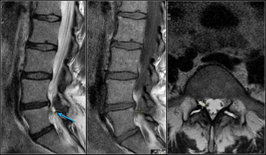
Use of a marker
When you notice an abnormality, it can be helpful to use a marker that correlates with the same location on the other series.
If you enlarge the image, you will see the small yellow cross, which indicates that you are looking at the exact same spot on the other series.Sử dụng bút đánh dấu
Khi bạn nhận thấy một bất thường, có thể hữu ích khi sử dụng một điểm đánh dấu tương quan với cùng một vị trí trên chuỗi khác.
Nếu bạn phóng to hình ảnh, bạn sẽ thấy chữ thập nhỏ màu vàng, cho thấy rằng bạn đang nhìn vào cùng một vị trí trên loạt ảnh khác.
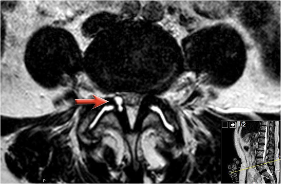
Here the L5 nerve on the right is compressed by a synovial cyst, which is the result of facet arthrosis with effusion within the intervertebral joints.Ở đây, dây thần kinh L5 bên phải bị chèn ép bởi một nang hoạt dịch, là kết quả của khớp mặt với tràn dịch trong các khớp gian đốt sống.
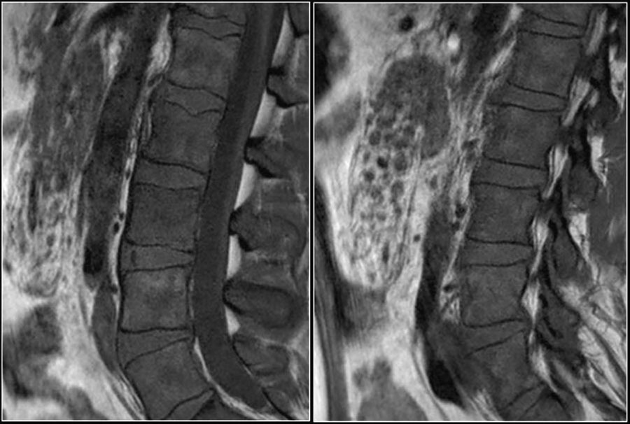
Prevertebral tissuesMô trước cột sống
Here a 25 year old patient who presented with low back pain.
Notice multiple small masses in the abdomen surrounding the mesenteric vessels and abnormal low signal intensity of the bone marrow.
The signal intensity of the discus is a little bit higher compared to the bone marrow (bright discus sign).Đây là một bệnh nhân 25 tuổi bị đau thắt lưng.
Lưu ý nhiều khối nhỏ ở bụng xung quanh các mạch mạc treo và cường độ tín hiệu thấp bất thường của tủy xương.
cường độ tín hiệu của đĩa cao hơn một chút so với tủy xương (dấu hiệu đĩa sáng).
This examination was the first indication of an abnormality in this patient.
This proved to be bone- and lymph node metastases of a carcinoma of the colon.Đây là lần đầu tiên có dấu hiệu bất thường trên bệnh nhân này.
Đây là di căn xương và hạch bạch huyết của ung thư biểu mô đại tràng.
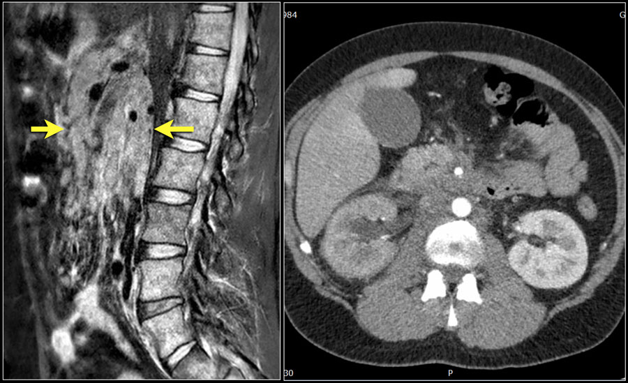
Here another patient with widespread lymphadenopathy in the mesenterium and retroperitoneum.Đây là một bệnh nhân khác có bệnh hạch lan rộng trong mạc treo và sau phúc mạc.
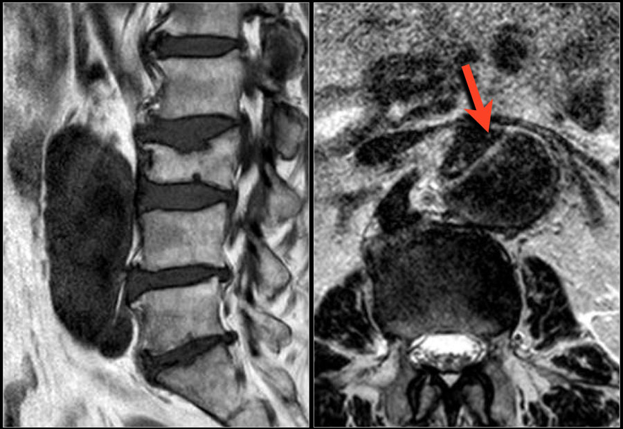
Here a patient, who presented with severe low back pain.
Notice the aortic dissection.Đây là một bệnh nhân, có biểu hiện đau thắt lưng nặng.
Chú ý đến sự tách thành động mạch chủ.
Sometimes these aortic abnormalities are incidental findings.Đôi khi những bất thường động mạch chủ này là những phát hiện tình cờ.
Disc herniationThóat vị đĩa đệm
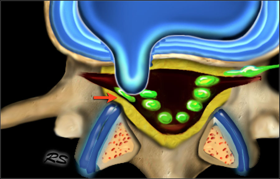
Disc herniation is displacement of disc material like nucleus pulposus, parts of the annulus fibrosus and cartilage, beyond the limits of the intervertebral disc space.Thóat vị đĩa đệm là sự dịch chuyển của vật liệu đĩa như nhân tuỷ, các phần của vành khuyên và sụn, vượt quá giới hạn của khe đĩa đệm.
It can be focal ( < 90º), broad-based ( 90º-180º) or caused by bulging of the disc (> 180º).Nó có thể là khu trú (< 90 o), rộng (90 o-180 o) hoặc do phình đĩa đệm (> 180 o).
Protrusion indicates that the distance between the edges of the disc herniation is less than the distance between the edges of the base.Sự nhô ra cho thấy khoảng cách giữa các cạnh của thóat vị đĩa đệm nhỏ hơn khoảng cách giữa hai cạnh của đáy.
Extrusion is present when the distance between the edges of the disc material is greater than the distance at the base.Đùn có mặt khi khoảng cách giữa các cạnh của vật liệu đĩa lớn hơn khoảng cách ở đáy.
Read Spine - Disc Nomenclature for more information about disc herniation nomenclature.Đọc thêm về danh pháp cột sống – đĩa đệm để biết thêm thông tin về danh pháp thóat vị đĩa đệm.
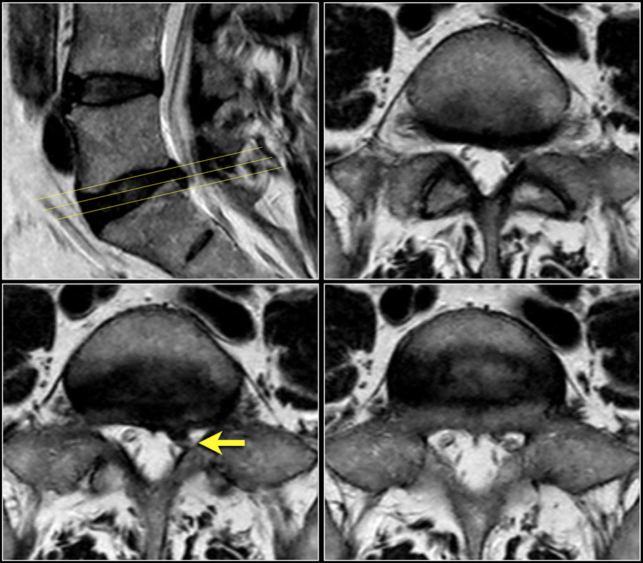
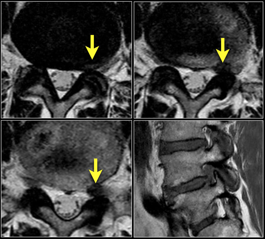
Here a focal protrusion at the L5S1 level.
The S1 nerve is compressed (arrow).Đây là một lồi khu trú ở mức L5S 1.
Dây thần kinh S1 bị chèn ép (mũi tên).
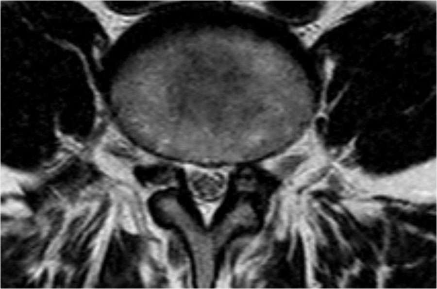
Scroll through the images and describe the findings.Cuộn qua các hình ảnh và mô tả các phát hiện.
Then continue reading.Sau đó tiếp tục đọc.
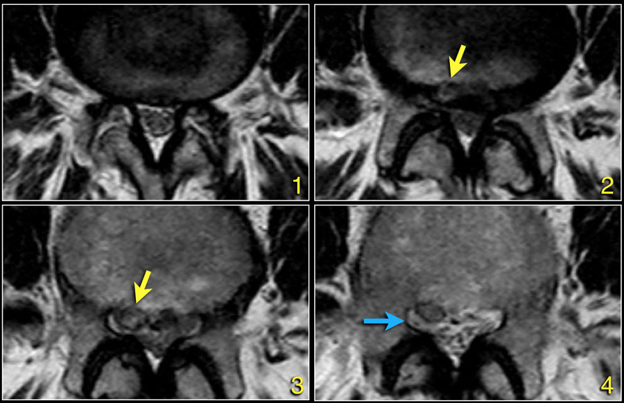
The findings at the different levels 1-4 are:Các phát hiện ở các mức độ khác nhau 1-4 là:
- At the level of the disc there is minimal spinal canal narrowing by bulging of the disc and facet arthrosis.Ở mức đĩa đệm có sự thu hẹp ống sống tối thiểu do phình đĩa đệm và khía cạnh khớp.
- At this level there is severe spinal canal stenosis due to bulging disc and facet arthrosis.
There is also an annular tear (high signal) through which the disc herniates (yellow arrow)Ở mức này có hẹp ống sống nặng do phình đĩa đệm và khía cạnh khớp.
Ngoài ra còn có một vết rách hình khuyên (tín hiệu cao) qua đó thóat vị đĩa đệm (mũi tên màu vàng) - At the level of the lateral recess, there is a focal herniation of disc material compressing the L5 nerve (yellow arrow).
This is called an extrusion, because the distance between the edges of the disc material is greater than the distance at the base.Ở mức độ lõm ngoài, có một thóat vị khu trú của vật liệu đĩa đệm chèn ép dây thần kinh L5 (mũi tên màu vàng).
Điều này được gọi là đùn, bởi vì khoảng cách giữa các cạnh của vật liệu đĩa lớn hơn khoảng cách ở đáy. - Compressed L5 nerve (blue arrow) within the lateral recess.
The herniated disc has migrated caudally and is seen as an oval structure anterior to the nerve.
The herniated disc is uncontained, i.e. not covered by fibers of the annulus.Dây thần kinh L5 bị chèn ép (mũi tên màu xanh) trong hốc bên.
Thóat vị đĩa đệm đã di chuyển ra phía sau và được xem như là một cấu trúc hình bầu dục phía trước dây thần kinh.
Đĩa thóat vị không bị che phủ, tức là không bị bao phủ bởi các sợi của vòng vành.
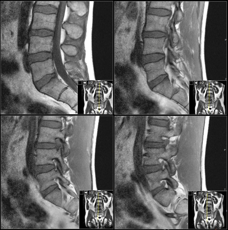
First study the images.Đầu tiên nghiên cứu hình ảnh.
Click to enlarge them.
Then continue reading.Nhấp để phóng to chúng.
Sau đó tiếp tục đọc.
The findings are:Kết quả nghiên cứu:
- Herniation of the L3L4 disc.Thóat vị đĩa đệm L3L4.
- Migration cranially.Di chuyển trong sọ.
- Compression of the L3 nerve in the foramen.Chèn ép dây thần kinh L3 trong lỗ.
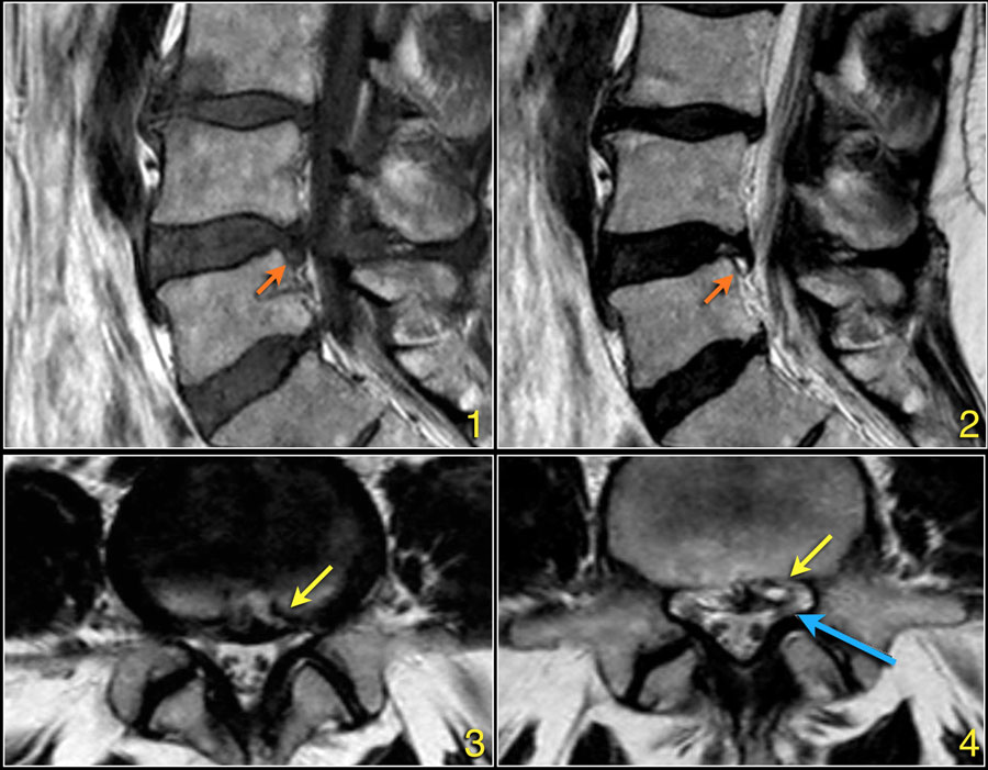 Click to enlargeNhấp để phóng to
The signal intensity of a hernia on T1W-images is usually intermediate, while on T2W-images it can be a high signal fresh herniated nucleus pulposus or a somewhat older low signal intensity hernia.Cường độ tín hiệu của thóat vị trên T1W thường là trung gian, trong khi trên T2W nó có thể là một thóat vị nhân tuỷ tươi có tín hiệu cao hoặc thóat vị cường độ tín hiệu thấp hơn một chút.
Here a herniation with high signal (yellow arrow on transverse images) sliding through the annular tear and compressing the left L5 nerve (blue arrow).Ở đây, một thóat vị với tín hiệu cao (mũi tên màu vàng trên hình ảnh ngang) trượt qua vết rách hình khuyên và chèn ép dây thần kinh L5 bên trái (mũI tên màu xanh).
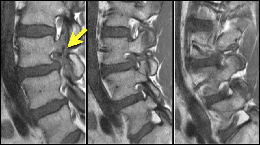
Here sagittal T1W-images demonstrating a structure of very low signal intensity at the L4L5 level (arrow) and at the L5S1 level.Ảnh T1W mặt phẳng đứng dọc cho thấy một cấu trúc tín hiệu rất thấp ở tầng L4–L5 (mũi tên) và tầng L5–S1.
Continue with the T2W-images.Tiếp tục với hình ảnh T2W.
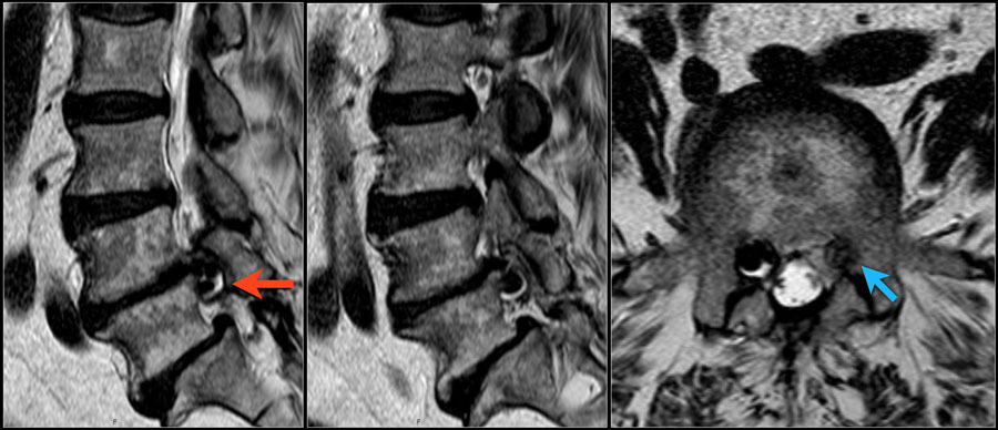 T2W-imagesHình ảnh T2W
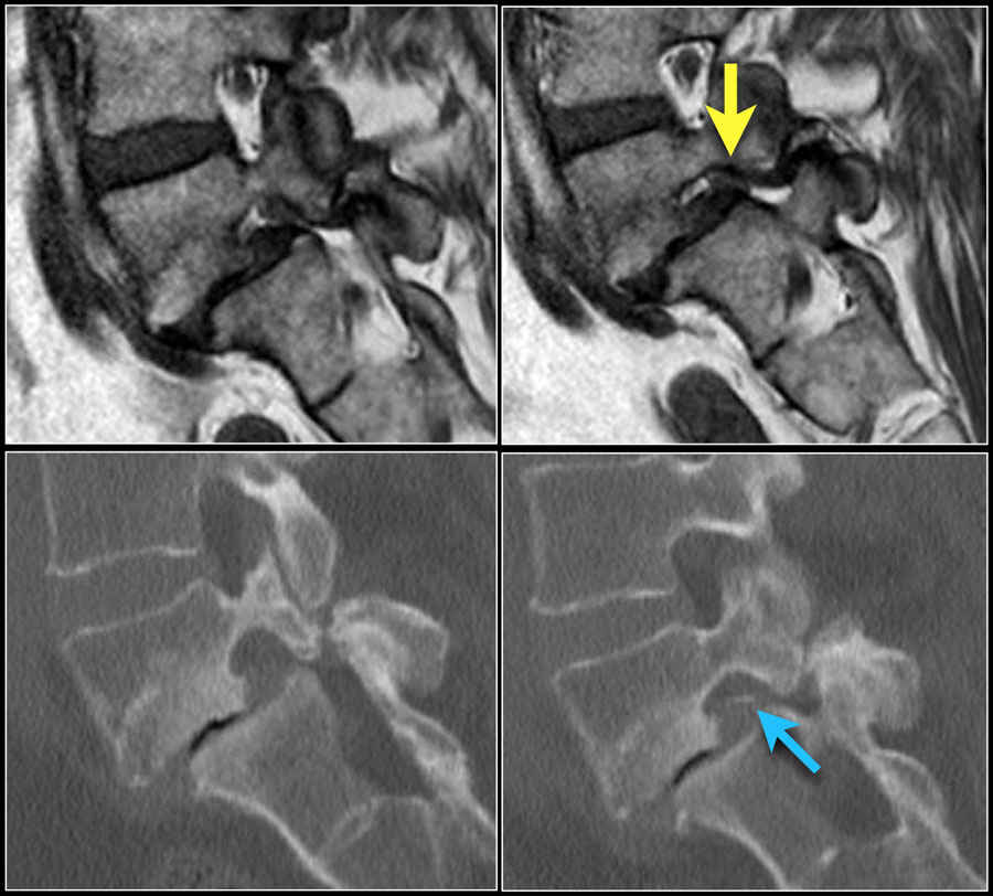
Also on the T2W-images the signal intensity is very low.Cũng trên hình ảnh T2W, cường độ tín hiệu rất thấp.
A CT-scan was performed to see if this could be a calcified herniated disc or some artifact.CT-scan được thực hiện để xem đây có thể là thóat vị đĩa đệm vôi hóa hoặc một số hiện vật.
Continue with the CT-images.Tiếp tục với hình ảnh CT.
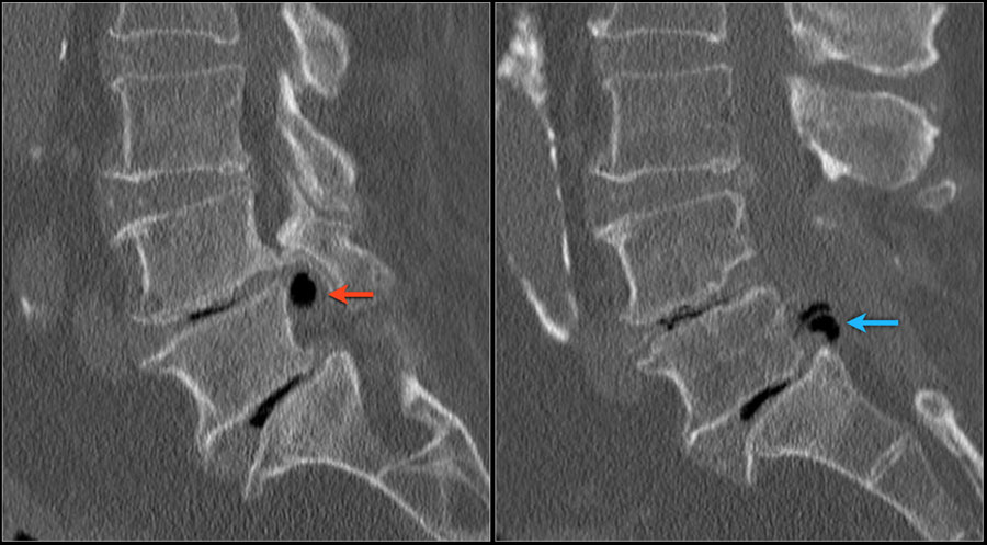
The low signal intensity could finally be explained by the vacuum phenomena due to nitrogen gas within the herniated disc both on L4L5 (red arrow) and L5S1 level (blue arrow)Cường độ tín hiệu thấp cuối cùng có thể được giải thích bởi hiện tượng chân không do khí nitơ trong đĩa đệm thóat vị cả trên mức L4L5 (mũi tên màu đỏ) và L5S1 (mũI tên màu xanh lam)
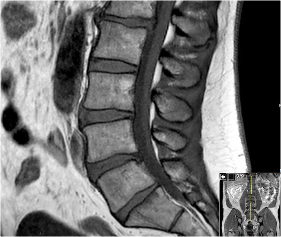
First scroll through these sagittal T1W-images.
What are the findings.Lướt qua các hình ảnh T1W dọc này.
Những phát hiện này là gì?
Then continue with the next series.Sau đó tiếp tục với loạt tiếp theo.
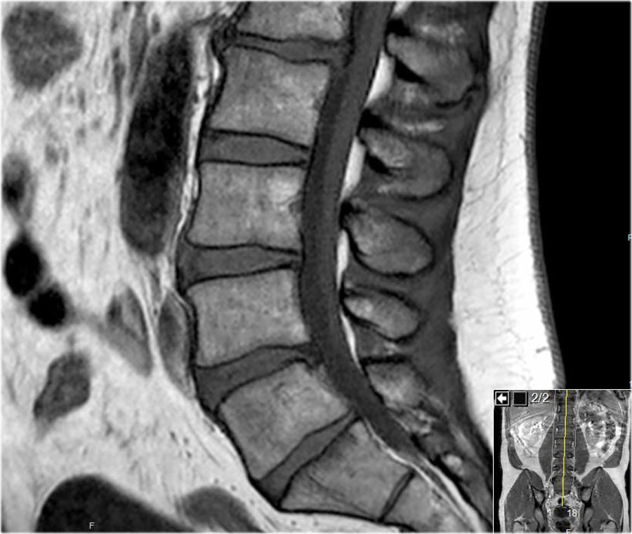
The most striking finding is a intermediate intensity structure posterior to the L3 vertebra (blue arrow).
It has the same signal intensity as the disc.Phát hiện nổi bật nhất là cấu trúc cường độ trung gian phía sau đốt sống L3 (mũi tên màu xanh).
Nó có cùng cường độ tín hiệu với đĩa.
This is a hernia that has migrated cranially compressing the L3 nerve on the right side.Đây là thóat vị đã di chuyển vào sọ chèn ép dây thần kinh L3 ở bên phải.
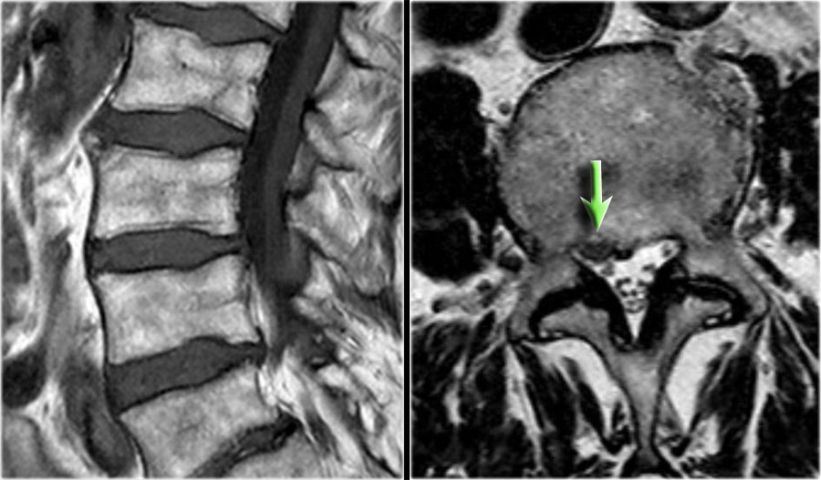
Here another example of disc migration.Đây là một ví dụ khác về di chuyển đĩa.
Notice how the disc herniation at the L3L4 level migrates caudally behind the body of L4.Lưu ý cách thóat vị đĩa đệm ở mức L3L4 di chuyển phía sau cơ thể của L 4.
Notice that at the L4L5 level the migrated disc simulates a nerve root (green arrow), while in fact the nerve is compressed posteriorly and subsequently moves to the foramen (yellow arrow).Lưu ý rằng ở mức L4L5, đĩa di chuyển mô phỏng một rễ thần kinh (mũi tên màu xanh lá cây), trong khi thực tế dây thần kinh bị chèn ép phía sau và sau đó di chuyển đến lỗ (mũa tên màu vàng).
Lateral recess stenosisHẹp lõm bên
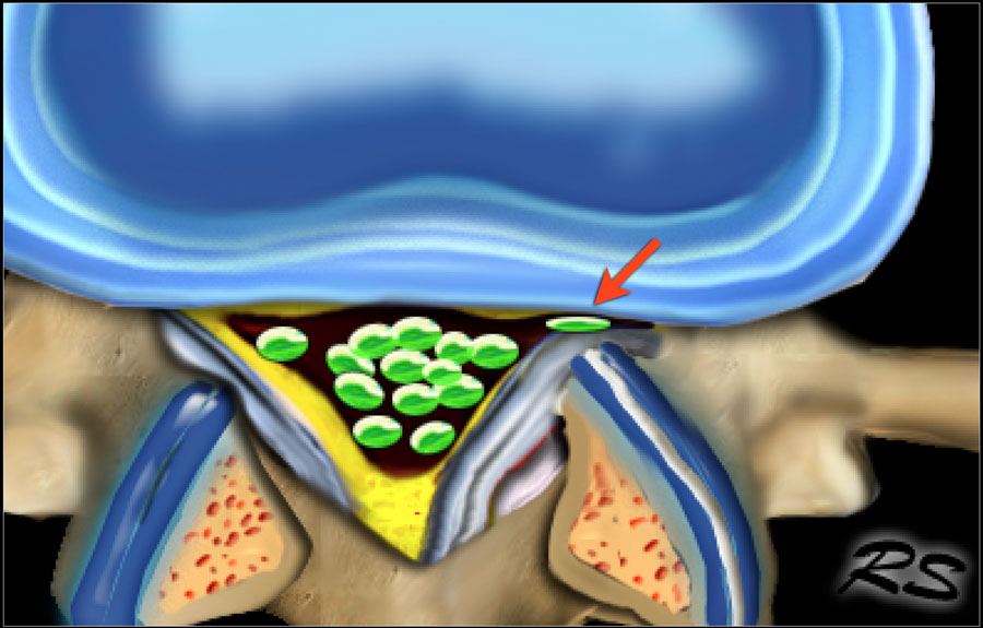
Stenosis of the lateral recess is a common problem especially in older patients.Hẹp ngách ngoài là một vấn đề thường gặp, đặc biệt ở người cao tuổi.
The stability of the vertebral column decreases, which results in instability.
This results in hypertrophy of the facet joints and arthrosis, bulging of the disc and more stress on the flavum ligament resulting in hypertrophy.
All these mechanisms lead to stenosis of the lateral recess (figure).
In advances cases of arthrosis a synovial cyst may form, which contributes to the narrowing.Sự ổn định của cột sống giảm, dẫn đến mất vững.
Điều này dẫn đến sự phì đại của các khớp mặt và khớp, phình đĩa đệm và căng thẳng hơn trên dây chằng flavum dẫn đến phì đại.
Tất cả các cơ chế này dẫn đến hẹp hốc ngoài (hình).
Trong các trường hợp tiến triển của bệnh khớp, nang hoạt dịch có thể hình thành, góp phần làm hẹp khớp.
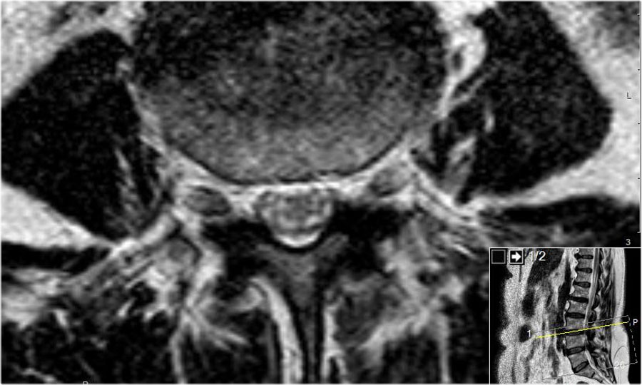
Here a patient with bilateral facet arthrosis resulting in narrowing of the lateral recess on both sides with nerve compression.Đây là một bệnh nhân bị khớp mặt hai bên dẫn đến hẹp hốc bên ở cả hai bên do chèn ép thần kinh.
Scroll through the images.Cuộn qua các hình ảnh.
Synovial cystNang hoạt dịch
Synovial cysts are frequently seen in combination with facet arthrosis.
Mostly they lead to stenosis of the lateral facet.
When they are very large they can protrude into the foramen and cause foraminal stenosis.Nang hoạt dịch khớp thường gặp kết hợp với viêm khớp dạng mặt.
Hầu hết các trường hợp đều dẫn đến hẹp mặt bên.
Khi chúng rất lớn, chúng có thể nhô ra lỗ và gây hẹp lỗ.
Here a patient with severe arthrosis of the facet joints.Đây là một bệnh nhân bị viêm khớp nặng.
Notice that there are many synovial cysts related to the arthrosis (red arrows).Lưu ý có nhiều nang hoạt dịch liên quan đến khớp (các mũi tên đỏ).
At the L5S1 level a large cyst on the right compresses the S1-nerve (yellow arrow).Ở mức L5S1, một nang lớn ở bên phải sẽ chèn ép dây thần kinh S 1 (mũi tên màu vàng).
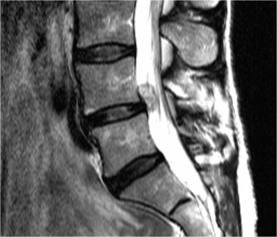
Here sagittal T2-weighted images of a patient with a synovial cyst, that completely fills the neuroforamen (arrow).Ở đây là hình ảnh xung T2W dọc của một bệnh nhân có nang hoạt dịch, lấp đầy hoàn toàn các lổ thần kinh (mũi tên).
Notice that at first glance the foramen looks normal, but the nerve iseems to be missing.
The nerve is compressed by the cyst.Lưu ý rằng thoạt nhìn lỗ này trông bình thường, nhưng dây thần kinh dường như bị mất.
Dây thần kinh bị chèn ép bởi u nang.
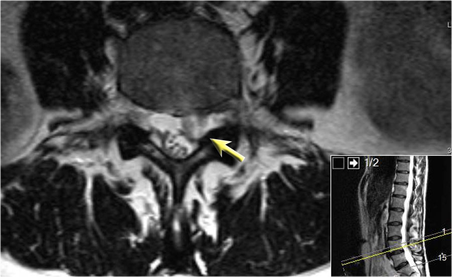
Here we have the axial images of the same patient.Ở đây chúng ta có ảnh mặt phẳng ngang của cùng một bệnh nhân.
Notice the dumbbell configuration of the synovial cyst at the level of the neuroforamen (yellow arrows).Chú ý hình dạng quả tạ của nang hoạt dịch ở mức của liềm thần kinh (các mũi tên màu vàng).
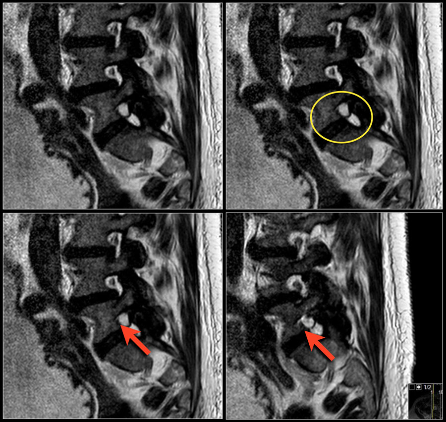
Synovial cysts can be easily overlooked.
On these T2W-images it looks as if the foramen is normal.
Notice that the nerve is missing.Nang hoạt dịch có thể dễ dàng bị bỏ qua.
Trên những hình ảnh T2W này, có vẻ như lỗ thông là bình thường.
Lưu ý rằng dây thần kinh bị mất.
In fact the neuroforamen is almost completely filled by the synovial cyst, which compresses the nerve root against the vertebra (arrows).Trên thực tế, lọ thần kinh gần như được lấp đầy hoàn toàn bởi nang hoạt dịch, nó nén rễ thần kinh vào đốt sống (các mũi tên).
The nerve can hardly be seen.Dây thần kinh hầu như không thể nhìn thấy.
Stenosis of the Spinal CanalHẹp của ống sống
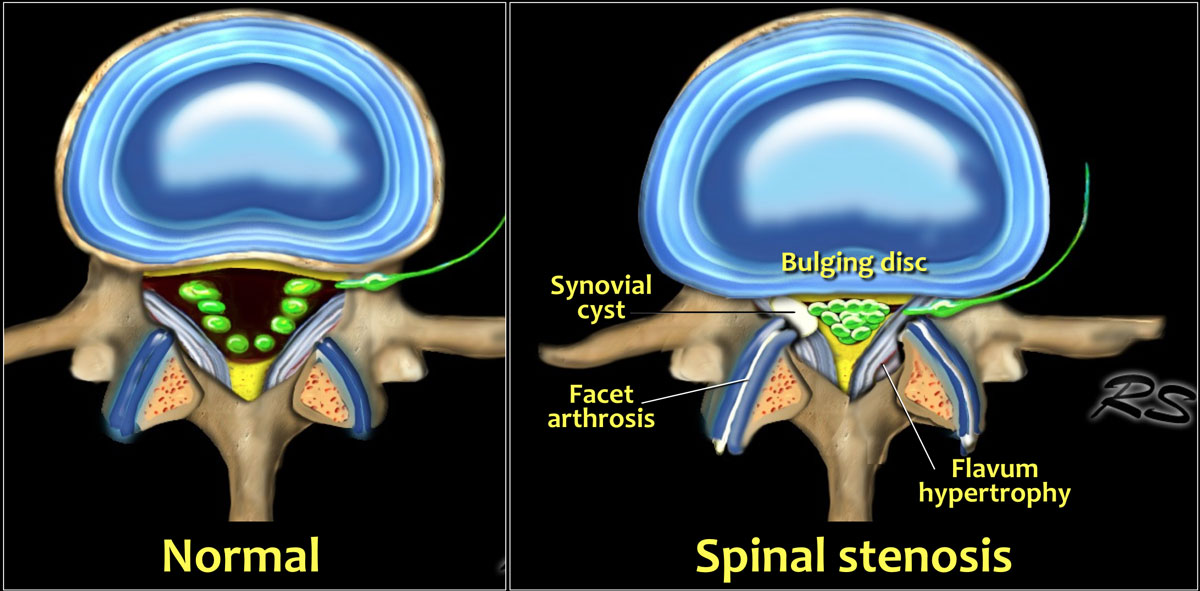

Causes of spinal stenosis:Nguyên nhân của hẹp ống sống:
Most commonPhổ biến nhất
- Bilateral facet arthrosis in combination with bulging of the disc and hypertrophy of the ligamentum flavumBệnh khớp mặt hai bên kết hợp với lồi đĩa đệm và phì đại dây chằng ức
Less commonÍt phổ biến hơn
- Congenital narrowing with short pediclesThu hẹp bẩm sinh có cuống ngắn
- Spinal injury and epidural hematomaChấn thương cột sống và máu tụ ngoài màng cứng
- Bone tumorsU xương
- Spondylodiscitis or epidural abscessViêm cột sống hoặc áp xe ngoài màng cứng
- SpondylolisthesisTrượt đốt sống
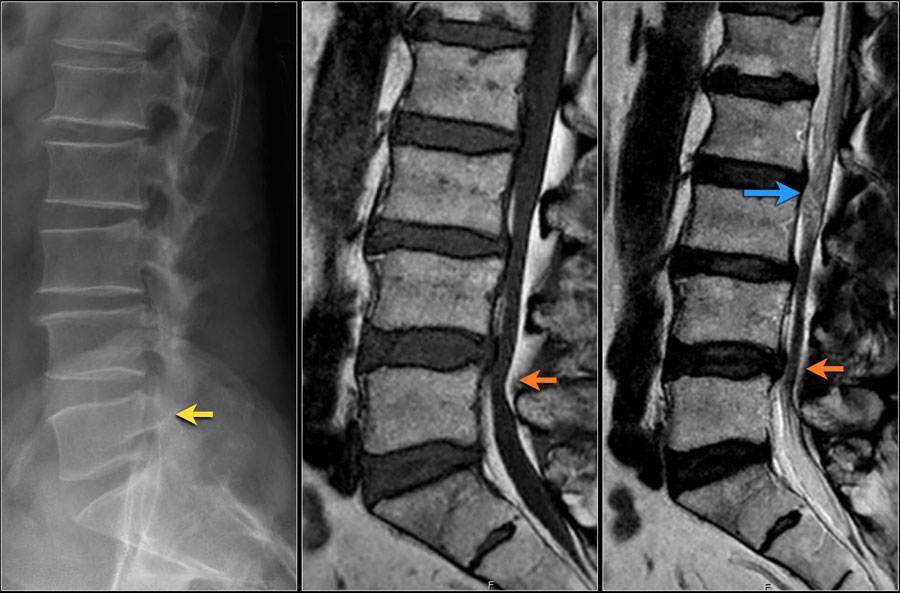
Notice the diffuse narrowing of the lumbar spinal canal.
It is best seen on the MR-images, but can already be suspected on the radiograph, because the pedicles are very short.Lưu ý sự thu hẹp lan toả của ống sống ở thắt lưng.
Nó được nhìn thấy rõ nhất trên hình ảnh MR, nhưng có thể đã bị nghi ngờ trên X quang, bởi vì các cuống mạch rất ngắn.
You may have to enlarge the image to appreciate this.Bạn có thể phải phóng to hình ảnh để đánh giá cao điều này.
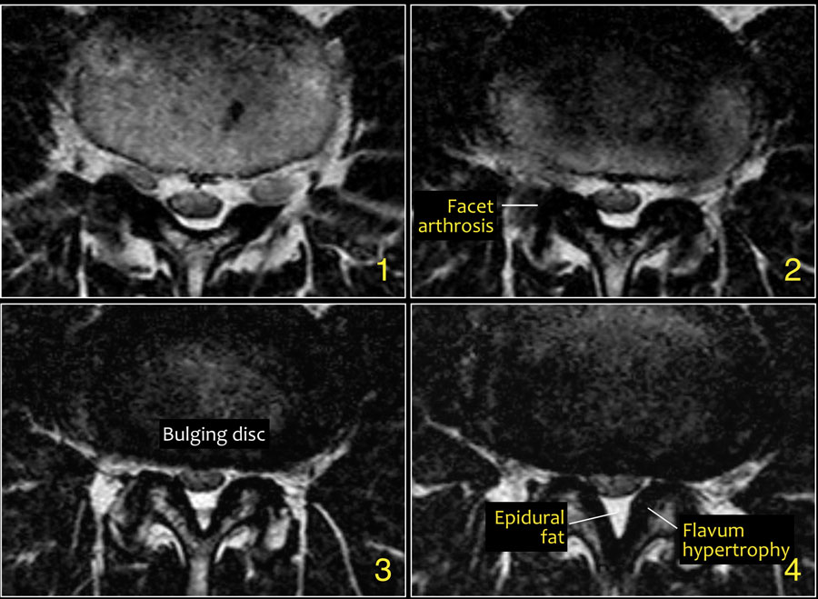

On the axial T2W-images you can see, that there is no CSF visible surrounding the nerve roots.
This means that there is a severe spinal stenosis.
The epidural fat compresses the nerves from posteriorly.Trên hình ảnh T2W dọc trục, bạn có thể thấy, không có dịch não tuỷ có thể nhìn thấy xung quanh rễ thần kinh.
Điều này có nghĩa là có hẹp ống sống nặng.
Mỡ ngoài màng cứng chèn ép các dây thần kinh từ phía sau.
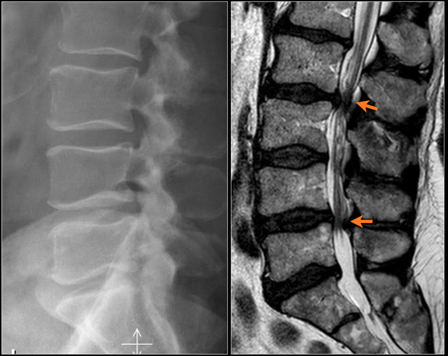
Here another example of spinal stenosis.Đây là một ví dụ khác của hẹp ống sống.
Again on the radiograph you can suspect congenital narrowing.
Usually a small hernia or bulging is enough to cause nerve compression in these patients.Một lần nữa trên phim X-quang, bạn có thể nghi ngờ hẹp bẩm sinh.
Thông thường, một thóat vị nhỏ hoặc phình ra là đủ để gây chèn ép thần kinh ở những bệnh nhân này.
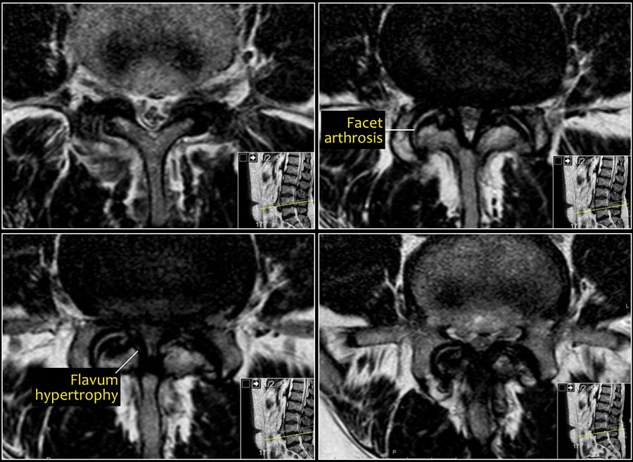

On the axial T2W-images a severe spinal stenosis is seen.
Notice the short pedicles in combination with facet arthrosis and flavum hypertrophy.Trên hình ảnh T2W dọc trục, thấy hẹp ống sống nặng.
Lưu ý các cuống ngắn kết hợp với khía cạnh khớp và phì đại flavum.
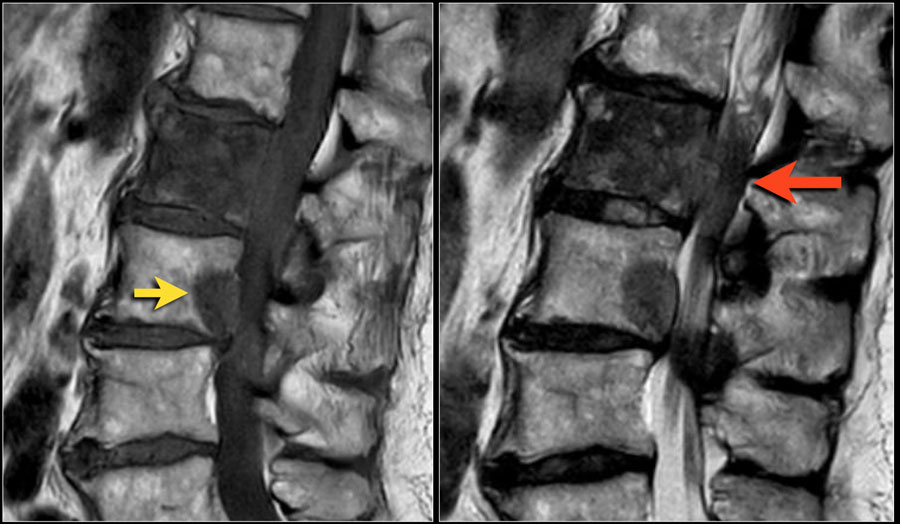
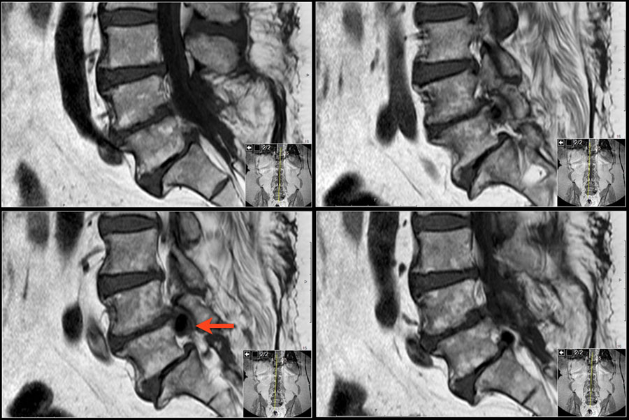
Here a patient with multiple osseus metastases.Đây là một bệnh nhân có di căn đa xương.
At the level of L1L2 this causes spinal stenosis with compression of the nerves (red arrow).Ở mức L1L2, điều này gây ra hẹp ống sống với sự chèn ép của các dây thần kinh (mũi tên màu đỏ).
It is not that common for metastases to cause nerve compression at the lumbar level.
At the thoracic and cervical level metastases frequently cause compression because there is not much CSF surrounding the myelum.Di căn gây chèn ép thần kinh ở mức độ thắt lưng không phải là phổ biến.
Ở cấp độ ngực và cổ, di căn thường gây chèn ép vì không có nhiều CSF xung quanh tuỷ.
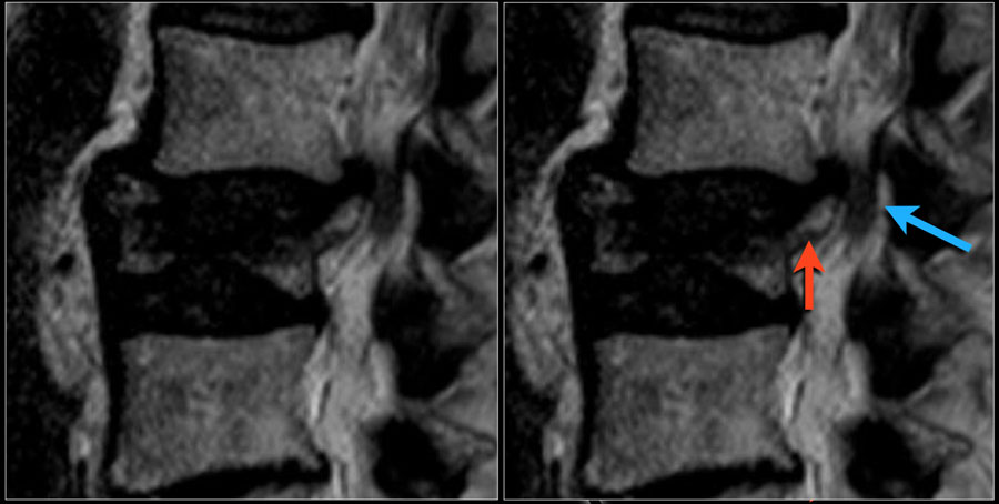
Fractures can cause stenosis of the spinal canal especially when there is displacement of bony structures like in burst fractures and fractures with rotation and translation.Gãy xương có thể gây hẹp ống sống đặc biệt khi có sự dịch chuyển của các cấu trúc xương như trong gãy vỡ và gãy xoay và dịch chuyển.
Here a patient with an old burst fracture.
There is retropulsion of the posterosuperior fragment (red arrow) compressing the cauda (blue arrow).Đây là một bệnh nhân bị gãy xương cũ.
Có sự đẩy ngược của mảnh sau trên (mũi tên màu đỏ) chèn ép vào khoang (mũa tên màu xanh).
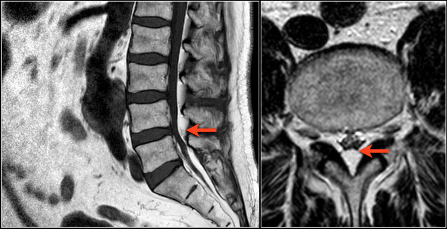
Epidural lipomatosisLipomatosis ngoài màng cứng
Epidural lipomatosis is excessive amount of fat within the epidural space compressing the thecal sac.
Patients present with symptoms of spinal canal stenosis.Lipomatosis ngoài màng cứng là lượng chất béo quá mức trong khoang ngoài màng cứng chèn ép túi canxi.
Bệnh nhân có triệu chứng hẹp ống sống.
It is seen in patients with obesity, like in this case and in patients who are treated with steroids.Nó được thấy ở những bệnh nhân béo phì, như trong trường hợp này và ở những bệnh nhi được điều trị bằng steroid.
Continue with the axial images of this patient.Tiếp tục với ảnh mặt phẳng ngang của bệnh nhân này.
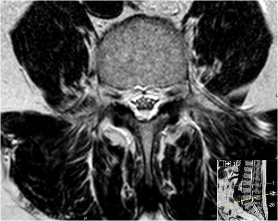
Scroll through the axial images.Cuộn qua ảnh mặt phẳng ngang.
Notice how the spinal canal is narrowed by the epidural fat.
Do not mistake the high signal intensity of the fat for CSF.Lưu ý mỡ ngoài màng cứng làm hẹp ống sống. Đừng nhầm tín hiệu cao của mỡ với dịch não tủy.
Foraminal stenosisHẹp trước sinh
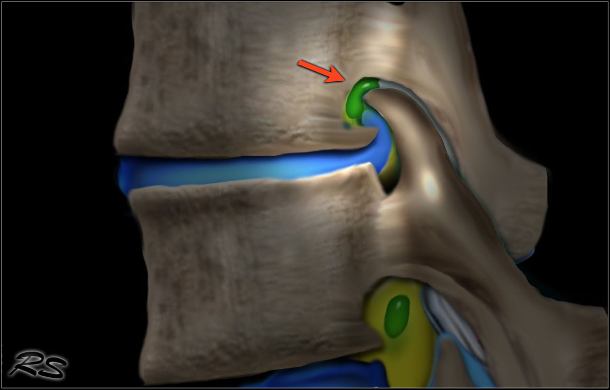
Causes of Foraminal stenosis:Nguyên nhân của hẹp hậu môn:
- Facet arthrosis Khía cạnh khớp
- Disc herniation with upward migrationThóat vị đĩa đệm với di chuyển lên trên
- Spondylolistesis
Stenosis of the neuroforamen is usually the result of a combination of upward disc herniation and facet arthrosis.
It is also frequently seen in patients with spondylolisthesis.
Spondylolisthesis is a condition in which one vertebra slips forward over the one below it, usually L4 upon L5.
The shift results from either spondylolysis (stress fracture of the pars interarticularis) or facetarthrosis with sliding of the facets.Hẹp lách thần kinh thường là kết quả của sự kết hợp của thóat vị đĩa đệm hướng lên trên và khớp mặt.
Nó cũng thường thấy ở những bệnh nhân bị trượt đốt sống.
Trượt đốt sống là tình trạng một đốt sống trượt về phía trước so với đốt sống bên dưới, thường là L4 trên L 5.
Sự dịch chuyển này là kết quả của thóai hóa cột sống (gãy căng thẳng của khớp liên khớp) hoặc viêm khớp mặt với sự trượt của các mặt.
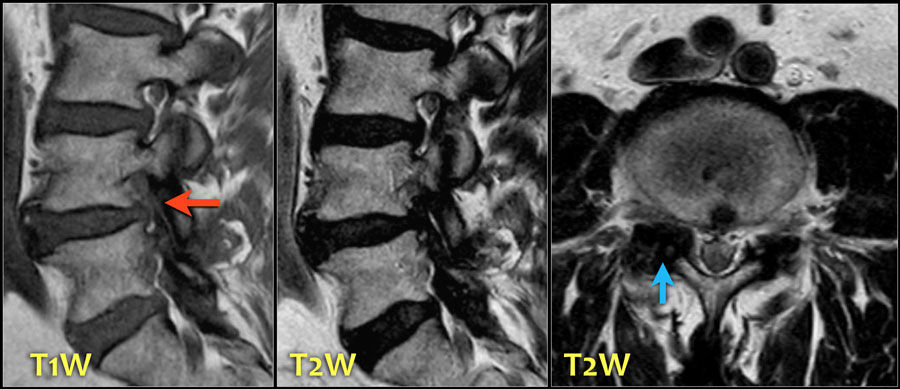
Here a patient with severe facet arthrosis on the right side at the L4L5 level.Đây là một bệnh nhân bị khớp mặt bên phải mức L4L5.
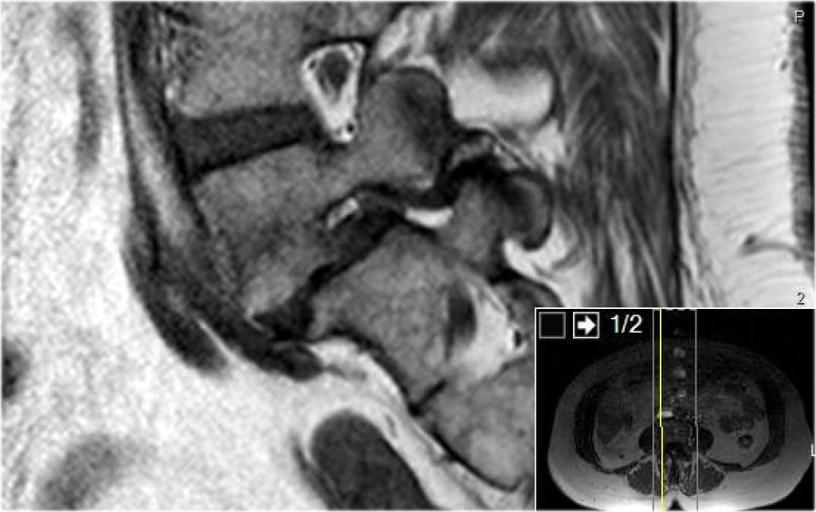
Scroll through the images of a patient with spondylolisthesis.Cuộn qua các hình ảnh của một bệnh nhân bị trượt đốt sống.
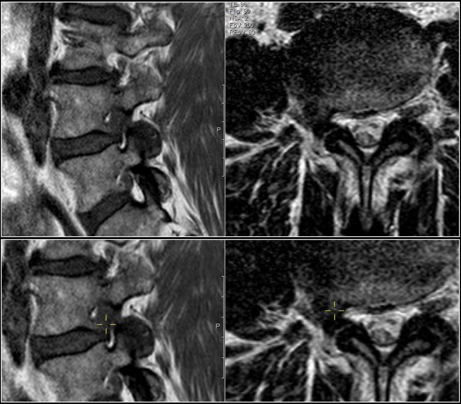
Here a disc herniation with upward migration.Đây là thóat vị đĩa đệm với di chuyển lên trên.
By using the marker, you can correlate the abnormality on the T1W-image with the axial T2W-images.Bằng cách sử dụng marker, bạn có thể tương quan các bất thường trên T1W-image với T2W-images trục.
On the T2W-image it is more obvious that this is a disc herniation.Trên hình ảnh T2W, rõ ràng hơn đây là thóat vị đĩa đệm.
The lower images are with the marker on.
you may have to enlarge the images to see it.Hình ảnh dưới cùng là khi có điểm đánh dấu.
bạn có thể phải phóng to hình ảnh để xem nó.
These foraminal disc herniations can be easily overlooked.Những thóat vị đĩa đệm lỗ này có thể dễ dàng bị bỏ qua.
Notice how subtle the findings are on the axial T2W-images.
The herniated disc can be easily mistaken for the nerve root.Chú ý đến sự tinh tế của các phát hiện trên hình ảnh T2W dọc trục.
Thóat vị đĩa đệm có thể dễ bị nhầm lẫn với rễ thần kinh.
The sagittal T1W-image shows the upward migration of the disc.Hình ảnh T1W dọc cho thấy sự di chuyển lên trên của đĩa đệm.
Here a patient with a combination of spondylolysis (arrow), minimal listhesis and disc herniation resulting in compression of the L3 nerve within the foramen.Đây là một bệnh nhân có kết hợp giữa giải phóng xơ đốt sống (mũi tên), liệt tối thiểu và thóat vị đĩa đệm dẫn đến chèn ép dây thần kinh L3 trong lỗ.
Here a patient with severe anterolisthesis due to bilateral spondylolysis.Đây là một bệnh nhân bị liệt trước khớp gối nặng do thóai hóa cột sống hai bên.
The anteroposition in combination with the upward movement of the disc has resulted in severe narrowong of the foramen and nerve compression (yellow arrow).Sự lệch trước kết hợp với sự di chuyển lên trên của đĩa đệm đã dẫn đến sự hẹp nghiêm trọng của lỗ và chèn ép thần kinh (mũi tên màu vàng).
Notice the calcification of the uppper part of the disc (blue arrow).Chú ý vôi hóa của phần trên của đĩa (mũi tên màu xanh).
Extraforaminal nerve compressionChèn ép thần kinh ngoài cùng
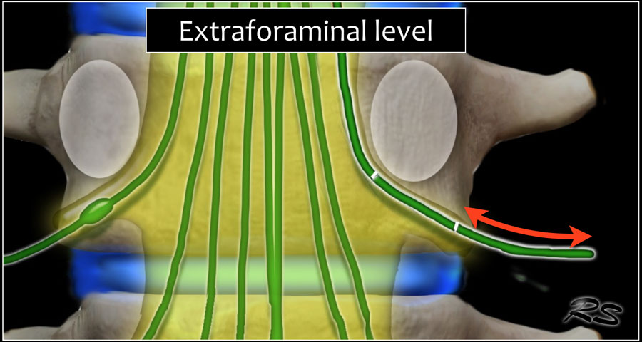
Extraforaminal nerve compression is seen in about 5% of cases.
Almost always it is a lateral disc herniation from a lower level that compresses the extraforaminal part of the nerve.Chèn ép dây thần kinh ngoài cùng được thấy trong khoảng 5% trường hợp.
Hầu như luôn luôn là một thóat vị đĩa đệm bên từ một mức thấp hơn chèn ép phần ngoài cùng của dây thần kinh.
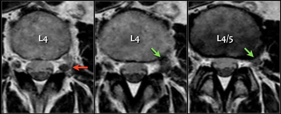
Here an example of a lateral disc herniation that produces compression of the superiorly exiting nerve root and ganglion.Đây là một ví dụ về một thóat vị đĩa đệm bên tạo ra sự nén của rễ thần kinh và hạch thóat ra phía trên.
Notice the L4 nerve (red arrow), which is being displaced posteriorly by a lateral disc herniation at the L4-5 level (green arrow).Chú ý dây thần kinh L4 (mũi tên màu đỏ), đang bị di lệch ra sau do thóat vị đĩa đệm bên ở mức L 4-5 (mũI tên màu xanh lá cây).