
Neonatal Chest X-RayX-quang ngực sơ sinh
Radiology, Nuclear medicine and Neonatology department of the Amsterdam University Medical CentreKhoa X quang, Y học hạt nhân và Sơ sinh, Trung tâm Y tế Đại học Amsterdam
Bản gốc trên radiologyassistant.nl ↗ · tải về lúc 2026-11-19 20:19 · bản dịch máy có chuẩn hóa thuật ngữ chẩn đoán hình ảnh
In this review we will discuss a systematic approach to the neonatal chest radiograph.
Close collaboration between neonatologists and radiologists is the key in achieving the correct diagnosis, since the radiographic features of many lung disorders overlap and the findings on the chest film can be rather subtle.
Clinical information like age of the neonate, gestational age and therapy with ventilation or surfactant is vital for the interpretation of the radiological findings.Trong bài tổng quan này, chúng tôi sẽ thảo luận về cách tiếp cận có hệ thống đối với phim X-quang ngực sơ sinh.
Sự hợp tác chặt chẽ giữa bác sĩ giải phẫu và bác sĩ X quang là chìa khóa để đạt được chẩn đoán chính xác, vì các đặc điểm X quang của nhiều bệnh lý phổi chồng chéo và các phát hiện trên phim ngực có thể khá tinh tế.
Thông tin lâm sàng như tuổi của trẻ sơ sinh, tuổi thai và liệu pháp thông khí hoặc surfactant là rất quan trọng để giải thích các dấu hiệu X quang.
We will first discuss a diagnostic approach based on radiographic and clinical findings and subsequently we will discuss the specific pulmonary diseases and complications.Trước hết, chúng ta sẽ thảo luận về cách tiếp cận chẩn đoán dựa trên X quang và các dấu hiệu lâm sàng, sau đó chúng ta sẽ bàn về các bệnh phổi và các biến chứng cụ thể.
Interpretations of tube positions, lines and catheter positions are discussed in Tubes and lines in neonates.Giải thích vị trí ống thông, đường truyền và vị trí Ống thông được thảo luận trong Ống và đường truyền ở trẻ sơ sinh.
IntroductionMở đầu
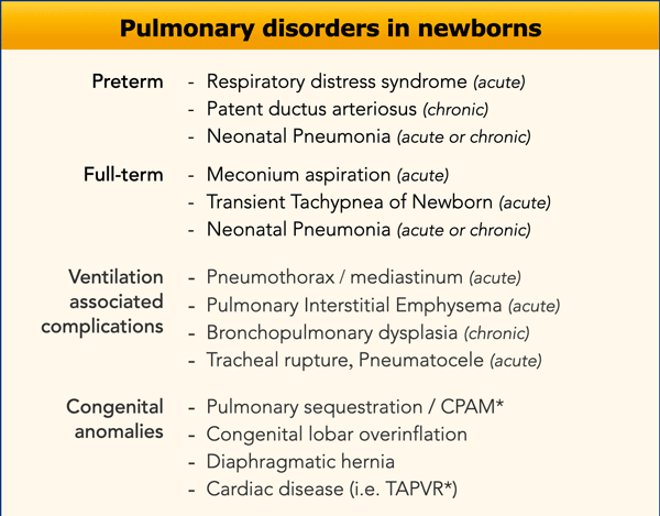
Preterm infants show different types of pathology compared to
term infants.
For example, respiratory distress syndrome (RDS) is almost
exclusively seen in preterm infants.
Meconium aspiration (MA) on the other
hand, is seen in full term or late term neonates in combination with
meconium-stained amniotic fluid during labor.
Infants born between 34 and 37 weeks of gestation can have
diseases occurring in both preterm and full-term infants. Trẻ sinh non có các loại bệnh lý khác nhau so với trẻ đủ tháng.
Ví dụ, hội chứng suy hô hấp (RDS) hầu như chỉ gặp ở trẻ sinh non.
Hít phân su (MA) thì ngược lại, được thấy ở trẻ sơ sinh đủ tháng hoặc muộn kết hợp với nước ối có màu phân su trong quá trình chuyển dạ.
Trẻ sơ sinh từ 34 đến 37 tuần tuổi thai có thể có bệnh xảy ra ở cả trẻ sinh non và trẻ đủ tháng.
Invasive mechanical ventilation and surfactant therapy will have a huge impact on the radiographic findings and are essential clinical information for the radiologist.Việc áp dụng thở máy xâm lấn và liệu pháp surfactant sẽ có tác động rất lớn đến các dấu hiệu trên X quang và là thông tin lâm sàng thiết yếu cho bác sĩ X quang.
Here is a list of common pulmonary disorders in neonates based on acute and chronic disease, complications of ventilation and congenital anomalies (Table).Dưới đây là danh sách các rối loạn phổi thường gặp ở trẻ sơ sinh dựa trên bệnh lý cấp tính và mạn tính, biến chứng của thông khí và dị tật bẩm sinh (Bảng).
* CPAM was previously referred to as congenital cystic adenomatoid malformation (CCAM) and presents as a mass of abnormal non-functional lung tissue
* CPAM trước đây được gọi là dị dạng nang tuyến bẩm sinh (CCAM) và biểu hiện dưới dạng một khối mô phổi bất thường không có chức năng.
* TAPVR is total anomalous pulmonary venous return. The pulmonary veins do not connect to the left atrium, but enter the systemic venous circulation. * TAPVR là bất thường hoàn toàn về tĩnh mạch phổi, các tĩnh mạch phổI không nối với nhĩ trái, nhưng đi vào tuần hoàn tĩnh mạch hệ thống.
The approach to the neonatal chest film starts by looking at the technique of the radiograph.
Then the position of lines and tubes is analyzed.
See Lines and tubes in Neonates.
After these steps, the chest film can be interpreted for pathology. Cách tiếp cận phim ngực sơ sinh bắt đầu bằng cách xem xét kỹ thuật của X quang.
Sau đó vị trí của các đường và ống được phân tích.
Xem Đường và ống ở trẻ sơ sinh.
Sau các bước này, phim ngực có thể được giải thích cho bệnh lý.
This is done in a stepwise manner:Điều này được thực hiện theo từng bước:
- Consider gestational age, postnatal age, and other clinical information, including respiratory support.Xem xét tuổi thai, tuổi sau sinh và các thông tin lâm sàng khác, bao gồm hỗ trợ hô hấp.
- Lung volume should interpreted, being either normal, hypo-inflated or hyper-inflated.Thể tích phổi nên được giải thích, là bình thường, giảm hoặc tăng.
- In ventilated neonates positive end expiratory pressure (PEEP) should be noted on the request. (Normal PEEP 5-8 cm H2O)
Ở trẻ sơ sinh thở máy, cần lưu ý áp lực dương cuối kỳ thở ra (PEEP) theo yêu cầu. (PEEP bình thường 5-8 cm H2O) - Interpretation of lung parenchyma, being either normal, lucent, or opacified. Giải thích nhu mô phổi, bình thường, sáng hoặc đục.
- Pathology unilateral or bilateral.
Bệnh lý một bên hoặc hai bên.
The postnatal age of the neonate in days or hours is important as some disorders are acute conditions and some disorders at a present later stage. In addition, the course of illness and therapy so far should be taken into account. Tuổi sau sinh của trẻ sơ sinh theo ngày hoặc giờ rất quan trọng vì một số rối loạn là tình trạng cấp tính và một số rối loại ở giai đoạn muộn hiện tại, ngoài ra, quá trình bệnh tật và điều trị cho đến nay cần được tính đến.
Pattern ApproachPhương pháp tiếp cận mẫu
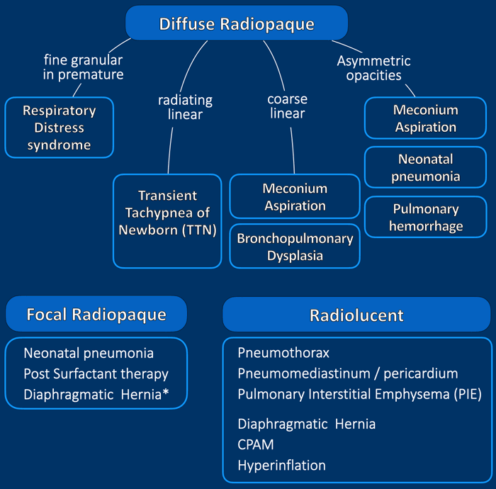
Chest abnormalities can be divided into:
Các bất thường ở ngực có thể được chia thành:
- Radiopaque
Other terms that are used to describe these abnormalities are high attenuation, or opacities. They can be fine or course linear, granular or reticulo-granular. They can be diffuse or focal.Xạ trị
Các thuật ngữ khác được sử dụng để mô tả những bất thường này là độ đậm đặc cao, hoặc độ mờ đục. Chúng có thể là dạng tuyến tính, dạng hạt hoặc dạng lưới. Chúng có thê được khuếch tán hoặc khu trú. - Radiolucent
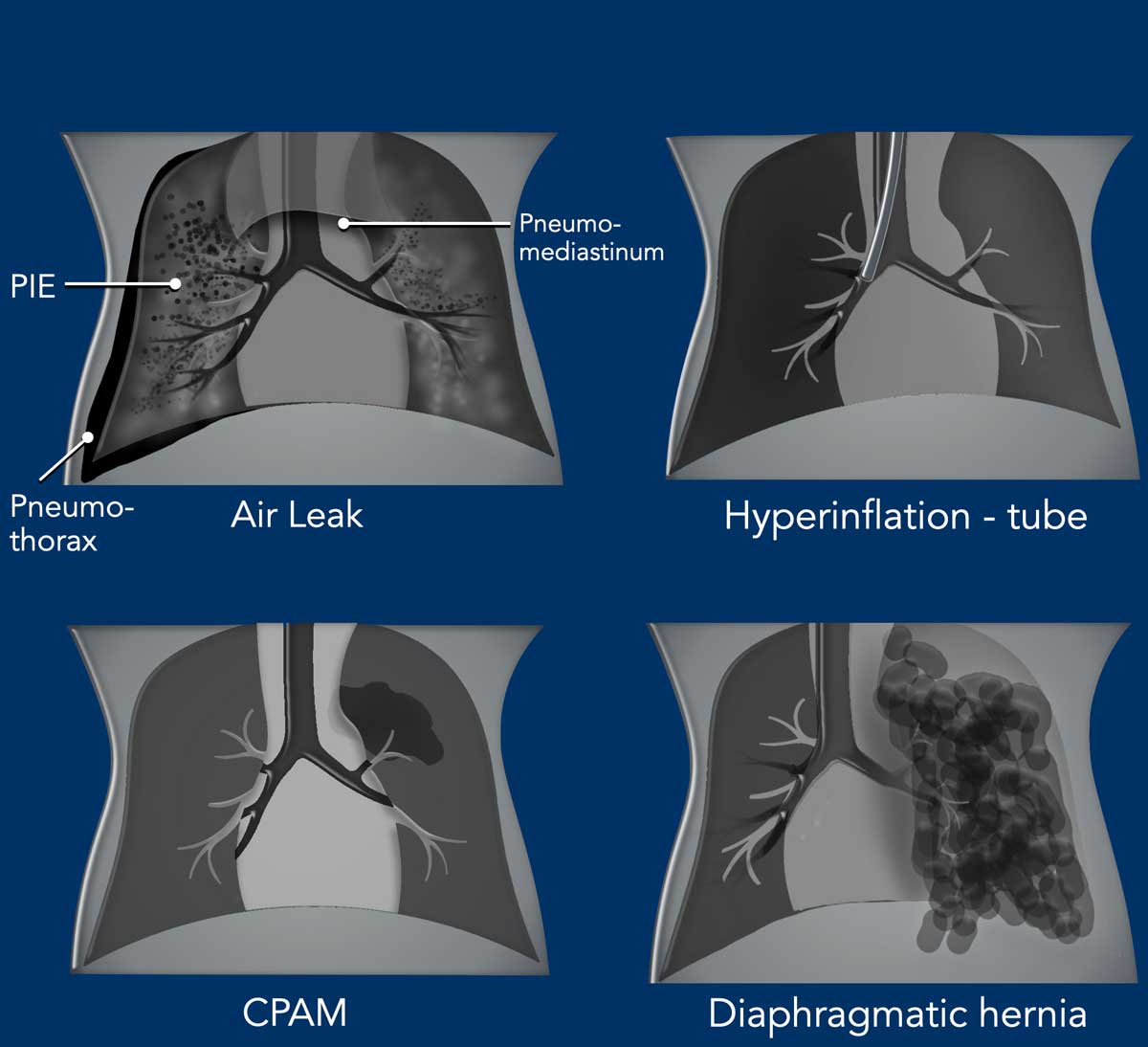
These are lucent abnormalities, being the result of hyperinflation of lung parenchyma, or abnormal collections of air. Depending on the location, these lucencies can follow pleura or mediastinal structures and are often sharply delineated.
Intraparenchymal abnormalities can be less circumscribed.
Phóng xạ
Đây là những bất thường về ánh sáng, là kết quả của sự căng phồng của nhu mô phổi, hoặc sự tập trung không khí bất thường. Tuỳ thuộc vào vị trí, những ánh sáng này có thể theo cấu trúc màng phổi hoặc trung thất và thường được phân định rõ ràng.
Các bất thường trong nhu mô có thể ít được xác định.
In some cases there is a combination of radiopaque and radiolucent abnormalities, Trong một số trường hợp có sự kết hợp của bất thường cản quang và cản quang,
which makes the interpretation of the radiographic findings challenging.
For example, pulmonary interstitial emphysema (PIE)
can be regarded as lucencies, but can also be interpreted as radiating linear radiopaque abnormalities.
Notice that a diaphragmatic hernia can be a focal radiopaque or radiolucent Điều này khiến cho việc giải thích các dấu hiệu trên X quang trở nên khó khăn.
Ví dụ, khí phế thũng kẽ phổi (PIE) có thể được coi là các điểm sáng, nhưng cũng có thể được hiểu là các bất thường về bức xạ tuyến tính.
Lưu ý rằng thóat vị hoành có thể là thóat vị khu trú hoặc thóat vị mờ
Radiopaque - High AttenuationĐộ xạ - Độ suy giảm cao
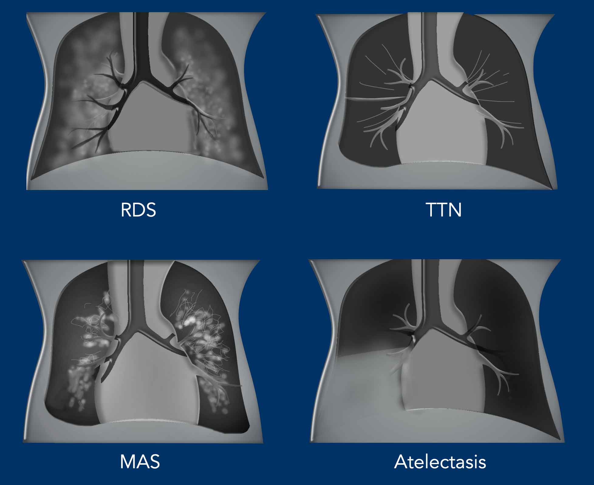
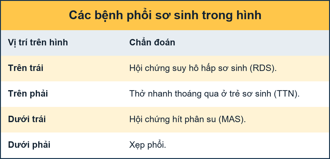
RDS
RDSName
Respiratory distress syndrome in preterm neonates presents with low lungvolume, air bronchograms and symmetric fine granular opacities, ranging from mild disease to complete white out of the lungs. Hội chứng suy hô hấp ở trẻ sơ sinh non tháng biểu hiện với thể tích phổi thấp, chụp phế quản không khí và các hạt mờ đối xứng, từ bệnh nhẹ đến phổi trắng hoàn toàn.
TTN
Transient tachypnea of the newborn is seen in full term neonates with mild respiratory distress and presents with subtle increased lung volume, parahilar linear densities and pleural effusions.TTN
Thở nhanh thóang qua của trẻ sơ sinh được thấy ở trẻ sơ sinh đủ tháng bị suy hô hấp nhẹ và biểu hiện với thể tích phổi tăng lên một cách tinh tế, mật độ tuyến tính cạnh ổ răng và tràn dịch màng phổi.
MAS
Meconium aspiration is seen in full term and late term neonates and presents with coarse bilateral densities, that can be asymmetrical. There can be areas of atelectasis and air-trapping.MAS
Hít phân su được thấy ở trẻ sơ sinh đủ tháng và muộn và có biểu hiện mật độ hai bên thô, có thể không đối xứng, có thể có các vùng xẹp phổi và bẫy không khí.
Atelectasis
Xẹp phổi
Atelectasis can be seen in tube malpositioning and as complication, e.g. in RDS and meconium aspiration. Xẹp phổi có thể được nhìn thấy trong việc đặt sai vị trí ống và biến chứng, ví dụ như trong RDS và hít phân su.
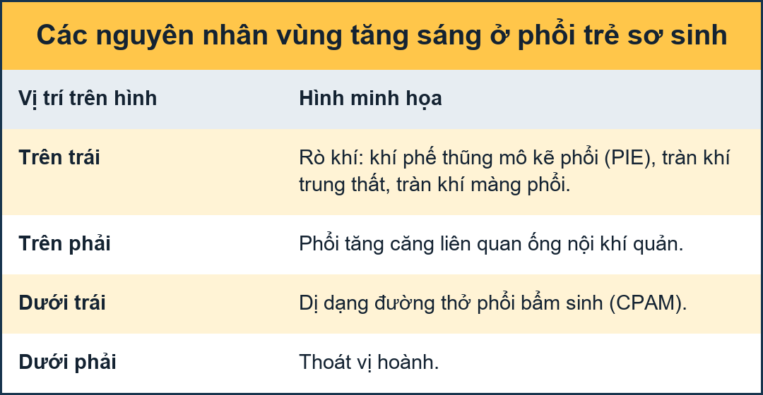
Radiolucent - Low attenuationChiếu sáng - Độ suy giảm thấp
Air Leak
Most radiolucent lung abnormalities are the result of air leakage usually as a complication of positive pressure ventilatory support and present as:Rò rỉ khí
Hầu hết các bất thường về phổi do ánh sáng là kết quả của rò rỉ khí thường là biến chứng của hỗ trợ thông khí áp lực dương và biểu hiện như sau:
- PneumothoraxTràn khí màng phổi
- Pneumomediastinum and pericardiumTràn khí trung thất và màng ngoài tim
- Pulmonary Interstitial Emphysema (PIE)Phổi kẽ khí phế thũng (PIE)
- Pneumatocele
Nang khí
Hyperinflation
It can be the result of malpositioning of the tube with hyperinflation of one lung or in the case of atelectasis, there can be compensatory hyperinflation of other lung parts.Siêu lạm phát
Nó có thể là kết quả của việc đặt sai vị trí của ống với sự căng phồng của một phổi hoặc trong trường hợp xẹp phổi, có thể có sự căng phình bù trừ của các phần khác của phổi.
CPAM
CPAM
is the most common congenital lung malformation, but is still a rare disease.
There is a microcystic and a macrocystic form. The latter presents as a
radiolucent abnormality. Often in the first hours the lesion is not containing
air yet and can present as radiopaque. CPAMComment
CPAM là dị tật phổi bẩm sinh phổ biến nhất, nhưng vẫn là một bệnh hiếm gặp, có dạng nang nhỏ và nang lớn, dạng nang lớn biểu hiện như một bất thường về quang học, thường trong những giờ đầu tổn thương chưa chứa khí và có thể biểu hiện như tổn thương do tia xạ.
Congenital diaphragmatic hernia
This is a birth defect of the diaphragm and can be a life threatening disease.
The herniation of abdominal organs into the chest results in underdevelopment of the lung.
Thóat vị hoành bẩm sinh
Đây là một dị tật bẩm sinh của cơ hoành và có thể là một bệnh đe doạ tính mạng.
Thóat vị các cơ quan trong ổ bụng vào ngực dẫn đến sự phát triển kém của phổi.
Respiratory Distress Syndrome (RDS)Hội chứng suy hô hấp (RDS)
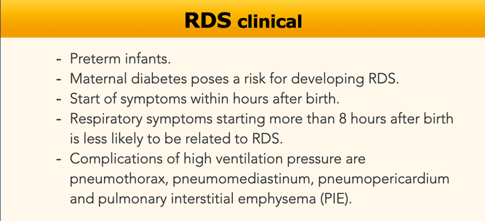
RDS is also known as hyaline membrane disease (HMD), idiopathic respiratory distress syndrome (IRDS) and surfactant deficiency disorder (SDD).
It is a result of deficiency of surfactant due to
immaturity of the lungs in preterm infants.RDS còn được gọi là bệnh màng hyaline (HMD), hội chứng suy hô hấp vô căn (IRDS) và rối loạn thiếu chất hoạt động bề mặt (SDD).
Đây là hậu quả của việc thiếu surfactant do phổi non ở trẻ non tháng.
Surfactant production starts between 24 to 28 week of gestational age.
The incidence of RDS is inversely related to the gestational age at birth. It ranges from 50% in newborns at 26-28 weeks gestation and decreases to 25% in newborns at 30-31 weeks.
As a result of the surfactant deficiency, the alveoli will not contain air and cause diffuse bilateral micro-atelectasis, in combination with fibrin and cellular debris due to alveolar damage, which leads to poor compliance of the lungs and prevents the newborn to expand the lungs properly.Sản xuất surfactant bắt đầu từ 24 đến 28 tuần tuổi thai.
Tỷ lệ mắc RDS có liên quan nghịch với tuổi thai khi sinh, dao động từ 50% ở trẻ sơ sinh ở tuần 26-28 và giảm xuống còn 25% ở trẻ sơ sanh ở tuần 30-31.
Do thiếu chất hoạt động bề mặt, phế nang sẽ không chứa không khí và gây ra vi xẹp phổi lan toả hai bên, kết hợp với fibrin và các mảnh vụn tế bào do tổn thương phế nang, dẫn đến sự tuân thủ kém của phổi và ngăn ngừa trẻ sơ sinh mở rộng phổi đúng cách.
Endogenous production of surfactant will begin at approximately 36 - 72 hours regardless of the gestational age of the patient.
Therefore, the diagnosis of RDS is restricted to the first week of life.Sản xuất surfactant nội sinh sẽ bắt đầu vào khoảng 36-72 giờ bất kể tuổi thai của bệnh nhân.
Vì vậy, chẩn đoán RDS chỉ giới hạn trong tuần đầu tiên của cuộc đời.
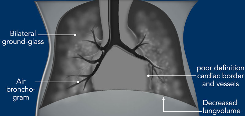

ImagingHình ảnh
- Decreased lung volume on chest radiograph unless the patient is on ventilator.Giảm thể tích phổi trên phim X-quang ngực trừ khi bệnh nhân đang thở máy.
- Bilateral symmetric ground glass opacities. Đục kính mờ đối xứng hai bên.
- Air bronchograms can be visible into the periphery. Chụp phế quản không khí có thể nhìn thấy ở ngoại vi.
- Poor definition of the cardiac silhouette and pulmonary vessels, as a result of less aerated lung parenchyma.Định nghĩa hình ảnh tim và mạch phổi kém, do nhu mô phổi ít khí hóa.
- Usually no pleural effusion.Radiological appearance can be asymmetric immediately after minimally invasive surfactant therapy (MIST) and can mimic for example neonatal pneumonia.Thông thường không có tràn dịch màng phổi. Hình ảnh X quang có thể không đối xứng ngay sau khi điều trị bằng surfactant xâm lấn tối thiểu (MIST) và có thể bắt chước ví dụ viêm phổi sơ sinh.
Imaging findings are often most
severe at birth, but may peak at 12- 24 hours after birth.
In many preterm neonates improvements
in treatment, including antenatal glucocorticoid administration, surfactant
replacement therapy and better ventilatory strategies have decreased the
prevalence of RDS.
When prolonged
mechanical ventilation is necessary, this increases the risk of lung injury and air
leak and can evolve into chronic lung disease.Các dấu hiệu hình ảnh thường nặng nhất khi sinh, nhưng có thể đạt đỉnh điểm 12-24 giờ sau khi sinh.
Ở nhiều trẻ sơ sinh non tháng, những cải thiện trong điều trị, bao gồm sử dụng glucocorticoid trước sinh, liệu pháp thay thế surfactant và các chiến lược thông khí tốt hơn đã làm giảm tỷ lệ mắc RDS.
Khi thở máy kéo dài là cần thiết, điều này làm tăng nguy cơ tổn thương phổi và rò rỉ khí và có thể tiến triển thành bệnh phổi mạn tính.
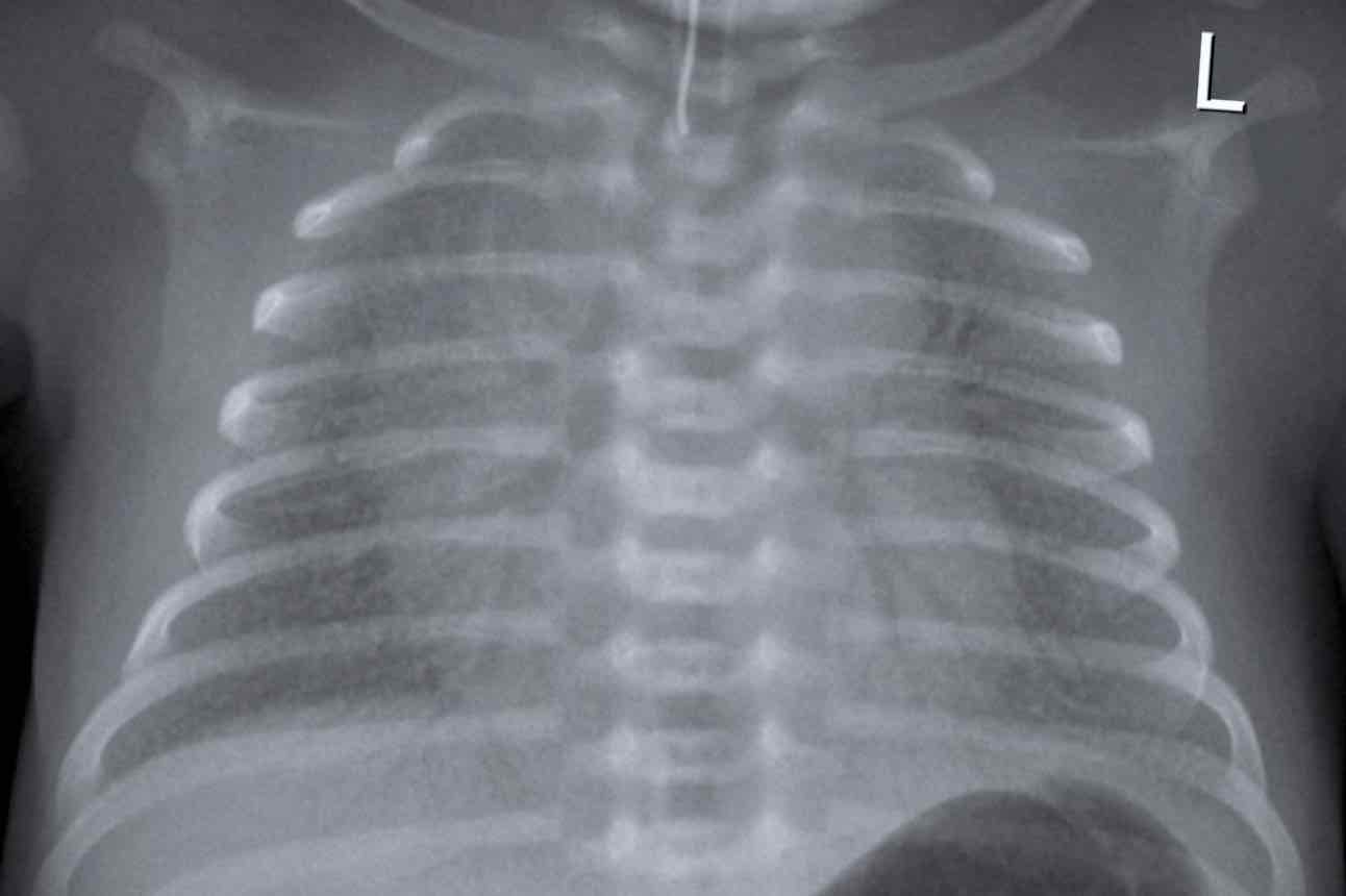
33 weeks + 5 weeks gestational age, day one.
First look at the image.
What are the findings?Tuổi thai 33 tuần + 5 tuần, ngày thứ nhất.
Đầu tiên hãy nhìn vào hình ảnh.
Những phát hiện này là gì?
Findings:Kết quả:
- Intubated patient.Bệnh nhân được đặt nội khí quản.
- Good position of endotracheal tube (ETT). Hyperinflation. Vị trí đặt ống nội khí quản tốt (ETT).
- Symmetric granular opacifications with air bronchograms.Đục hạt đối xứng với hình ảnh khí phế quản.
- Definition of heart and vessels is diminished. Định nghĩa về tim và mạch máu bị giảm.
This is a typical case of RDS.Đây là một trường hợp điển hình của RDS.
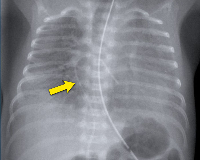
29 weeks + 1, day one. CPAP.
First look at the image.
What are the findings?29 tuần + 1 ngày, CPAP.
Đầu tiên hãy nhìn vào hình ảnh.
Những phát hiện này là gì?
Findings:Kết quả:
- Reticulogranular opacification of lungsĐục dạng hạt võng mạc ở phổi
- Air bronchogram Hình phế quản hơi
- Consolidation in the right lower lobeĐông đặc ở thuỳ dưới bên phải
- Heart, vessels and diaphragm are poorly defined.Tim, mạch và cơ hoành chưa được xác định rõ ràng.
- Malposition of umbilical vein catheter (arrow), probably in a pulmonary vein. Đặt sai vị trí catheter tĩnh mạch rốn (mũi tên), có thể là ở tĩnh mạch phổi.
- Nasogastric tube (NG tube) in good position.Ống thông mũi dạ dày (ống NG) ở vị trí tốt.
This is a severe case of RDS.
The differential diagnosis
includes pulmonary infection due to the asymmetric consolidation.Đây là một trường hợp nặng của RDS.
chẩn đoán phân biệt bao gồm nhiễm trùng phổi do đông đặc không đối xứng.
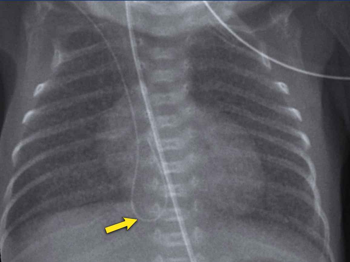
GradingPhân loại
Grading of RDS can be performed but is not used when the patient is on invasive mechanical ventilation support. Phân loại RDS có thể được thực hiện nhưng không được sử dụng khi bệnh nhân đang thở máy xâm lấn.
There are 4 grades of staging RDS.
Có 4 mức độ RDS.
- Only mild ground glass or mild granular opacification of the lungs.Chỉ có kính mờ nhẹ hoặc đục hạt nhẹ của phổi.
- Granular opacification with air bronchograms.Đục hạt với chụp phế quản khí.
- Increasing consolidation of lung tissue leading to obscured borders of the heart and diaphragm.Tăng đông đặc mô phổi dẫn đến che khuất bờ tim và cơ hoành.
- Complete pulmonary white-out.Bạch cầu toàn bộ phổi.
As the lungs cannot expand
properly in RDS, hyperinflation in a preterm infant without mechanical
ventilation makes the diagnosis of RDS highly unlikely. Vì phổi không thể giãn nở đúng cách trong RDS, tình trạng quá căng phồng ở trẻ sinh non không có thông khí cơ học làm cho chẩn đoán RDS rất khó xảy ra.
Image
One day old neonate, 27 weeks of gestational age.
Granular opacification of both lungs.
Vessels and cardiac silhouette are well depicted.
Hình ảnh
Một trẻ sơ sinh một ngày tuổi, 27 tuần tuổi thai.
Đục hạt cả hai phổi.
Hình ảnh mạch máu và hình bóng tim được mô tả rõ.
Conclusion: RDS grade 1.Kết luận: RDS độ 1.
Peripherally inserted central catheter (PICC) line, curled in the right atrium. The PICC should be pulled back to the level of the superior vena cava and right atrium.Đường catheter trung tâm (PICC) được đặt ngoại vi, cuộn tròn ở tâm nhĩ phải. PICC nên được kéo trở lại mức của tĩnh mạch chủ trên và tâm nhĩ bên phải.
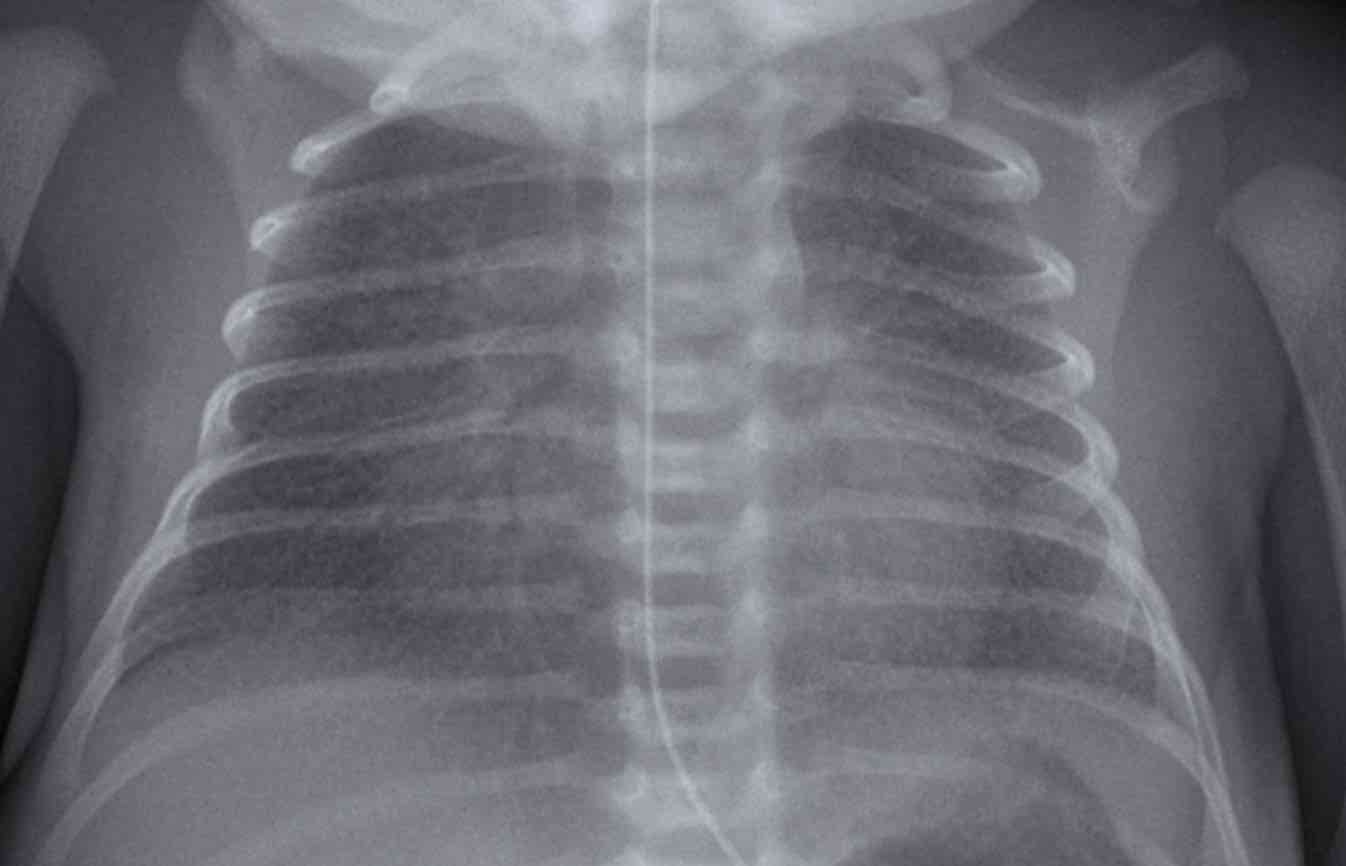
One day old neonate, 29 weeks of gestational age.Trẻ sơ sinh một ngày tuổi, 29 tuần tuổi thai.
ImageHình ảnh
- Hyperinflation due to CPAP.Tăng áp lực tĩnh mạch phổi do CPAP.
- Granular opacification of both lungs with air bronchograms. Đục hạt cả hai phổi với chụp phế quản không khí.
- NG tube in good position.Ống NG ở vị trí tốt.
- Vessels and cardiac silhouette are harder to distinguish.Mạch máu và hình bóng tim khó phân biệt hơn.
Conclusion: RDS grade 2. Kết luận: RDS độ 2.
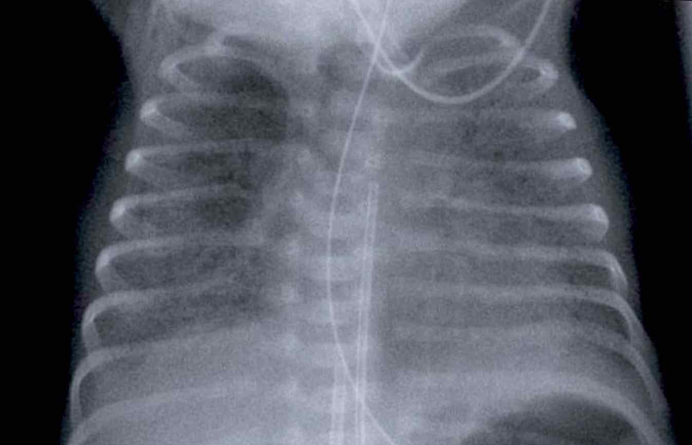
One day old neonate, 26 weeks of gestational age.Một trẻ sơ sinh một ngày tuổi, 26 tuần tuổi thai.
ImageHình ảnh
- Granular opacification of both lungs.Đục hạt cả hai phổi.
- Vessels and cardiac silhouette are hard to distinguish.Rất khó phân biệt được hình ảnh mạch máu và hình bóng tim.
- Umbilical venous line properly positioned. Đường tĩnh mạch rốn được định vị đúng.
- Deep position of umbilical artery line, which should be pulled back to the level of T6.Vị trí sâu của đường động mạch rốn, nên được kéo trở lại mức T 6.
- NG tube in situ.Ống NG tại chỗ.
Conclusion: RDS grade 3.Kết luận: RDS độ 3.
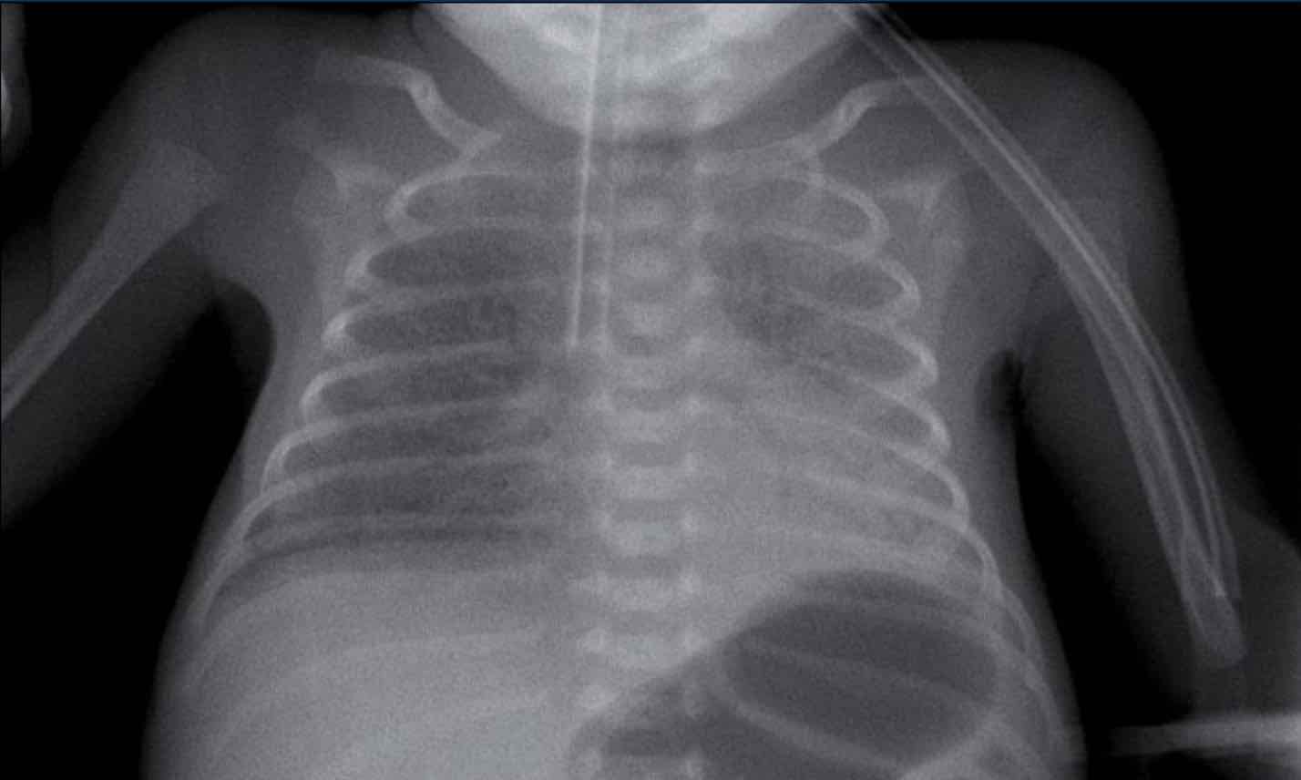 Respiratory distress syndromeHội chứng suy hô hấp
One week old neonate, born at 27 weeks of gestational age. Trẻ sơ sinh một tuần tuổi, sinh ra ở tuổi thai 27 tuần.
Image
Hình ảnh
- Deep position of the tracheal tube, which should be repositioned to the mid of the trachea.Vị trí sâu của ống khí quản, nên được đặt lại vị trí ở giữa khí quản.
- Diffuse granular opacification of both lungs.Đục hạt lan toả ở cả hai phổi.
- Air bronchograms.Chụp phế quản không khí.
- Pumonal vessels are not recognized anymore, but cardiac silhouette is still recognizable. Các mạch máu phổi không còn được nhận ra nữa, nhưng hình bóng tim vẫn có thể nhận ra được.
- Opacification of the left lower lobe caused by atelectasis.Đục thuỳ dưới trái do xẹp phổi.
No grading because this neonate is on mechanical ventilation.Không có sự phân độ vì trẻ sơ sinh này đang thở máy.
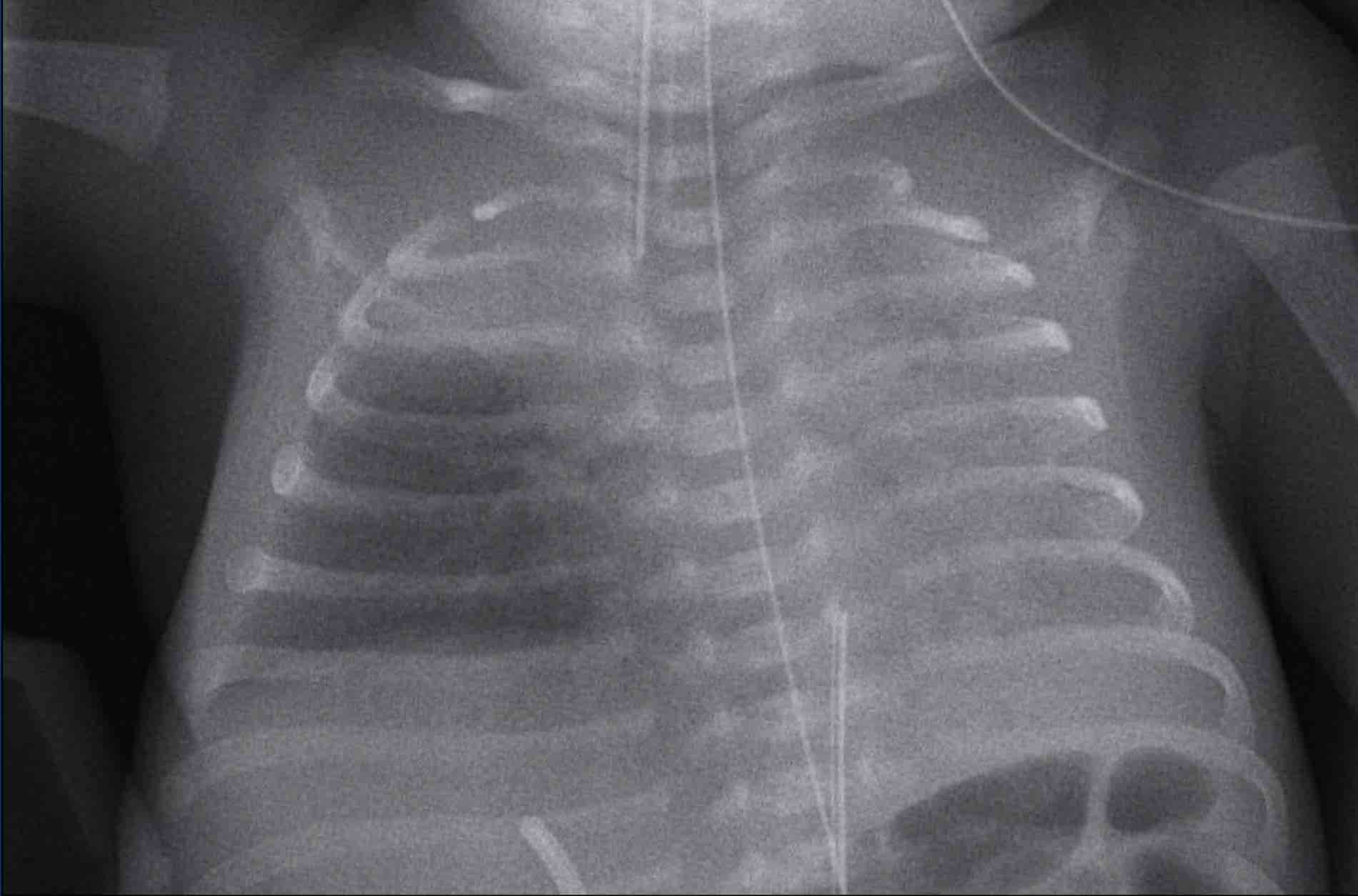 Respiratory distress syndrome - surfactant treatmentHội chứng suy hô hấp - điều trị surfactant
TreatmentĐiều trị
Preventive treatment of RDS consists of antenatal
corticosteroid administration in women at risk for preterm
delivery.
After birth, RDS may require treatment with exogenous
surfactant.
If the infant is supported with ventilation, surfactant is administered intra-tracheally
as a liquid bolus.
The clinical and radiological result of this treatment can often
be seen shortly after the administration of the surfactant.
The surfactant is often not evenly distributed, which can lead to more patchy aeration of the lung parenchyma.
Differentiation from other entities such as neonatal pneumonia
can be difficult.
Điều trị dự phòng RDS bao gồm sử dụng corticosteroid trước sinh ở phụ nữ có nguy cơ sinh non.
Sau khi sinh, RDS có thể cần điều trị bằng surfactant ngoại sinh.
Nếu trẻ sơ sinh được hỗ trợ thông khí, surfactant được tiêm vào khí quản dưới dạng bolus lỏng.
Kết quả lâm sàng và X quang của phương pháp điều trị này thường có thể được nhìn thấy ngay sau khi sử dụng chất hoạt động bề mặt.
Chất hoạt động bề mặt thường không được phân bố đều, có thể dẫn đến sự sục khí nhiều hơn của nhu mô phổi.
Sự khác biệt với các thực thể khác như viêm phổi sơ sinh có thể khó khăn.
Image
One day old boy, gestational age 25 weeks and 5 days.
Hình ảnh
Một bé trai, tuổi thai 25 tuần và 5 ngày.
- Grade 4 RDS with complete opacification of left lung and right upper lobe.RDS độ 4 với đục thuỳ phổi trái và thuỳ trên phải hoàn toàn.
- After one bolus of surfactant the right lower lobe and the middle lobe are aerated.Sau một liều surfactant, sục khí thuỳ dưới bên phải và thuỳ giữa.
- Good position of ETT and umbilical artery line.Vị trí tốt của động mạch mũ và đường động mạch rốn.
- Malposition of the umbilical vein line in the portal vein. This should be removed.Sự sai lệch của đường tĩnh mạch rốn trong tĩnh mạch cửa. Điều này cần được loại bỏ.
Transient Tachypnea of the Newborn (TTN)Thở nhanh thóang qua ở trẻ sơ sinh (TTN)
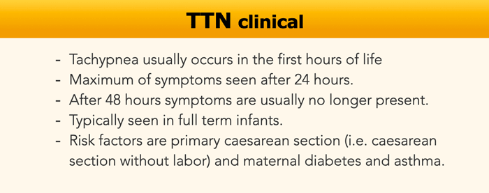
Transient Tachypnea of the Newborn (TTN) is also known as wet lung or retained fetal lung liquid.Thở nhanh thóang qua ở trẻ sơ sinh (TTN) còn được gọi là phổi ướt hoặc chất lỏng phổi thai nhi bị giữ lại.
TTN is a diffuse lung
disorder that results from delayed clearance of fetal lung fluid after birth,
leading to relative surfactant efficacy.
It is seen more frequently – but not
exclusively – in full term neonates after cesarean delivery.
Delayed clearance of fetal
lung fluid causes transient respiratory distress that improves within 48–72
hours after birth.TTN là một rối loạn phổi lan toả do chậm thải dịch phổi thai nhi sau sinh, dẫn đến hiệu quả của surfactant tương đối.
Nó được thấy thường xuyên hơn - nhưng không phải là duy nhất - ở trẻ sơ sinh đủ tháng sau khi sinh mổ.
Sự thanh thải chậm trễ của dịch phổi thai nhi gây ra suy hô hấp thóang qua cải thiện trong vòng 48-72 giờ sau khi sinh.
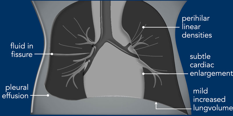

Imaging
Hình ảnh
- Mild increased lung volume.Tăng thể tích phổi nhẹ.
- Interstitial edema resulting in perihilar linear densities.Phù mô kẽ dẫn đến mật độ tuyến tính quanh răng.
- Subtle enlargement of the cardiac silhouette.Hình bóng tim to lên một cách tinh tế.
- Pleural effusions and fluid in the fissures.Tràn dịch màng phổi và dịch trong các vết nứt.
- The radiological findings may be asymmetrical. Các dấu hiệu X quang có thể không đối xứng.
In many cases the clinical
presentation is mild and there is no need for a chest radiograph.
Trong nhiều trường hợp, biểu hiện lâm sàng nhẹ và không cần phim X-quang ngực.
Only in some cases a chest
x-ray is performed to rule out complications.
The imaging findings may be
similar to those of RDS, showing diffuse granular opacities, or of pneumonia
with more coarse opacities. Chỉ trong một số trường hợp, X-quang ngực được thực hiện để loại trừ các biến chứng.
Các dấu hiệu hình ảnh có thể tương tự như RDS, cho thấy các đốm mờ dạng hạt lan toả, hoặc viêm phổi với các đố mầm mờ hơn.
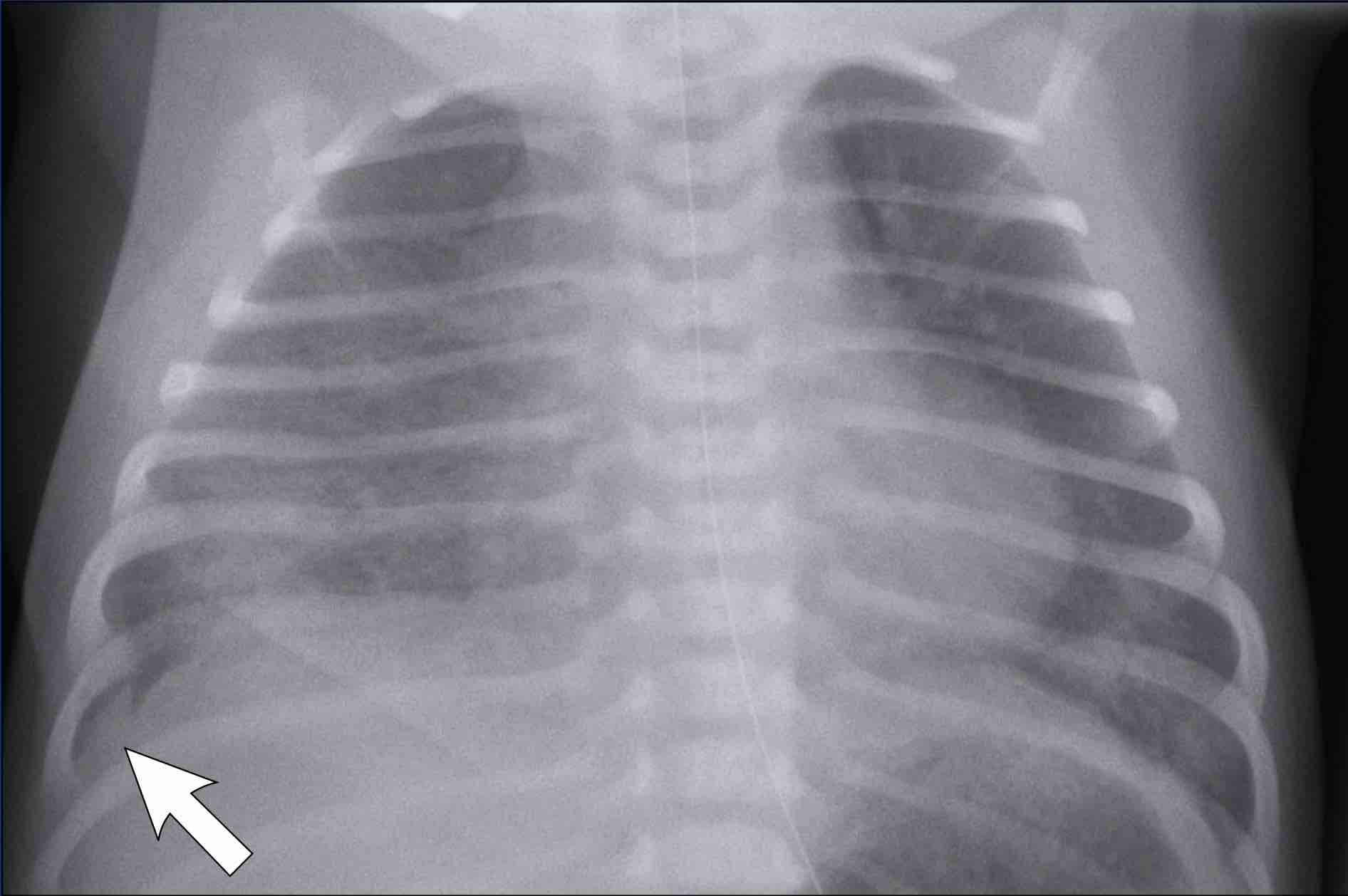
Full term infant, 2 hours after elective caesarean section with some respiratory distress.Trẻ đủ tháng, 2 giờ sau mổ lấy thai có suy hô hấp.
Image
Hình ảnh
- Mild hyperinflationTăng phồng nhẹ
- Subtle interstitial linings on both sidesLớp lót kẽ ở cả hai bên
- Some pleural fluid on the right side (arrow).Một số dịch màng phổi bên phải (mũi tên).
- Skin fold on the right sideNếp gấp da bên phải
After
supportive therapy the respiratory distress disappeared the next day. Sau khi điều trị hỗ trợ, tình trạng suy hô hấp biến mất vào ngày hôm sau.
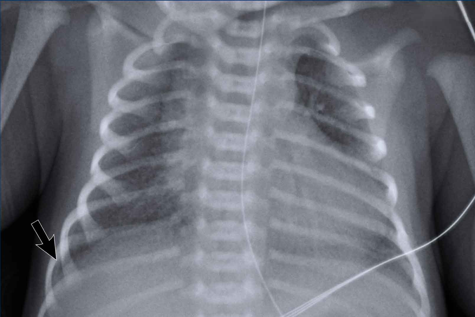
Image of a neonate with gestational age of 41 weeks with mild distress after birth.
No need for ventilatory
support.Hình ảnh một trẻ sơ sinh có tuổi thai 41 tuần với tình trạng suy dinh dưỡng nhẹ sau sinh.
Không cần hỗ trợ thông khí.
ImageHình ảnh
- Hyperinflation of both lungsSiêu lạm phát cả hai phổi
- Subtle interstitial markingsDấu hiệu kẽ tinh tế
- Pleural fluid on the right side (arrow). This is very subtle.Dịch màng phổi bên phải (mũi tên) rất tinh tế.
- NG tube in situỐng NG tại chỗ
Clinical follow up was uneventfulTheo dõi lâm sàng không có biến cố.
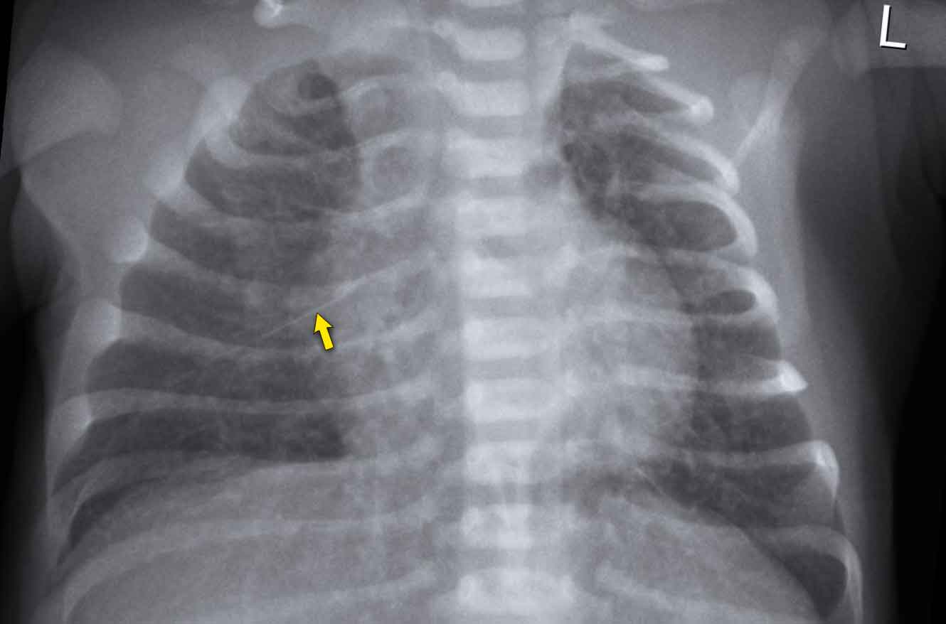
41 Weeks neonate. 24 hours old.
Respiratory distress, no
ventilatory support41 tuần tuổi, 24 giờ tuổi.
Suy hô hấp, không hỗ trợ thông khí
ImageHình ảnh
- Marked hyperinflation of both lungsDấu hiệu căng phồng cả hai phổi
- Increased vascular markings and interstitial markingsTăng dấu hiệu mạch máu và dấu hiệu kẽ
- Some interfissural fluid (arrow).Một số dịch liên xương (mũi tên).
Spontaneous improvement within 48 hours.
Cải thiện tự phát trong vòng 48 giờ.
Meconium AspirationHút phân su
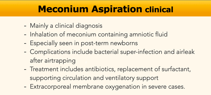
Meconium aspiration results in diffuse pulmonary disease and it is the most common cause of significant morbidity and mortality among full-term and post-term neonates.Hít phân su dẫn đến bệnh phổi lan toả và là nguyên nhân phổ biến nhất gây ra bệnh tật và tử vong đáng kể ở trẻ sơ sinh đủ tháng và sau sinh.
When intra-uterine hypoxia occurs, usually during labor, this
can lead to the fetus prenatally excreting meconium.
Inhalation of the meconium containing amniotic fluid results in a
chemical bronchiolitis with obstruction of the smaller airways
and surfactant dysfunction resulting in air trapping and
atelectasis.
Meconium aspiration can impede the transition from prenatal fetal
circulation to postnatal neonatal circulation.
In 10–15% of births meconium staining of the amniotic fluid is present, but only in 5% of these cases it will result in meconium aspiration.Khi tình trạng thiếu oxy trong tử cung xảy ra, thường là trong quá trình chuyển dạ, điều này có thể dẫn đến thai nhi bài tiết phân su trước khi sinh.
Hít phải phân su có chứa nước ối dẫn đến viêm tiểu phế quản do hóa chất gây tắc nghẽn đường thở nhỏ hơn và rối loạn chức năng chất hoạt động bề mặt dẫn đến bẫy khí và xẹp phổi.
Hít phân su có thể cản trở sự chuyển đổi từ tuần hoàn thai nhi trước khi sinh sang tuần hoàn sơ sinh sau sinh.
Trong 10–15% số ca sinh, có hiện tượng nhuộm phân su trong nước ối, nhưng chỉ trong 5% số trường hợp này, nó sẽ dẫn đến hít phân su.
The term Meconium Aspiration Syndrome is used to describe the
combination of sterile chemical pneumonitis and persistent fetal circulation or
persistent pulmonary hypertension of the newborn (PPHN).
Usually, this condition
presents within a few hours after birth.Thuật ngữ Hội chứng hít phân su được sử dụng để mô tả sự kết hợp của viêm phổi hóa học vô trùng và tuần hoàn thai nhi dai dẳng hoặc tăng áp phổi dai dẳNG của trẻ sơ sinh (PPHN).
Thông thường, tình trạng này xuất hiện trong vòng vài giờ sau khi sinh.
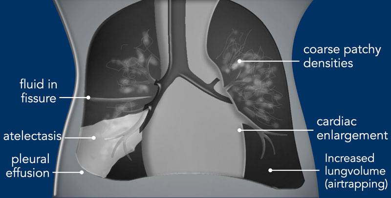

ImagingHình ảnh
- Increased lung volume due to air trapping.Tăng thể tích phổi do bẫy khí.
- Bronchiolitis can lead to atelectasis and air trapping.Viêm tiểu phế quản có thể dẫn đến xẹp phổi và bẫy khí.
- Complete obstruction of the bronchus can lead to atelectasis.Tắc nghẽn hoàn toàn phế quản có thể dẫn đến xẹp phổi.
- Coarse, diffuse bilateral patchy or more linear opacification.Đục hai bên lan toả, thô hoặc đục nhiều hơn.
- Can present asymmetrical.Có thể biểu hiện bất đối xứng.
- Pleural effusions can be seen. Tràn dịch màng phổi có thể được nhìn thấy.
- In a later stage persistent pulmonary hypertension present as cardiomegaly on the x-ray.Trong giai đoạn sau, tăng áp phổi dai dẳng biểu hiện như tim to trên phim X-quang.
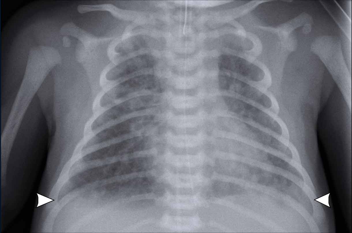
Chest film of a full term newborn with meconium stained amniotic fluid. Phim ngực của một trẻ sơ sinh đủ tháng có nước ối nhiễm phân su.
Image
Hình ảnh
- HyperinflationSiêu lạm phát
- Course diffuse patchy consolidations on both sides. Tất nhiên là có sự đông đặc lan toả ở cả hai bên.
- Some subtle pleural fluid on both sides (arrowheads).Một số dịch màng phổi ở cả hai bên (các đầu mũi tên).
- Good position ETT. Superficial position NG tube, still in esophagus. Vị trí ETT tốt Vị trí bề mặt ống NG, vẫn còn trong thực quản.
- Deep position of umbilical vein line with tip in right atrium. Vị trí sâu của đường tĩnh mạch rốn với đầu ở tâm nhĩ phải.
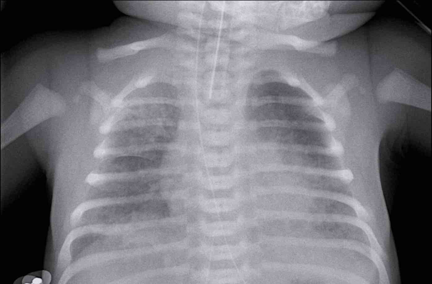
Chest film of a full term newborn with meconium stained amniotic fluid. Phim ngực của một trẻ sơ sinh đủ tháng có nước ối nhiễm phân su.
ImageHình ảnh
- Good position of ETT, NG tube and umbilical vein line.Vị trí tốt của ống ETT, NG và đường tĩnh mạch rốn.
- Course patchy consolidations with relative sparing of the left apical lobe and right middle lobe. Tất nhiên là có sự đông đặc loang lổ với sự tương đối của thuỳ đỉnh trái và thuỳ giữa phải.
- Mild hyperinflation.Tăng áp lực nhẹ.
Conclusion: chest radiograph in
keeping with meconium aspiration. Without staining of the amniotic fluid the
differential diagnosis would include neonatal pneumonia or TTN.Kết luận: phim X-quang ngực phù hợp với chọc hút phân su, không làm ố nước ối, chẩn đoán phân biệt có thể gây viêm phổi sơ sinh hoặc TTN.
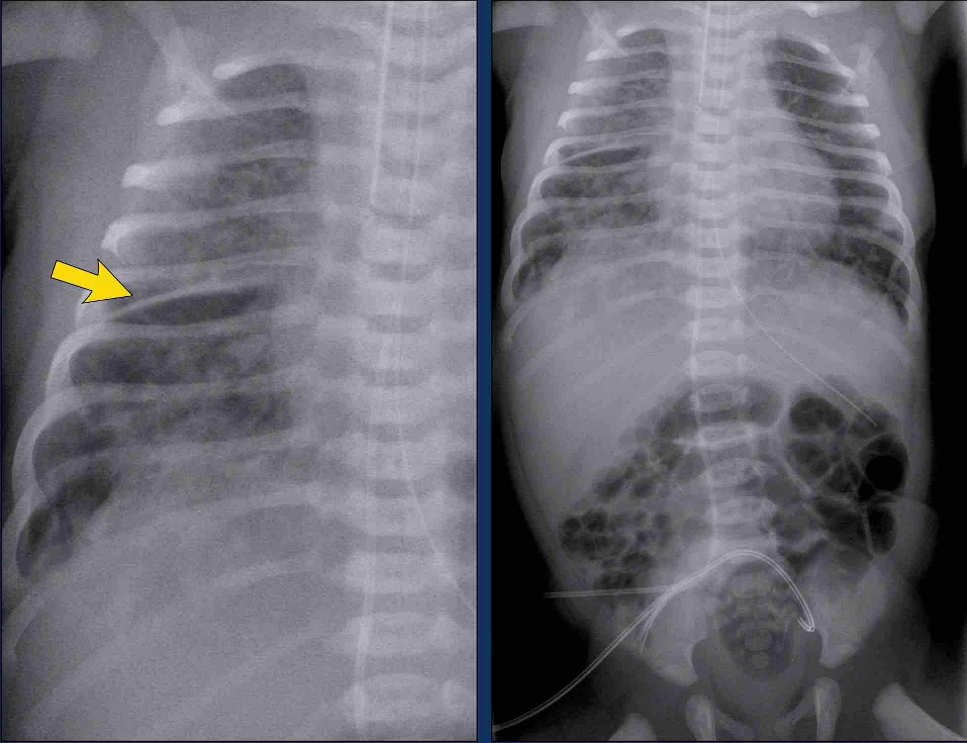
X-ray of a full term newborn with meconium stained amniotic fluid. X-quang một trẻ sơ sinh đủ tháng có nước ối nhiễm phân su.
ImageHình ảnh
- Hyperinflation. Siêu lạm phát.
- Asymmetrical patchy consolidation. Không đối xứng đông đặc.
- Pleural fluid in the fissure on the right (arrow).Dịch màng phổi ở khe nứt bên phải (mũi tên).
- Deep position of the tracheal tube. Vị trí sâu của ống khí quản.
- Malposition of the umbilical vein in right atrium and artery line in distal iliac artery.Tắc tĩnh mạch rốn ở nhĩ phải và đường động mạch ở đầu xa động mạch chậu.
Pulmonary hemorrhageXuất huyết phổi
Pulmonary hemorrhage is uncommon in neonates.
Premature neonates are most at risk.
Other associated
factors are a persistent ductus arteriosus, surfactant treatment and mechanical
ventilation.
The exact etiology remains unclear.
The
radiographic signs are nonspecific and difficult to distinguish from other
disorders such as RDS.
This means that the diagnosis can only be made, when there is leakage of red blood or pink effusion from the
endotracheal tube.Xuất huyết phổi không phổ biến ở trẻ sơ sinh.
Trẻ sơ sinh non tháng có nguy cơ cao nhất.
Các yếu tố liên quan khác là ống động mạch tồn tại dai dẳng, điều trị surfactant và thở máy.
Nguyên nhân chính xác vẫn chưa rõ ràng.
Các dấu hiệu trên X quang không đặc hiệu và khó phân biệt với các rối loạn khác như RDS.
Điều này có nghĩa là chẩn đoán chỉ có thể được thực hiện, khi có rò rỉ máu đỏ hoặc tràn dịch hồng từ ống nội khí quản.
Supportive measurements consist of ventilator support and sometimes xylometazoline or adrenalin, which is administered via the tracheal tube.Các biện pháp hỗ trợ bao gồm hỗ trợ thông khí và đôi khi xylometazoline hoặc adrenalin, được dùng qua ống khí quản.
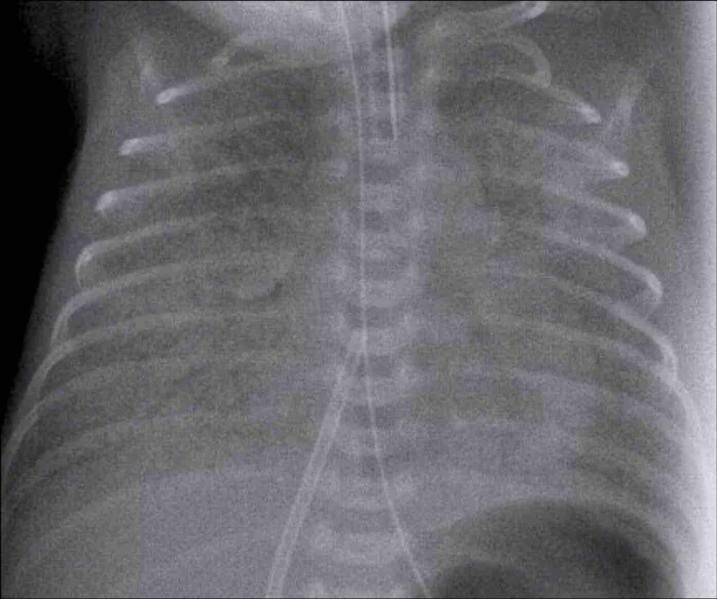
Mild hemorrhage can be hard to distinguish from RDS.
Massive bleedings show complete opacification of one or both lungs.Xuất huyết nhẹ có thể khó phân biệt với RDS.
Chảy máu nhiều cho thấy đục hoàn toàn một hoặc cả hai phổi.
Image Hình ảnh
- Premature infant, gestational age 33 weeks, with a history of RDS and blood via the endotracheal tube.Trẻ sinh non, tuổi thai 33 tuần, có tiền sử RDS và có máu qua ống nội khí quản.
- Extensive consolidation of both lungs. Đông đặc mở rộng của cả hai phổi.
- Malposition of umbilical vein line. The line is positioned in the right atrium.Vị trí sai của đường tĩnh mạch rốn Đường này nằm ở tâm nhĩ phải.
- Good position of ETT, and NG tubeVị trí tốt của ống ETT, NG
The most likely diagnosis of this chest x-ray is RDS, but
given the clinical information of blood via the tracheal tube, the diagnosis is
pulmonary hemorrhage, possibly in combination with RDS.Chẩn đoán xác định nhất của X-quang ngực này là RDS, nhưng với thông tin lâm sàng của máu qua ống khí quản, chẩn đoán là xuất huyết phổi, có thể kết hợp với RDS.
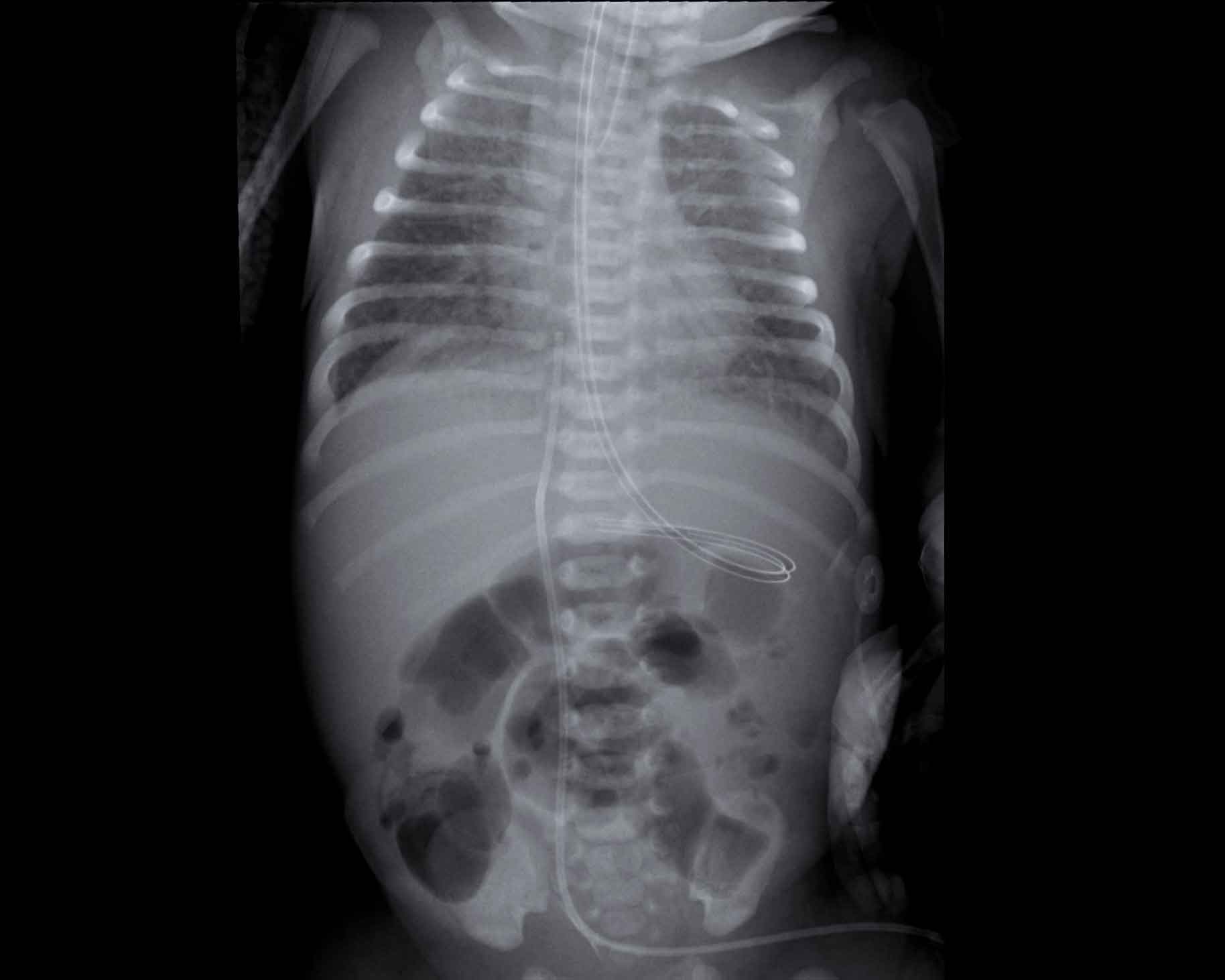
Image of a neonate with gestational age of 41 weeks.
After 24 hours intubated for respiratory distress.
Blood via the endotracheal tube.Hình ảnh một trẻ sơ sinh có tuổi thai 41 tuần.
Sau 24 giờ đặt nội khí quản cho suy hô hấp.
Máu qua ống nội khí quản.
Image
Hình ảnh
- Good position of ETT, umbilical vein line, and NG tubeVị trí tốt của ETT, đường tĩnh mạch rốn, và ống NG
- Heterogeneous aeration of the lungs with hyperinflation of lower lobes. Sục khí không đồng nhất của phổi với sự căng phồng của thuỳ dưới.
- Diffuse interstitial
markings, more pronounced in right upper lobe and lower lobe.
Các dấu hiệu kẽ lan toả, rõ rệt hơn ở thuỳ trên bên phải và thuỳ dưới.
The differential
diagnosis includes pneumonia and RDS in maternal diabetes.
In case of meconium
stained amniotic fluid, mecoinum aspiration could be considered as well, but this
is often more coarse. Chẩn đoán phân biệt bao gồm viêm phổi và RDS ở mẹ đái tháo đường.
Trong trường hợp dịch ối nhiễm phân su, có thể xem xét việc hút mecoinum, nhưng điều này thường thô hơn.
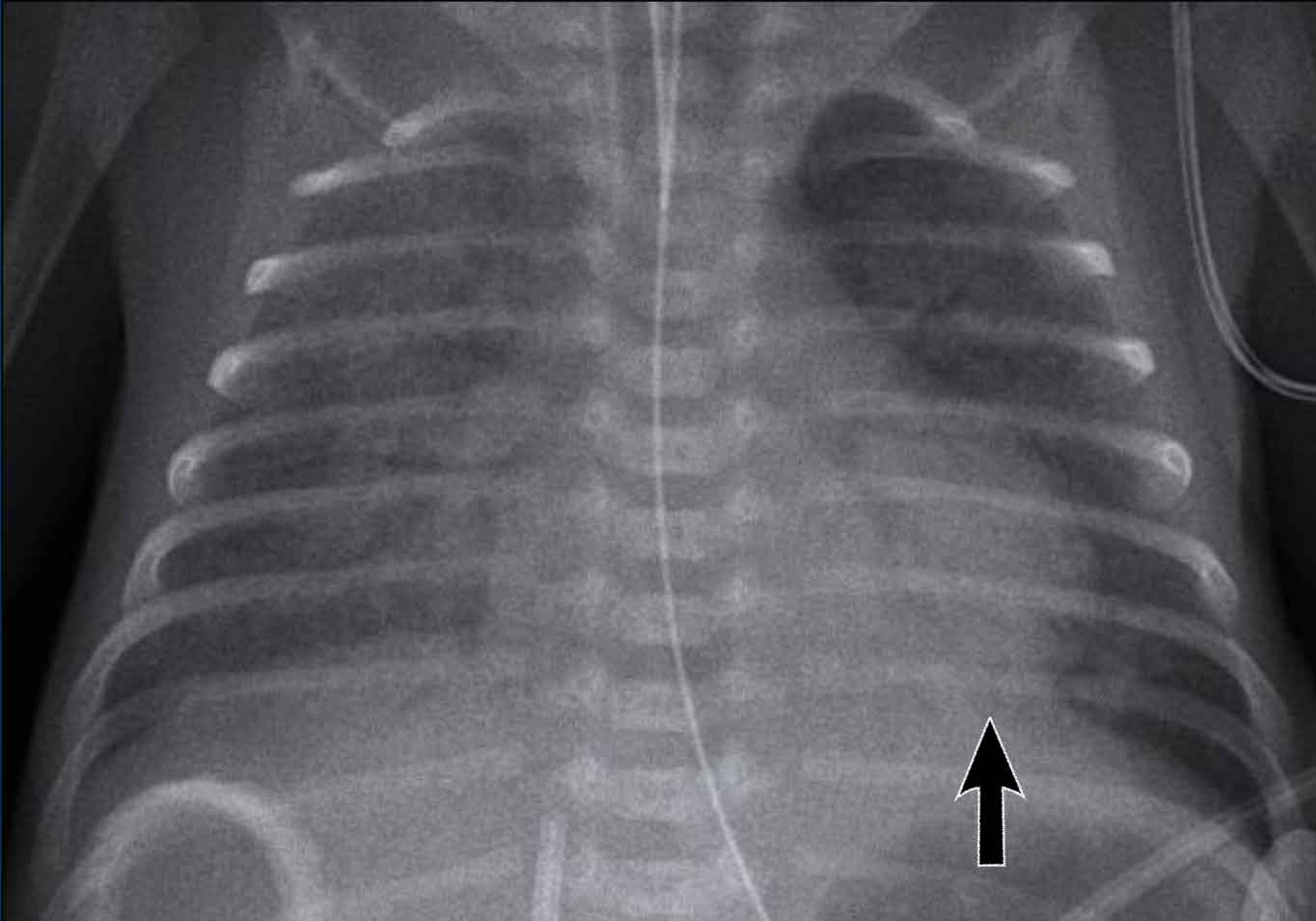
Premature, 28 weeks of gestational age treated for RDS.
Blood via the tracheal tube.Điều trị RDS sớm, 28 tuần tuổi thai.
Máu qua ống khí quản.
Image Hình ảnh
- Patchy opacifications in the right lung, due to hemorrhage.Đục nhiều chỗ ở phổi phải, do xuất huyết.
- Atelectasis of the left lower lobe (arrow) with mild hyperinflation of the left upper lobe. Xẹp phổi thuỳ dưới trái (mũi tên) với sự tăng phình nhẹ của thuỳ trên trái.
- Good position of ETT. Vị trí ETT tốt.
- Low position of umbilical vein line, NG tube in situ.Vị trí thấp của đường tĩnh mạch rốn, ống NG tại chỗ.
Persistent ductus arteriosusỐng động mạch dai dẳng
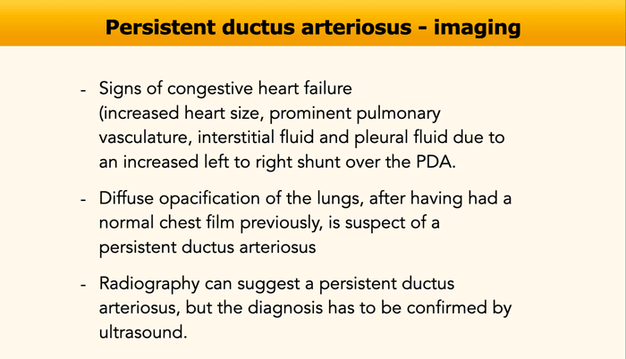
The ductus arteriosus is
the connection between the pulmonary artery and the aorta.
Normally the ductus is open before birth and closes in term infants within
the first day after birth as arterioles feeding the wall of the ductus contract
in reaction to oxygen.Ống động mạch là nơi nối giữa động mạch phổi và động mạch chủ.
Thông thường ống động mạch mở trước khi sinh và đóng lại ở trẻ đủ tháng trong ngày đầu tiên sau sinh khi các tiểu động mạch nuôi thành ống động kinh co lại để phản ứng với oxy.
Preterm infants have fewer of these arterioles and sometimes an increase in oxygen does not lead to closure of the duct, which might result
in a high pulmonary blood flow.
The diagnosis usually is suspected 5 - 7 days after birth, when there is pulmonary overflow or systemic steal.Trẻ sinh non có ít tiểu động mạch hơn và đôi khi tăng oxy không dẫn đến đóng ống dẫn, có thể dẫn đến lưu lượng máu phổi cao.
Chẩn đoán thường được nghi ngờ sau sinh 5-7 ngày, khi có tràn dịch phổi hoặc ăn cắp hệ thống.
Prostaglandin inhibitors
Prostaglandin inhibitors can be used to close the open duct.
In some cases, surgical closure can be achieved either via a lateral
thoracotomy with a clip, or later in life via an intra-arterial approach using
a coil.Thuốc ức chế prostaglandin
Thuốc ức chế prostaglandin có thể được sử dụng để đóng ống dẫn.
Trong một số trường hợp, phẫu thuật đóng có thể được thực hiện thông qua mở ngực bên bằng kẹp clip, hoặc sau này thông qua đường tiếp cận trong động mạch bằng cách sử dụng một cuộn dây.
Prostaglandins
In neonates with a ductal-dependent cardiac disorder Prostaglandins can be used to keep the ductus arteriosus
patent, which can be life-saving. Prostaglandin
Ở trẻ sơ sinh có rối loạn tim phụ thuộc ống động mạch, Prostaglandin có thể được sử dụng để giữ ống động tĩnh mạch, có thể cứu sống trẻ.
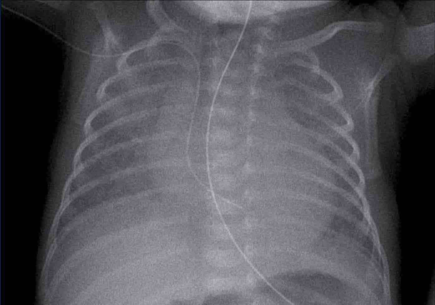
Premature with previously normal chest radiographs.X quang ngực trước đó bình thường.
Image
Hình ảnh
- Moderate enlargement of the heart in combination with ground glass changes of the lungs, in keeping with pulmonary edema. Phì đại trung bình tim kết hợp với thay đổi kính mờ của phổi, phù phổi.
- Deep position of the CVC in the right atrium, normal position of gastric tube.Vị trí sâu của CVC ở tâm nhĩ phải, vị trí bình thường của ống dạ dày.
Ultrasound confirmed a persistent ductus arteriosus. Siêu âm xác nhận ống động mạch tồn tại dai dẳng.
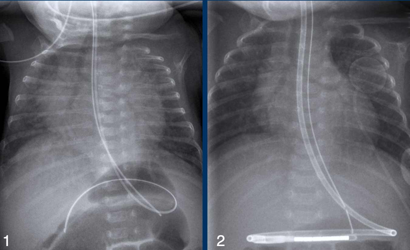
Premature of 25 weeks and 5 days, now 2 month old. Treatement for RDS
Điều trị RDS sớm 25 tuần và 5 ngày, hiện nay là 2 tháng tuổi
- The first chest radiograph shows hyperinflation, and a
markedly enlarged heart with increased vascular markings and interstitial
markings.
NG tube and duodenal tube in situ.Phim X-quang ngực đầu tiên cho thấy sự căng phồng, và tim to rõ rệt với các dấu hiệu mạch máu và các dấu hiệu kẽ tăng lên.
Ống NG và ống tá tràng tại chỗ. - After 5 days of treatment there is less hyperinflation and normalization of the heart size and vascular markings.
There still remain some interstitial markings, in keeping with BPD.Sau 5 ngày điều trị, ít có tình trạng tăng căng phồng và bình thường hóa kích thước tim và các dấu hiệu mạch máu.
Vẫn còn một số dấu hiệu kẽ, phù hợp với BPD.
Bronchopulmonary DysplasiaLoạn sản phế quản phổi
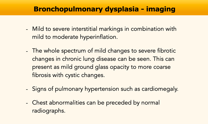
Bronchopulmonary dysplasia (BPD) also known as chronic lung
disease of the premature, is a disorder of lung injury and repair originally
ascribed to positive-pressure mechanical ventilation and oxygen toxicity.
BPD is nowadays a purely clinical diagnosis characterized by the requirement of oxygen for at least 28 days in an infant born at less than 30 weeks of gestation. Loạn sản phế quản phổi (BPD) còn được gọi là bệnh phổi mạn tính của trẻ sinh non, là một rối loạn tổn thương và phục hồi phổi ban đầu được quy cho thông khí cơ học áp suất dương và độc tính oxy.
BPD hiện nay là một chẩn đoán lâm sàng đơn thuần đặc trưng bởi nhu cầu oxy trong ít nhất 28 ngày ở trẻ sơ sinh sinh ra ở dưới 30 tuần tuổi thai.
Before the advent of surfactant replacement therapy, chest
radiographs of infants with classic BPD demonstrated coarse reticular lung
opacities, cystic lucencies, and markedly disordered lung aeration that
reflected alternating regions of alveolar septal fibrosis and hyperinflated
normal lung.
Nowadays after the introduction of prenatal steroids and
postnatal surfactant and more sophisticated ventilatory support, BPD is
infrequently seen in infants with a birth weight of more than 1200 grams and
over 32 weeks of gestational age.
However, despite these advances in neonatal care, the
prevalence of BPD has changed little over the last decades due to the treatment
and improved survival rate of even more very preterm infants.
Trước khi có liệu pháp thay thế surfactant, X quang ngực của trẻ sơ sinh có BPD cổ điển cho thấy các đám mờ dạng lưới ở phổi, các nang sáng và sự rối loạn khí phổi rõ rệt phản ánh các vùng xen kẽ của xơ hóa vách phế nang và phổi bình thường tăng áp.
Ngày nay sau khi giới thiệu steroid trước sinh và surfactant sau sinh và hỗ trợ thông khí tinh vi hơn, BPD không thường xuyên thấy ở trẻ sơ sinh có trọng lượng lúc sinh trên 1200 gram và trên 32 tuần tuổi thai.
Tuy nhiên, mặc dù có những tiến bộ trong chăm sóc trẻ sơ sinh, tỷ lệ hiện mắc BPD đã thay đổi rất ít trong những thập kỷ qua do việc điều trị và cải thiện tỷ lệ sống sót của trẻ sơ sinh rất non tháng.
As a result of these changes, the international criteria for the diagnosis BPD were changed from 28 days postnatal age to 36 weeks postmenstrual age. Therefore, the term BPD should be avoided before this postmenstrual age.Do những thay đổi này, các tiêu chuẩn quốc tế về chẩn đoán BPD đã thay đổi từ 28 ngày sau sinh đến 36 tuần sau kinh nguyệt, do đó, nên tránh thuật ngữ BPD trước tuổi sau kinh nguyệt này.
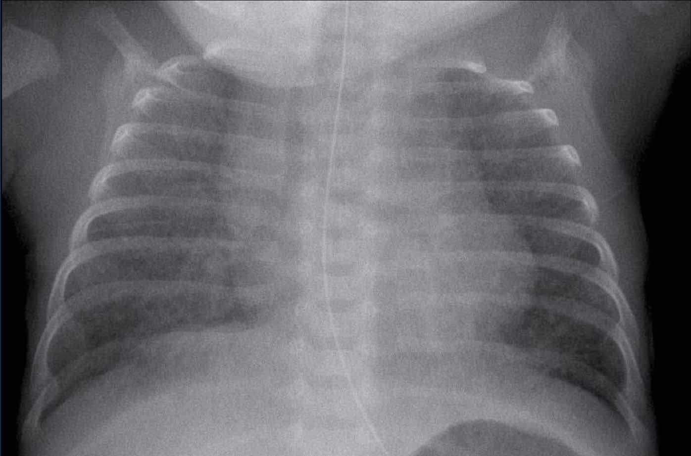
Premature, born at 27 weeks of gestational age. Now 6 weeks of age.
History of intubation and mechanical airway support.Sinh non, 27 tuần tuổi thai, 6 tuần tuổi.
Tiền sử đặt nội khí quản và hỗ trợ đường thở cơ học.
Image
Bilateral perihilar opacification and increased interstitial markings.Hình ảnh
Đục quanh răng hai bên và tăng dấu hiệu kẽ.
Given the history, BPD is the most likely diagnosis. Với tiền sử, BPD là chẩn đoán có khả năng nhất.
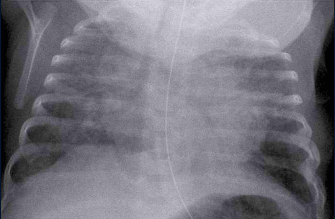
Premature, born at 27 weeks of gestational age. Now 8 weeks
of age.
History of extensive mechanical ventilation with prolonged
need for oxygen support.Sinh non, 27 tuần tuổi thai, 8 tuần tuổi.
Tiền sử thở máy kéo dài với nhu cầu hỗ trợ oxy kéo dài.
Image
Bilateral perihilar opacifications with a coarse interstitial pattern as a sign of chronic small airway disease. NG tube in situ.Hình ảnh
Đục quanh rốn phổi hai bên với mô kẽ thô là dấu hiệu của bệnh lý đường thở nhỏ mạn tính.
Given the history in combination with the radiological findings BPD is the most likely diagnosis. Với tiền sử kết hợp với các dấu hiệu X quang, BPD là chẩn đoán có khả năng nhất.
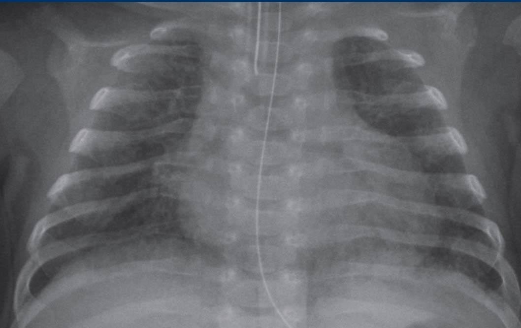
Premature at 28 weeks, now 7 weeks of age, need for ventilatory support.Trẻ đẻ non 28 tuần, nay 7 tuần tuổi, cần hỗ trợ thông khí.
Image
Bilateral diffuse
interstitial markings, with some atelectasis on the left. Hình ảnh
Dấu hiệu kẽ lan toả hai bên, với một số xẹp phổi ở bên trái.
Given the history in combination with the radiological findings, BPD is the most likely diagnosis. Với tiền sử kết hợp với các dấu hiệu X quang, BPD là chẩn đoán có khả năng nhất.
AtelectasisXẹp phổi
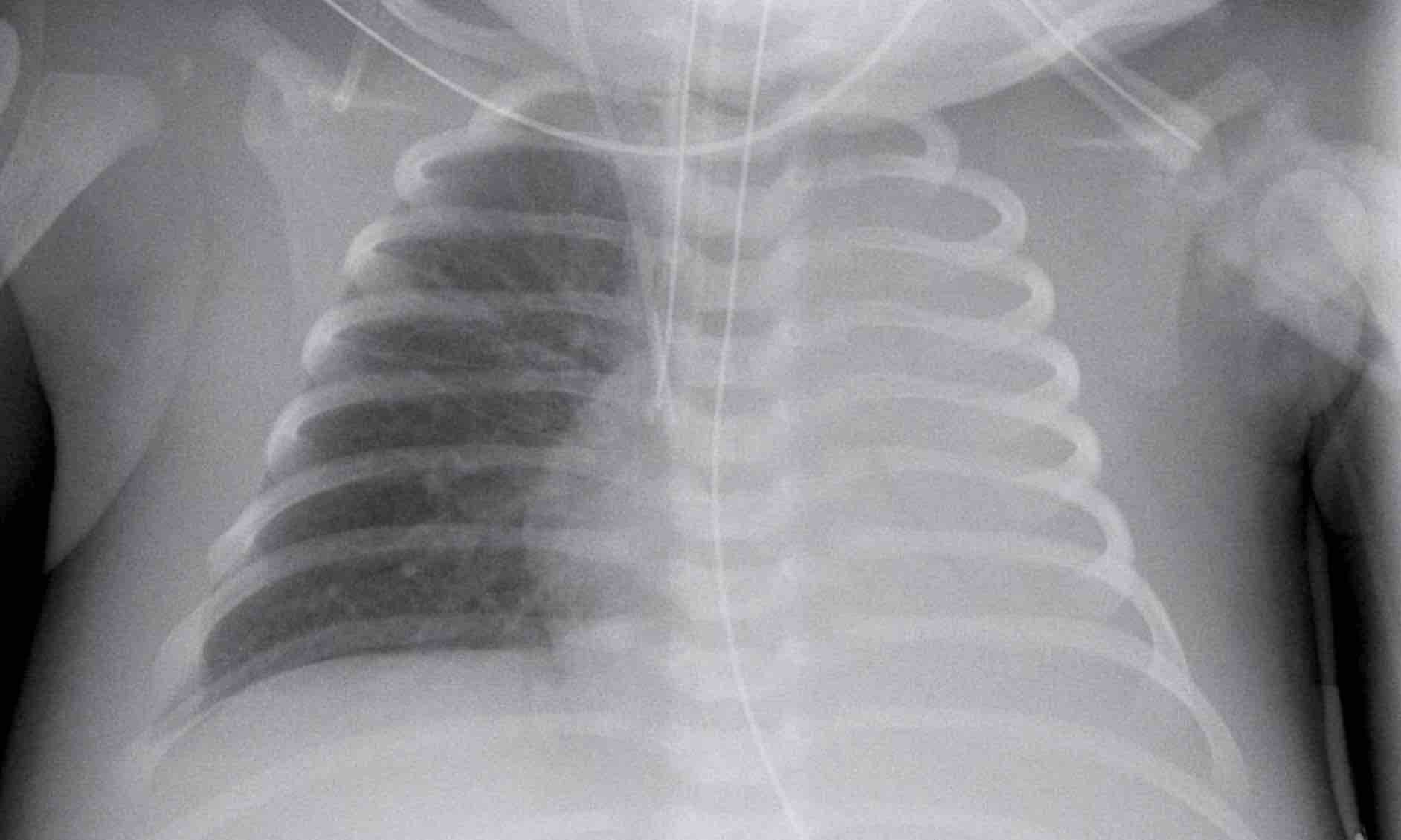 Complete atelectasis of the left lungXẹp phổi toàn phần của phổi trái
Atelectasis often occurs due to malposition of tracheal
tubes, or to low positive pressure when using invasive mechanical ventilation.
Failure to clear mucus or secretion can cause atelectasis by
plugging of mucus.
Deficiency of surfactant can cause micro-atelectasis, leading
to diffuse atelectasis.
Xẹp phổi thường xảy ra do sự sai lệch vị trí của ống khí quản, hoặc do áp lực dương thấp khi sử dụng thông khí cơ học xâm lấn.
Không làm sạch chất nhầy hoặc bài tiết có thể gây ra xẹp phổi bằng cách cắm chất nhầY.
Thiếu chất hoạt động bề mặt có thể gây ra vi xẹp phổi, dẫn đến xẹp phổi lan toả.
Treatment depends on the cause of the atelectasis, such as change of tracheal tube position, change in ventilatory support (pressure), alternating position from side to side, or in case of surfactant deficiency, intratracheal administration of surfactant.Điều trị phụ thuộc vào nguyên nhân của xẹp phổi, chẳng hạn như thay đổi vị trí ống khí quản, thay đổi hỗ trợ thông khí (áp lực), vị trí luân phiên từ bên này sang bên kia, hoặc trong trường hợp thiếu surfactant, sử dụng surfactant trong ống dẫn lưu.
Image
Complete atelectasis of the
left lung due to selective intubation on the right. PICC line in situ with tip
in superior vena cava. NG tube in situ. Hình ảnh
Xẹp phổi trái hoàn toàn do đặt nội khí quản chọn lọc bên phải đường PICC tại chỗ có đầu ở tĩnh mạch chủ trên ống NG tại chỗ.
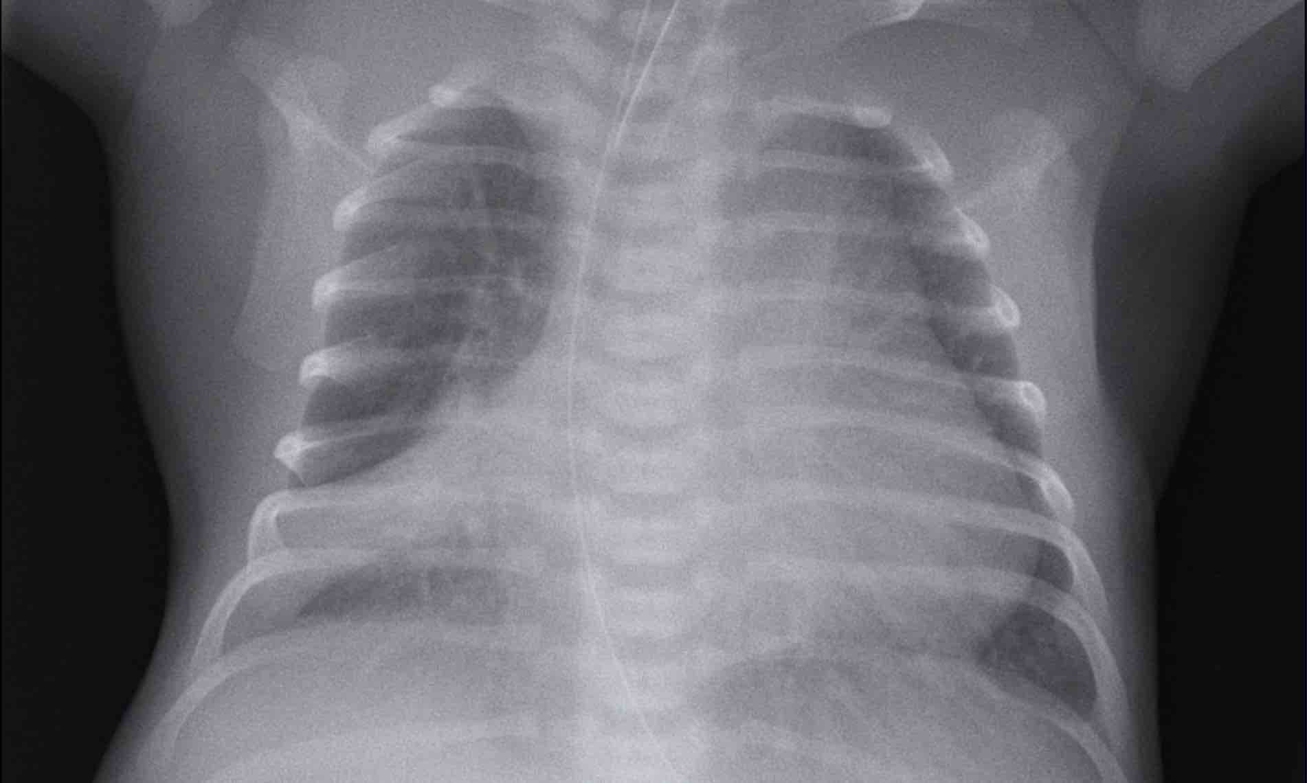 Atelectasis of the right middle lobeXẹp phổi của thuỳ giữa bên phải
This preterm neonate was treated for respiratory distress, probably due to pneumonia.Trẻ sơ sinh non tháng này được điều trị suy hô hấp, có thể do viêm phổi.
Image
Bilateral perihilar streaking can be seen, making the diagnosis of pneumonia most likely.
There is atelectasis
of the right middle lobe, probably due to mucus impaction of the bronchus. Hình ảnh
Có thể thấy vệt quanh rốn phổi hai bên, làm cho chẩn đoán viêm phổi rất có thể xảy ra.
Có xẹp phổi của thuỳ giữa bên phải, có thể là do sự ngấm chất nhầy của phế quản.
Notice the position of the nasogastric catheter indicating mediastinal shift to
the right.
Chú ý vị trí của ống thông mũi dạ dày cho thấy trung thất dịch chuyển sang phải.
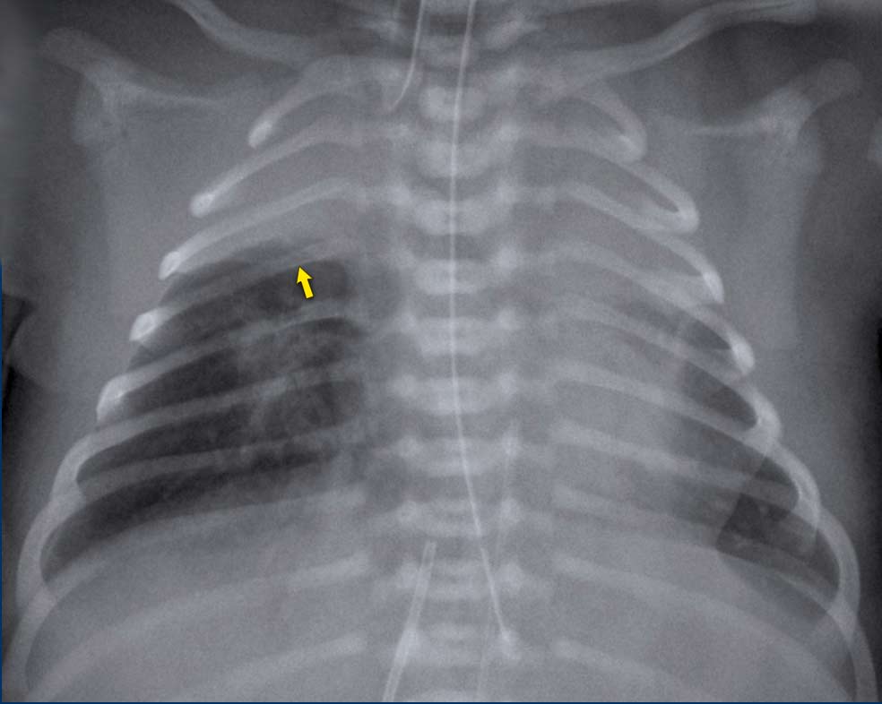
This is a neonate of 41 weeks gestational age, who was treated for asphyxia, including hypothermia treatment. Sudden respiratory distress.Đây là một trẻ sơ sinh 41 tuần tuổi thai, được điều trị ngạt, hạ thân nhiệt, suy hô hấp cấp.
Image
Bilateral opacification of the upper lobes most likely due to atelectasis.
There is some subtle streaky opacity most
pronounced retrocardial in the left lower lobe. This could be some atelectasis aswell.Hình ảnh
Đục thuỳ trên hai bên nhiều khả năng do xẹp phổi.
Có một số vệt mờ sau tim rõ rệt nhất ở thuỳ dưới bên trái, có thể là một số xẹp phổi.
Neonatal PneumoniaViêm phổi sơ sinh
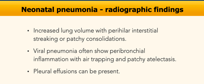
Pneumonia can be difficult
to distinguish from other entities such as RDS or bronchopulmonary dysplasia.
In the majority of cases the clinical course, together with intratracheal sputum cultures and biochemical parameters are leading in the diagnosis of pneumonia.
Without signs of infection, a consolidation on a chest radiograph is unlikely to be caused by pneumonia.Viêm phổi có thể khó phân biệt với các bệnh khác như RDS hoặc loạn sản phế quản phổi.
Trong đa số các trường hợp, lâm sàng, kết hợp với nuôi cấy đàm trong màng phổi và các thông số sinh hóa là những yếu tố hàng đầu trong chẩn đoán viêm phổi.
Không có dấu hiệu nhiễm trùng, đông đặc trên phim X-quang ngực không có khả năng là do viêm phổi.
Risk factors for neonatal pneumonia are:Các yếu tố nguy cơ viêm phổi sơ sinh là:
- Preterm infants with ventilatory support and intubation.Trẻ sinh non được hỗ trợ thông khí và đặt nội khí quản.
- Full-term newborns after prolonged rupture of membranes or after maternal infection.Trẻ sơ sinh đủ tháng sau khi vỡ ối kéo dài hoặc sau khi nhiễm trùng từ mẹ.
- Secondary after meconium aspiration. Thứ phát sau hút phân su.
- Transplacental (TORCH) infectionsNhiễm trùng qua nhau thai (TORCH)
- Perinatal infection, such as E. coli or group B streptococcus, viral. Miscellaneous agents as Chlamydia trachomatis can be found. Nhiễm trùng chu sinh, chẳng hạn như E. coli hoặc liên cầu nhóm B, vi rút Các tác nhân khác như Chlamydia trachomatis có thể được tìm thấy.
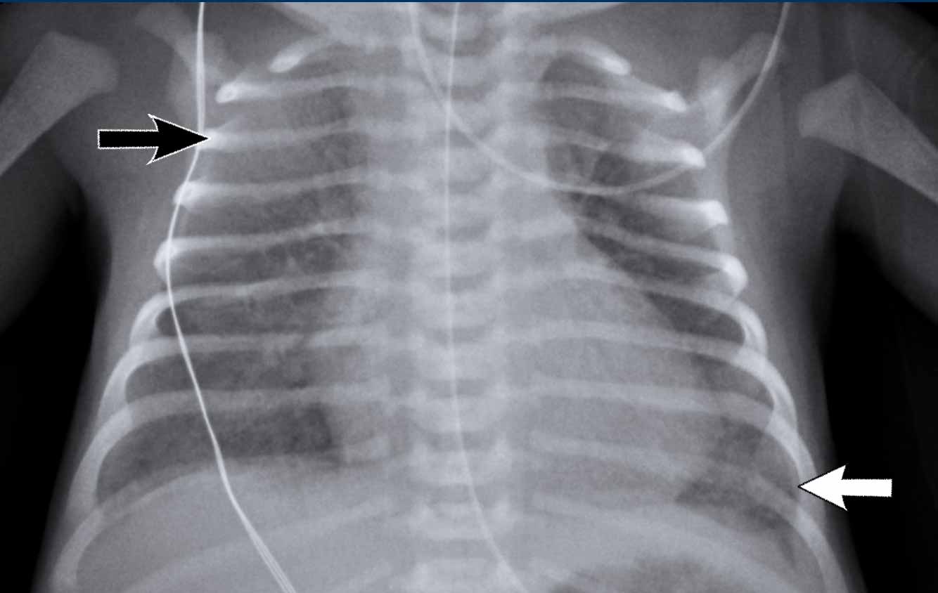
Neonate, GA 37+6 weeks, respiratory distress, treated with CPAP after delivery. History of maternal infection.Trẻ sơ sinh, tuần sinh 37 + 6, suy hô hấp, được điều trị thở máy CPAP sau sinh, tiền sử nhiễm trùng mẹ.
First study the image.
What are the findings.Đầu tiên nghiên cứu hình ảnh.
Những phát hiện này là gì?
Image
Bilateral increased lung volume with asymmetric increased opacification of the lungs with subtle consolidation of right upper lobe (black arrow) and left lower lobe (white arrow).
Hyperinflation of left upper lobe.Hình ảnh
Thể tích phổi tăng lên hai bên với sự tăng đục không đối xứng của phổi với sự đông đặc tinh tế của thuỳ trên bên phải (mũi tên đen) và thuỳ dưới bên trái (mũI tên trắng).
Tăng phồng thuỳ trên trái.
This child developed signs of infection, both clinically and in the laboratory findings.
The radiographic findings were attributed to neonatal pneumonia.Trẻ này có dấu hiệu nhiễm trùng, cả trên lâm sàng và trong các xét nghiệm.
X quang cho thấy viêm phổi sơ sinh.
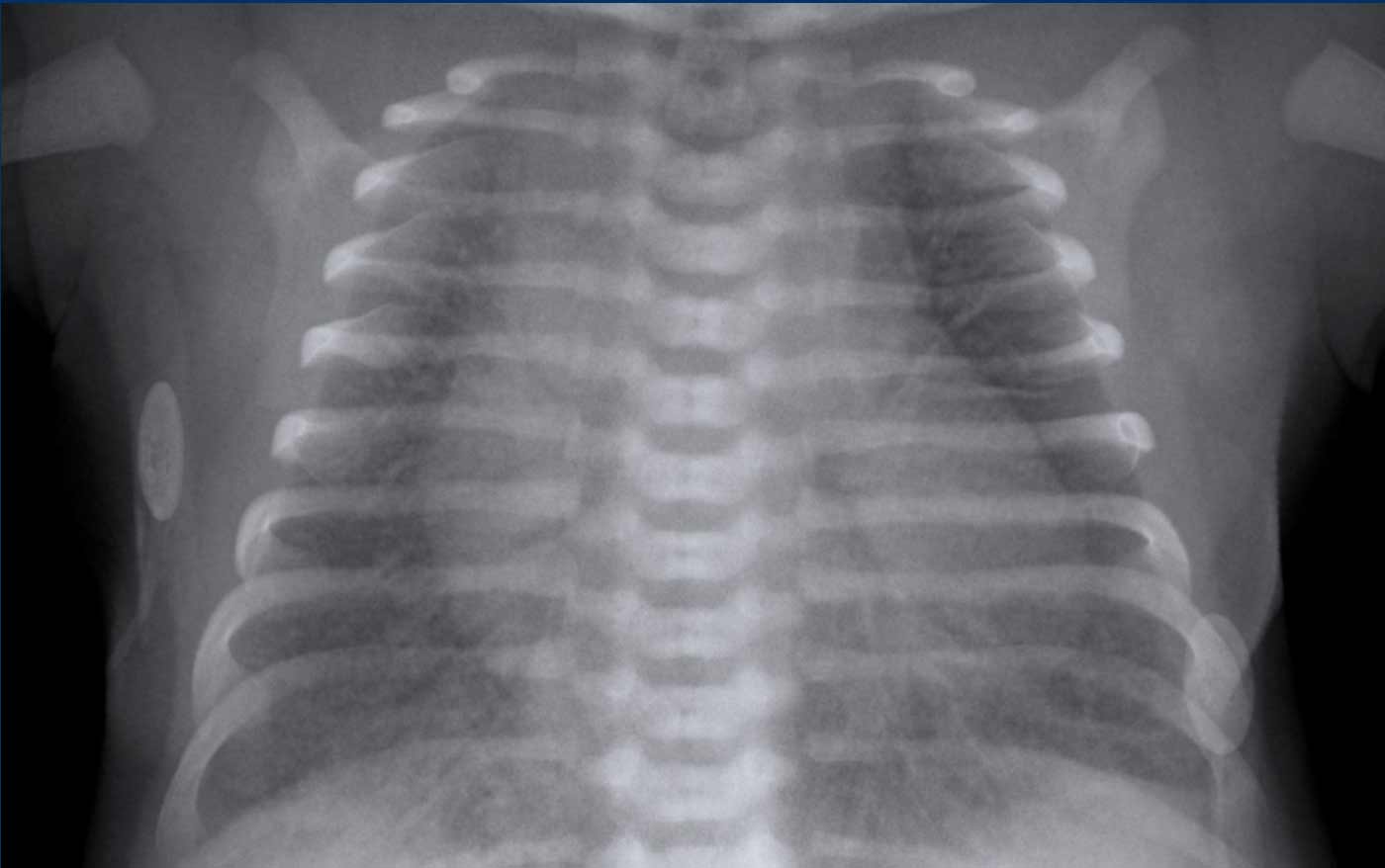
A full term neonate, with respiratory distress after 24 hours.Trẻ sơ sinh đủ tháng, suy hô hấp sau 24 giờ.
Image
Hyperinflation of both lungs and cardiac enlargement with increased
interstitial markings and vascular markings. No pleural fluid.Hình ảnh
Tăng căng phồng phổi và tim phì đại với dấu hiệu kẽ và dấu hiệu mạch máu tăng lên Không có dịch màng phổi.
The differential diagnosis includes TTN and neonatal pneumonia. After 48 hours there was no improvement of the respiratory distress, and the neonate developed signs of infection. Chẩn đoán phân biệt gồm TTN và viêm phổi sơ sinh, sau 48 giờ không có cải thiện tình trạng suy hô hấp, trẻ sơ sinh có dấu hiệu nhiễm trùng.
One
might argue that there could be a combination of TTN and pneumonia.
Người ta có thể cho rằng có thể có sự kết hợp của TTN và viêm phổi.
Ventilation associated complicationsBiến chứng liên quan thở máy
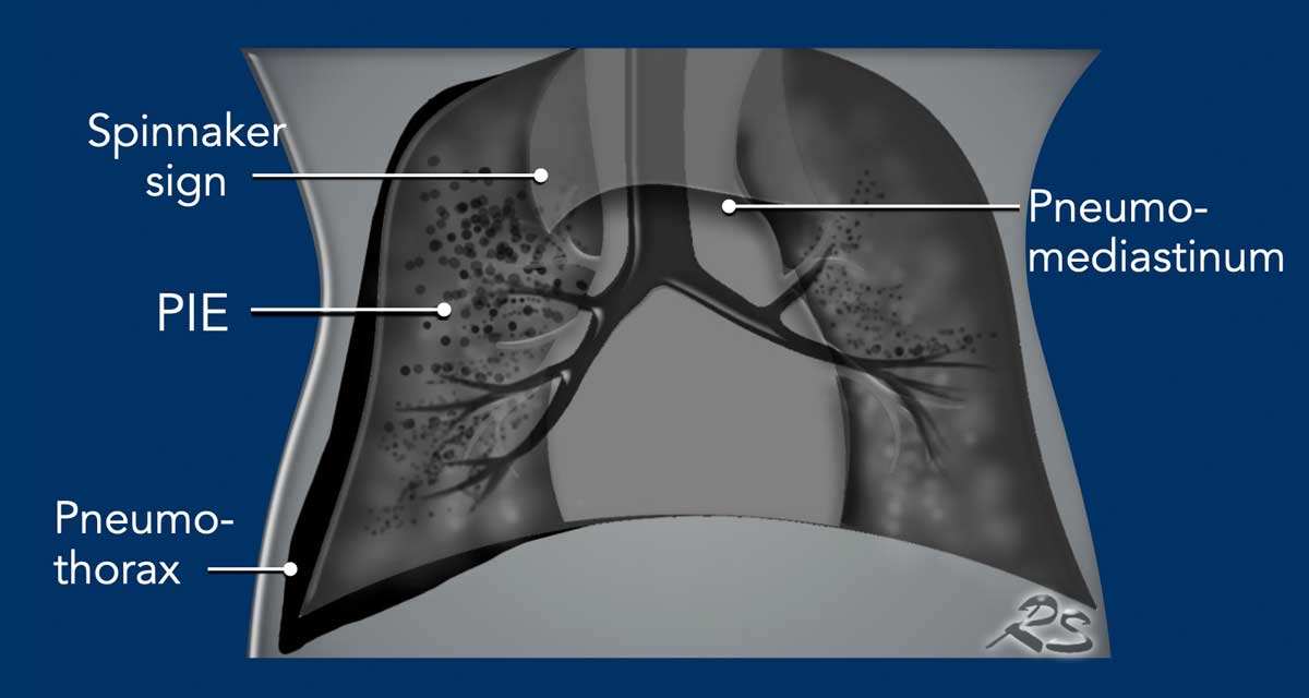
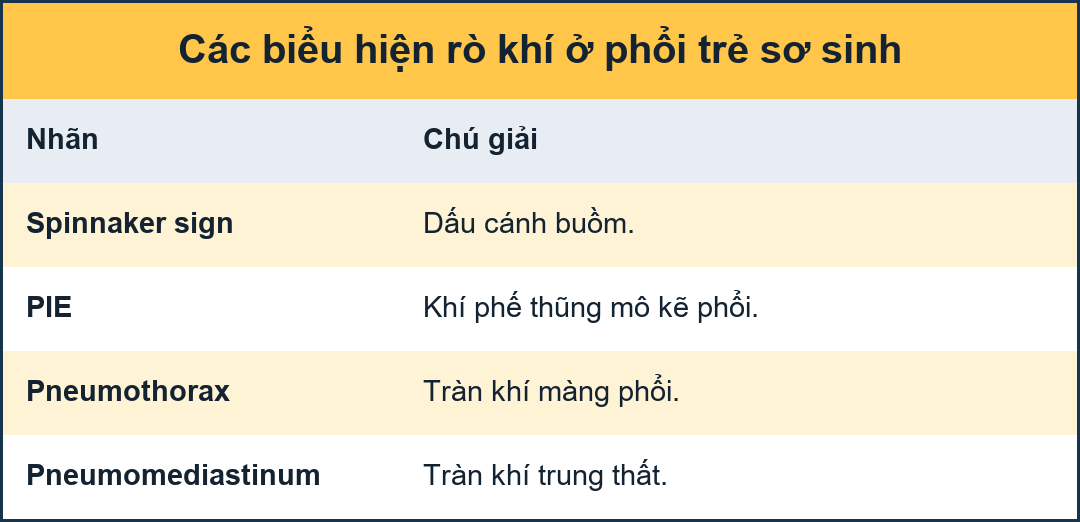
Air leakage as a result of barotrauma in newborns present as:Rò khí là hậu quả của chấn thương khí áp ở trẻ sơ sinh có biểu hiện như sau:
- PneumothoraxTràn khí màng phổi
- Pneumomediastinum and pericardiumTràn khí trung thất và màng ngoài tim
- Pulmonary Interstitial Emphysema (PIE)Phổi kẽ khí phế thũng (PIE)
- Tracheal ruptureVỡ khí quản
- Pneumatocele
Nang khí
Pneumothorax can occur spontaneously or as a complication of
positive pressure ventilatory support.
The introduction of surfactant treatment and improved
ventilatory support has significantly decreased the incidence.Tràn khí màng phổi có thể xảy ra tự phát hoặc là một biến chứng của hỗ trợ thông khí áp lực dương.
Việc sử dụng surfactant và hỗ trợ hô hấp cải tiến đã làm giảm đáng kể tỷ lệ này.
Rupture of terminal airways results from high pressure
insufflation of collapsed lung tissue. This causes air leakage into the
pulmonary interstitium, lymphatic system or pleural space.
A specific finding on neonatal chest films is air leak
tracking along the bronchi termed pulmonary interstitial emphysema (PIE).
Air within the interstitium can cause stiff and incompliant
lungs and diminished blood flow, leading to a reduced blood oxygenation.
Pneumothorax can cause collapse of a lung, but in newborns the lungs are
relatively non-compliant and volume loss is often limited.Vỡ đường thở cuối cùng là kết quả của sự bơm phồng áp lực cao của mô phổi bị xẹp Điều này gây ra sự rò rỉ không khí vào mô kẽ phổi, hệ thống bạch huyết hoặc khoang màng phổi.
Một dấu hiệu cụ thể trên phim chụp ngực sơ sinh là dấu rò khí dọc theo phế quản được gọi là khí phế thũng (PIE) mô kẽ phổi.
Không khí trong khoảng kẽ có thể gây ra phổi cứng và không tương thích và giảm lưu lượng máu, dẫn đến giảm oxy máu.
tràn khí màng phổi có thể gây xẹp phổi, nhưng ở trẻ sơ sinh phổi tương đối không tuân thủ và mất thể tích thường hạn chế.
When additional pneumomediastinum is present, a ‘lifted thymus’ can be seen, also known as the ‘Spinnaker Sign’.Khi có thêm tràn khí trung thất, có thể thấy một 'cơn đau tuyến ức', còn được gọi là 'Dấu hiệu Spinnaker'.
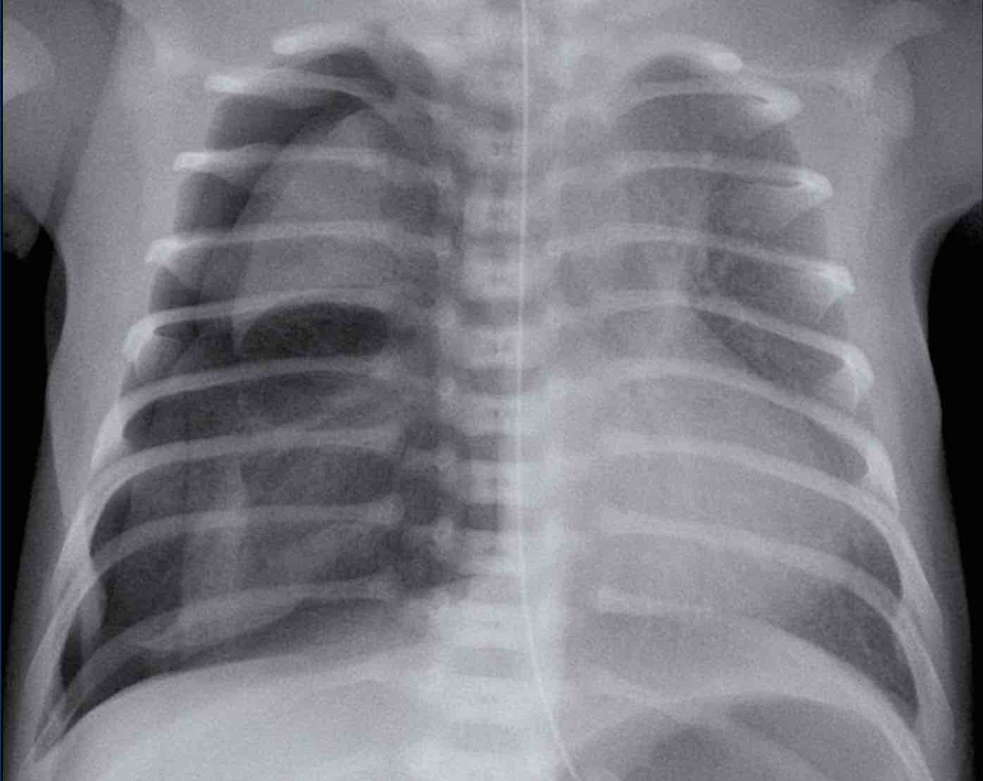
PneumothoraxTràn khí màng phổi
Neonate 3 days old with RDS. Gestational age: 34 weeks.
First study the image.
What are the findings?Trẻ sơ sinh 3 ngày tuổi có RDS. Tuổi thai: 34 tuần.
Đầu tiên nghiên cứu hình ảnh.
Những phát hiện này là gì?
ImageHình ảnh
- Increased lucency of the right pleural space.Tăng độ sáng của khoang màng phổi phải.
- Increased density of the partially collapsed right lung.Tăng mật độ của phổi phải bị xẹp một phần.
- Mediastinal shift to the left as a result of the pneumothorax under tension.Dịch chuyển trung thất sang trái do tràn khí màng phổi bị căng.
- Pneumomediastinum causing ‘lifting’ of the thymus.Tràn khí trung thất gây ra tình trạng “nâng ” tuyến ức.
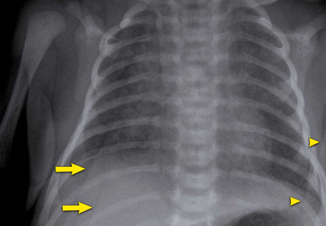
Pitfall
In newborns an important pitfall is the presence of a skin fold, which can be mistaken for a pneumothorax. Cạm bẫy
Ở trẻ sơ sinh, một cạm bẫy quan trọng là sự hiện diện của nếp gấp da, có thể bị nhầm lẫn với tràn khí màng phổi.
Skinfolds do no follow anatomical borders and can be traced outside the chest cavity or cross-over pulmonary vessels.Các nếp gấp da không theo đường viền giải phẫu và có thể được tìm thấy bên ngoài khoang ngực hoặc các mạch máu phổi.
Image
A term newborn with an abdominal sepsis.
There are signs of
fluid overload with accentuated blurry vessels. Skinfolds project over the right lower lung (arrows).
The lines cross anatomical borders, e.g. diaphragm and do
not follow pleura or lungs.Hình ảnh
Một trẻ sơ sinh đủ tháng bị nhiễm trùng bụng.
Có dấu hiệu quá tải dịch với các mạch máu mờ nổi bật. Nếp gấp da lồi lên trên phổi dưới bên phải (các mũi tên).
Các đường thẳng vượt qua ranh giới giải phẫu, ví dụ như cơ hoành và không đi theo màng phổi hoặc phổi.
Left arm projecting over hemi-thorax, resulting in sharp radiopaque line (arrowhead).Cánh tay trái chiếu qua màng tim, tạo đường đứt gãy sắc nét (đầu mũi tên).
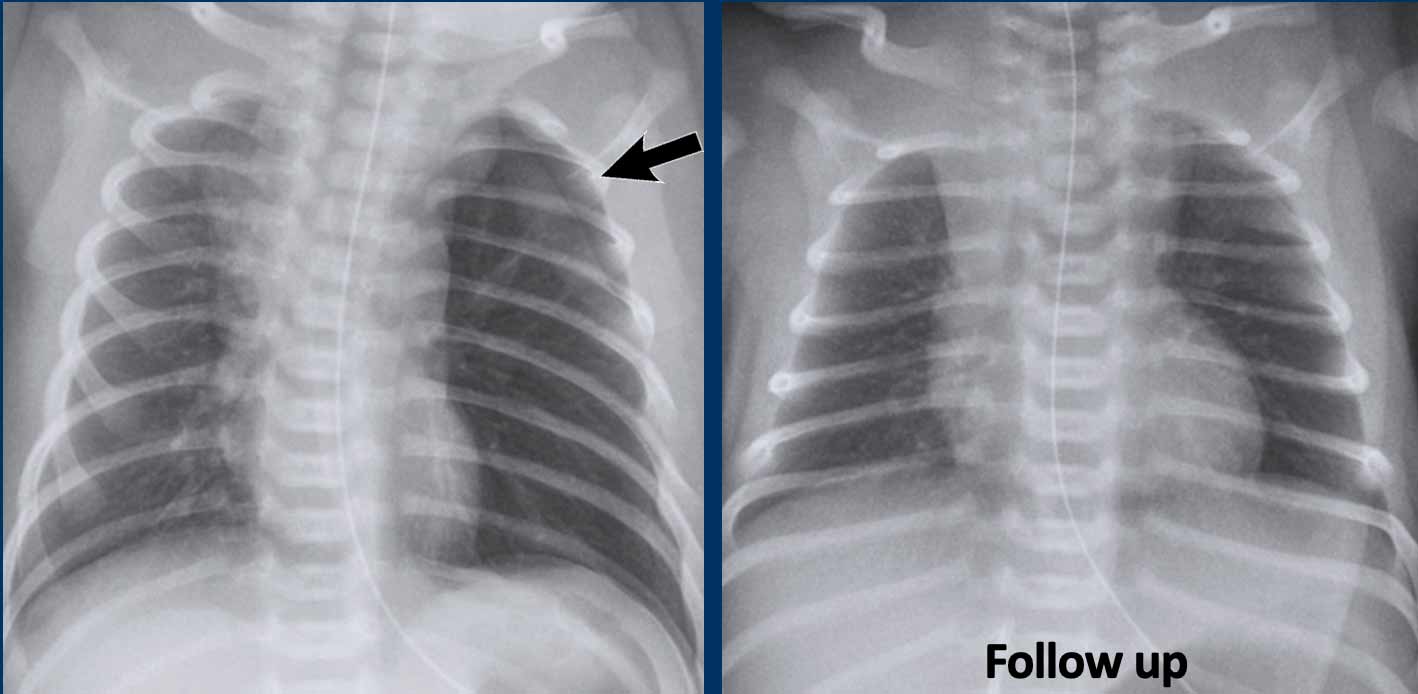
Images of a neonate with respiratory distress.
37 weeks gestational age
After primary caesarean section. Hình ảnh của một trẻ sơ sinh bị suy hô hấp.
37 tuần tuổi thai
Sau mổ lấy thai.
Left image
Hyperinflation on the left side.
Mild displacement of the midline
structures to the right.
A pneumothorax is visible on the left side (arrow)Hình ảnh bên trái
Siêu lạm phát ở bên trái.
Chuyển vị nhẹ các cấu trúc đường giữa sang phải.
Một tràn khí màng phổi có thể nhìn thấy ở phía bên trái (mũi tên)
Right image
On the follow up chest x-ray, the pneumothorax has spontaneously resolved.Hình ảnh bên phải
Theo dõi X-quang ngực, tràn khí màng phổi đã tự giải quyết.
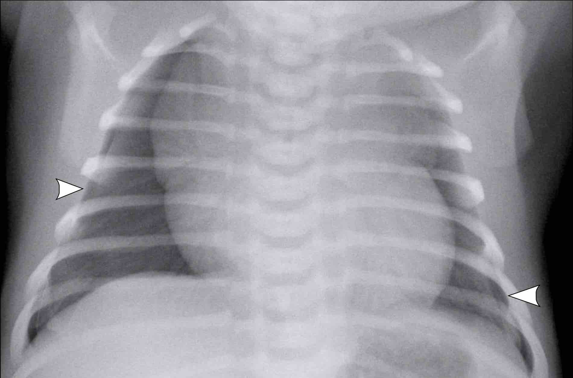
A pneumothorax can be very subtle, since in many neonatal
pulmonary disorders the lungs are not compliant and will not collapse.
Often in
a supine neonate, the pneumothorax only manifests ventrally.
Sometimes only sharpening of the mediastinal structures with
unilateral lucency is noted.Tràn khí màng phổi có thể rất tinh tế, vì trong nhiều bệnh phổi sơ sinh, phổi không tuân thủ và sẽ không bị xẹp.
Thông thường ở trẻ sơ sinh nằm ngửa, tràn khí màng phổi chỉ biểu hiện ở tâm thất.
Đôi khi chỉ cần ghi nhận sự mài sắc của các cấu trúc trung thất với độ sáng một bên.
Image
Full term infant shortly after birth with mild pulmonary
distress. Hình ảnh
Trẻ đủ tháng ngay sau sinh với suy hô hấp nhẹ.
- Mild diffuse linear opacification of both lungs in keeping with TTN.Đục tuyến tính lan toả nhẹ ở cả hai phổi phù hợp với TTN.
- Bilateral pneumothorax (arrowheads).Hai bên tràn khí màng phổi (các đầu mũi tên).
- Sharpening of the mediastinal structures on both sides as a result of interface between mediastinum and pneumothorax.
Nghiên cứu mài sắc cấu trúc trung thất hai bên qua đường tiếp giáp giữa trung thất và tràn khí màng phổi.
Due to fluid in the lung parenchyma these lungs were stiff and did not collapse. Do có dịch trong nhu mô phổi nên phổi bị cứng và không xẹp.
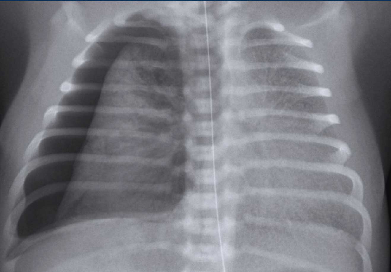
Neonate 32 weeks gestational age. Treated for RDS via CPAP, with sudden respiratory distress.Trẻ sơ sinh 32 tuần tuổi thai, được điều trị suy hô hấp cấp bằng CPAP, có suy hô hấp đột ngột.
First study the image.
What are the findings?Đầu tiên nghiên cứu hình ảnh.
Những phát hiện này là gì?
Image
There is a a pneumothorax on the right side.
Midline structures are displaced to the left.
The left lungs
shows reticulonodular markings in keeping with RDS.
The right lung is not completely collapsed due to stiffness of the parenchyma in RDS and fluid.Hình ảnh
Có một tràn khí màng phổi ở bên phải.
Cấu trúc đường giữa bị dịch chuyển sang trái.
Phổi trái có dấu hiệu hạt mạng lưới phù hợp với RDS.
Phổi phải không bị xẹp hoàn toàn do độ cứng của nhu mô trong RDS và dịch.
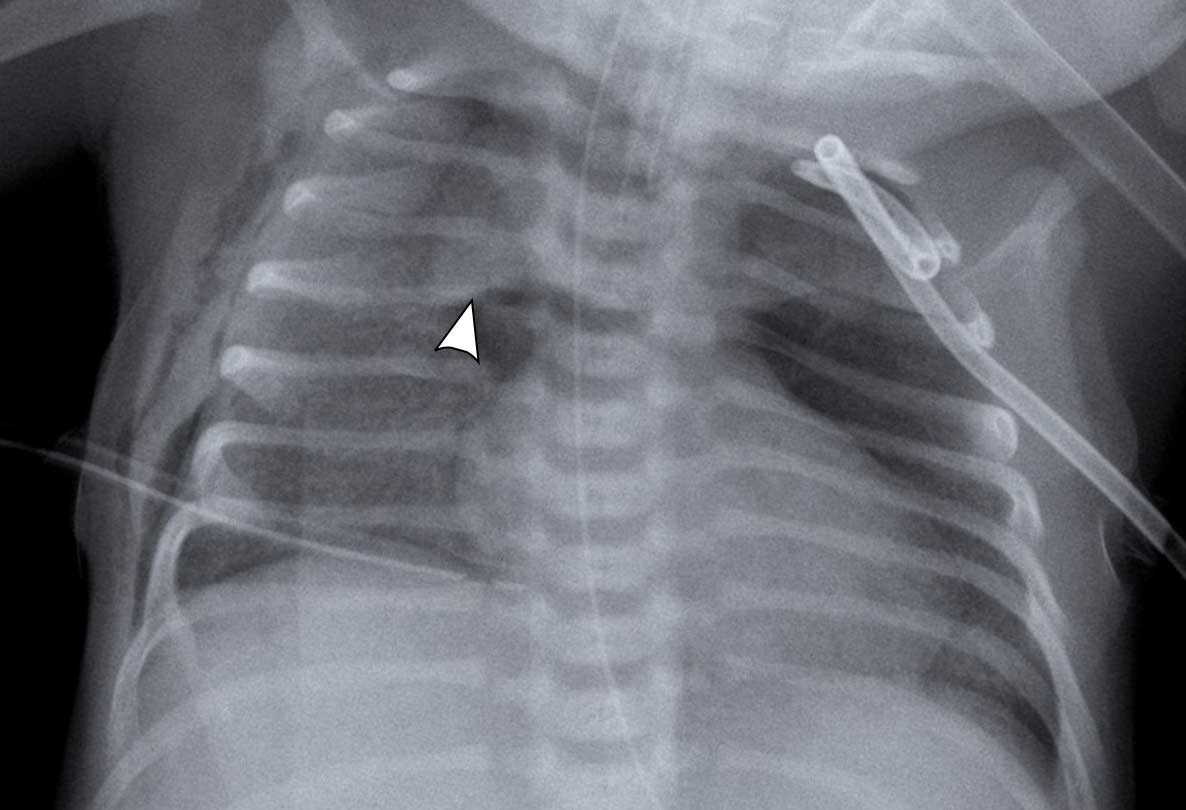
PneumomediastinumTràn khí trung thất
Pneumomediastinum is recognized as air inside the
mediastinum.
The classical sign is the so-called spinnaker sign
(arrowhead).
This is caused by the thymus being ‘lifted up’ from the lower
mediastinum by the mediastinal air. Tràn khí trung thất được công nhận là không khí bên trong trung thất.
Dấu hiệu cổ điển là cái gọi là dấu hiệu spinnaker (đầu mũi tên).
Điều này là do tuyến ức được không khí trung thất nâng lên từ trung thất dưới.
First study the image.
What are the findings?Đầu tiên nghiên cứu hình ảnh.
Những phát hiện này là gì?
Image
Hình ảnh
- A residual ventral pneumothorax can be recognized by the sharp paracardial and mediastinal contour on the left side.Một tràn khí màng phổi tồn dư ở bụng có thể được nhận ra bởi đường viền cạnh tâm thất và trung thất sắc nét ở bên trái.
- On the right
side there is a drain in situ, with no apparent residual pneumothorax, but subcutaneous emphysema, due to air leak with drain.
Bên phải có ống dẫn lưu tại chỗ, không có tràn khí màng phổi còn lại rõ ràng, nhưng khí phế thũng dưới da, do rò khí có ống thông. - Pneumomediastinum is recognized as air inside the mediastinum, where there is air between the thymus and heart figure (arrowhead).Tràn khí trung thất được công nhận là không khí bên trong trung thất, nơi có không khí giữa tuyến ức và hình trái tim (đầu mũi tên).
- Skinfold over right hemi-thorax.Gạc da trên nửa ngực phải.
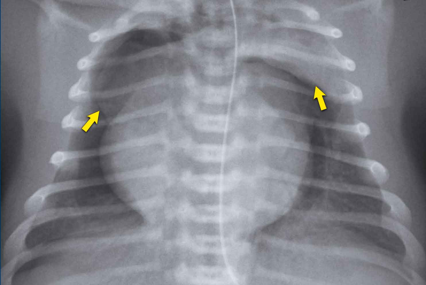
Image
Air can be recognized
between the thymus and the heart, which indicates a pneumomediastinum.
No apparent pneumothorax is
recognized.
The thymus
is uplifted on both sides (arrows).Hình ảnh
Không khí có thể được nhận ra giữa tuyến ức và tim, cho thấy có tràn khí trung thất.
Không có tràn khí màng phổi rõ ràng được công nhận.
Tuyến ức được nâng lên ở cả hai bên (các mũi tên).
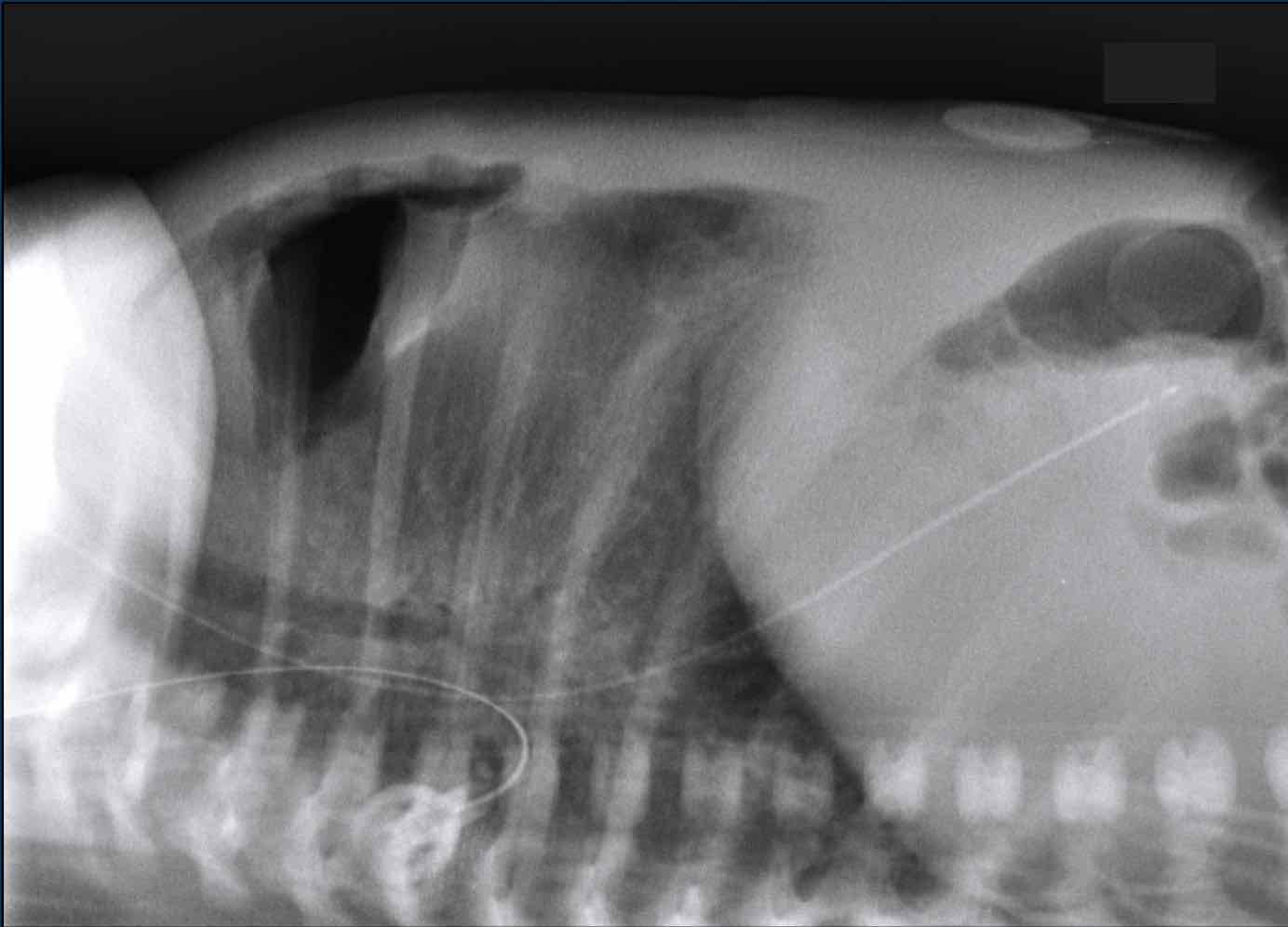
A cross table lateral view can help
to confirm the presence of a pneumothorax or a pneumomediastinum.
Mặt cắt ngang của bảng chéo có thể giúp xác nhận sự hiện diện của tràn khí màng phổi hoặc tràn khí trung thất.
Image
Air can be recognized between the
thymus and the heart, which indicates a pneumomediastinum.
No apparent pneumothorax is recognized.Hình ảnh
Không khí có thể được nhận ra giữa tuyến ức và tim, cho thấy có tràn khí trung thất.
Không có tràn khí màng phổi rõ ràng được công nhận.
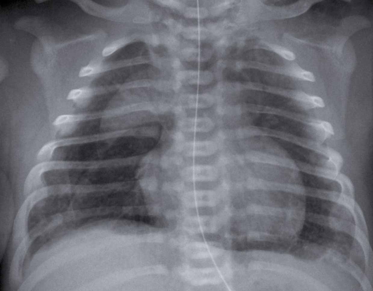
A full term neonate after meconium aspiration. Một trẻ sơ sinh đủ tháng sau hút phân su.
Pneumomediastium, in combination with subcutaneous emphysema. NG tube in situ.Mất trung thất, kết hợp với đặt ống khí phế thũng.NG dưới da tại chỗ.
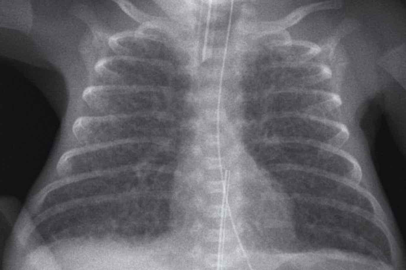
Pulmonary Interstitial Emphysema (PIE)Phổi kẽ khí phế thũng (PIE)
Pulmonary interstitial emphysema (PIE) is leakage of air into
the perivascular and peribronchial spaces as a result of rupture at the
bronchiolo-alveolar junctions.
PIE is recognized as either small bubbles or linear air
collections along the bronchovascular bundle radiating from hilum to the
periphery.
PIE can be bilateral or unilateral.
Once PIE is established, air may dissect centrifugally along
bronchovascular sheaths or lymphatic channels to form subpleural blebs, which
may rupture into the pleural space and produce a pneumothorax.Khí phế thũng kẽ phổi (PIE) là sự rò rỉ không khí vào khoang quanh mạch máu và khoang quanh phế quản do vỡ ở các điểm nối phế quản phế nang.
PIE được công nhận là các bong bóng nhỏ hoặc các luồng khí dọc theo bó phế quản-mạch máu lan từ rốn phổi đến ngoại vi.
PIE có thể là hai bên hoặc một bên.
Khi PIE được thiết lập, không khí có thể phân tách ly tâm dọc theo vỏ bọc phế quản hoặc các kênh bạch huyết để tạo thành các khối dưới màng phổi, có thể vỡ vào khoang màng phổi và tạo ra tràn khí màng phổi.
Image
Preterm infant born at 27 weeks gestational age.
Now 6 weeks old.
Mechanical ventilation for RDS.Hình ảnh
Trẻ sinh non 27 tuần tuổi thai, hiện tại 6 tuần tuổi.
Thông khí cơ học trong điều trị suy hô hấp cấp.
First study the image.
What are the findings?Đầu tiên nghiên cứu hình ảnh.
Những phát hiện này là gì?
Findings:Kết quả:
- Bubbly air configurations bilateral indicating PIE.Hình thái khí có bọt hai bên cho thấy PIE.
- Note the small
mediastinal silhouette, caused by shrinking of the thymus as a result of
stress.
Lưu ý hình bóng trung thất nhỏ, do sự co lại của tuyến ức là kết quả của sự căng thẳng. - Deep position umbilical vein line, tip likely in left atrium or pulmonary vein. Vị trí đường tĩnh mạch rốn sâu, đầu có thể ở tâm nhĩ trái hoặc tĩnh mạch phổi.
- Good position ETT. NG tube in situ.Vị trí đặt ống ETT. NG tại chỗ tốt.
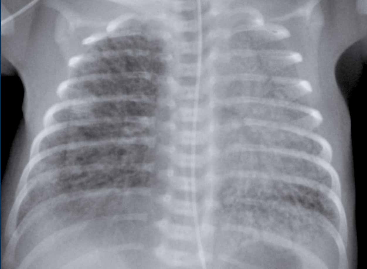
Neonate, gestational age 30 weeks, day 1, sudden deterioration after MIST.Trẻ sơ sinh, tuổi thai 30 tuần, ngày 1, suy dinh dưỡng sau mổ.
First study the image.
What are the findings?Đầu tiên nghiên cứu hình ảnh.
Những phát hiện này là gì?
Image
Bilateral radiating bubbly lucencies, with
hyperinflation of both lungs.
These are typical findings in PIE. Hình ảnh
Các bóng mờ hai bên, với sự căng phồng của cả hai phổi.
Đây là những dấu hiệu điển hình trong PIE.
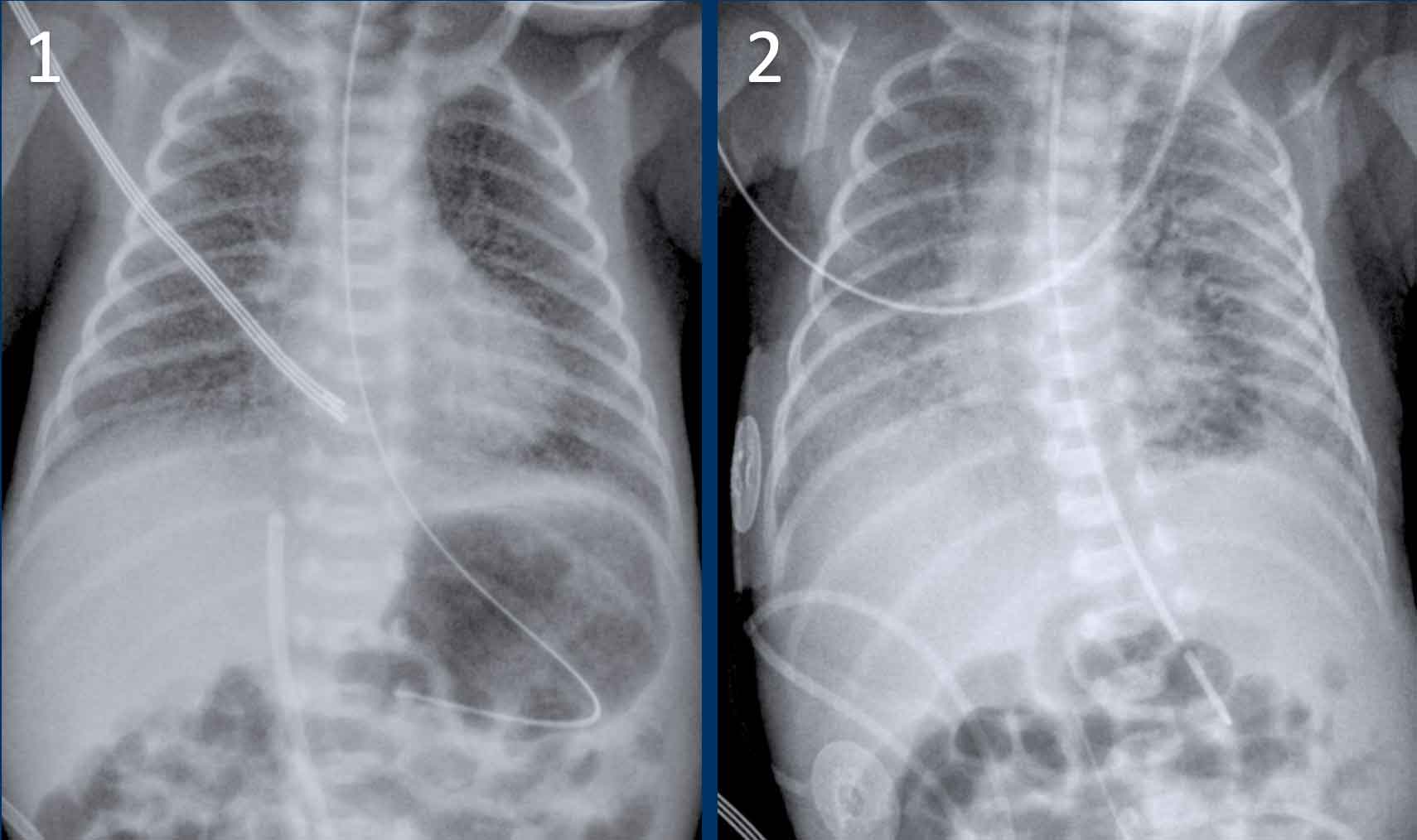
Neonate, gestational age 27 weeks, treated for RDS.
At age of 2 days sudden deterioration.Trẻ sơ sinh, tuổi thai 27 tuần, điều trị RDS.
Ở tuổi 2 ngày, bệnh đột ngột xấu đi.
First study the images.
What are the findings?Đầu tiên nghiên cứu hình ảnh.
Những phát hiện này là gì?
Image 1
Bilateral reticulonodulair opacities in keeping with IRDS,
treated via CPAP.Hình 1
Bệnh nhân có biểu hiện đục hai bên theo IRDS, điều trị bằng CPAP.
Image 2
At the age of 2 days the X-ray
shows radiating lucencies in the left lung as a result of PIE.Hình 2
Ở tuổi 2 ngày, X-quang cho thấy các tia sáng trong phổi trái do hậu quả của PIE.
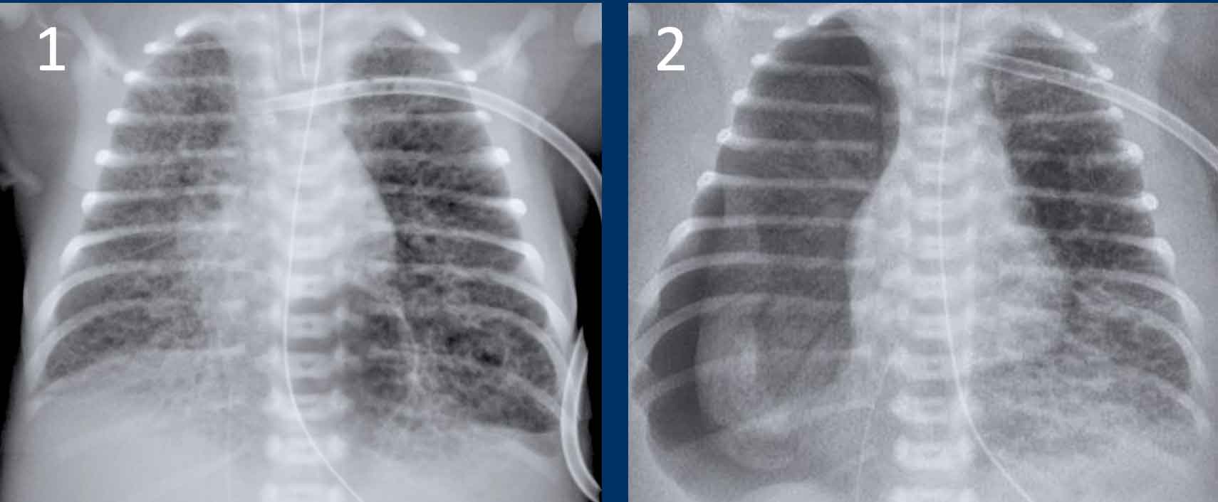
These images are of a neonate gestational age 32 weeks, who was treated for RDS.
Những hình ảnh này là của một trẻ sơ sinh 32 tuần tuổi thai, được điều trị RDS.
Several complications developed, including a pneumothorax on the left side, which was drained.
After
drainage there was a deterioration.Một số biến chứng đã xuất hiện, bao gồm tràn khí màng phổi ở bên trái, được dẫn lưu.
Sau khi dẫn lưu có sự suy giảm.
Image 1
The radiograph shows bilateral radiating bubbly lucencies due to bilateral PIE.
This is more pronounced on the left side.Hình 1
Phim X-quang cho thấy các luồng sáng phát ra từ hai bên do PIE hai bên.
Điều này rõ rệt hơn ở bên trái.
Image 2
In follow up the child also developed a pneumothorax on the right side. Hình 2
Theo dõi trẻ cũng phát hiện được tràn khí màng phổi ở bên phải.
Congenital anomaliesDị tật bẩm sinh
The most common congenital anomalies in neonates are:Các dị tật bẩm sinh phổ biến nhất ở trẻ sơ sinh là:
- Congenital pulmonary airway malformation (CPAM)Dị dạng đường thở phổi bẩm sinh (CPAM)
- Pulmonary sequesterMáy cô lập phổi
- Congenital lobar emphysemaThuỳ bẩm sinh khí phế thũng
- Diafragmatic herniationThóat vị đĩa đệm
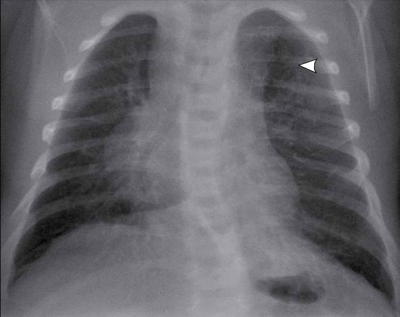
Congenital pulmonary airway malformationDị dạng đường thở phổi bẩm sinh
Congenital pulmonary airway malformation (CPAM) was until recently know as congenital cystic adenomatoid malformation (CCAM).
It is a spectrum of bronchopulmonary foregut malformations.Dị dạng đường thở phổi bẩm sinh (CPAM) cho đến gần đây được biết đến như là dị dạng nang tuyến (CCAM) bẩm Sinh.
Đây là một phổ của dị dạng phế quản phổi.
There are three histological types:Có ba loại mô học:
- Type I (75% of cases) is composed of variable sized cysts, with at least one dominant cyst larger than 2 cm in diameter.Loại I (75% trường hợp) bao gồm các nang có kích thước thay đổi, với ít nhất một nang trội có đường kính lớn hơn 2 cm.
- Type 2 (10-15% of cases) is composed of smaller multiple cysts of the same size, less than 1 cm in diameter. Loại 2 (10-15% trường hợp) bao gồm nhiều nang nhỏ hơn có cùng kích thước, đường kính dưới 1 cm.
- Type 3 presents as a solid mass composed of micro cysts.Loại 3 biểu hiện dưới dạng một khối rắn bao gồm các nang vi mô.
Image
1 week old child, born at 41 weeks gestational age.
On prenatal routine imaging, a cystic lesion was seen in the upper left
lobe.
The radiograph shows a delineated lucent area in the apical part of the
left upper lobe (arrow).
There is a slight mediastinal shift to the right.Hình ảnh
Đứa trẻ 1 tuần tuổi, sinh lúc 41 tuần tuổi thai.
Trên hình ảnh thường quy trước sinh, một tổn thương dạng nang được nhìn thấy ở thuỳ trên bên trái.
Chụp X-quang cho thấy một vùng sáng được phác thảo ở phần đỉnh của thuỳ trên bên trái (mũi tên).
Có một sự dịch chuyển nhẹ về phía trung thất phải.
Radiographic findings in CPAMĐặc điểm X quang trong CPAM
- In type I and II, multiple thin walled cysts containing air and varying in size are seen.Ở type I và II, có nhiều nang có thành mỏng chứa không khí và kích thước khác nhau.
- In type III a solid lesion is seen.Ở type III, có tổn thương đặc.
- Infection of the cysts can cause air fluid levels. Nhiễm trùng nang có thể gây ra mức dịch không khí.
- Large lesions cause mass effect with mediastinal shift and atelectasis of adjacent lung parenchyma.
Tổn thương lớn gây ra hiệu ứng khối với sự dịch chuyển trung thất và xẹp phổi của nhu mô phổi lân cận. - CPAM usually involves one lobe.
CPAM thường liên quan đến một thuỳ.
Contrast
enhanced CT scan is essential in the analysis of CPAM and sequesters.
Given that CPAM and sequester often are hybrid lesions,
feeding arterial vessels need to be visualized or ruled out before surgical
intervention.
Chụp CT có tiêm thuốc cản quang là cần thiết trong phân tích CPAM và sequesters.
Do CPAM và cô lập thường là tổn thương lai, nên cần phải quan sát các mạch máu nuôi dưỡng hoặc loại trừ trước khi can thiệp phẫu thuật.
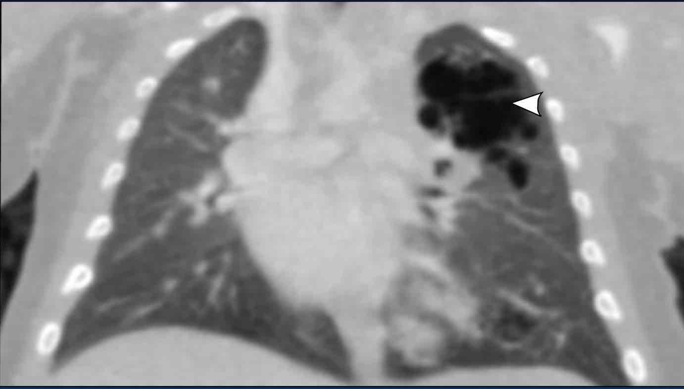
Image
CT scan of the same patient as above.
The lucent lesion in the left upper lobe has a multicystic aspect.
Because the largest cyst has a diameter of more than 2 cm, this is classified as a
type I CPAM.Hình ảnh
Chụp CT của cùng một bệnh nhân như trên.
Tổn thương dạng bóng ở thuỳ trên bên trái có hình thái đa nang.
Vì nang lớn nhất có đường kính trên 2 cm, đây được phân loại là CPAM loại I.
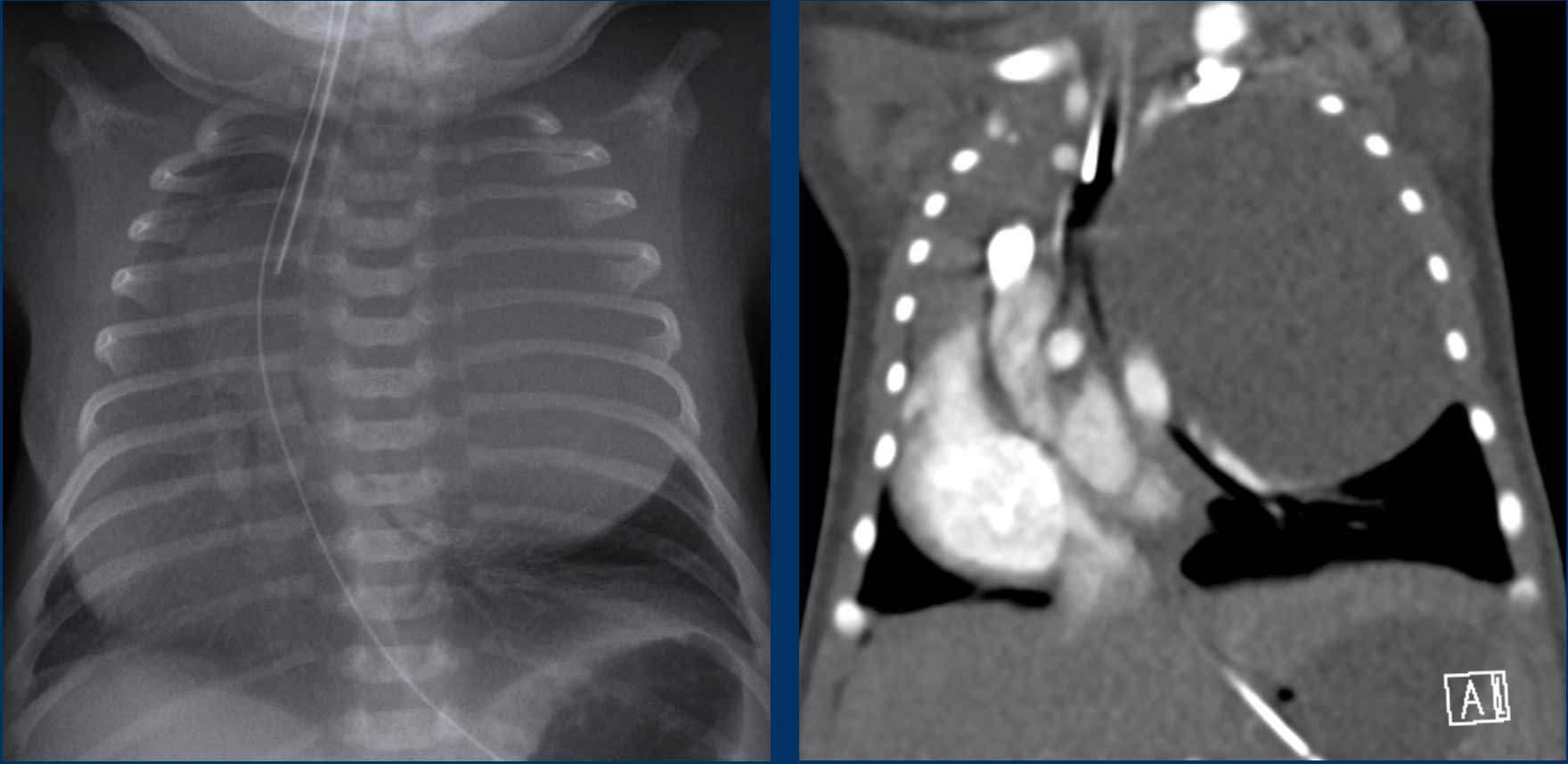
Images of a neonate, 40 weeks gestational age.
Antenatal suspicion of large CPAM on the left side.
At birth respiratory distressHình ảnh của một trẻ sơ sinh, 40 tuần tuổi thai.
Nghi ngờ trước sinh CPAM lớn ở bên trái.
Khi sinh ra suy hô hấp
Images
Radiograph shows a large round opacified lesion.
Severe displacement of
midline structures with atelectasis of the right lung.
Deep position ETT. NG tube in situ.Hình ảnh
X quang cho thấy một tổn thương tròn lớn đục.
Sự dịch chuyển nặng nề của các cấu trúc đường giữa với xẹp phổi của phổi phải.
Ống ETT. NG vị trí sâu tại chỗ.
Because of the need of direct intervention a CT after IV contrast was
performed.
CT shows a large cystic lesion in the upper lobe of
the left lung, with displacement of vascular and bronchial structures.
The lesion has relation to any systemic vessel, Which excludes a sequester.
This is a CPAM, which is not yet aerated.Do nhu cầu can thiệp trực tiếp nên CT sau tiêm thuốc cản quang được thực hiện.
CT cho thấy một tổn thương dạng nang lớn ở thuỳ trên của phổi trái, với sự dịch chuyển của các cấu trúc mạch máu và phế quản.
Tổn thương có liên quan đến bất kỳ mạch hệ thống nào, loại trừ một mạch máu bị cô lập.
Đây là CPAM, chưa được sục khí.
Pulmonary sequestrationPhổi biệt lập
A pulmonary sequestration is a segment of dysplastic lung
tissue, which is separate from the rest of the lung. It receives an anomalous
systemic vascular supply.
The most common type is the intralobar type, which is
situated within a normal lobe and has no own visceral pleura.
Usually there is
a normal venous return via the pulmonary veins.
In contrast the extralobar type is situated outside of the
normal lung and has a separate visceral pleural and a systemic venous return.Phổi biệt lập là một phần của mô phổi loạn sản, tách biệt với phần còn lại của phổi.Nó nhận được một nguồn cung cấp mạch máu hệ thống bất thường.
Loại phổ biến nhất là loại trong thuỳ, nằm trong thuỳ bình thường và không có màng phổi nội tạng.
Thông thường có tĩnh mạch trở lại bình thường qua các tĩnh mạch phổi.
Ngược lại, loại ngoài thuỳ nằm ngoài phổi bình thường và có màng phổi nội tạng riêng biệt và tĩnh mạch trở lại hệ thống.
Most sequestrations are detected on antenatal ultrasound and present as a mass in the basal lobes, usually paramedian on the left.Hầu hết các khối cô lập được phát hiện trên siêu âm trước sinh và có dạng khối ở thuỳ đáy, thường là cạnh trong bên trái.
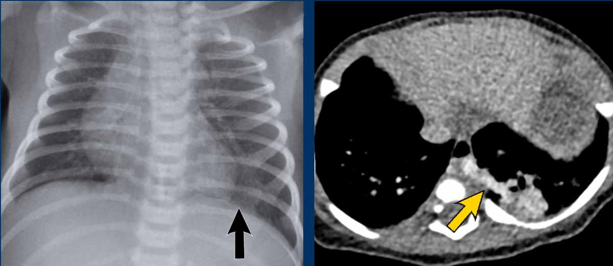
Image
Neonate, 39 weeks gestational age, antenatal suspicion of CPAM left lower
lobe.
Radiograph shows a subtle, not well circumscribed lesion on the
left lower lobe.
CT at age of one month shows a mixed lesion, both cystic and
solid, with a large feeding artery from the descending aorta, in keeping with
sequester.Hình ảnh
Trẻ sơ sinh, 39 tuần tuổi thai, tiền sản nghi ngờ CPAM thuỳ dưới trái.
X quang cho thấy một tổn thương kín đáo, không được bao quanh tốt ở thuỳ dưới bên trái.
CT ở tuổi một tháng cho thấy một tổn thương hỗn hợp, cả nang và đặc, với một động mạch nuôi lớn từ động mạch chủ xuống, phù hợp với động mạch chủ.
ImagingHình ảnh
- On radiographs a sequestration is often not detected. Trên phim X quang, sự cô lập thường không được phát hiện.
- Prenatal
ultrasound often raises the suspicion of a sequestration, which is seen as a hyperechoic mass.
Siêu âm trước khi sinh thường làm tăng nghi ngờ về sự cô lập, được xem như là một khối tăng âm.
Management is controversial. Some advocate surgical resection because of the risk of infection
and cardiac failure due to the left to right shunting.
Others advocate a wait and
see approach, as these sequestrations can resolve spontaneously.
Before surgical management MR or CT is needed to analyze the blood supply and the type of the sequestration.Điều trị còn nhiều tranh cãi, một số người ủng hộ phẫu thuật cắt bỏ vì nguy cơ nhiễm trùng và suy tim do shunt trái sang phải.
Những người khác ủng hộ cách tiếp cận chờ đợi và xem xét, vì những sự cô lập này có thể tự giải quyết.
Trước khi phẫu thuật, cần phải có MR hoặc CT để phân tích nguồn cung cấp máu và loại cô lập.
Congenital Lobar EmphysemaBẩm sinh thuỳ khí phế thũng
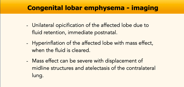
Congenital lobar emphysema, now known as congenital lobar
overinflation, is a condition in which there is hyperexpansion of a lobe of the
lungs.
Narrowing or weakness of a lobar bronchus causing a
check-valve mechanism is the most likely cause.Thuỳ phổi bẩm sinh khí phế thũng, hiện được gọi là quá tải thuỳ phổi, là tình trạng có sự giãn nở quá mức của thuỳ phổI.
Hẹp hoặc yếu phế quản thuỳ gây ra cơ chế kiểm tra van là nguyên nhân có khả năng nhất.
Symptoms depend on the degree of lobar hyperexpansion.
A mildly hyperexpanded lobe may be asymptomatic and decrease
in size with time and close follow-up is warranted in these cases. Progressive
hyperexpansion of a lobe can occur and cause significant, sometimes life-threatening,
symptoms. Those cases are treated with lobectomy.Các triệu chứng phụ thuộc vào mức độ giãn nở thuỳ.
Thuỳ phổi giãn nở nhẹ có thể không có triệu chứng và giảm kích thước theo thời gian và cần theo dõi chặt chẽ trong những trường hợp này. Phát triển quá mức của thuỳ phổi có thể xảy ra và gây ra các triệu chứng đáng kể, đôi khi đe doạ tính mạng, những trường hợp đó được điều trị bằng phẫu thuật cắt thuỳ phổI.
Usually congenital lobar emphysema is detected on prenatal ultrasound. Thông thường, khí phế thũng thuỳ bẩm sinh được phát hiện trên siêu âm trước sinh.
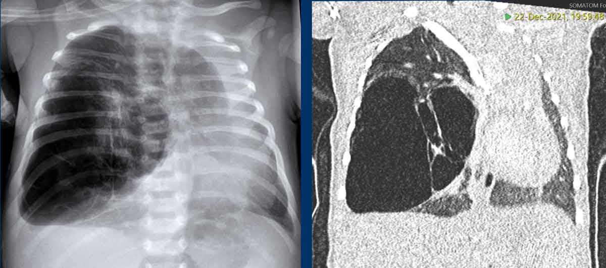
Images of a term neonate with mild respiratory distress.
Antenatal there was suspicion
of a CPAM.
The chest radiograph and CT show hyperinflation of the right upper lobe with architectural distortion.
The right lower lobe is compressed, but probably has a normal architecture.
Displacement of
mediastinum and heart and atelectasis of the left lung.Hình ảnh một trẻ sơ sinh đủ tháng bị suy hô hấp nhẹ.
Có nghi ngờ CPAM trong thai kỳ.
phim X-quang ngực và CT cho thấy sự tăng căng phồng của thuỳ trên bên phải với biến dạng cấu trúc.
Thuỳ dưới bên phải bị chèn ép, nhưng có thể có cấu trúc bình thường.
Sự dịch chuyển trung thất và tim và xẹp phổi của phổi trái.
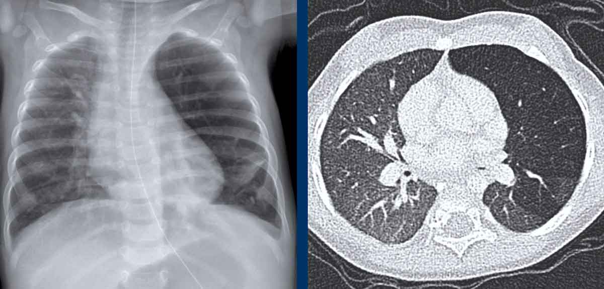
Images of a 6 months old neonate with mild respiratory symptoms. Hình ảnh một trẻ sơ sinh 6 tháng tuổi có triệu chứng hô hấp nhẹ.
Radiograph reveals hyperinflation of left upper lobe with displacement of midline structures and some atelectasis of the lower lobe.X quang cho thấy sự tăng phình của thuỳ trên bên trái với sự dịch chuyển của các cấu trúc đường giữa và một số xẹp phổi của thuỳ dưới.
The CT confirms overinflation of the right upper lobe.Chụp CT xác nhận tình trạng phồng quá mức của thuỳ trên bên phải.
Diaphragmatic herniaThóat vị hoành
There are two types of hernia, the Bochdalek and the Morgagni type.
The diagnosis is often made prenatally.Có hai loại thóat vị, loại Bochdalek và loại Morgagni.
Chẩn đoán thường được thực hiện trước khi sinh.
The
Bochdalek hernia is seen in about 95% of cases and located on the
posterior side of the chest. Most often it is left sided (90%).
The
Morgagni hernia, also known as retrosternal or parasternal hernia, is located
parasternally, where the foramina of Morgagni are situated. Thóat vị Bochdalek gặp ở khoảng 95% các trường hợp và nằm ở phía sau ngực, thường gặp nhất là bên trái (90%).
Thóat vị Morgagni, còn được gọi là thóat vị sau xương ức hoặc thóat vị cạnh xương ức, nằm ở cạnh ức, nơi có lỗ Morgagni.
The severity of the disease is based on the degree of hypoplasia of the lung. Mức độ nghiêm trọng của bệnh dựa trên mức độ giảm sản của phổi.
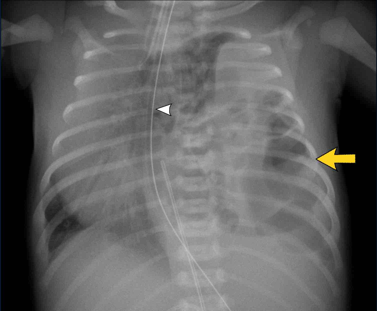
Imaging findings
Chẩn đoán hình ảnh
- Total or partial opacification of one side of the thorax, with no or little aeration of the ipsilateral lung. Đục một phần hoặc toàn bộ một bên ngực, không có hoặc ít sục khí phổi cùng bên.
- Shift of the mediastinum to contralateral side. Dịch chuyển trung thất sang bên đối diện.
- The finding of part of the digestive tract in the chest cavity is pathognomic for the diagnosis. Việc phát hiện một phần của đường tiêu hóa trong khoang ngực là bệnh lý để chẩn đoán.
- Direct postpartum the digestive tract does not contain air, so it may go unnoticed or simulate an abnormality within the lungs.
Trực tiếp sau khi sinh, đường tiêu hóa không chứa không khí, do đó nó có thể không được chú ý hoặc mô phỏng một bất thường trong phổi.
Image
Full term neonate with shortness of breath.
Partial opacification of the left upper hemithorax.
Bowel structures are seen in the left hemithorax (arrow).
Shift of midline structures seen as deviation of the nasogastric tube (arrowhead)Hình ảnh
Trẻ sơ sinh đủ tháng có khó thở.
Đục một phần thành ngực trái.
Cấu trúc ruột được nhìn thấy trong tràn máu màng phổi trái (mũi tên).
Sự thay đổi cấu trúc đường giữa được xem là độ lệch của ống thông mũi dạ dày (đầu mũi tên)
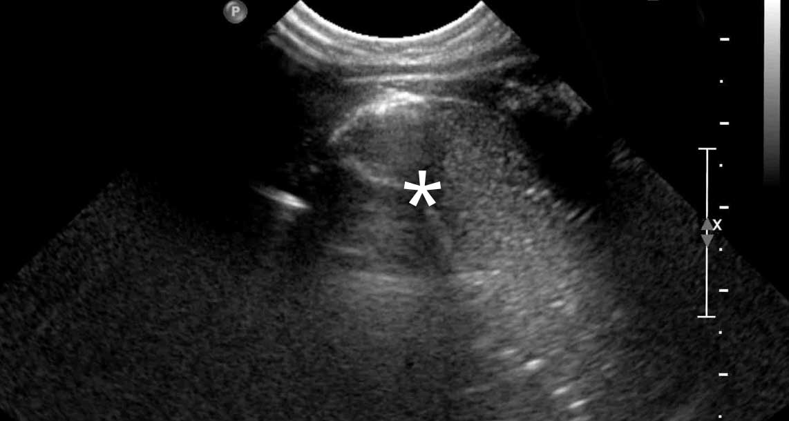
Ultrasound demonstrated peristalsis in air and fluid containing structures confirming a large diafragmatic hernia . Siêu âm cho thấy có nhu động trong không khí và các cấu trúc chứa dịch, xác nhận thóat vị hoành lớn.
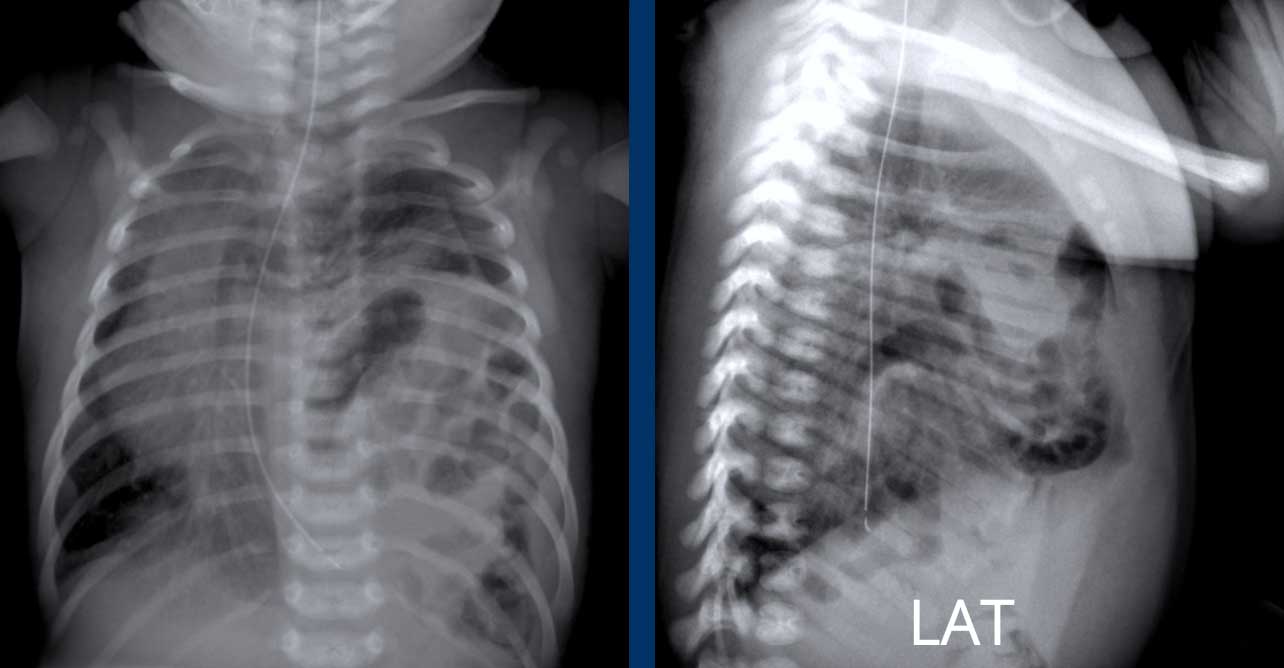
These images are of a full-term neonate with shortness of breath.Những hình ảnh này là của một trẻ sơ sinh đủ tháng bị khó thở.
Images
Partial opacification of
the left upper hemithorax.
Bowel structures are seen
in the left hemithorax.
Shift of midline structures seen as deviation of the nasogastric tube
(arrowhead).
The lateral chest film also demonstrates bowel loops in the chest. Hình ảnh
Đục một phần thành ngực trái.
Cấu trúc ruột được nhìn thấy trong tràn máu màng phổi trái.
Sự thay đổi cấu trúc đường giữa được coi là độ lệch của ống thông mũi dạ dày (đầu mũi tên).
phim ngực bên cũng cho thấy các quai ruột ở ngực.

Charity
Hoạt động từ thiện
All the profits of the Radiology Assistant go to Medical Action Myanmar which is run by Dr. Nini Tun and Dr. Frank Smithuis sr, who is a professor at Oxford university and happens to be the brother of Robin Smithuis.Toàn bộ lợi nhuận của Radiology Assistant được chuyển cho Medical Action Myanmar, do bác sĩ Nini Tun và bác sĩ Frank Smithuis Sr. điều hành. Frank Smithuis Sr. là giáo sư tại Đại học Oxford và là anh em ruột của Robin Smithuis.
Click here to watch the video of Medical Action Myanmar and if you like the Radiology Assistant, please support Medical Action Myanmar with a small giftNhấp vào đây để xem video về Medical Action Myanmar và nếu bạn thích Trợ lý X quang, vui lòng hỗ trợ Medical Action Myanmar bằng một món quà nhỏ
ReferencesTài liệu tham khảo
-
Lung Disease in Premature Neonates: Radiologic-Pathologic Correlation
Bệnh phổi ở trẻ sơ sinh non tháng: Mối tương quan về X quang và mô bệnh học
by Agrons GA, Courtney SE, Stocker JT, Markowitz RI.
From the archives of the AFIP.
Radiographics 2005; 25:1047-1073
của Agrons GA, Courtney SE, Stocker JT, Markowitz RI. -
An approach to the asymmetric neonatal chest radiograph.
Phương pháp tiếp cận phim X quang ngực không đối xứng ở trẻ sơ sinh.
Alford BA, McIlhenny J.
Radiol Clin North Am 1999; 37:1079-1092.
-
Thorax, Mediastinum, Heart, and Great Vessels.
Lưng, trung thất, tim và các mạch máu lớn.
In: Van Rijn RR, Blickman JG eds.
Differential Diagnosos in Pediatric Imaging. 1th ed: Thieme, 2011; 1-138.
Trong: Van Rijn RR, Blickman JG biên tập. -
Radiological imaging of the Neonatal Chest.
Hình ảnh X quang lồng ngực sơ sinh.
Donoghue V, Baert AL, Knauth M, Sartor K,.
2nd revised Edition, Springer-Verlag, 2008, 261-273.
-
The neonatal chest X-ray.
X-quang ngực sơ sinh.
Arthur R.
Paediatr Respir Rev 2001; 2:311-323.
A. R.