
Special Ankle FracturesGãy xương mắt cá chân đặc biệt
Detection of 'Not So Obvious' FracturesPhát hiện gãy xương 'không rõ ràng'
Radiology Department of the Rijnland Hospital, Leiderdorp, the NetherlandsKhoa Chẩn đoán hình ảnh Bệnh viện Rijnland, Leiderdorp, Hà Lan
Ngày đăng / cập nhật: 2012-10-01
Bản gốc trên radiologyassistant.nl ↗ · tải về lúc 2026-11-19 20:19 · bản dịch máy có chuẩn hóa thuật ngữ chẩn đoán hình ảnh
The ankle is the most frequently injured joint.
Management decisions are based on the interpretation of the AP and lateral X-rays.
In this article we will focus on detection of fractures, that may not be so obvious at first sight.
Before you read this article, you need to understand the classification of ankle fractures and exorotation injuries that were highlighted in Ankle - Fractures 1 and 2.Mắt cá chân là khớp thường bị tổn thương nhất.
Các quyết định điều trị dựa trên sự giải thích của AP và X-quang bên.
Trong bài viết này chúng tôi sẽ tập trung vào việc phát hiện các loại gãy xương, có thể không rõ ràng ngay từ cái nhìn đầu tiên.
Trước khi đọc bài viết này, bạn cần hiểu về phân loại gãy cổ chân và các tổn thương vùng hở được nêu bật trong Gãy cổ chân 1 và 2.
Posterior malleolus fracturesGãy mắt cá sau
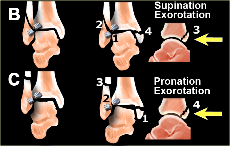
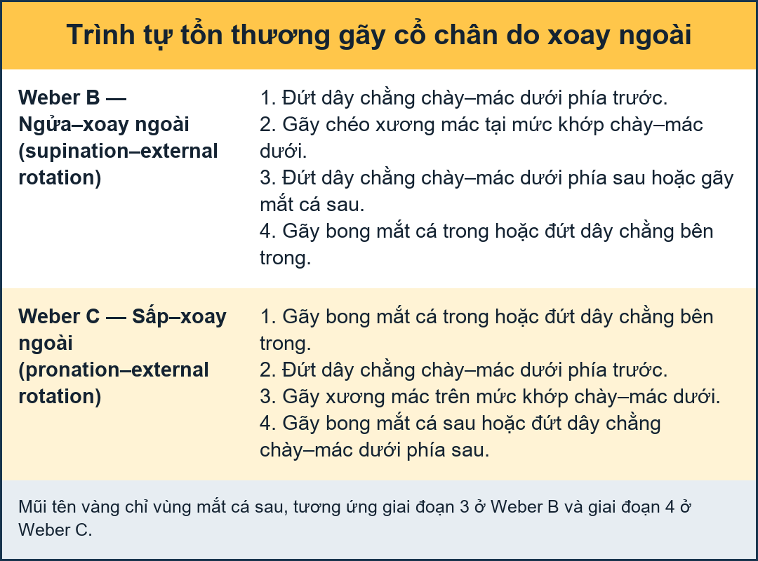
Almost all fractures of the posterior malleolus are part of a rotational injury resulting in a Weber B or Weber C fracture.
The tertius fracture is stage 3 in Weber B and stage 4 in Weber C (figure).
In some cases the tertius fractures are easily seen on the x-rays, but frequently they can be difficult to detect.
It is important to find these fractures, since a tertius fracture can be the only clue to an unstable ankle injury.Hầu như tất cả các gãy mắt cá sau là một phần của chấn thương xoay dẫn đến gãy Weber B hoặc Weber C.
Gãy xương bánh chè ở Weber B là giai đoạn 3 và Weber C là giai đoạn 4 (hình).
Trong một số trường hợp, gãy xương bánh chè có thể dễ dàng nhìn thấy trên phim X-quang, nhưng thường thì rất khó phát hiện.
Điều quan trọng là tìm ra những gãy xương này, vì gãy xương tertius có thể là manh mối duy nhất cho thấy chấn thương cổ chân không ổn định.
Ankle - Fractures 2 - Fracture mechanism and RadiographyGãy cổ chân 2 - Cơ chế gãy và X quang
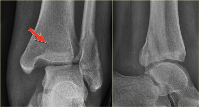 Linear lucency indicating a fracture of the posterior malleolusĐèn tuyến tính cho thấy gãy mắt cá sau
When we study the radiographs of a patient with an ankle injury, we have to study the region of the posterior malleolus very carefully.
In many cases there is only a small gap between the fracture parts and detection depends on optimal radiography and a high level of suspicion.Khi nghiên cứu hình ảnh X quang của một bệnh nhân chấn thương cổ chân, chúng ta phải nghiên cứu vùng mắt cá sau rất cẩn thận.
Trong nhiều trường hợp chỉ có một khoảng cách nhỏ giữa các phần gãy xương và việc phát hiện phụ thuộc vào X quang tối ưu và mức độ nghi ngờ cao.
The images show an obvious Weber B fracture.
On the AP-view the linear lucency is the clue to a tertius fracture (red arrow).
It results from subtle malalignment of the fracture fragment.
Likewise in some cases malalignment can result in a linear density.Hình ảnh cho thấy gãy Weber B rõ ràng.
Trên mặt cắt AP, ánh sáng tuyến tính là đầu mối cho gãy xương tertius (mũi tên màu đỏ).
Nó là kết quả của sự lệch lạc tinh tế của mảnh gãy.
Tương tự như vậy trong một số trường hợp, sự lệch trục có thể dẫn đến mật độ tuyến tính.
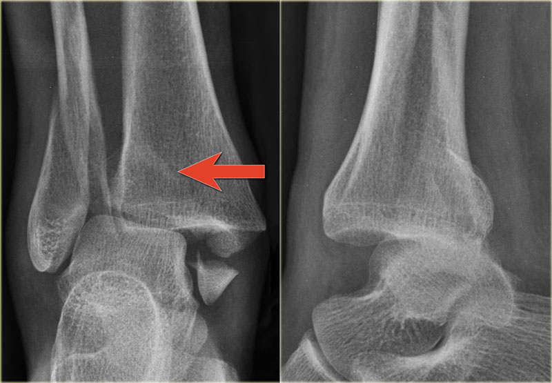 Trimalleolar Weber B fractureGãy Weber B dạng tam giác nhỏ
In this case there is a Weber B fracture with avulsion of the medial malleolus.
The bright line on the AP-view indicates a large tertius fracture fragment.
This tertius fracture can also be seen on the lateral view, but in many cases we need all the information of both the lateral and AP-view to diagnose a tertius fracture.Trong trường hợp này có gãy Weber B kèm theo bong mắt cá trong.
Đường sáng trên mặt cắt ngang cho thấy một mảnh gãy xương lớn.
Gãy xương bánh chè này cũng có thể thấy trên phim nghiêng, nhưng trong nhiều trường hợp chúng ta cần tất cả các thông tin của cả hai bên và phim nghiêng để chẩn đoán gãy xương bánh răng.
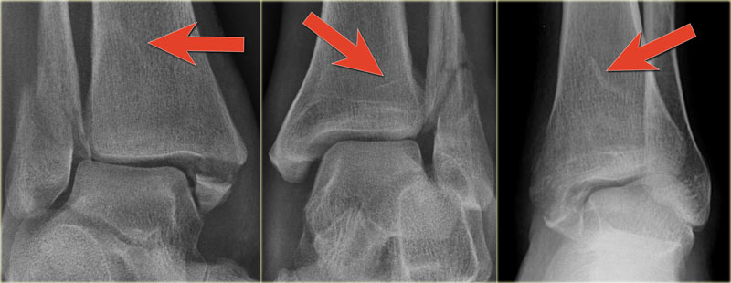
Here more examples of the bright line that indicates a tertius fracture.Ở đây có thêm các ví dụ về đường sáng cho thấy gãy xương tertius.
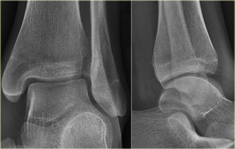
In some cases a fracture of the posterior malleolus is barely or not visible on the radiographs and can only be seen on CT.
First study the radiographs and then continue with the CT.
By the way....there are two fractures.
You can enlarge the images by clicking on them.Trong một số trường hợp, gãy mắt cá sau hầu như không nhìn thấy hoặc không nhìn thấy được trên phim X-quang và chỉ có thể nhìn thấy trên CT.
Đầu tiên nghiên cứu các hình ảnh X quang và sau đó tiếp tục với CT.
Nhân tiện...có hai vết gãy.
Bạn có thể phóng to hình ảnh bằng cách nhấp vào chúng.
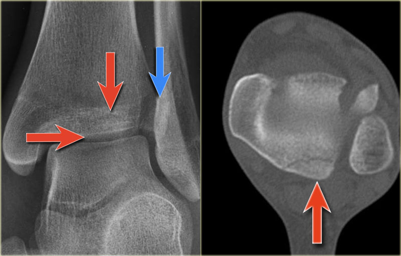
The CT shows an avulsion of the tertius at the insertion of the posterior syndesmosis (red arrows).
The alignment is so perfect, that you do not see the fracture on the radiographs.
Maybe the fracture is seen on the AP-view as indicated by the red arrows, but this is questionable.
Notice that there is also an avulsion at the tibial insertion of the anterior syndesmosis, i.e. Tillaux fracture.
This combination of findings implicate that the ankle is unstable.
A syndesmotic screw has to be inserted.CT cho thấy một chỗ nhô ra của xương bánh chè ở vị trí chèn vào của khớp sợi chày–mác phía sau (các mũi tên màu đỏ).
Sự sắp xếp rất hoàn hảo, bạn không thấy vết nứt trên phim X-quang.
Có thể gãy xương được nhìn thấy trên chế độ xem AP như được chỉ ra bởi các mũi tên màu đỏ, nhưng điều này là đáng nghi ngờ.
Lưu ý rằng cũng có một chỗ nhô ra ở chỗ chèn xương chày của phần trước khớp chày–mác dưới, tức là gãy xương Tillaux.
Sự kết hợp các dấu hiệu này cho thấy cổ chân không ổn định.
Một vít dính khớp phải được đưa vào.
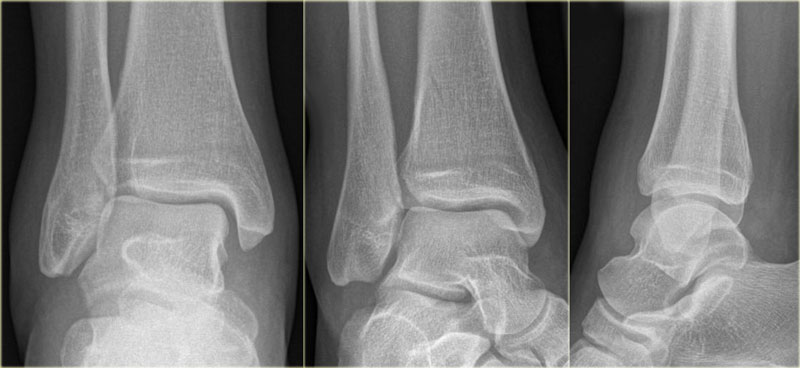 .
Here we have images of an extremely difficult case.
This woman had a distortion of the ankle and had pain on both medial and lateral side.
She was referred to the radiology department by her general practitioner.
The technician made the standard AP-, Mortise- and lateral view and showed them to the radiologist, who was a little bit puzzled.
First study the images and then continue reading. Ở đây chúng tôi có hình ảnh của một trường hợp cực kỳ khó khăn.
Bệnh nhân này bị biến dạng cổ chân và đau cả bên trong và bên ngoài.
Bác sĩ đa khoa đã giới thiệu cô đến khoa X quang.
Kỹ thuật viên đã tạo ra các hình ảnh AP, Mortise và mặt bên tiêu chuẩn và cho bác sĩ X quang xem, người có một chút bối rối.
Đầu tiên nghiên cứu hình ảnh và sau đó tiếp tục đọc.
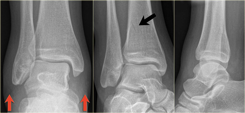
The findings are: Kết quả nghiên cứu:
-
Soft tissue swelling both medial and lateral (red arrows).
Especially the medial swelling should make you consider a pronation exorotation injury (Weber C).
Mô mềm sưng cả trong và ngoài (các mũi tên đỏ).
Đặc biệt là sưng trong nên làm cho bạn xem xét chấn thương sấp ngửa (Weber C). - Lucent line on Mortise view (black arrow) and lateral view. This should make you consider a tertius fracture.
Đường gân nổi trên mặt cắt xương (mũi tên màu đen) và mặt cắt ngoài, điều này nên được xem xét khi gãy xương tertius.
The radiologist decided first to order a CT to find out if there really was a tertius fracture.
Continue with the CT and be amazed.Bác sĩ chẩn đoán hình ảnh quyết định chỉ định chụp CT để xác định gãy xương Tertius.
Tiếp tục với CT và hãy ngạc nhiên.
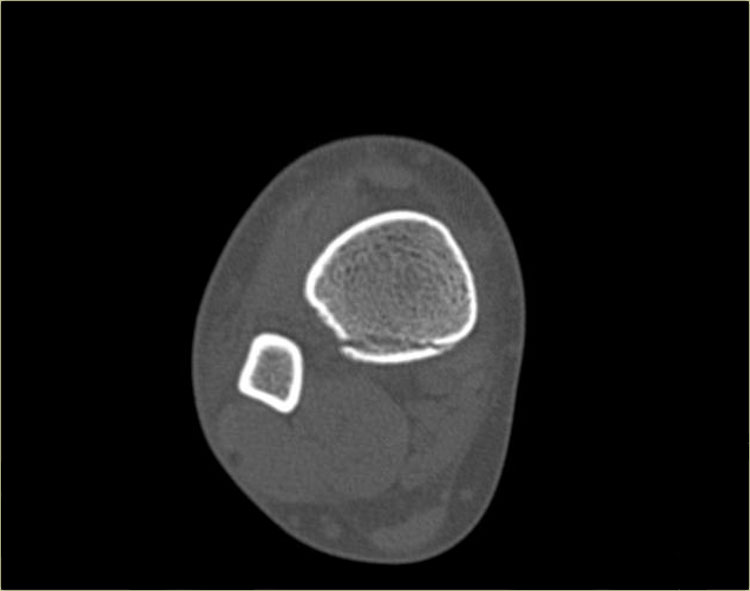
Scroll through the images.
It is amazing, that such a large tertius fragment is so difficult to see on the radiographs.
Also notice the soft tissue swelling on the medial side indicating rupture of the medial collateral ligaments (arrow). Cuộn qua các hình ảnh.
Thật đáng kinh ngạc, một mảnh tertius lớn như vậy rất khó nhìn thấy trên phim X quang.
Cũng nhận thấy mô mềm sưng ở phía trong cho thấy đứt dây chằng bên trong (mũi tên).
Do you have an idea what kind of injury this is?Anh có biết đây là loại thương tích gì không?
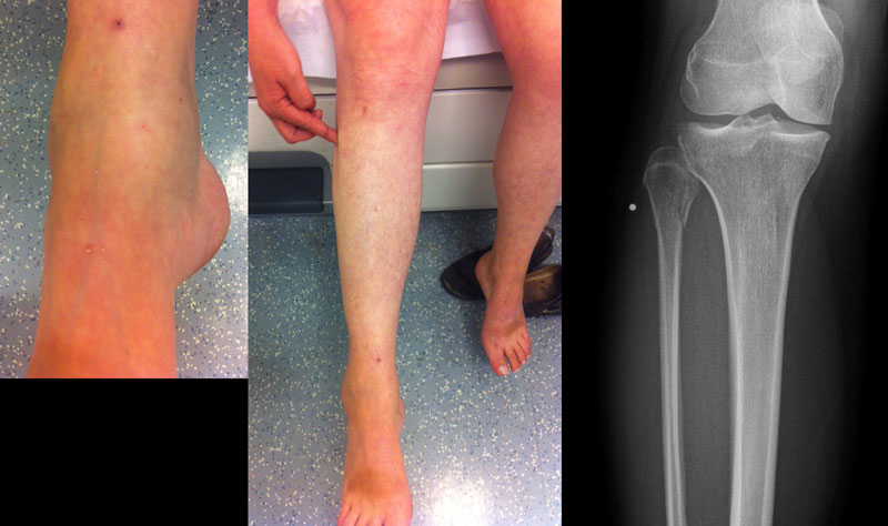
Medial soft tissue swelling and a tertius fracture are both indications of a Weber C or Pronation Exorotation injury.
Since there is no fibula fracture seen on the x-rays of the ankle, there must be a high fibular fracture.Sưng mô mềm trong và gãy xương bánh chè đều là dấu hiệu của chấn thương Weber C hoặc Pronation Exorotation.
Vì không có gãy xương mác trên phim X-quang cổ chân, nên phải có gãy cao xương mác.
At physical exam there was some swelling on the medial side and although the patient did not complain of any pain higher in the lower leg, there was some tenderness when the fibula was palpated.
This spot was marked and a fracture was found.
This case illustrates the importance of medial soft tissue swelling aswell as the finding of a tertius fracture.Khi khám lâm sàng, bên trong có sưng nề, tuy không đau nhiều ở cẳng chân nhưng khi sờ nắn xương mác có cảm giác đau.
Vị trí này được đánh dấu và tìm thấy một vết gãy.
Trường hợp này cho thấy tầm quan trọng của sưng mô mềm trong xương cũng như phát hiện gãy xương tertius.
According to Lauge Hansen we can conclude that this patient first had a rupture of the medial collateral ligaments (stage 1), followed by a rupture of the anterior syndesmosis (stage 2) and a high fibula fracture (stage 3) and finally an avulsion of the posterior malleolus, i.e. PE stage 4.Theo Lauge Hansen chúng ta có thể kết luận rằng bệnh nhân này đầu tiên bị đứt dây chằng bên trong (giai đoạn 1), tiếp theo là đứt phần trước khớp chày–mác dưới (giai đoạn 2) và gãy xương mác cao (giai đoạn 3) và cuối cùng là nhổ mắt cá sau, tức là PE giai đoạn 4.
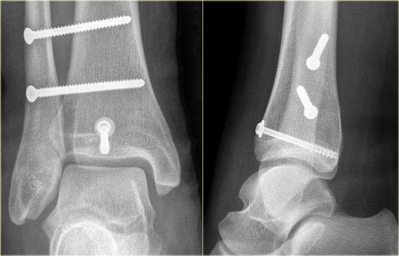
At surgery the ankle was found to be unstable and syndesmosis screws were inserted.
There was an indication for fixing the posterior malleolar fracture, since the fragment involved more than 25% of the articular surface of the distal tibia.Khi phẫu thuật, cổ chân được phát hiện là không ổn định và vít khớp sợi chày–mác được đưa vào.
Có chỉ định cố định gãy mắt cá sau, vì mảnh gãy chiếm hơn 25% diện tích mặt khớp chày đầu xa.
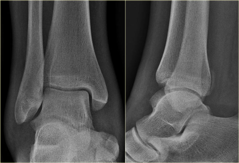
This patient had a twisted ankle and the only abnormality is seen on the lateral view.
This was thought to be an avulsion of the posterior malleolus.
Knowing that this can be the only clue to a high Weber C, additional radiographs were taken.
Continue with the images of the lower leg.Bệnh nhân này bị xoắn cổ chân và bất thường duy nhất được nhìn thấy trên phim nghiêng.
Đây được cho là một sự nhổ của mắt cá sau.
Biết rằng đây có thể là manh mối duy nhất cho Weber C cao, các phim X quang bổ sung đã được thực hiện.
Tiếp tục với hình ảnh của cẳng chân.
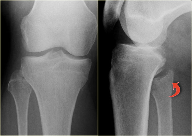
A subtle high fibula fracture is seen (arrow).
Final diagnosis is a Weber C fracture or according to Lauge Hansen: Pronation Exorotation injury stage 4.Một vết nứt tinh vân cao tinh tế được nhìn thấy (mũi tên).
Chẩn đoán cuối cùng là gãy Weber C hoặc theo Lauge Hansen: Pronation Exorotation injury stage 4.

Isolated Tertius fractureGãy đơn độc xương Tertius
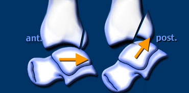
A fracture of the posterior malleolus as an isolated finding is very uncommon.
It is seen when someone's foot hits the ground and a fragment of the posterior malleolus is pushed off by the talus.
The size of this fragment depends on the direction of the force (figure).Gãy mắt cá sau là một phát hiện đơn độc rất hiếm gặp.
Nó được nhìn thấy khi bàn chân của một người nào đó chạm đất và một mảnh của mắt cá sau bị đẩy ra bởi xương sên.
Kích thước của mảnh này phụ thuộc vào hướng của lực tác dụng (hình).
Salter-Harris fracturesGãy Salter-Harris
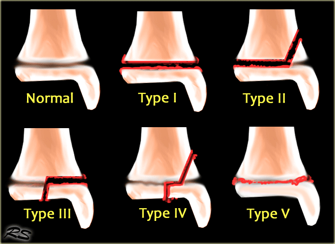
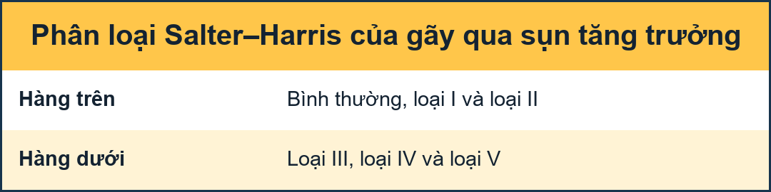
The Salter-Harris classification describes fractures that involve the epiphyseal plate or growth plate.
The most common is type II, which accounts for 75%.Phân loại Salter-Harris mô tả các loại gãy xương liên quan đến nẹp đầu xương hoặc nẹp tăng trưởng.
Phổ biến nhất là type II, chiếm 75%.
- Type I - transverse fracture through the growth plate or physis Loại I - gãy ngang qua tấm tăng trưởng hoặc physis
- Type II - fracture through the growth plate and the metaphysis, sparing the epiphysis Loại II - gãy xuyên qua mảng tăng trưởng và xương bàn đạp, không gãy xương đầu chi
- Type III - fracture through growth plate and epiphysis, sparing the metaphysis Loại III - gãy xương qua mảng tăng trưởng và mảng thượng ngành, không gãy xương mảng siêu ngành
- Type IV - fracture through all three elements of the bone, the growth plate, metaphysis, and epiphysis Loại IV - gãy xương qua cả ba yếu tố của xương, tấm tăng trưởng, xương, và xương
- Type V - compression fracture of the growth plate Type V - gãy lún của đĩa tăng trưởng
These Salter-Harris fractures can be easily missed.
In many cases there is only minimal or no displacement.
The fracture through the growth plate is usually obscure and difficult to differentiate from normal variations of the growth plate.
And finally we tend not to look carefully at the epiphysis.
Những gãy Salter-Harris này có thể dễ dàng bị bỏ qua.
Trong nhiều trường hợp chỉ có di lệch tối thiểu hoặc không di lệch.
Vết gãy qua tấm tăng trưởng thường mờ và khó phân biệt với các biến thể bình thường của tấm tăng trưởng và cuối cùng chúng ta có xu hướng không xem xét cẩn thận về mặt ngoại sinh.
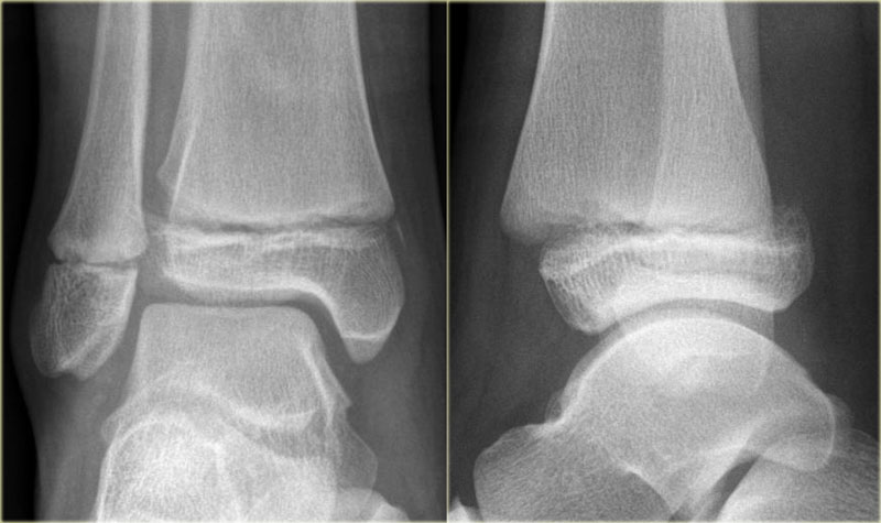
Type ILoại I
Type I Salter-Harris fractures tend to occur in younger children (5).
It is a transverse fracture through the cartilage of the growth plate or physis.
Often, x-rays of a child with a type I growth plate fracture will appear normal.
Most type I growth plate injuries are treated with a cast.
Healing of type I fractures tends to be rapid and complications are rare.Gãy xương Salter-Harris loại I có xu hướng xảy ra ở trẻ nhỏ hơn (5).
Đây là một gãy ngang xuyên qua sụn của tấm tăng trưởng hoặc physis.
Thông thường, phim X-quang của một đứa trẻ bị gãy xương loại I sẽ có vẻ bình thường.
Hầu hết các chấn thương đĩa tăng trưởng loại I được điều trị bằng bó bột.
Chữa lành gãy loại I có xu hướng nhanh chóng và biến chứng hiếm gặp.
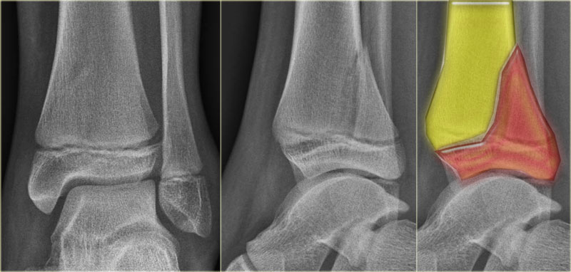
Type IILoại II
A type II growth plate fracture starts across the growth plate, but the fracture then continues up through the metaphysis.
This is the most common type of growth plate fracture, and tends to occur in older children. Often type II growth plate fractures must be repositioned under anesthesia, but healing is usually quick and complications are uncommon.
Gãy xương mảng tăng trưởng loại II bắt đầu qua mảng tăng triển, nhưng gãy xương sau đó tiếp tục đi lên qua mảng siêu xương.
Đây là loại gãy xương tăng trưởng phổ biến nhất, và có xu hướng xảy ra ở trẻ lớn hơn. Thường gãy xương tấm tăng trưởng loại II phải được đặt lại vị trí dưới gây mê, nhưng việc lành thương thường nhanh và biến chứng không phổ biến.
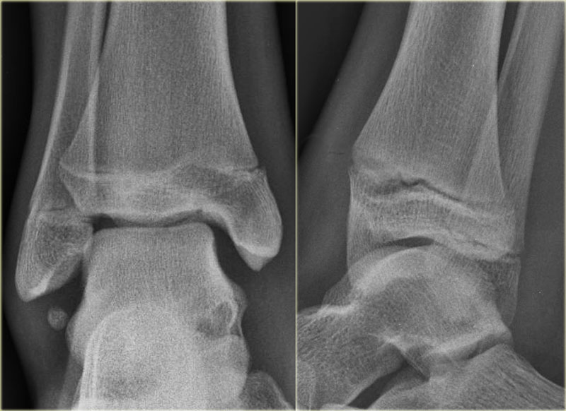
Type IIILoại III
Type III is a fracture through the growth plate and epiphysis sparing the metaphysis.
A type III fracture also starts through the growth plate, but turns and exits through the end of the bone, and into the adjacent joint.
These injuries can be concerning because the joint cartilage is disrupted by the fracture.
Proper positioning is essential after a type II growth plate fracture.
These injuries also tend to affect older children in whom the growth plate is partially closed.
Study the images and then scroll to the next images.Loại III là gãy xuyên qua mảng tăng trưởng và mảng thượng bội, không có mảng siêu bội.
Gãy loại III cũng bắt đầu qua đĩa tăng trưởng, nhưng quay và thóat ra qua đầu xương, và vào khớp liền kề.
Những tổn thương này có thể đáng lo ngại vì sụn khớp bị gãy.
Định vị đúng là rất cần thiết sau khi gãy xương loại II.
Những chấn thương này cũng có xu hướng ảnh hưởng đến trẻ lớn hơn, những trẻ mà đĩa tăng trưởng bị đóng một phần.
Nghiên cứu các hình ảnh và sau đó cuộn đến các hình ảnh tiếp theo.
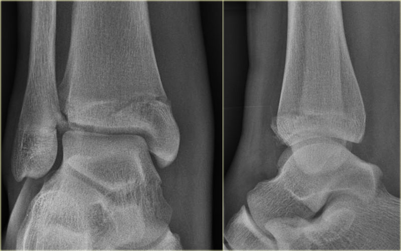
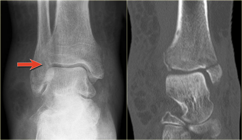
The fracture through the epiphysis can be easily missed (blue arrow).
The fracture through the growth plate is only seen on CT.
Continue with the CT images.Vết gãy qua đầu xương có thể dễ dàng bị bỏ sót (mũi tên màu xanh).
Vết gãy xuyên qua đĩa tăng trưởng chỉ thấy trên CT.
Tiếp tục với hình ảnh CT.
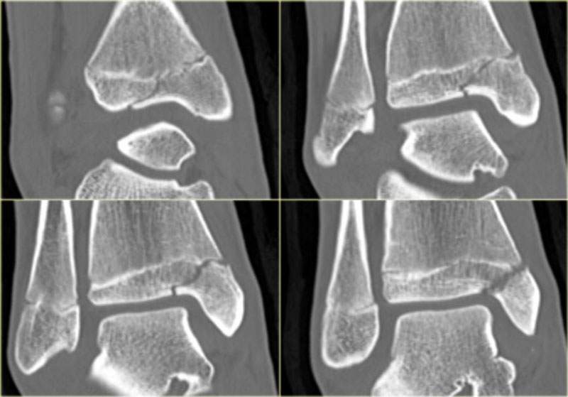
The CT-images nicely display the fracture through the growth plate and the epiphysis.Hình ảnh CT cho thấy rõ ràng gãy xương qua mảng tăng trưởng và mảng đầu xương.
Study the images and then scroll to the next images.Nghiên cứu các hình ảnh và sau đó cuộn đến các hình ảnh tiếp theo.
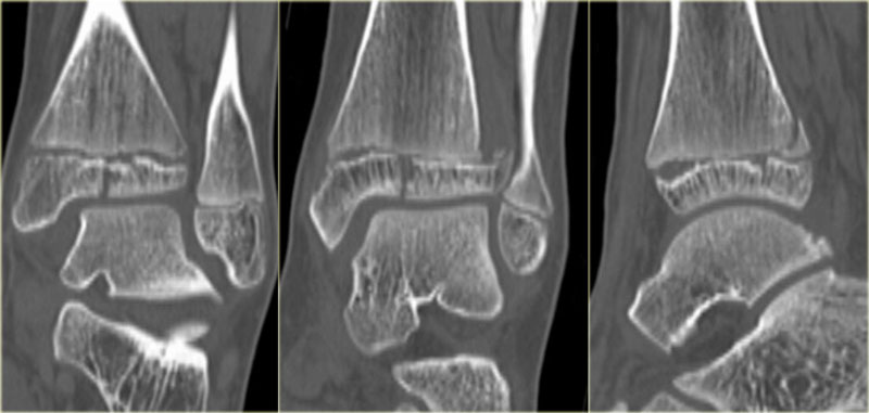
This is also a Salter-Harris type III fracture.
Notice that there is also a Tillaux fracture.
We will discuss these fractures in a moment.Đây cũng là gãy Salter-Harris loại III.
Lưu ý rằng cũng có một gãy Tillaux.
Chúng ta sẽ thảo luận về những loại gãy này trong một thời điểm.
Type IVLoại IV
Type IV is a fracture through all three elements of the bone, the growth plate, metaphysis and epiphysis.
Notice that the epiphyseal fracture is in the sagittal plane, the fracture through the growth plate is in the axial plane and the metaphyseal fracture is in the coronal plane.
These fractures are also named triplane fractures.
These are discussed in the next chapter.Loại IV là gãy xuyên qua cả ba yếu tố của xương, mảng tăng trưởng, mảng chuyển tiếp và mảng đầu xương.
Lưu ý rằng gãy xương hành xương nằm trong mặt phẳng đứng dọc, gãy xương xuyên qua mảng tăng trưởng nằm trong mặt bằng dọc trục và gãy xương xương hành xương ở mặt phẳng vành.
Những loại gãy này cũng được đặt tên là gãy triplane.
Những vấn đề này sẽ được thảo luận trong chương tiếp theo.
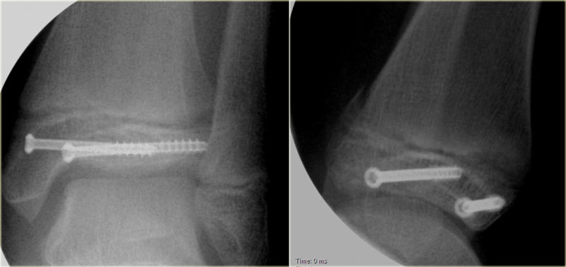
Proper positioning is also essential with type IV growth plate fractures, and surgery may be needed to hold the bone fragments in proper position.Định vị đúng cũng rất cần thiết với gãy xương loại IV, và phẫu thuật có thể cần thiết để giữ các mảnh xương ở vị trí thích hợp.
Type VLoại V
Type V growth plate injuries occur with the growth plate is crushed.
Type V growth plate fractures carry the most concerning prognosis as bone alignment and length can be affected.
These types of fractures may permanently injure the growth plate, requiring later treatment to restore alignment of the limb.Chấn thương đĩa tăng trưởng loại V xảy ra khi đĩa tăng trưởng bị nghiền nát.
Gãy xương đĩa tăng trưởng loại V mang lại tiên lượng đáng lo ngại nhất vì sự liên kết xương và chiều dài có thể bị ảnh hưởng.
Những loại gãy xương này có thể làm tổn thương vĩnh viễn tấm tăng trưởng, cần điều trị sau này để khôi phục lại sự liên kết của chi.
Triplane fractureGãy Triplane
 Triplane fractureGãy Triplane
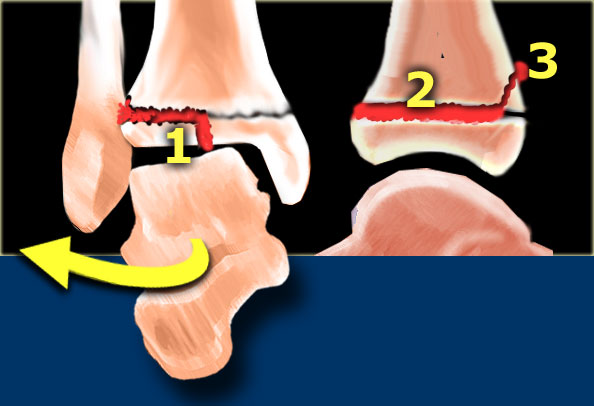
This fracture is named triplane because it occurs in the coronal, sagittal and axial plane.
It is actually a Salter-Harris type IV.
It is seen exclusively in young adolescents in the period, when the medial tibial epiphysis is closed, while the lateral portion is still open leaving it vulnerable to injury.
As the force cannot continue into the medial part of the growth plate since this is already closed, the epiphysis will fracture.
As in most ankle fractures the mechanism is external rotation.
Gãy này được đặt tên là triplane vì nó xảy ra ở vành, dọc và mặt phẳng ngang.
Nó thực sự là một loại Salter-Harris IV.
Nó chỉ được nhìn thấy ở thanh thiếu niên trong giai đoạn này, khi xương chày trong đóng lại, trong khi phần bên vẫn còn mở để lại nó dễ bị tổn thương.
Vì lực không thể tiếp tục vào phần giữa của tấm tăng trưởng vì nó đã được đóng lại, nên phần đầu xương sẽ bị gãy.
Giống như trong hầu hết các loại gãy cổ chân, cơ chế là xoay ngoài.
The injury results in: Tổn thương dẫn đến:
- Epiphyseal fracture in the sagittal plane Gãy xương đầu trên xương cánh tay mặt phẳng đứng dọc
- Injury to the growth plate in the axial plane Tổn thương tấm tăng trưởng trong mặt phẳng ngang
- Metaphyseal fracture in the coronal plane Gãy xương trên xương cánh tay mặt phẳng đứng ngang
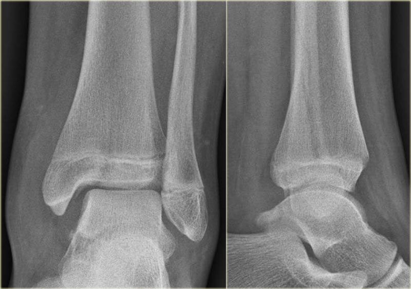
Study the images and then continue reading.Nghiên cứu các hình ảnh và sau đó tiếp tục đọc.
 Triplane fractureGãy Triplane
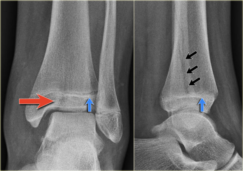
At first this looks like a Weber B fracture with an oblique fracture in the fibula as seen on the lateral view (black arrows).
Notice however that this fracture line stops at the level of the epiphyseal plate.
So this is the fracture of the metaphysis in the coronal plane.
On the AP-view there is a lucency within the epiphysis, which is the epiphyseal fracture in the sagittal plane.
Notice also that the medial epiphysis is already closed, while the lateral portion is still open(blue arrows).
We have to assume that there is an epiphysiolysis of this lateral portion. Đầu tiên, điều này trông giống như một gãy Weber B với một vết gãy chéo ở xương mác như nhìn thấy trên mặt cắt ngang (các mũi tên màu đen).
Tuy nhiên, lưu ý rằng đường gãy này dừng lại ở mức của nẹp đầu xương.
Vậy đây là gãy xương của xương bàn đạp trong mặt phẳng đứng ngang.
Trên góc nhìn của AP có một điểm sáng trong mỏm xương đầu, đó là gãy xương đầu trong mặt phẳng đứng dọc.
Cũng lưu ý rằng phần đầu trong đã đóng, trong khi phần bên vẫn mở (các mũi tên màu xanh).
Chúng ta phải giả định rằng có một sự phân chia theo chiều dọc của phần bên này.
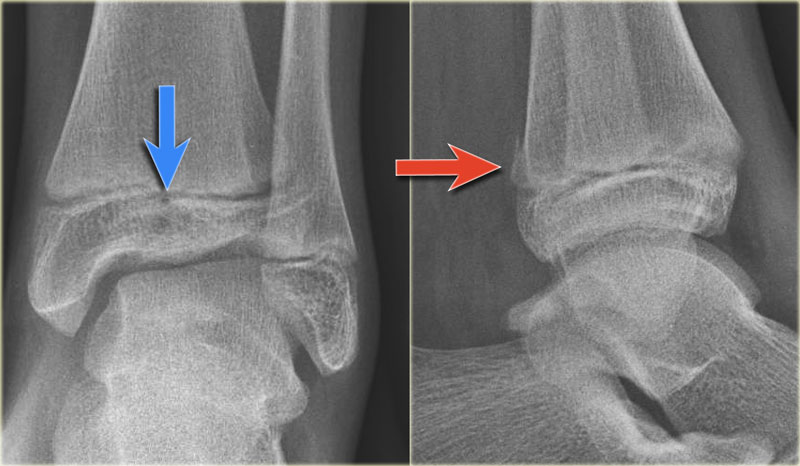
Here another example.
There is only a small metaphyseal fragment, which is usually the case (red arrow).
The fracture through the epiphysis is indicated by the blue arrow.Đây là một ví dụ khác.
Chỉ có một mảnh xương nhỏ, thường là trường hợp (mũi tên màu đỏ).
Vết gãy qua đầu xương được chỉ định bởi mũi tên màu xanh.
Maisonneuve fractureGãy Maisonneuve
In 1840 Maisonneuve described a frature of the proximal shaft of the fibula, which was caused by exorotation force applied to the ankle.
It is a high Weber C fracture.
These fractures are easily overlooked because the patients rarely complain of pain in the region of the proximal fibula, since the ankle is most painful.Năm 1840, Maisonneuve đã mô tả một trường hợp gãy đầu gần xương mác, nguyên nhân là do lực tác động lên mắt cá chân.
Đây là gãy Weber C cao.
Những gãy xương này dễ bị bỏ qua vì bệnh nhân hiếm khi phàn nàn về đau ở vùng đầu gần xương mác, vì cổ chân là đau nhất.
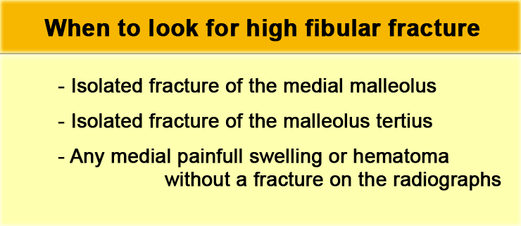
There are three situations in which we should suspect a high Weber C or Maisonneuve fracture:Có ba tình huống mà chúng ta nên nghi ngờ gãy Weber C hoặc Maisonneuve cao:
- Isolated fracture of the medial malleolus Gãy đơn độc mắt cá trong
- Isolated fracture of the posterior malleolus without a fracture on the lateral sideGãy mắt cá sau đơn thuần không gãy xương bên ngoài
- Any painful swelling or hematoma on the medial side without a fracture on the radiographsTrên phim X-quang có bất kỳ dấu hiệu sưng đau hoặc tụ máu ở mặt trong mà không có gãy xương
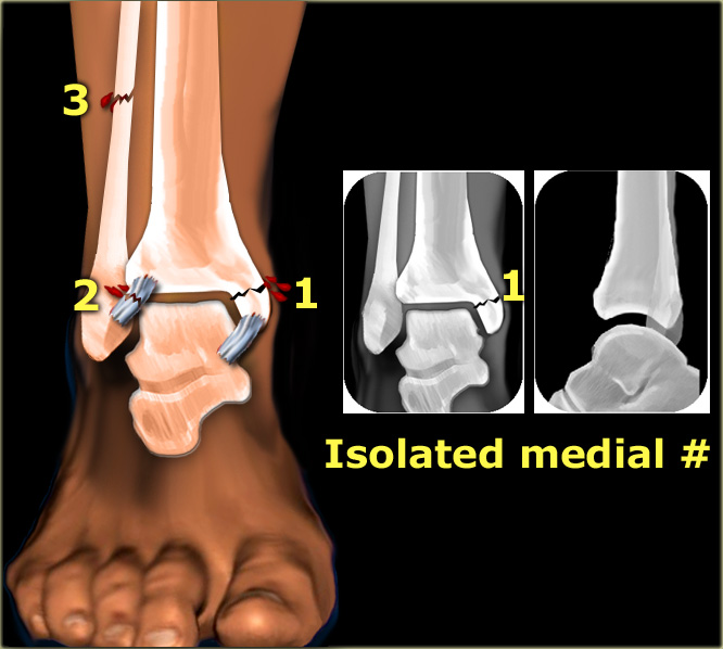

Isolated fracture of the medial malleolus
According to Lauge-Hansen this is the first stage of a pronation exorotation injury, which results in a Weber C fracture.
So we have to look for higher stages.
Gãy đơn độc mắt cá trong
Theo Lauge-Hansen đây là giai đoạn đầu tiên của chấn thương sấp ngửa, dẫn đến gãy Weber C.
Vì vậy, chúng ta phải tìm kiếm các giai đoạn cao hơn.
The injury can continue to the following: Tổn thương có thể tiếp tục như sau:
- stage 2: rupture of the anterior syndesmosis Giai đoạn 2: vỡ phần trước khớp chày–mác dưới
- stage 3: high fibular fracture Giai đoạn 3: gãy xương mác cao
- stage 4: rupture of the posterior syndesmosis Giai đoạn 4: vỡ khớp sợi chày–mác sau
In all these subsequent stages, purely ligamentous injury will not be visible on the radiographs of the ankle.
So even in a Weber C stage 4 sometimes only a fracture of the medial malleolus will be visible.Trong tất cả các giai đoạn tiếp theo này, tổn thương dây chằng hoàn toàn sẽ không nhìn thấy trên phim X quang cổ chân.
Vì vậy, ngay cả trong Weber C giai đoạn 4 đôi khi chỉ có thể nhìn thấy một vết nứt của mắt cá trong.
In the illustration we see the fractures and ligamentous injury on the left and the resulting x-rays on the right.Trong hình minh hoạ, chúng ta thấy gãy xương và tổn thương dây chằng bên trái và kết quả chụp X-quang bên phải.
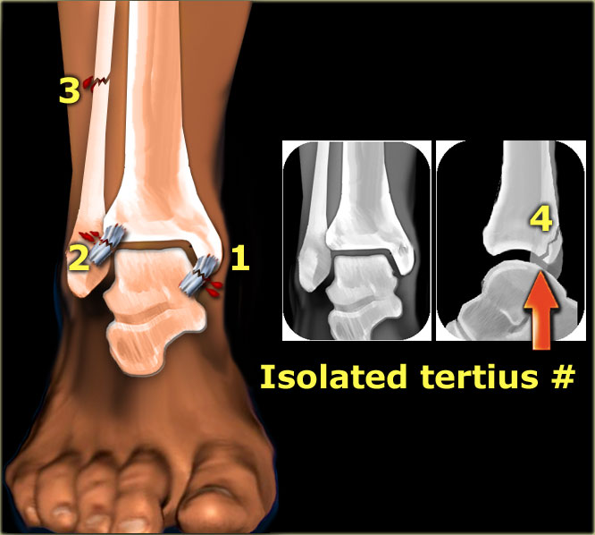

Isolated fracture of the posterior malleolus
Truely isolated fractures of the posterior malleolus are very uncommon.
Most fractures of the posterior malleolus are part of a complex ankle injury, either Weber B or Weber C.
A Weber B fracture is easily detected because of the characteristic oblique fracture.
So if there is a tertiu sfracture and no sign of a Weber B fracture, then we have to start looking for a high Weber C fracture.Gãy mắt cá sau đơn thuần
Gãy đơn độc mắt cá sau rất hiếm gặp.
Hầu hết các gãy mắt cá sau là một phần của chấn thương phức tạp ở cổ chân, Weber B hoặc Weber C.
Gãy Weber B dễ phát hiện do đặc điểm gãy chéo.
Vì vậy, nếu có một gãy xương lớn và không có dấu hiệu của gãy Weber B, thì chúng ta phải bắt đầu tìm kiếm gãy Webers C cao.
In that case we have the following combination:Trong trường hợp đó chúng ta có sự kết hợp sau:
- stage 1: rupture of the medial collateral ligament stage 2: rupture of the anterior syndesmosisGiai đoạn 1: đứt dây chằng bên trong Giai đoạn 2: đứt phần trước khớp chày–mác dưới
- stage 3: high fibular fractureGiai đoạn 3: gãy xương mác cao
- stage 4: tertius fractureGiai đoạn 4: gãy xương bánh chè
An isolated tertius fracture on the ankle radiographs indicates the presence of an unstable ankle injury.Gãy xương cổ chân đơn độc trên phim X-quang cho thấy có tổn thương cổ chân không ổn định.
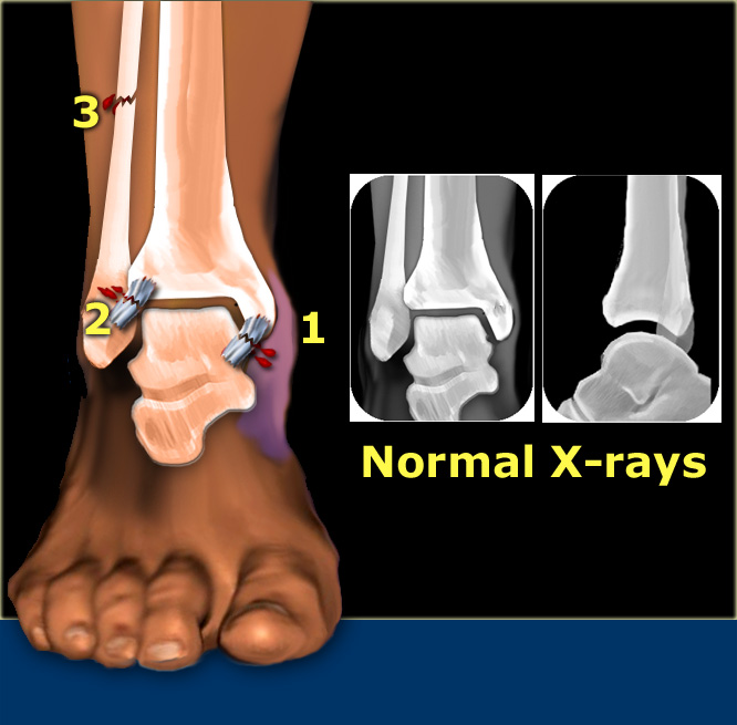

Any medial painful swelling or hematoma
Normal radiographs do not rule out a Weber C fracture.Bất kỳ sưng đau hoặc tụ máu trong
Chụp X-quang bình thường không loại trừ gãy Weber C.
We may have the following combination: Chúng ta có thể có sự kết hợp sau:
- stage 1: rupture of the medial collateral ligament, which causes the swelling and hematoma Giai đoạn 1: đứt dây chằng bên trong, gây sưng và tụ máu.
- stage 2: rupture of the anterior syndesmosis Giai đoạn 2: vỡ phần trước khớp chày–mác dưới
- stage 3: high fibular fracture - not visible on the radiographs of the ankleGiai đoạn 3: gãy xương mác cao - không thấy trên phim X-quang cổ chân
- stage 4: rupture of the posterior syndesmosisGiai đoạn 4: vỡ khớp sợi chày–mác sau
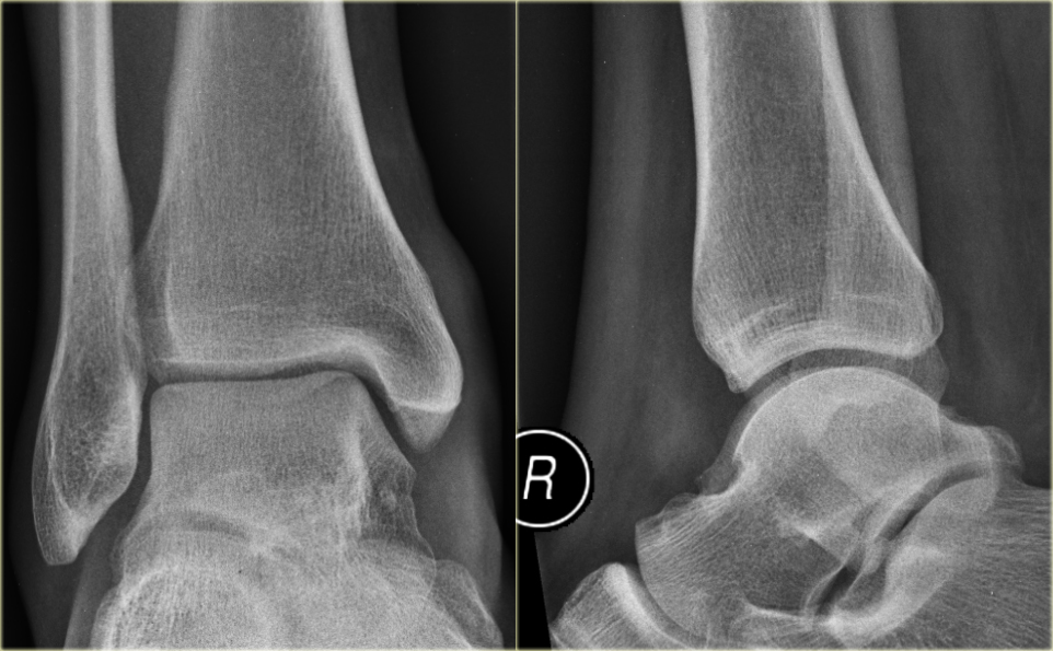
Example 1
On the left images of a patient with a hematoma on the medial side.
Ví dụ 1
Trên hình ảnh bên trái của một bệnh nhân có máu tụ ở phía trong.
- First impression
We can exclude a Weber A or B fracture, because we see no fracture.
A high Weber C is still a possibility, i.e. Ấn tượng đầu tiên
Chúng ta có thể loại trừ gãy Weber A hoặc B, vì chúng ta không thấy gãy xương.
Weber C cao vẫn là một khả năng, tức là- medial ligament rupture Vỡ dây chằng trong
- high fibular fracture gãy xương mác cao
- posterior syndesmosis rupture.Vỡ khớp sợi chày–mác sau.
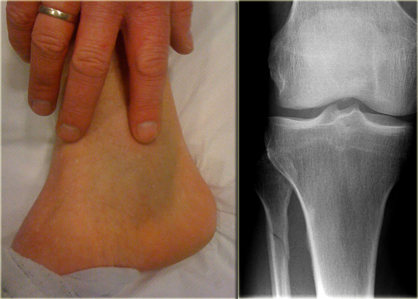
- Re-examination
Additional radiographs of the lower leg were taken and demonstrated a high fibular fracture, also known as Maisonneuve fracture. Khám lại
Chụp X-quang bổ sung cẳng chân được thực hiện và cho thấy gãy xương mác cao, còn được gọi là gãy Maisonneuve. - Final report
Weber C stage 4, i.e. medial collateral ligamentous rupture, rupture of the anterior syndesmosis, high fibular fracture and probably a rupture of the posterior syndesmosis.Báo cáo tổng kết
Weber C giai đoạn 4, tức là đứt dây chằng bên trong, đứt dính trước, gãy xương mác cao và có thể là đứT dính sau.
This case demonstrates that there can be an unstable ankle injury that needs surgery even when the radiographs of the ankle do not show a fracture.
In any patient with an ankle injury you should always ask yourself the question......can I exclude a high Weber C fracture or do I need additional imaging.Trường hợp này chứng tỏ có thể có tổn thương cổ chân không ổn định cần phẫu thuật ngay cả khi phim X quang cổ chân không thấy gãy xương.
Ở bất kỳ bệnh nhân nào bị chấn thương cổ chân, bạn nên luôn tự hỏi mình câu hỏi......tôi có thể loại trừ gãy Weber C cao hay tôi cần thêm hình ảnh.
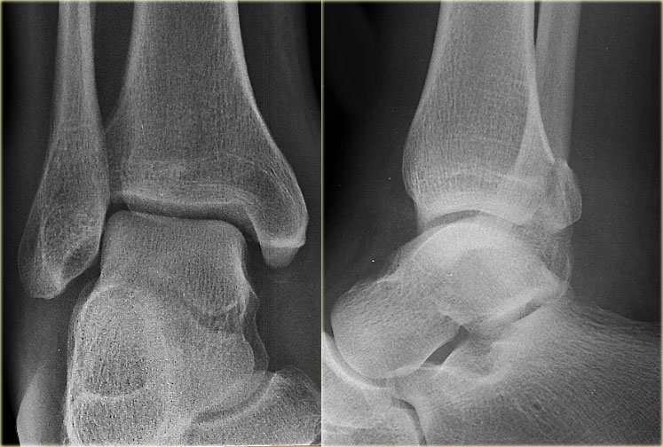
Example 2 Ví dụ 2
- First impression
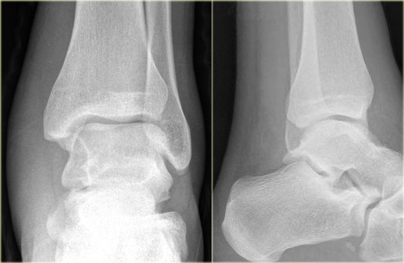
There is a fracture of the posterior malleolus.
Classification according to Weber is not possible.
An isolated fracture of the posterior malleolus is uncommon, but as part of a supination exorotation (Weber B) or pronation exorotation injury (Weber C) it is quite common.
So we have to re-examine the films to look for signs of a Weber B or C fracture.Ấn tượng đầu tiên
Có gãy mắt cá sau.
Không thể phân loại theo Weber.
Gãy đơn độc mắt cá sau là không phổ biến, nhưng là một phần của chấn thương khi ngửa (Weber B) hoặc sấp khi ngửA (Weber C) thì khá phổ biến.
Vì vậy chúng tôi phải kiểm tra lại phim để tìm dấu hiệu gãy Weber B hoặc C.
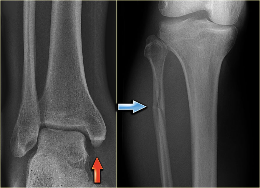
- Re-examination
No sign of an oblique fracture of the lateral malleolus, so we can exclude a Weber B fracture.
There is still the possibility of a Weber C fracture, i.e. medial rupture or avulsion, high fibular fracture and finally a posterior malleolar fracture.
Now we notice the subtle avulsion of the medial malleolus (red arrow).
Additional radiographs of the lower extremity demonstrate a high fibular fracture (blue arrow). Khám lại
Không có dấu hiệu gãy chéo mắt cá ngoài, do đó có thể loại trừ gãy Weber B.
Vẫn còn khả năng gãy Weber C, tức là vỡ trong hoặc bong xương, gãy xương mác cao và cuối cùng là gãy mắt cá sau.
Bây giờ chúng ta nhận thấy sự nhả ra tinh tế của mắt cá trong (mũi tên màu đỏ).
Chụp X-quang bổ sung ở chi dưới cho thấy gãy xương mác cao (mũi tên màu xanh). - Final report
Weber C stage 4. Báo cáo tổng kết
Weber C giai đoạn 4.
Example 3
In this case no fracture is seen, but only soft tissue swelling on the medial side.
In such a case, you have to rule out a Maisonneuve fracture, which is a high Weber C fracture.
Additional x-rays of the lower leg were taken.
Ví dụ 3
Trong trường hợp này không thấy gãy xương, chỉ thấy sưng mô mềm ở mặt trong.
Trong trường hợp này, bạn phải loại trừ gãy Maisonneuve, là gãy Weber C cao.
Chụp X-quang bổ sung vùng cẳng chân.
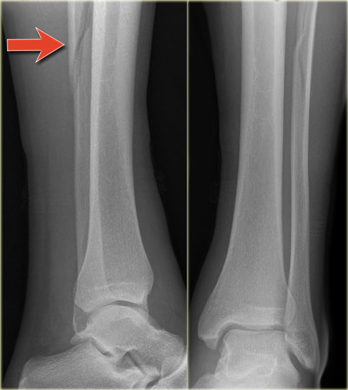
There is a high fibula fracture.
Có một vết gãy xương mác cao.
Tillaux fractureGãy Tillaux
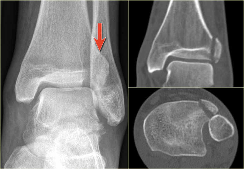
External rotation injury of the ankle is the most common ankle injury and can lead to a Weber B or Weber C fracture.
One of the first stages in this injury is rupture of the anterior tibiofibular ligament (or anterior syndesmosis).
Less frequently it leads to an avulsion of the anterolateral tibial epiphysis.
Whenever you see such a fracture, you have to look for higher stages of this exorotation injury.Tổn thương xoay ngoài của cổ chân là tổn thương cổ chân phổ biến nhất và có thể dẫn đến gãy Weber B hoặc Weber C.
Một trong những giai đoạn đầu tiên của tổn thương này là đứt dây chằng chày mác trước (phần trước khớp chày–mác dưới).
Ít thường xuyên hơn, nó dẫn đến sự nhổ của xương chày trước bên.
Bất cứ khi nào bạn thấy một vết gãy như vậy, bạn phải tìm kiếm các giai đoạn cao hơn của tổn thương này.
The x-ray shows a subtle Tillaux fracture, which is better appreciated on the CT-images.X-quang cho thấy một gãy Tillaux tinh tế, được đánh giá tốt hơn trên hình ảnh CT.
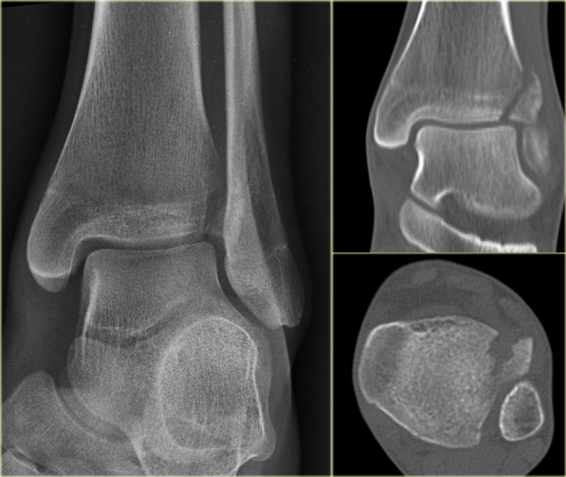
Study these images carefully and remember the stages of an exorotation injury.
What is going on here?Nghiên cứu những hình ảnh này một cách cẩn thận và ghi nhớ các giai đoạn của chấn thương khi thở ra.
Chuyện gì đang xảy ra ở đây?
There is a Tillaux fracture due to avulsion of the anterolateral part of the distal tibia by the anterior syndesmosis.
This can be a stage 2 of a Weber C fracture.
Stage 1 is rupture of the medial collateral ligaments and stage 3 is a fibula fracture above the level of the syndesmosis.
So now we start looking for stage 4, which is rupture or avulsion of the posterior syndesmosis.Có gãy Tillaux do xương chày đầu dưới bên trước xương chày bị bong ra bởi phần trước khớp chày–mác dưới.
Đây có thể là giai đoạn 2 của gãy Weber C.
Giai đoạn 1 là đứt dây chằng bên trong và giai đoạn 3 là gãy xương mác trên mức khớp sợi chày–mác.
Vì vậy, bây giờ chúng tôi bắt đầu tìm kiếm giai đoạn 4, đó là vỡ hoặc nhổ ra khớp sợi chày–mác phía sau.
Do you now see the tertius fracture on the axial CT-image?
This patient has an unstable ankle injury and a syndesmotic screw needs to be inserted.Bây giờ bạn có thấy gãy xương tertius trên hình ảnh CT dọc trục không?
Bệnh nhân này có tổn thương cổ chân không ổn định và cần phải đặt vít dính.
Stages of exorotation injuries of the ankleCác giai đoạn tổn thương cổ chân
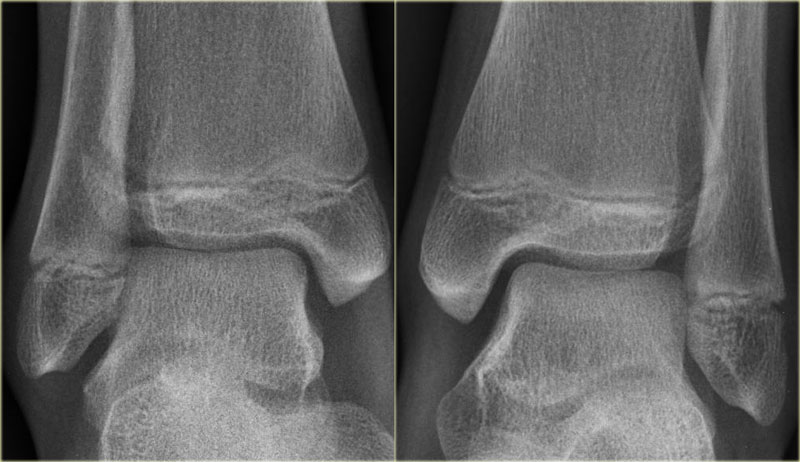
Another Tillaux in a patient with a strange combination of findings. There is an avulsion of the lateral malleolus, a Tillaux and a medial malleolar fracture.Một Tillaux khác ở một bệnh nhân có sự kết hợp kỳ lạ của các dấu hiệu Có một sự nhổ của mắt cá ngoài, một Tillaux và một gãy xương mắt cá trong.
juvenile TillauxTillaux vị thành niên
A Tillaux fracture is more commonly seen in adolescents at the age of 12 -15 years.
At that age it is a fracture through the growth plate and is then called a juvenile Tillaux.
It occurs before the distal tibial epiphysis has completely fused.
The fracture occurs when the medial epiphysis has fused and the lateral part becomes avulsed at the attachment of the anterior tibiofibular ligament (or syndesmosis).Gãy Tillaux thường gặp ở thanh thiếu niên từ 12 -15 tuổi.
Ở tuổi đó, nó là một vết gãy xuyên qua tấm tăng trưởng và sau đó được gọi là Tillaux vị thành niên.
Nó xảy ra trước khi xương chày đầu xa đã hợp nhất hoàn toàn.
Gãy xương xảy ra khi đầu xương chày trong đã hợp nhất và phần bên bị bong ra ở chỗ bám của dây chằng chày mác trước (hoặc khớp sợi chày–mác).
Study the images and then continue reading.
You can enlarge the images by clicking on them.Nghiên cứu các hình ảnh và sau đó tiếp tục đọc.
Bạn có thể phóng to hình ảnh bằng cách nhấp vào chúng.
There is a subtle widening of the lateral part of the growth plate of the right ankle.
There is also a very subtle fracture through the epiphysis.
Continue with the CT.Có sự giãn rộng một cách tinh tế của phần bên của đĩa tăng trưởng của mắt cá chân phải.
Ngoài ra còn có một vết nứt rất tinh tế qua đầu xương.
Tiếp tục với CT.
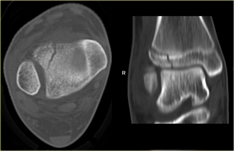
The CT-images show a epiphysiolysis fracture Salter Harris type 3.
This juvenile Tillaux is especially seen in young athletes.
Always look for higher stages of an exorotation injury.Hình ảnh CT cho thấy gãy đầu dưới xương cánh tay Salter Harris loại 3.
Loài Tillaux vị thành niên này đặc biệt thấy ở các vận động viên trẻ.
Luôn tìm kiếm các giai đoạn cao hơn của chấn thương vùng thắt lưng.
Charity
Hoạt động từ thiện
All the profits of the Radiology Assistant go to Medical Action Myanmar which is run by Dr. Nini Tun and Dr. Frank Smithuis sr, who is a professor at Oxford university and happens to be the brother of Robin Smithuis.Toàn bộ lợi nhuận của Radiology Assistant được chuyển cho Medical Action Myanmar, do bác sĩ Nini Tun và bác sĩ Frank Smithuis Sr. điều hành. Frank Smithuis Sr. là giáo sư tại Đại học Oxford và là anh em ruột của Robin Smithuis.
Click here or on the image below to watch the video of Medical Action Myanmar and if you like the Radiology Assistant, please support Medical Action Myanmar with a small gift.Nhấp vào đây hoặc vào hình ảnh bên dưới để xem video về Medical Action Myanmar và nếu bạn thích Trợ lý X quang, vui lòng hỗ trợ Medical Action Myanmar bằng một món quà nhỏ.
ReferencesTài liệu tham khảo
-
Fractures of the ankle, combined experimental-surgical and experimental-roentgenologic investigationsby N. Lauge-Hansen (1948) Gãy xương cổ chân, kết hợp thực nghiệm-phẫu thuật và thực nghiệm-tổn thương họcbởi N. Lauge-Hansen (1948)
- Website of the Ottawa Ankle rules Website của Ottawa Ankle rules
-
Ankle fractures
Gãy cổ chân
East Lancashire Foot and Ankle Hyperbook East Lancashire Foot and Ankle Hyperbook (bằng tiếng Anh)
-
Free AO Surgery Reference
Phẫu thuật AO miễn phí
The AO Surgery Reference is a huge online repository of surgical knowledge, consisting of more than 7000 pages. AO Surgery Reference là một kho lưu trữ trực tuyến khổng lồ về kiến thức phẫu thuật, bao gồm hơn 7000 trang.
-
Types of Growth Plate Fractures
Các loại gãy xương tăng trưởng
By Jonathan Cluett, M.D., About.com Guide Bởi Jonathan Cluett, M.D., About.com Hướng dẫn